Embed Size (px)
Citation preview
ORIGINAL ARTICLE
CRH-R1 and CRH-R2 differentially modulate dendriticoutgrowth of hippocampal neurons
Hui Sheng • Yongjun Xu • Yanming Chen •
Yanmin Zhang • Xiaohui Xu • Cheng He •
Xin Ni
Received: 15 August 2011 / Accepted: 5 January 2012 / Published online: 17 January 2012
� Springer Science+Business Media, LLC 2012
Abstract Corticotropin-releasing hormone (CRH) has
been implicated to be involved in the development of
dendrites in brain. In the present study, we examined the
effect of CRH on dendrite outgrowth in primary cultured
hippocampal neurons and defined the specific CRH
receptor subtype involved. Treatment of neurons with
increasing concentration of CRH resulted in an increase in
the total dendritic branch length (TDBL) of neurons com-
pared with untreated neurons over 2–4 days period of
treatment. These effects can be reversed by the specific
CRH-R1 antagonist antalarmin but not by the CRH-R2
antagonist astressin 2B. Treatment of neurons with uro-
cortin II, the exclusive CRH-R2 agonist, significantly
decreased TDBL of the cultured neurons. These effects can
be reversed by the CRH-R2 antagonist astressin 2B. Our
results suggest that CRH-R1 and CRH-R2 differentially
modulate the dendritic growth of hippocampal neurons in
culture.
Keywords Corticotropin-releasing hormone �Corticotropin-releasing hormone receptors � Dendrite �Hippocampus
Introduction
During nervous system development, formation of a net-
work of neuronal circuitries has profound functional
implications. At the level of a single neuron, the structural
organization of the postsynaptic surface, the dendritic
arbor, is one of the critical determinants of neuronal cir-
cuitry besides the axon projection of presynaptic neurons
[1]. The dendritic patterns of a neuron determine the
number and arrangement of its synaptic inputs and the way
a neuron integrates and processes its inputs [2, 3]. Thus, it
is important to understand the mechanisms controlling the
dendritic branch number, distribution, and length.
The dendritic arbor development is controlled by the
intrinsic genetic programs as well as extracellular signals
[4, 5]. Intrinsic factors determine the extent and pattern of
dendritic outgrowth [6, 7]. However, extracellular factors
including neurotransmitters, neurotrophins, and hormones
also regulate extent and rate of dendritic extension,
retraction, or stabilization [8–10]. For instance, glucocor-
ticoids can induce retraction of apical dendrites of hippo-
campal neurons [11, 12].
Corticotropin-releasing hormone (CRH), a 41-amino-
acid polypeptide, is a critical neurotransmitter and/or
neuromodulator in the brain, which regulates a wide
spectrum of autonomic, electrophysiological, and behav-
ioral responses to stress [13]. CRH has also been impli-
cated to regulate synaptic plasticity including long-term
potentiation and dendritic development [14]. There may be
conflict regarding the effect of CRH on dendrites devel-
opment. Cibelli et al. [15] have shown that CRH stimulates
neurite outgrowth of a catecholaminergic immortalized
neuron in culture. In contrast, Chen et al. [16] reported that
CRH suppresses the dendrites growth in hippocampal
organotypic cultures.
H. Sheng � Y. Xu � Y. Chen � Y. Zhang � X. Ni (&)
Department of Physiology, Second Military Medical University,
800 Xiangyin Road, Shanghai 200433, People’s Republic
of China
e-mail: [email protected]
H. Sheng � Y. Xu � Y. Chen � Y. Zhang � X. Xu � C. He � X. Ni
The Key Laboratory of Molecular Neurobiology of Ministry of
Education, Second Military Medical University, Shanghai
200433, People’s Republic of China
X. Xu � C. He
Department of Neurobiology, Second Military Medical
University, Shanghai 200433, People’s Republic of China
123
Endocrine (2012) 41:458–464
DOI 10.1007/s12020-012-9603-5
Corticotropin-releasing hormone exerts its actions via
two G-protein coupled receptors, CRH receptor type 1
(CRH-R1) and type 2 (CRH-R2). More recently, Chen
et al. [16, 17] reported that CRH-R1 knockout results in
abnormal shape of dendritic trees in hippocampus, sug-
gesting that CRH-R1 is critical for dendritic development.
So far, the role of CRH-R2 in the dendritic development
remains unknown. Thus, we conducted the experiments to
explore the effect of CRH on dendrite outgrowth in cul-
tured hippocampal neurons and elucidate the role of CRH-
R1 and CRH-R2 in the regulation of dendrite development.
Materials and methods
Chemicals
Corticotropin-releasing hormone, antalarmin, and astressin
2B were purchased from Sigma-Aldrich (St Louis, MO).
Urocortin II was obtained from Bachem California Inc.
(Torrance, CA). DMEM, B27, neurobasal medium, fetal
bovine serum (FBS), horse serum, and trypsin were supplied
by Life Technologies (Grand Island, NY). Antibody against
microtubule-associated protein 2 (MAP2) was purchased
from Neomarkers Biotechnology (Neomarkers, Fremont,
CA). Antibody against NMDA receptor subunit 2A (NR2A)
was from Santa Cruz Biotechnology (Santa Cruz, CA).
Preparation of hippocampal neuron cultures
All animal procedures were approved by the Ethical
Committee of Experimental Animals of Second Military
Medical University, China. Procedures were designed to
minimize the number of animals used and their suffering.
Primary hippocampal neurons were cultured as described
previously [18]. Briefly, the hippocampi were dissected
from neonatal (P1) Sprague-Dawley rats in ice-cold dis-
section solution containing sucrose/glucose/HEPES
(136 mM NaCl, 5.4 mM KCl, 0.2 mM Na2HPO4, 2 mM
KH2PO4, 16.7 mM glucose, 20.8 mM sucrose, 0.0012%
phenol red, and 10 mM HEPES, pH 7.4), and then incu-
bated with 0.125% trypsin at 37�C for 20 min. Single cell
suspension was obtained by mechanically dissociation
using a Pasteur pipette with a fire-narrowed tip in DMEM
containing 10% heat-inactivated FBS and 10% horse
serum. Cells were then plated at a density of 1 9 105 cells/
cm2 on poly-L-lysine coated 6-well plates or glass cover-
slips. Culture was maintained in 5% CO2 at 37�C in
DMEM containing 10% heat-inactivated FBS and 10%
horse serum overnight, and then the culture media were
changed to serum-free B27/neurobasal medium. Half of the
medium was replaced with fresh medium every 3 days.
Neurons were cultured 7 days before use in experiments.
Transient transfection
Transient transfections were performed using SofastTM
(Sunma Biotech, Xiamen, China) cationic polymer trans-
fection reagent according to the manufacturer’s protocols.
Enhanced green fluorescent protein-N3 (EGFP-N3) expres-
sion vector was purchased from Clontech (Mountain View,
CA). At 7 days in vitro (7DIV), the hippocampal neurons
were transfected with EGFP-N3 plasmid DNA (0.4 lg
DNA/well). Three hours later, culture media were changed
to fresh media. After 24 h, the neurons were treated with
CRH, antalarmin, astressin 2B, or urocortin II for 2 or 4 days,
and then the length of dendrite was analyzed.
Immunofluorescence analysis
The cells were fixed in 4% paraformaldehyde for 1 h after
washing with PBS. Fixed cells were washed with PBS and
incubated with 10% BSA for 1 h. Then cells were incubated
with a mouse monoclonal antibody against MAP2 (Neo-
markers, ms-249-p0) or a rabbit antibody against NR2A
(Santa Cruz, sc-9056) at a dilution of 1:500 overnight at
4�C. All dilutions were made in PBS containing 1% BSA.
Subsequently, the specimens were washed with PBS three
times and then incubated with rhodamine-conjugated anti-
mouse or rhodamine-conjugated anti-rabbit IgG (1:100) at
37�C for 1 h in the dark. For negative controls, the primary
antibody was substituted with a normal IgG in same dilu-
tion. Results were viewed under fluorescent microscope
using appropriate filters. To determine the number of cells
expressing MAP2 and NR2A in cultured hippocampal
neurons, a threshold of average cytoplasmic density level of
immunoreactive product was set and determined using an
image of negative control neurons (normal IgG control).
The areas of all measured cells were obtained at the same
time, and for each cell, the optical density of immunore-
active product and the cell areas were plotted.
Microscope
The low efficacy of transfection (*0.1%) allowed single-
labeled neuron to be observed under a fluorescence micro-
scope. Neurons expressing EGFP were imaged at 9200
using an Olympus Optical IX70 microscope (Tokyo, Japan)
with a Roper Scientific CCD cooled camera and RS Image
software. The total dendritic branch length (TDBL) was
analyzed with Image J software (http://rsb.info.nih.gov/ij/)
manually, and data from axons were not included. Dendrites
were distinguished from axons by the following criteria
[19]: (i) dendrites emit branches at an angle \45�, while
axons typically bifurcate at an angle close to 90�; (ii) the
dendrites are usually shorter than axons, and axons are
usually finer and much longer; and (iii) dendrites are
Endocrine (2012) 41:458–464 459
123
positive for MAP2 immunostaining. Only neurons with the
defined dendrites were used for further analysis. At the end
of each experiment, the dendrites measured were verified by
immunostaining with MAP2. The dendrites initiating from
the cell soma were defined as primary dendrites [20]. In
each culture, we picked up the cells that were clearly
staining for EGFP calculating TDBL. A total of 120 neu-
rons from three independent cultures were analyzed in each
treatment. In addition, the neurons with MAP2 staining
were also used to measure the TDBL. We randomly picked
up 30-40 neurons clearly staining for MAP2 for calcula-
tion of TDBL in each culture. A total of 120 neurons from
three independent cultures were analyzed in each treatment.
Statistically analysis
All data were expressed as mean ± SEM. One-way
ANOVA plus Dunnett’s test was used to compare the mean
values of TDBL. P \ 0.05 was considered significant.
Results
CRH increases dendritic outgrowth in cultured
hippocampal neurons
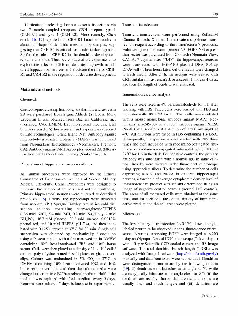
Immunofluorescence analysis showed that a lot of neurons
were MAP2 positive. More than 100 neurons were calcu-
lated. There were about 91% (153 positive cells/168 total
cells) were MAP2 positive. In addition, the staining for
NR2A, a subunit of N-methyl-D-aspartate (NMDA) recep-
tors, was examined to define the cell type. It was found that
84% (147 positive cells/174 total cells) of the cells were
NR2A positive (Fig. 1).
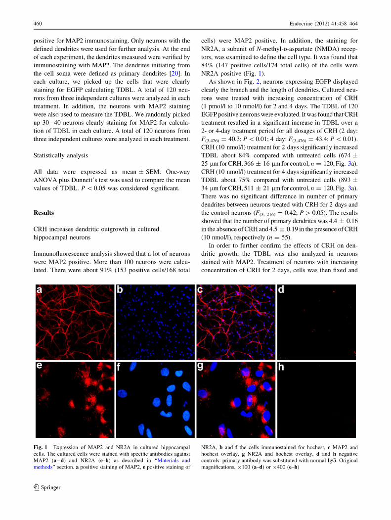
As shown in Fig. 2, neurons expressing EGFP displayed
clearly the branch and the length of dendrites. Cultured neu-
rons were treated with increasing concentration of CRH
(1 pmol/l to 10 nmol/l) for 2 and 4 days. The TDBL of 120
EGFP positive neurons were evaluated. It was found that CRH
treatment resulted in a significant increase in TDBL over a
2- or 4-day treatment period for all dosages of CRH (2 day:
F(3,476) = 40.3; P \ 0.01; 4 day: F(3,476) = 43.4; P \ 0.01).
CRH (10 nmol/l) treatment for 2 days significantly increased
TDBL about 84% compared with untreated cells (674 ±
25 lm for CRH, 366 ± 16 lm for control, n = 120, Fig. 3a).
CRH (10 nmol/l) treatment for 4 days significantly increased
TDBL about 75% compared with untreated cells (893 ±
34 lm for CRH, 511 ± 21 lm for control, n = 120, Fig. 3a).
There was no significant difference in number of primary
dendrites between neurons treated with CRH for 2 days and
the control neurons (F(3, 216) = 0.42; P [ 0.05). The results
showed that the number of primary dendrites was 4.4 ± 0.16
in the absence of CRH and 4.5 ± 0.19 in the presence of CRH
(10 nmol/l), respectively (n = 55).
In order to further confirm the effects of CRH on den-
dritic growth, the TDBL was also analyzed in neurons
stained with MAP2. Treatment of neurons with increasing
concentration of CRH for 2 days, cells was then fixed and
Fig. 1 Expression of MAP2 and NR2A in cultured hippocampal
cells. The cultured cells were stained with specific antibodies against
MAP2 (a-d) and NR2A (e–h) as described in ‘‘Materials and
methods’’ section. a positive staining of MAP2, e positive staining of
NR2A, b and f the cells immunostained for hochest, c MAP2 and
hochest overlay, g NR2A and hochest overlay, d and h negative
controls: primary antibody was substituted with normal IgG. Original
magnifications, 9100 (a–d) or 9400 (e–h)
460 Endocrine (2012) 41:458–464
123
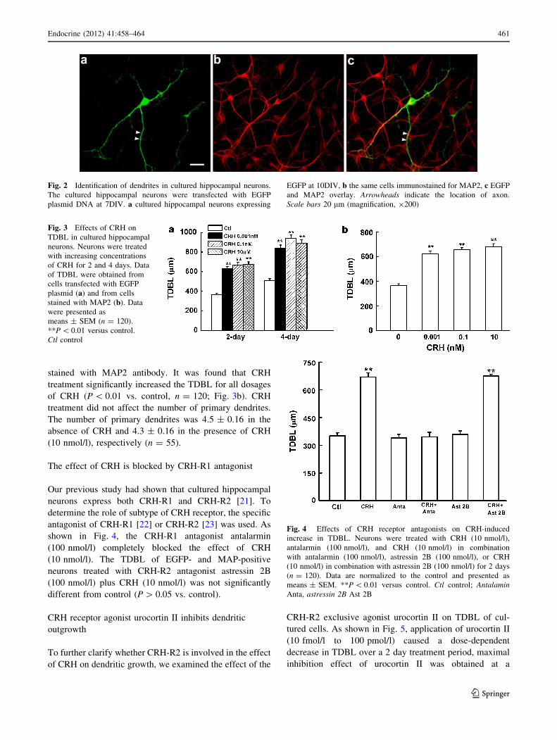
stained with MAP2 antibody. It was found that CRH
treatment significantly increased the TDBL for all dosages
of CRH (P \ 0.01 vs. control, n = 120; Fig. 3b). CRH
treatment did not affect the number of primary dendrites.
The number of primary dendrites was 4.5 ± 0.16 in the
absence of CRH and 4.3 ± 0.16 in the presence of CRH
(10 nmol/l), respectively (n = 55).
The effect of CRH is blocked by CRH-R1 antagonist
Our previous study had shown that cultured hippocampal
neurons express both CRH-R1 and CRH-R2 [21]. To
determine the role of subtype of CRH receptor, the specific
antagonist of CRH-R1 [22] or CRH-R2 [23] was used. As
shown in Fig. 4, the CRH-R1 antagonist antalarmin
(100 nmol/l) completely blocked the effect of CRH
(10 nmol/l). The TDBL of EGFP- and MAP-positive
neurons treated with CRH-R2 antagonist astressin 2B
(100 nmol/l) plus CRH (10 nmol/l) was not significantly
different from control (P [ 0.05 vs. control).
CRH receptor agonist urocortin II inhibits dendritic
outgrowth
To further clarify whether CRH-R2 is involved in the effect
of CRH on dendritic growth, we examined the effect of the
CRH-R2 exclusive agonist urocortin II on TDBL of cul-
tured cells. As shown in Fig. 5, application of urocortin II
(10 fmol/l to 100 pmol/l) caused a dose-dependent
decrease in TDBL over a 2 day treatment period, maximal
inhibition effect of urocortin II was obtained at a
Fig. 2 Identification of dendrites in cultured hippocampal neurons.
The cultured hippocampal neurons were transfected with EGFP
plasmid DNA at 7DIV. a cultured hippocampal neurons expressing
EGFP at 10DIV, b the same cells immunostained for MAP2, c EGFP
and MAP2 overlay. Arrowheads indicate the location of axon.
Scale bars 20 lm (magnification, 9200)
Fig. 3 Effects of CRH on
TDBL in cultured hippocampal
neurons. Neurons were treated
with increasing concentrations
of CRH for 2 and 4 days. Data
of TDBL were obtained from
cells transfected with EGFP
plasmid (a) and from cells
stained with MAP2 (b). Data
were presented as
means ± SEM (n = 120).
**P \ 0.01 versus control.
Ctl control
Fig. 4 Effects of CRH receptor antagonists on CRH-induced
increase in TDBL. Neurons were treated with CRH (10 nmol/l),
antalarmin (100 nmol/l), and CRH (10 nmol/l) in combination
with antalarmin (100 nmol/l), astressin 2B (100 nmol/l), or CRH
(10 nmol/l) in combination with astressin 2B (100 nmol/l) for 2 days
(n = 120). Data are normalized to the control and presented as
means ± SEM. **P \ 0.01 versus control. Ctl control; AntalaminAnta, astressin 2B Ast 2B
Endocrine (2012) 41:458–464 461
123
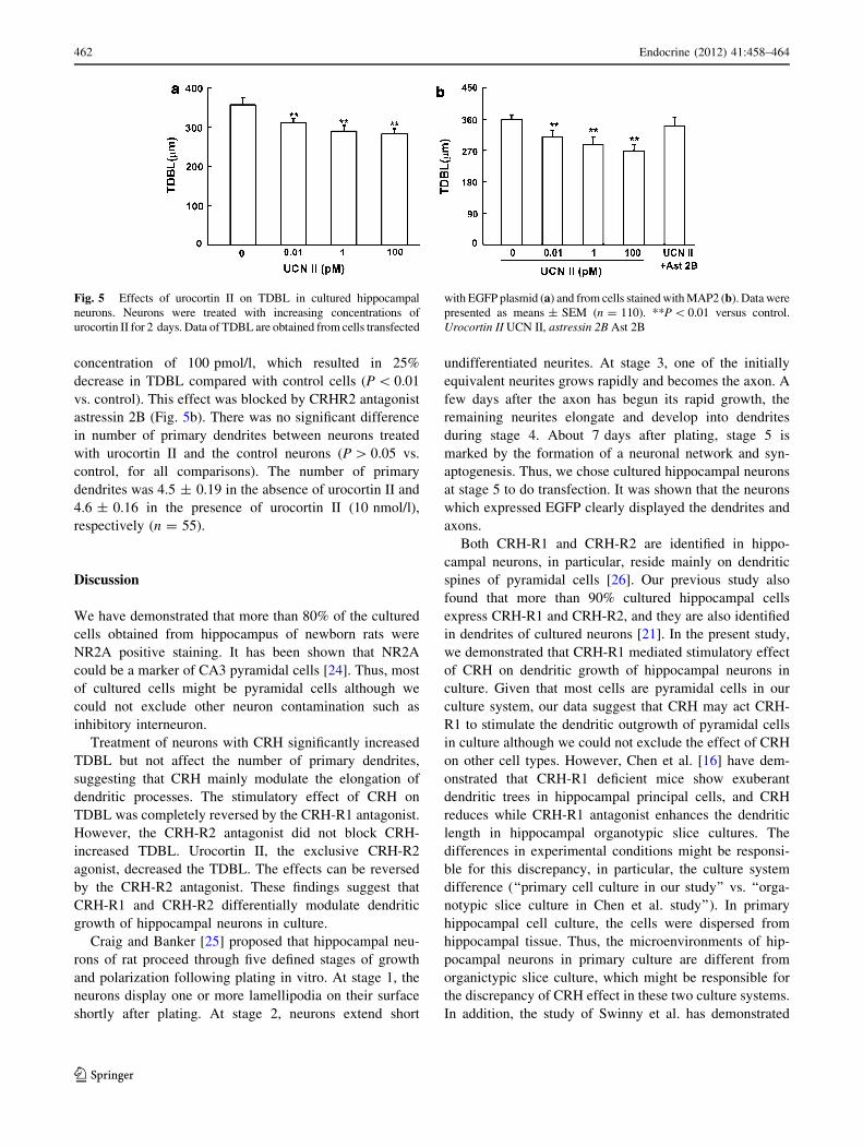
concentration of 100 pmol/l, which resulted in 25%
decrease in TDBL compared with control cells (P \ 0.01
vs. control). This effect was blocked by CRHR2 antagonist
astressin 2B (Fig. 5b). There was no significant difference
in number of primary dendrites between neurons treated
with urocortin II and the control neurons (P [ 0.05 vs.
control, for all comparisons). The number of primary
dendrites was 4.5 ± 0.19 in the absence of urocortin II and
4.6 ± 0.16 in the presence of urocortin II (10 nmol/l),
respectively (n = 55).
Discussion
We have demonstrated that more than 80% of the cultured
cells obtained from hippocampus of newborn rats were
NR2A positive staining. It has been shown that NR2A
could be a marker of CA3 pyramidal cells [24]. Thus, most
of cultured cells might be pyramidal cells although we
could not exclude other neuron contamination such as
inhibitory interneuron.
Treatment of neurons with CRH significantly increased
TDBL but not affect the number of primary dendrites,
suggesting that CRH mainly modulate the elongation of
dendritic processes. The stimulatory effect of CRH on
TDBL was completely reversed by the CRH-R1 antagonist.
However, the CRH-R2 antagonist did not block CRH-
increased TDBL. Urocortin II, the exclusive CRH-R2
agonist, decreased the TDBL. The effects can be reversed
by the CRH-R2 antagonist. These findings suggest that
CRH-R1 and CRH-R2 differentially modulate dendritic
growth of hippocampal neurons in culture.
Craig and Banker [25] proposed that hippocampal neu-
rons of rat proceed through five defined stages of growth
and polarization following plating in vitro. At stage 1, the
neurons display one or more lamellipodia on their surface
shortly after plating. At stage 2, neurons extend short
undifferentiated neurites. At stage 3, one of the initially
equivalent neurites grows rapidly and becomes the axon. A
few days after the axon has begun its rapid growth, the
remaining neurites elongate and develop into dendrites
during stage 4. About 7 days after plating, stage 5 is
marked by the formation of a neuronal network and syn-
aptogenesis. Thus, we chose cultured hippocampal neurons
at stage 5 to do transfection. It was shown that the neurons
which expressed EGFP clearly displayed the dendrites and
axons.
Both CRH-R1 and CRH-R2 are identified in hippo-
campal neurons, in particular, reside mainly on dendritic
spines of pyramidal cells [26]. Our previous study also
found that more than 90% cultured hippocampal cells
express CRH-R1 and CRH-R2, and they are also identified
in dendrites of cultured neurons [21]. In the present study,
we demonstrated that CRH-R1 mediated stimulatory effect
of CRH on dendritic growth of hippocampal neurons in
culture. Given that most cells are pyramidal cells in our
culture system, our data suggest that CRH may act CRH-
R1 to stimulate the dendritic outgrowth of pyramidal cells
in culture although we could not exclude the effect of CRH
on other cell types. However, Chen et al. [16] have dem-
onstrated that CRH-R1 deficient mice show exuberant
dendritic trees in hippocampal principal cells, and CRH
reduces while CRH-R1 antagonist enhances the dendritic
length in hippocampal organotypic slice cultures. The
differences in experimental conditions might be responsi-
ble for this discrepancy, in particular, the culture system
difference (‘‘primary cell culture in our study’’ vs. ‘‘orga-
notypic slice culture in Chen et al. study’’). In primary
hippocampal cell culture, the cells were dispersed from
hippocampal tissue. Thus, the microenvironments of hip-
pocampal neurons in primary culture are different from
organictypic slice culture, which might be responsible for
the discrepancy of CRH effect in these two culture systems.
In addition, the study of Swinny et al. has demonstrated
Fig. 5 Effects of urocortin II on TDBL in cultured hippocampal
neurons. Neurons were treated with increasing concentrations of
urocortin II for 2 days. Data of TDBL are obtained from cells transfected
with EGFP plasmid (a) and from cells stained with MAP2 (b). Data were
presented as means ± SEM (n = 110). **P \ 0.01 versus control.
Urocortin II UCN II, astressin 2B Ast 2B
462 Endocrine (2012) 41:458–464
123
that intermittent exposure of CRH and urocortin induces
more dendrite outgrowth of Purkinje cells in organotypic
cerebellar slice culture. Conversely, constant exposure to
CRH and urocortin inhibits dendritic outgrowth [27]. It
may suggest that the effects of CRH and its related peptides
be dependent on the type of neurons.
Corticotropin-releasing hormone receptors are also
activated by urocortin family of peptides, with urocortin I
having equal affinity for both CRH-R1 and CRH-R2, and
urocortin II and urocortin III having exclusive affinity for
the CRH-R2 [28]. It was found that CRH-R1 activation
exerts the inhibitory effect on the activity of N-methyl-D-
aspartate (NMDA) in hippocampal neurons [21]. CRH-R2
activation potentiates the activity of NMDARs in dopa-
mine neuron in the ventral tegmental area [29]. Our pre-
vious study has demonstrated that the cultured
hippocampal neurons express both CRH-R1 and CRH-R2
[21]. The present study showed that urocortin II inhibited
dendritic growth, and this effect was blocked by CRH-R2
antagonist. Thus, CRH-R1 and CRH-R2 in hippocampal
neurons mediate opposite effect on dendrite growth. At
current stage, the mechanisms responsible for differential
regulation of dendrite growth by CRH-R1 and CRH-R2 are
not known. It has been demonstrated that CRH-R could
couple to multiple G proteins including Gs, Gi, and Gq/11
and then go on to induce multiple signaling pathways [30].
The signaling pathways responsible for the effects of CRH-
R1 and CRH-R2 on dendrite growth should be investigated
in following studies. In addition, it was found that CRH
had much stronger stimulating effect on dendritic out-
growth by doubling the TDBL than urocortin II which only
reduced dendritic elongation by about 20%. It is of inter-
esting to explore the discrepancy in such effects of CRH
and urocortin II in the future.
CRH-R1 and CRH-R2 have been implicated in modu-
lation of learning and memory in responses to stress [30,
31]. In addition, studies by Chen et al. indicate that CRH is
involved in memory defects and hippocampal dendritic
spine loss after acute stress [16, 17, 32]. Our findings that
CRH-R1 and CRH-R2 activation have differential actions
on dendritic growth in hippocampal neurons suggest that
CRH and its related peptides be involved in modulation of
hippocampal synaptic connectivity. Nevertheless, the
effects of CRH-R1 and CRH-R2 on dendritic development
in vivo need to be done in the CRH-R1 and CRH-R2
deficient mice.
In conclusion, CRH stimulates dendritic outgrowth of
cultured hippocampal neurons via CRH-R1. In contrast,
CRH-R2 activation results in an inhibition in dendritic
growth. Our results suggest that CRH-R1 and CRH-R2
differentially modulate the dendritic growth of hippocam-
pal neurons in culture.
Acknowledgment This study was supported by the National Basic
Research Program of China (2007CB512303), Natural Science Foun-
dation of China (No. 30900434 and No. 31100840), and Technology
Commission of Shanghai Municipals (09XD1405600, 09ZR1439800,
and 11ZR1446700). All the experiments in this manuscript comply with
Chinese laws.
Conflict of interest None.
References
1. D.B. Chklovskii, Synaptic connectivity and neuronal morphol-
ogy: two sides of the same coin. Neuron 43, 609–617 (2004)
2. R. Yuste, D.W. Tank, Dendritic integration in mammalian neu-
rons, a century after Cajal. Neuron 16, 701–716 (1996)
3. Y.N. Jan, L.Y. Jan, Branching out: mechanisms of dendritic
arborization. Nat. Rev. Neurosci. 11, 316–328 (2010)
4. H.T. Cline, Dendritic arbor development and synaptogenesis.
Curr. Opin. Neurobiol. 11, 118–126 (2001)
5. L.I. Zhang, M.M. Poo, Electrical activity and development of
neural circuits. Nat. Neurosci. 4(Suppl), 1207–1214 (2001)
6. F. Metzger, Molecular and cellular control of dendrite maturation
during brain development. Curr. Mol. Pharmacol. 3, 1–11 (2010)
7. A.W. Moore, Intrinsic mechanisms to define neuron class-specific
dendrite arbor morphology. Cell Adhes. Migr. 2, 81–82 (2008)
8. F. Henle, C. Fischer, D.K. Meyer, J. Leemhuis, Vasoactive
intestinal peptide and PACAP38 control N-methyl-D-aspartic
acid-induced dendrite motility by modifying the activities of Rho
GTPases and phosphatidylinositol 3-kinases. J. Biol. Chem. 281,
24955–24969 (2006)
9. A.M. Magarinos, C.J. Li, J.G. Toth, K.G. Bath, D. Jing, F.S. Lee,
B.S. McEwen, Effect of brain-derived neurotrophic factor hap-
loinsufficiency on stress-induced remodeling of hippocampal
neurons. Hippocampus (2010) (Epub ahead of print)
10. P.R. Moult, J. Harvey, Hormonal regulation of hippocampal
dendritic morphology and synaptic plasticity. Cell Adhes. Migr.
2, 269–275 (2008)
11. D.N. Alfarez, A. De Simoni, E.H. Velzing, E. Bracey, M. Joels,
F.A. Edwards, H.J. Krugers, Corticosterone reduces dendritic
complexity in developing hippocampal CA1 neurons. Hippo-
campus 19, 828–836 (2009)
12. D.A. Tata, B.J. Anderson, The effects of chronic glucocorticoid
exposure on dendritic length, synapse numbers and glial volume
in animal models: implications for hippocampal volume reduc-
tions in depression. Physiol. Behav. 99, 186–193 (2010)
13. M.J. Owens, C.B. Nemeroff, Physiology and pharmacology of
corticotropin-releasing factor. Pharmacol. Rev. 43, 425–473
(1991)
14. J.P. Gallagher, L.F. Orozco-Cabal, J. Liu, P. Shinnick-Gallagher,
Synaptic physiology of central CRH system. Eur. J. Pharmacol.
583, 215–225 (2008)
15. G. Cibelli, P. Corsi, G. Diana, F. Vitiello, G. Thiel, Corticotropin-
releasing factor triggers neurite outgrowth of a catecholaminergic
immortalized neuron via cAMP and MAP kinase signalling
pathways. Eur. J. Neurosci. 13, 1339–1348 (2001)
16. Y. Chen, R.A. Bender, K.L. Brunson, J.K. Pomper, D.E. Gri-
goriadis, W. Wurst, T.Z. Baram, Modulation of dendritic differ-
entiation by corticotropin-releasing factor in the developing
hippocampus. Proc. Natl. Acad. Sci. USA 101, 15782–15787
(2004)
17. Y. Chen, C.M. Dube, C.J. Rice, T.Z. Baram, Rapid loss of den-
dritic spines after stress involves derangement of spine dynamics
Endocrine (2012) 41:458–464 463
123
by corticotropin-releasing hormone. J. Neurosci. 28, 2903–2911
(2008)
18. T.E. Nelson, D.L. Gruol, The chemokine CXCL10 modulates
excitatory activity and intracellular calcium signaling in cultured
hippocampal neurons. J. Neuroimmunol. 156, 74–87 (2004)
19. C.G. Dotti, C.A. Sullivan, G.A. Banker, The establishment of
polarity by hippocampal neurons in culture. J. Neurosci. 8,
1454–1468 (1988)
20. P. Salama-Cohen, M.A. Arevalo, R. Grantyn, A. Rodrıguez-
Tebar, Notch and NGF/p75NTR control dendrite morphology and
the balance of excitatory/inhibitory synaptic input to hippocampal
neurones through Neurogenin 3. J. Neurochem. 97, 1269–1278
(2006)
21. H. Sheng, Y. Zhang, J. Sun, L. Gao, B. Ma, J. Lu, X. Ni, Cor-
ticotropin-releasing hormone (CRH) depresses N-methyl-D-
aspartate receptor-mediated current in cultured rat hippocampal
neurons via CRH receptor type 1. Endocrinology 149, 1389–1398
(2008)
22. I.R. Young, E.C. Chan, R. Smith, G.P. Chrousos, J.D. Veldhuis,
B.J. Canny, Effect of antalarmin, a novel corticotropin-releasing
hormone antagonist, on the dynamic function of the preterm
ovine fetal hypothalamo-pituitary-adrenal axis. Neuroendocri-
nology 76, 47–54 (2002)
23. M.W. Lewis, G.E. Hermann, R.C. Rogers, R.A. Travagli, In vitro
and in vivo analysis of the effects of corticotropin releasing factor
on rat dorsal vagal complex. J. Physiol. 543, 135–146 (2002)
24. J.M. Fritschy, O. Weinmann, A. Wenzel, D. Benke, Synapse-
specific localization of NMDA and GABA(A) receptor subunits
revealed by antigen-retrieval immunohistochemistry. J. Comp.
Neurol. 390, 194–210 (1998)
25. A.M. Craig, G. Banker, Neuronal polarity. Annu. Rev. Neurosci.
17, 267–310 (1994)
26. Y. Chen, K.L. Brunson, G. Adelmann, R.A. Bender, M. Frot-
scher, T.Z. Baram, Hippocampal corticotropin releasing hor-
mone: pre- and postsynaptic location and release by stress.
Neuroscience 126, 533–540 (2004)
27. J.D. Swinny, F. Metzger, J. IJkema-Paassen, N.V. Gounko, A.
Gramsbergen, J.J. van der Want, Corticotropin-releasing factor
and urocortin differentially modulate rat Purkinje cell dendritic
outgrowth and differentiation in vitro. Eur. J. Neurosci. 19,
1749–1758 (2004)
28. K. Gysling, M.I. Forray, P. Haeger, C. Daza, R. Rojas, Cortico-
tropin-releasing hormone and urocortin: Redundant or distinctive
functions? Brain Res. Brain Res. Rev. 47, 116–125 (2004)
29. M.A. Ungless, V. Singh, T.L. Crowder, R. Yaka, D. Ron, A.
Bonci, Corticotropin-releasing factor requires CRF binding pro-
tein to potentiate NMDA receptors via CRF receptor 2 in dopa-
mine neurons. Neuron 39, 401–407 (2003)
30. T. Blank, I. Nijholt, D.K. Grammatopoulos, H.S. Randeva, E.W.
Hillhouse, J. Spiess, Corticotropin-releasing factor receptors cou-
ple to multiple G-proteins to activate diverse intracellular signaling
pathways in mouse hippocampus: role in neuronal excitability and
associative learning. J. Neurosci. 23, 700–707 (2003)
31. A. Schierloh, J. Deussing, W. Wurst, W. Zieglgansberger, G.
Rammes, Corticotropin-releasing factor (CRF) receptor type
1-dependent modulation of synaptic plasticity. Neurosci. Lett.
416, 82–86 (2007)
32. Y. Chen, C.S. Rex, C.J. Rice, C.M. Dube, C.M. Gall, G. Lynch,
T.Z. Baram, Correlated memory defects and hippocampal den-
dritic spine loss after acute stress involve corticotropin-releasing
hormone signaling. Proc. Natl. Acad. Sci. USA 107, 13123–13128
(2010)
464 Endocrine (2012) 41:458–464
123