Embed Size (px)
Citation preview
CRISPR-Cas9 Based Engineering of Actinomycetal GenomesYaojun Tong,† Pep Charusanti,†,‡ Lixin Zhang,∥ Tilmann Weber,*,† and Sang Yup Lee*,†,§
†The Novo Nordisk Foundation Center for Biosustainability, Technical University of Denmark, Kogle Alle 6, Hørsholm 2970,Denmark‡Department of Bioengineering, University of California, San Diego, La Jolla, California 92093, United States§Metabolic and Biomolecular Engineering National Research Laboratory, Department of Chemical and Biomolecular Engineering(BK21 Plus Program), Center for Systems and Synthetic Biotechnology, Institute for the BioCentury, Korea Advanced Institute ofScience and Technology (KAIST), Daejeon 305-701, Republic of Korea∥Chinese Academy of Sciences, Key Laboratory of Pathogenic Microbiology and Immunology, Institute of Microbiology, Beijing100190, China
*S Supporting Information
ABSTRACT: Bacteria of the order Actinomycetales are one ofthe most important sources of pharmacologically active andindustrially relevant secondary metabolites. Unfortunately,many of them are still recalcitrant to genetic manipulation,which is a bottleneck for systematic metabolic engineering. Tofacilitate the genetic manipulation of actinomycetes, wedeveloped a highly efficient CRISPR-Cas9 system to deletegene(s) or gene cluster(s), implement precise gene replace-ments, and reversibly control gene expression in actino-mycetes. We demonstrate our system by targeting two genes,actIORF1 (SCO5087) and actVB (SCO5092), from theactinorhodin biosynthetic gene cluster in Streptomyces coelicolorA3(2). Our CRISPR-Cas9 system successfully inactivated the targeted genes. When no templates for homology-directed repair(HDR) were present, the site-specific DNA double-strand breaks (DSBs) introduced by Cas9 were repaired through the error-prone nonhomologous end joining (NHEJ) pathway, resulting in a library of deletions with variable sizes around the targetedsequence. If templates for HDR were provided at the same time, precise deletions of the targeted gene were observed with near100% frequency. Moreover, we developed a system to efficiently and reversibly control expression of target genes, deemedCRISPRi, based on a catalytically dead variant of Cas9 (dCas9). The CRISPR-Cas9 based system described here comprises apowerful and broadly applicable set of tools to manipulate actinomycetal genomes.
KEYWORDS: CRISPR-Cas9, CRISPRi, DNA repair, actinomycetes, genome engineering
Actinomycetes are Gram-positive bacteria with the capacityto produce a wide variety of medically and industrially
relevant secondary metabolites,1−3 including many well-knownantibiotics, herbicides, chemotherapeutics, and immunosup-pressants, such as vancomycin, bialaphos, doxorubicin, andrapamycin, respectively. It is very challenging to find novelsecondary metabolites with properties suitable as drug leads asthe same known molecules are often rediscovered repeatedlywhen using traditional approaches. However, there currently isa renaissance in investigating this group of bacteria as studiesemploying modern genome mining techniques4 indicate thatthey still harbor a huge unexploited potential to producesecondary metabolites with novel structures.5 One of the mainchallenges of this genome-driven approach is to metabolicallyengineer the strains to express the biosynthetic pathways and toproduce the compounds in high titers.6 Unfortunately, geneticmanipulation of actinomycetes is much more difficult comparedto model organisms, such as Escherichia coli and Saccharomycescerevisiae, due in part to their more diverse genomic contents
and the extremely high GC content of their genomes. Thecommon gene replacement technique for actinomycetes usesRecA mediated double-crossover events with nonreplicative ortemperature sensitive plasmids.7 While the efficacy of thisapproach has been drastically increased by using templates withvery long homology regions, such as in the “ReDirectmethod”,8 or using meganuclease I-SceI to introduce DNADSBs as an additional positive selection marker,9 the protocolsstill are labor-intensive and relatively slow.In other organisms, several alternative methods that allow
precise genome editing have been developed. For example, zincfinger nucleases can be engineered to target custom sequencesin human cell lines.10 Transcription activator-like effectors(TALEs) are another example; they possess a highly modularDNA binding motif that can be engineered to bind to specificsequences in the target genomes.11 More recently, genetic
Received: February 22, 2015Published: March 25, 2015
Research Article
pubs.acs.org/synthbio
© 2015 American Chemical Society 1020 DOI: 10.1021/acssynbio.5b00038ACS Synth. Biol. 2015, 4, 1020−1029

manipulation systems based on the clustered regularlyinterspaced short palindromic repeats (CRISPR)/CRISPR-associated (Cas) proteins belonging to the bacterial adaptiveimmune system have garnered widespread attention. There arethree different types of CRISPR systems.12 Type I and type IIICRISPR systems require multiple Cas proteins to induce thecleavage of their target DNA. Type II CRISPR systems, such asthose found in Streptococcus pyogenes, in contrast only require asingle Cas protein, Cas9 (an endonuclease), for activity. Thetarget sequences are defined in the CRISPR loci containingshort repeats separated by “spacer” sequences that exactlymatch the genomes of bacteriophages and other mobile geneticelements offering adaptive immunity to the previously exposedbacteriophages or other mobile genetic elements, and thus areregarded as bacterial adaptive immune systems.13−17 In thenative type II CRISPR system, the spacer sequence of theCRISPR array transcribes to CRISPR RNA (crRNA).Subsequently, an associated trans-activating CRISPR RNA(tracrRNA) hybridizes with the crRNA, forming an RNAduplex, which is cleaved and further processed by endogenousRNase III and possibly other, yet unknown nucleases.18 ThecrRNA-tracrRNA duplex interacts with Cas9 to form acomplex, and scans the target genome for the presence of
trinucleotide protospacer adjacent motifs (PAMs) (Figure 1A).When this complex finds a PAM that has a 5′ sequencecomplementary to the spacer sequence in the crRNA-tracrRNAduplex, it binds to this position and then triggers the nucleaseactivity by activating the HNH and RuvC domains of Cas9.19,20
In further studies, Cas9 has shown to be a highly versatiletool to also introduce DNA DSBs into other genomic DNA bycoexpression with customized single guide RNAs (sgRNAs),which are artificially generated chimeras of the crRNA and thetracrRNA found in the native CRISPR systems17 (Figure 1A).This engineered type II CRISPR is referred to as CRISPR-Cas9system.21 Because of its modularization and easy handling, theCRISPR-Cas9 system has been successfully applied as agenome editing tool in a wide range of organisms such as S.cerevisiae,22 some plants,23 Caenorhabditis elegans,24 Drosophi-la,25 Chinese hamster ovary (CHO) cells,26 mice,27 rats,28
rabbits,29 and human cells.30 Studies also demonstrated that theCRISPR-Cas9 system can be multiplexed by introducing abattery of sgRNAs that simultaneously target multiple differentregions in the genome.31 Recently, a CRISPR based system,pCRISPomyces was developed for streptomycetes geneticmanipulation.32 However, this method only harnesses theDSBs introduced by Cas9 nuclease to mediate the homologous
Figure 1. Schematic illustration of the CRISPR-Cas9 system. (A) Diagram of the Cas9 and sgRNA complex. The Cas9 HNH and RuvC-likedomains each cleave one strand of the sequence targeted by the sgRNA; the PAM is shown in red; the 20 nt target sequence is shown in green; thesgRNA core structure is shown in orange. (B) Map of pCRISPR-Cas9, the backbone is a temperature sensitive pSG5 replicon, cas9 is controlled bythe thiostrepton inducible tipA promoter, the sgRNA cassette is under control of the ermE* promoter, apramycin serves as the selection marker. Thisplasmid can shuttle between E. coli and actinomycetes. (C) Magnification of the design of sgRNA scaffold in pCRISPR-Cas9.
ACS Synthetic Biology Research Article
DOI: 10.1021/acssynbio.5b00038ACS Synth. Biol. 2015, 4, 1020−1029
1021
recombination events, but does not fully implement thefeatures of CRISPR-Cas9 system.Besides introduction of DSBs, it was shown that mutating the
HNH and RuvC domains of Cas9 (D10A and H840A) resultedin a catalytically dead Cas9 (dCas9) that did not haveendonuclease activity, but could still form a complex withsgRNA and efficiently bind to the target DNA.17 This effect canbe used to sequence-specifically interfere with transcription andthus control gene expression. In analogy to eukaryotic RNAinterference RNAi, this system was named as CRISPRi.33
Here we report a CRISPR-Cas9 system for manipulation ofactinomycetal genomes. As in the previous report,32 our systemcan be used to create precise deletions in the genomes ofstreptomycetes via double-crossover with homologous recom-bination templates. Moreover, we report additional capabilitiesof our system, specifically that it functions even in the absenceof homology templates, which results in a distribution offragment deletion sizes, and that it can be redesigned toreversibly control gene expression.
■ RESULTS AND DISCUSSION
Design of a One Plasmid-Based CRISPR-Cas9 System.The temperature sensitive vector pGM1190, a derivative ofpGM190,34 whose selection marker is apramycin instead of
kanamycin, was used as a backbone to harbor both componentsof our CRISPR-Cas9 system, the nuclease Cas9/dCas9 and the“homing device” sgRNA, which was constructed by fusing acrRNA and an associated tracrRNA17 (Figure 1A). Thereplication systems of this E. coli-actinomycetes shuttle vectorare based on pMB1 and pSG5.35 The nuclease Cas9/dCas9 isunder control of the thiostrepton inducible tipA promoter,which requires the presence of the thiostrepton responsiveactivator TipA.36 The gene encoding this protein, tipA, is notpresent in pGM1190, but is present in the genomes of mostStreptomyces, including Streptomyces coelicolor.37 Therefore, itwould be necessary to clone tipA into pGM1190 or change thepromoter system prior to the use of our system inactinomycetes lacking a native tipA gene. For easy handling,the 20 nt target sequence within the sgRNA scaffold wasdesigned to be inserted into the NcoI and SnaBI restriction sites(Figure 1C). The StuI restriction site within the pCRISPR-Cas9vector is available for inserting other elements (Figure 1B), forinstance to subclone a homologous recombination template forprecise gene deletion, or the ScaligD expression cassette toreconstitute the incomplete NHEJ pathway. Because of itstemperature sensitivity, the plasmids can be eliminated fromactinomycete cells by incubating cells above 34 °C.
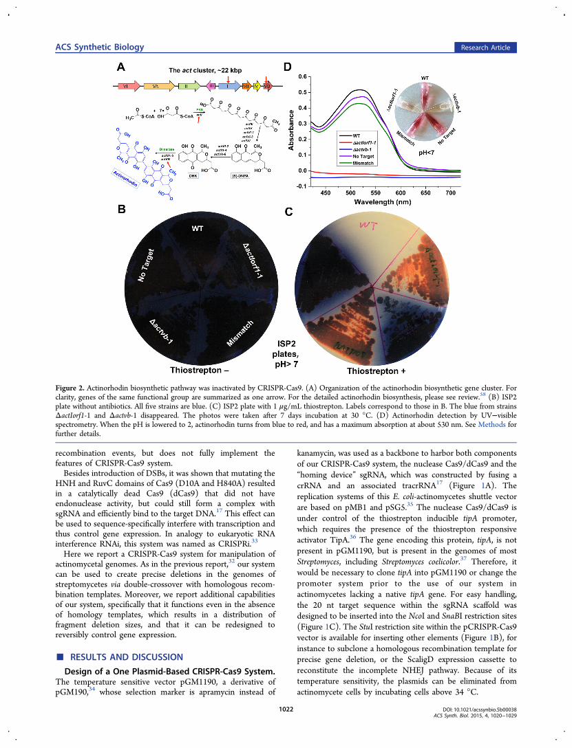
Figure 2. Actinorhodin biosynthetic pathway was inactivated by CRISPR-Cas9. (A) Organization of the actinorhodin biosynthetic gene cluster. Forclarity, genes of the same functional group are summarized as one arrow. For the detailed actinorhodin biosynthesis, please see review.58 (B) ISP2plate without antibiotics. All five strains are blue. (C) ISP2 plate with 1 μg/mL thiostrepton. Labels correspond to those in B. The blue from strainsΔactIorf1-1 and Δactvb-1 disappeared. The photos were taken after 7 days incubation at 30 °C. (D) Actinorhodin detection by UV−visiblespectrometry. When the pH is lowered to 2, actinorhodin turns from blue to red, and has a maximum absorption at about 530 nm. See Methods forfurther details.
ACS Synthetic Biology Research Article
DOI: 10.1021/acssynbio.5b00038ACS Synth. Biol. 2015, 4, 1020−1029
1022
Actinorhodin Biosynthesis Was Successfully Interrup-ted by Targeting Genes within the Pathway with theCRISPR-Cas9 System. To validate the CRISPR-Cas9 system,we chose to inactivate two genes, actIORF1 and actVB, from thebiosynthetic pathway of the blue-pigmented polyketide anti-biotic actinorhodin in S. coelicolor (Figure 2A). sgRNAs wereidentified with a modified version of the software CRISPRy26
(http://staff.biosustain.dtu.dk/laeb/crispy_scoeli/). On thebasis of the predictions, 6 sgRNAs with no predicted off-targeteffects were selected for each of the two genes (Table S3,Supporting Information). The sgRNAs were subcloned intopCRISPR-Cas9, and after sequence validation, one correctplasmid for each sgRNA was randomly selected to betransferred into the ET12567/pUZ8002 E. coli strain forconjugation. No template for HDR was used at this stage. Inaddition, three negative controls were used. The first one wasan “empty vector” in which the 20 nt target sequence of sgRNAis missing, resulting in no expected target match in the genome.This control was named “No Target”. The second onecontained a 23 nt sgRNA composed of the 20 nt targetsequence of actIORF1 extended by the 3 nt PAM motif “NGG”.The inclusion of the PAM as part of the sgRNA abolishescorrect recognition of the genomic target and therefore wascalled “Mismatch”. The last one is the wild-type (WT).After conjugation, 30−80 colonies transformed with each
sgRNA were randomly picked onto two different sets of ISP2plates. Only one set was supplemented with 1 μg/mLthiostrepton that induces Cas9 expression. The ratio of clonesthat lost actinorhodin production over the total number ofclones on the induction plates was used to calculate theefficiency for each sgRNA. The inactivation efficiency variedfrom 3 to 54%, depending on the different sgRNAs (Table 1).
Two clones for each target gene that lost blue pigmentformation after induction were randomly selected, namedΔactIorf1-1, ΔactIorf1-2 and Δactvb-1, Δactvb-2, respectively,and were further investigated together with the three negativecontrols, “No Target”, “Mismatch”, and the WT (Table S1).When they were transferred to new agar plates without theinducer thiostrepton, still no blue pigment was observed for thestrains carrying sgRNAs targeting actIORF1 and actVB, whilethe control strains were dark blue (only ΔactIorf1-1, Δactvb-1and 3 controls are shown in Figure 2, and Figure S1). Thevisual analysis on the agar plates was also confirmed bycultivating the clones in liquid ISP2 medium and detectingactinorhodin production with UV−visible spectroscopy (Figure2D). These observations indicated that the CRISPR-Cas9system indeed inactivated the actIORF1 and actVB genes, sothat the actinorhodin biosynthesis was interrupted.As mentioned above, the efficiency with which the different
sgRNAs yielded clones with abolished actinorhodin productionvaried from 3 to 54% (Table 1). This low efficiency was due tothe absence of a homology-directed repair template. Survivalwas most likely due to repair of the DNA DSBs with theimperfect NHEJ pathway.
Random Sized Deletions Caused by Imperfect NHEJin Actinomycetes. To unambiguously confirm that theobserved phenotype is due to the targeted inactivation of thetwo actinorhodin biosynthetic genes, but not due to unwantedside- or off-target effects, we sequenced the whole genomes ofthe above selected 7 strains using Illumina technology in orderto map the sites of mutation. Comparison of the S. coelicolorA3(2) strain used in this study (WT) with the S. coelicolorA3(2) reference sequence (Genbank: AL645882), resulted in95 SNPs (Table S4) and 1 fragment (5797650−5818686)deletion, which corresponds to the loss of an integrated plasmidin our WT strain. Subsequently, the sequence of our WT strainwas used as the mapping reference for the other strains. Thedetailed mapping results of the analysis are shown in Table S5and Figure 3A.Interestingly, the whole genome sequencing analysis
indicated the inactivation of actIORF1 and actVB was notuniform. In one of the investigated clones where actIORF1 wastargeted, the inactivation was caused by a 1 bp insertion (Figure3B). In another case (actVB was targeted), a deletion of morethan 30 000 bp around the DSB position was obtained (Figure3A). In other words, the deletions were precisely placed at thetargeted sites, but varied in size. Importantly, this observationconfirmed that a NHEJ pathway indeed exists in actinomycetes,but its efficiency is much lower than in eukaryoticorganisms.38,39 Thus, this feature can be exploited to easilygenerate a set of random-sized deletions around a preciselydefined target position through a single experiment using theCRISPR-Cas9 system.
Optimizing the Intrinsic NHEJ Pathway by Coex-pression of a LigD Ligase. It was hypothesized that theefficiency of the CRISPR-Cas9 system could be improved in S.coelicolor by coexpressing a DNA ligase D (LigD). Analysis ofactinomycetal genome sequences showed that most actino-mycetes lack LigD, a core component of the NHEJ pathway,which is present, for example, in Mycobacterium tuberculosis.A BLAST search using the M. tuberculosis LigD (MtbligD) as
a query against the NCBI nonredundant database revealed thatthere currently is only one known LigD homologue ofstreptomycete origin, which is found in Streptomyces carneus(NCBI GenPept ID WP_033244577.1). The amino acid
Table 1. Inactivation Efficiency of Different sgRNAs withDifferent DSB Repair Pathways
colony countaefficiency
(%)
ways of DSBrepair sgRNAs
nogrowth redb blue total red/total
incompleteNHEJ
ActIorf1-1 NT 20 31 30 81 38
ActIorf1-2 T 3 1 7 11 9ActIorf1-3 T 7 18 49 74 24ActIorf1-4 T 43 10 1 54 19ActIorf1-5 T 8 18 8 34 53Actvb-1 NT 10 20 22 52 38Actvb-3 T 17 6 40 63 10Actvb-4 T 30 6 5 41 15Actvb-5 NT 7 20 10 37 54Actvb-6 NT 1 1 30 32 3ActIorf1-6 T 10 18 12 40 45Actvb-2 NT 20 13 2 35 37
reconstitutedNHEJ
ActIorf1-6 T 0 24 7 31 77
Actvb-2 NT 0 18 8 26 69HDR (withhomologytemplates)
ActIorf1-6 T 0 52 0 52 100
Actvb-2 NT 0 35 1 36 97
aDenotes the number of colonies with the indicated phenotype afterinduction with thiostrepton. bActinorhodin is blue. Upon loss ofactinorhodin production, the red color of the 2nd pigmentedantibiotic, undecylprodigiosin, becomes visible.
ACS Synthetic Biology Research Article
DOI: 10.1021/acssynbio.5b00038ACS Synth. Biol. 2015, 4, 1020−1029
1023

identity and similarity to MtbligD is 56 and 67%, respectively.To coexpress it in our CRIPSR-Cas9 system, the expressioncassette ScaligD was designed. An internal NcoI site wasremoved from the S. carneus ligD gene by introducing thesynonymous substitution C606G. The construct was synthe-sized by Genscript and subcloned into the StuI site ofpCRISPR-Cas9 by Gibson assembly. This modified systemtogether with one sgRNA for each of the two target genes(sgRNA: ActIorf1-6 T for actIORF1, and sgRNA: Actvb-2 NT
for actVB) were used to investigate how the inclusion ofScaligD affected the inactivation efficiency; the efficiencyincreased from 45 to 77% for the sgRNA ActIorf1-6 T andfrom 37 to 69% for Actvb-2 NT (Table 1). To further validatethe observation, primers were designed to amplify the ∼600 bpfragment containing the theoretical cleavage sites of the usedsgRNAs. Eight red clones for each gene were randomly selectedfor colony PCR and subsequent Sanger sequencing. No largedeletions were found in any the 16 sequenced clones; most had
Figure 3. Analysis of the sequencing data. (A) Heatmap of the 7 mapped sequencing samples to the S. coelicolor A3(2) reference genome in a nativeNHEJ system. Dark colors represent a high read coverage, white represents low/no coverage. Displayed is the region spanning 5508800 to 5557230of the S. coelicolor genome. The actinorhodin gene cluster is denoted by red brackets; the target sites of the actIORF1 and actVB sgRNAs aredisplayed as red and blue arrows. The deletion sizes are shown on the map. (B) Alignment of the sequence traces of ΔactIorf1-1 with the WT. Theblue arrow indicates the genomic target site of the sgRNA: ActIorf1-6 T. The PAM sequence is shown in red. (C, D) DNA sequences of 8 randomlyselected red clones aligned to the WT genomic sequence of actIORF1 and actVB, respectively, in a reconstituted NHEJ system. The blue arrowindicates the genomic target sites of the related sgRNAs. The PAM sequences are shown in red. Green, yellow and light blue colors indicateinsertions, deletions and substitutions, respectively.
Figure 4. HDR pathway to repair the DNA DSBs caused by CRISPR-Cas9 system. (A, B) Diagrams of the CRISPR-Cas9 vectors with homologousrecombination templates for actIORF1 and actVB. (C, D) Colony PCR of 10 randomly selected clones that lost actinorhodin production to confirmdeletion of actIORF1 (C) and actVB (D) after use of the two vectors in A and B.
ACS Synthetic Biology Research Article
DOI: 10.1021/acssynbio.5b00038ACS Synth. Biol. 2015, 4, 1020−1029
1024

1 to 3 bp deletions, substitutions, or insertions (Figure 3C, andFigure 3D). For comparison, three out of four clones that lostactinorhodin production were found to have large deletionswhen ScaligD was not included (Figure 3A). These resultsindicated that the incomplete NHEJ pathway was successfullyreconstituted by complementing the missing DNA ligase D.Precise Gene Deletion in Actinomycetes Using HDR.
We next investigated whether the presence of a homologousrecombination template could improve the efficiency andfidelity of the CRISPR-Cas9 system even further, ashomologous recombination is used regularly to target genereplacement and deletion in a wide range of organismsincluding actinomycetes.40 A ∼2.2 kbp homologous recombi-nation template for each of the two target genes (actIORF1 andactVB) was amplified by PCR, assembled and subcloned intothe StuI site of pCRISPR-Cas9. The same sgRNAs (sgRNA:ActIorf1-6 T for actIORF1, and sgRNA: Actvb-2 NT for actVB)were selected for testing. All 52 clones randomly picked foractIORF1, and 35 out of 36 clones randomly picked for actVBlost actinorhodin production after induction (Table 1). Ten redclones were randomly selected for colony PCR validation forboth genes. The sizes of all 20 PCR products obtained in thereactions corresponded to the predicted sizes of the genedeletion (Figure 4), which were confirmed by Sangersequencing. Importantly, the CRISPR-Cas9 system using thehomologous recombination templates showed even higherefficiency and precision when compared to the gene deletionsystem involving ScaligD (Table 1).An Effective and Reversible CRISPRi System for Gene
Expression Controls in S. coelicolor. In some instances, thereversible repression of gene expression is preferred over genedeletion. As such, we tested whether a modified system,referred to as CRISPRi, could be adapted for use to controlgene expression in actinomycetes. Again, the actIORF1 genewas selected as the test case. Since the CRISPRy software usedto identify sgRNAs for gene deletion can only identify targetsequences within the coding regions, a different sgRNA finder
software, sgRNAcas9,41 was used to identify the sgRNAs fromnoncoding regions for the CRISPRi system. Six sgRNAs thattargeted the promoter region and another six targeting the ORFof actIORF1 were selected. In both cases, three of six targetedthe template strand DNA and the other three targeted thenontemplate strand (Table S3 and Figure 5A). In clonescarrying sgRNAs targeting the promoter region, actinorhodinproduction was abolished or dramatically reduced regardless ofwhether they targeted the template or nontemplate strand. Inclones carrying sgRNAs that targeted the ORF region, loss of orreduced actinorhodin production was only observed when thesgRNAs targeted the nontemplate strand (Figure 5B, C). Totest whether the effect was reversible, the loss of the pCRISPR-dCas9 plasmid was induced by increasing the incubationtemperature to 37 °C for 24 h and then transferring thecultures to fresh ISP2 plates without antibiotic selection. Afterincubation for 5 days at 37 °C, the colonies had regained theirdark blue color (Figure 5C, D), indicating that the repression ofactinorhodin biosynthesis by the CRISPR-dCas9 (CRISPRi)system was abrogated and all genes of the actinorhodinbiosynthetic pathway were expressed again.
Discussion. The classical method for actinomycete genemanipulation is double-crossover based gene replacements withnonreplicative or temperature sensitive plasmids.7 Theefficiency of the mutagenesis can be highly improved byusing long homology regions in the gene inactivation plasmids.8
In this protocol, cosmids/fosmids with inserts covering thetarget region are engineered in E. coli using Rec/ETrecombineering and then used as very long templates (up to20 kbp homology arms) for homologous recombinationmediated gene replacement. Although this method increasesthe frequency of obtaining double-crossovers significantly, thegeneration of such constructs is very laborious and the overallefficiency is still relatively low. Thus, alternative systems are ofgreat interest for the actinomycetes research community.One system that has gained recent attention is based on
CRISPR-Cas9, which is an antiphage defense mechanism in
Figure 5. CRISPRi effectively silences actIORF1 expression in a reversible manner. (A) Location of the 12 sgRNAs for CRISPRi. Half were designedto target the promoter region, while the other half were designed to target the ORF. In addition, half target the template strand and half target thenontemplate strand. (B) The 530 nm absorbance of extracts from cultures tested with the 12 sgRNAs shown in A relative to the wild-type control.Mean values from three independent extractions are shown. Error bars represent the standard deviation from three independent extractions. (C, D)Reversibility of the CRISPRi system. Red clones become blue when the incubation temperature is increased to 37 °C, indicating that the CRISPRieffect has gone away.
ACS Synthetic Biology Research Article
DOI: 10.1021/acssynbio.5b00038ACS Synth. Biol. 2015, 4, 1020−1029
1025

bacteria and archaea. Two core components of this system arethe nuclease Cas9 and its “homing device” sgRNA. The sgRNAsequence originally corresponded to phage sequences, but it hasbeen shown that it can be replaced by a sequence of interest toreguide the Cas9 nuclease for genome editing.21 Because of itsmodularity and simplicity, CRISPR-Cas9 system has beensuccessfully adapted for genome editing in an increasingnumber of organisms, mainly eukaryotic hosts and somebacteria.32
We observed nearly 100% efficiency in precise gene deletionin S. coelicolor when CRISPR-Cas9 was used in tandem with ahomology-based repair template (Figure 4). The observedefficiency is in very good agreement with data reported fromthe recently developed pCRISPomyces-system,32 anotherCRISPR-based system for actinomycetes that uses a similarstrategy.DNA DSBs of the chromosome are lethal events in both
prokaryotic and eukaryotic organisms unless the lesion isrepaired by HDR or NHEJ.42,43 Therefore, the use of CRISPR-Cas9 to cleave the host’s chromosome was recently proposed asan antimicrobial application44−47 as many pathogenic bacteriacannot repair chromosomal DSBs by NHEJ. However, in thisstudy, we observed that S. coelicolor can survive after the DNADSBs introduced by Cas9 even in the absence of a homologousrecombination template. Resulting colonies, however, con-tained deletions of variable sizes around the target site. In ourstudy for instance, one single sgRNA generated deletionsranging from 1 bp to more than 10 000 bp. This property canbe exploited and used to generate a set of random sizeddeletions originating from a single sgRNA-directed target onthe genome, which would be valuable to generate genomeminimized hosts or to identify essential genes in actinomycetes.When a DNA ligase D homologue from S. carneus wascoexpressed, the efficiency of the NHEJ pathway wassignificantly improved. This enables the use of the CRISPR-Cas9 system in an analogous way as in eukaryotes, where nohomologous template has to be provided to generate targetedmutations.In addition to using CRISPR-Cas9 system as a genome
editing tool, it has recently been engineered to transientlycontrol gene expression, a system referred to as CRISPRi inanalogy to the eukaryotic RNAi technology. It was shown thattwo single amino acid substitutions in Cas9 (D10A andH840A) result in Cas9 variants that lack the endonucleaseactivity but retain the ability to target DNA depending on thecoexpressed sgRNA. This targeted binding, which no longerresults in DSBs, can now be used to sterically blocktranscription.33 By introducing the modifications (D10A andH840A) into our CRISPR-Cas9 system, it was possible toefficiently knock down gene expression in S. coelicolor. TheCRISPRi we describe here does not rely on complex regulationfactors but only requires the dCas9 protein and an sgRNA tocontrol the expression of the target gene(s) of actinomycetes.Because of the easy cloning procedures required to generate theCRISPRi plasmids, this system is well suited as an alternative tothe other tools available to control gene expression inactinomycetes, for example the integration of heterologouspromoters, riboswitches,48 or using antisense RNA.49 As oursystem is encoded on a temperature sensitive plasmid under thecontrol of an inducible promoter, the repression can be easilyinduced and removed by eliminating the plasmid. Besides thegene silencing effects of CRISPRi, it can also be extended toother applications. For instance, combining it with a blue light
sensor allows for quantitative interrogation of cellularactivities,50 and together with the CRISPR activators (CRISP-Ra) system it can enable systematic investigation of the cellularconsequences of repressing or inducing individual transcripts,which can provide rich and complementary information formapping complex pathways.51
It is expected that the CRISPR-Cas9 gene deletion systemand the CRISPRi based gene expression control systemdeveloped in this study will accelerate the metabolic engineer-ing of actinomycetes, mutational/expression analyses of specificbiosynthetic pathways, the in vivo generation of secondarymetabolite derivatives, and the creation of genome minimizedplatform strains for heterologous expression of biosyntheticgene clusters (Figure S2). The system will be readily applicablealso to other streptomycetes and related actinomycetes.
■ METHODSStrains, Plasmids, Media and Growth Condition. The
strains and plasmids used in this study are listed in Table S1. S.coelicolor strains were grown in ISP2 medium (agar and liquid),and conjugation was carried out on mannitol soya flour (MS)agar supplemented with 10 mM MgCl2. E. coli strains weregrown in LB medium (agar and liquid). Appropriate antibioticswere added to the media when needed at the followingconcentrations: apramycin, 50 μg/mL; nalidixic acid, 50 μg/mL; thiostrepton, 1 μg/mL; kanamycin, 25 μg/mL andchloramphenicol, 25 μg/mL.
Construction of the CRISPR-Cas9 Vectors. TheCRISPR-Cas9 system includes 2 different vectors: pCRISPR-Cas9 for gene deletion or replacement, and pCRISPR-dCas9for gene expression control. The temperature sensitive plasmidpGM119035 was selected as the backbone. The sgRNA scaffoldwas designed by fusing tracrRNA and crRNA into a chimericsgRNA.17 The 20 nt target sequence within the sgRNA scaffoldis flanked by two unique restriction sites, NcoI and SnaBI, toallow facile insertion. Expression of the sgRNA is under controlof the constitutive ermE* promoter. The sgRNA scaffold wassubcloned into the SnaBI site of pGM1190 using Gibsonassembly in front of the to terminator, named pCRISPR-sgRNA. The to terminator served as a secondary terminator forthe sgRNA scaffold. The cas9 used in our system has beencodon optimized using Genscript’s OptimumGene algorithm toaccount for the large difference in the GC content (35 vs 72%)and codon bias between S. pyogenes and S. coelicolor. Theoptimized cas9 was subcloned into pGM1190-sgRNA withNdeI and XbaI sites, under control of the thiostrepton inducibletipA promoter,52 resulting in the final construct: pCRISPR-Cas9. To utilize the CRISPRi effects for gene expressioncontrol, both nuclease domains (the RuvC1 and the HNH) inthe codon optimized cas9 gene were mutated (D10A andH840A),17 resulting in a catalytically dead variant (dCas9)without endonuclease activity. The Cas9 of the pCRISPR-Cas9was replaced by dCas9, resulting in pCRISPR-dCas9, whichused for CRISPRi system. In order to reconstitute theincomplete NHEJ pathway, a ScaligD expression cassette wasdesigned and synthesized by Genscript, which is under controlof an ermE* promoter, and with a to terminator. Thisexpression cassette was amplified by the primers ScaligD-Fand ScaligD-R (Table S2), then subcloned into the StuI site ofpCRISPR-Cas9 by Gibson assembly to make pCRISPR-Cas9-ScaligD.The only requirement to use our CRISPR-Cas9 related
plasmids is insertion of the 20 nt target sequence into the
ACS Synthetic Biology Research Article
DOI: 10.1021/acssynbio.5b00038ACS Synth. Biol. 2015, 4, 1020−1029
1026

sgRNA scaffold, which can be achieved by changing the N20region of the forward primer (sgRNA-F) CATGCCATGG-N20GTTTTAGAGCTAGAAATAGC (N20 represents the 20 nttarget sequence). The reverse primer (sgRNA-R) alwaysremains the same (Table S2). PCR was used to generate thesgRNA with target sequence using pCRISPR-Cas9 as thetemplate. Then, the PCR product was digested with NcoI andSnaBI, and ligated to NcoI and SnaBI predigested pCRISPR-Cas9, pCRISPR-Cas9-ScaligD, or pCRISPR-dCas9. All theplasmids were maintained in Mach1-T1 E. coli (LifeTechnologies, U.K.). The N20 target sequences were identifiedby S. coelicolor modified version of CRISPy;26 http://staff.biosustain.dtu.dk/laeb/crispy_scoeli/, and sgRNAcas9.41 Thelist of the sgRNAs used in this study is shown in Table S3. Therelevant primers used to construct the functional sgRNA areshown in Table S2. The plasmids were transferred into S.coelicolor A3(2) by conjugation. Colony PCR and Sangersequencing were performed to screen for the correct constructs.Construction of Homologous Recombination Tem-
plates. PCR was used to amplify the ∼1 kbp fragments of the5′ and the 3′ regions out of the targeted genes with the primers(Table S2) orf1-5′F, orf1-5′R, orf1-3′F, orf1-3′R, and VB-5′F,VB-5′R, VB-3′F, VB-3′R, for actIORF1 and actVB, respectively.The orf1-5′F and VB-5′F primers contain a 20 bp overhangregion of the 5′ end of the StuI site from the pCRISPR-Cas9plasmid, and the orf1-3′R and VB-3′R primers contain a 20 bpoverhang region of the 3′ end of the StuI site from thepCRISPR-Cas9 plasmid. The orf1-5′R and VB-5′R primerscontain a 20 bp overhang of the orf1-3′ fragment and VB-3′fragment, respectively. After gel purification of the fragments,orf1-5′, orf1-3′, and the StuI digested pCRISPR-Cas9 plasmid,and VB-5′, VB-3′, and the StuI digested pCRISPR-Cas9plasmid were assembled with the Gibson Assembly kit (NewEngland Biolabs, US). The transformants were screened bycolony PCR using primers orf1-check-F, orf1-check-R and VB-check-F, VB-check-R for the homologous recombinationtemplates for actIORF1 and actVB, respectively, and finallyconfirmed by sequencing.DNA Manipulation. Primers used in this study are listed in
Table S2. Standard procedures were used for DNA purification,PCR, subcloning, and molecular analysis. PCR was performedusing Maxima Hot Start Taq DNA Polymerase (ThermoScientific, US), and Phusion High-Fidelity PCR Kit (ThermoScientific, US). Other enzymes used in this study were fromThermo Scientific. All kits and enzymes were used according tothe manufacturers’ recommendations.Transfer of the Plasmids from E. coli to S. coelicolor by
Conjugation. The relevant plasmids first were transferred intoE. coli ET12567/pUZ8002 cells by calcium chloride trans-formation. Conjugation between E. coli ET12567/pUZ8002and S. coelicolor A3(2) was carried out as described previously.7
The plates were incubated for 3−7 days at 30 °C, or untilconjugates became visible.Genomic DNA Extraction, MiSeq Library Construc-
tion, Sequencing and Sequence Analysis. Genomic DNAwas extracted from 10 mL culture (shaking for 7 days at 30 °C)for each strain using Blood & Cell Culture DNA Kit(QIAGEN, Germany). The genomic libraries were generatedusing the TruSeq Nano DNA LT Sample Preparation Kit(Illumina Inc., US). Briefly, 100 ng of genomic DNA diluted in52.5 μL of TE buffer was fragmented in Covaris Crimp Capmicrotubes on a Covaris E220 ultrasonicator (Covaris, U.K.)with 5% duty factor, 175 W peak incident power, 200 cycles/
burst, and 50 s duration under frequency sweeping mode at 5.5to 6 °C (Illumina recommendations for a 350 bp averagefragment size). The ends of fragmented DNA were repaired byT4 DNA polymerase, Klenow DNA polymerase, and T4polynucleotide kinase. The Klenow exominus enzyme was thenused to add an “A” base to the 3′ end of the DNA fragments.After the ligation of the adapters to the ends of the DNAfragments, DNA fragments ranging from 300 to 400 bp wererecovered by bead purification. Finally, the adapter-modifiedDNA fragments were enriched by 3 cycle-PCR. Finalconcentration of each library was measured by Qubit 2.0Fluorometer and Qubit DNA Broad range assay (LifeTechnologies, U.K.). Average dsDNA library sizes weredetermined using the Agilent DNA 7500 kit on an Agilent2100 Bioanalyzer. Libraries were normalized and pooled in 10mM Tris-Cl, pH 8.0, plus 0.05% Tween 20 to the finalconcentration of 10 nM, and denaturated in 0.2 N NaOH. A 10pM pool of 20 libraries in 600 μL of ice-cold HT1 buffer wasloaded onto the flow cell provided in the MiSeq Reagent kit v2(300 cycles) and sequenced on a MiSeq (Illumina Inc., US)platform with a paired-end protocol. The reads obtained fromthe sequencing of samples were mapped to the S. coelicolorA3(2) reference genome53 using the software BWA54 with theBWA-mem algorithm. The data was inspected and visualizedusing readXplorer55 and Artemis.56
Colony PCR for Actinomycetes. In order to quicklyidentify the genetic changes in the actinomycetes, mycelia ofthe selected colonies were scraped from the plates using asterile toothpick into 10 μL pure DMSO in PCR tubes. Thetubes were shaken vigorously for 10 min at 100 °C in a heatingblock. After this step, the solution was spun down at top speedfor 10 s, 1 μL of the supernatant was used as the PCR templatein a 20 μL reaction.
Detection of Actinorhodin. This method was modifiedfrom Bystrykh et al. (1996).57 The S. coelicolor clones (fromISP2 plates with thiostrepton) were inoculated in 100 mL ISP2liquid medium, and incubated with shaking for 7 days at 30 °C.A 30 mL culture for each strain was used to extractactinorhodin. The cultures were centrifuged at 8000g for 10min at room temperature, the supernatant was transferred to a50 mL tube, the pH was adjusted to about 2 with 1 M HCl, andthen chloroform was added (1/4 volume). The solution wasvortexed vigrously and then centrifuged at 8000g for 5 min atroom temperature. The chloroform phase was collected, andthe solvent was evaporated. The dried samples were redissolvedusing 2 mL solvent (methanol:chloroform = 1:1). Thesolutions were analyzed using the Evolution 220 UV−visibleSpectrophotometers (Thermo Scientific, US). Actinorhodinhad a peak absorption at ∼530 nm.The Genbank accession numbers for the vector pCRISPR-
Cas9 and the codon optimized dcas9 are KR011749 andKR011748, respectively.
■ ASSOCIATED CONTENT*S Supporting InformationFive additional tables and two figures. The SupportingInformation is available free of charge on the ACS Publicationswebsite at DOI: 10.1021/acssynbio.5b00038.
■ AUTHOR INFORMATIONCorresponding Authors*E-mail: [email protected].*E-mail: [email protected].
ACS Synthetic Biology Research Article
DOI: 10.1021/acssynbio.5b00038ACS Synth. Biol. 2015, 4, 1020−1029
1027

Author ContributionsY. Tong, T. Weber and S. Y. Lee designed the experiments; Y.Tong performed the experiments; Y. Tong, P. Charusanti andL. Zhang analyzed the data; Y. Tong, T. Weber and S. Y. Leewrote the manuscript.NotesThe authors declare no competing financial interest.
■ ACKNOWLEDGMENTSThis work was funded by a grant from the Novo NordiskFoundation. S.Y.L acknowledges support from the IntelligentSynthetic Biology Center through the Global Frontier Project(2011-0031963) of the Ministry of Science, ICT & FuturePlanning through the National Research Foundation of Korea.We thank Gunther Muth at University of Tubingen forproviding the pGM1190 vector, and Hyun Uk Kim at KAISTfor critical discussion.
■ REFERENCES(1) Berdy, J. (2005) Bioactive microbial metabolitesA personalview. J. Antibiot. 58, 1−26.(2) Berdy, J. (2012) Thoughts and facts about antibiotics: Where weare now and where we are heading. J. Antibiot. 65, 385−395.(3) Hwang, K. S., Kim, H. U., Charusanti, P., Palsson, B. O., and Lee,S. Y. (2014) Systems biology and biotechnology of Streptomycesspecies for the production of secondary metabolites. Biotechnol. Adv.32, 255−268.(4) Blin, K., Medema, M. H., Kazempour, D., Fischbach, M. A.,Breitling, R., Takano, E., and Weber, T. (2013) antiSMASH 2.0aversatile platform for genome mining of secondary metaboliteproducers. Nucleic Acids Res. 41, W204−212.(5) Weber, T., Charusanti, P., Musiol-Kroll, E. M., Jiang, X., Tong, Y.,Kim, H. U., and Lee, S. Y. (2015) Metabolic engineering of antibioticfactories: new tools for antibiotic production in actinomycetes. TrendsBiotechnol. 33, 15−26.(6) Lee, S. Y., Kim, H. U., Park, J. H., Park, J. M., and Kim, T. Y.(2009) Metabolic engineering of microorganisms: general strategiesand drug production. Drug Discovery Today 14, 78−88.(7) Kieser, T., Bibb, M., Buttner, M., Chater, K., and Hopwood, D.(2000) Practical Streptomyces Genetics, John Innes Foundation,Norwich, U.K.(8) Gust, B., Chandra, G., Jakimowicz, D., Yuqing, T., Bruton, C. J.,and Chater, K. F. (2004) Lambda red-mediated genetic manipulationof antibiotic-producing Streptomyces. Adv. Appl. Microbiol. 54, 107−128.(9) Fernandez-Martinez, L. T., and Bibb, M. J. (2014) Use of theMeganuclease I-SceI of Saccharomyces cerevisiae to select for genedeletions in actinomycetes. Sci. Rep. 4, 7100.(10) Urnov, F. D., Miller, J. C., Lee, Y. L., Beausejour, C. M., Rock, J.M., Augustus, S., Jamieson, A. C., Porteus, M. H., Gregory, P. D., andHolmes, M. C. (2005) Highly efficient endogenous human genecorrection using designed zinc-finger nucleases. Nature 435, 646−651.(11) Boch, J., Scholze, H., Schornack, S., Landgraf, A., Hahn, S., Kay,S., Lahaye, T., Nickstadt, A., and Bonas, U. (2009) Breaking the codeof DNA binding specificity of TAL-type III effectors. Science 326,1509−1512.(12) Haft, D. H., Selengut, J., Mongodin, E. F., and Nelson, K. E.(2005) A guild of 45 CRISPR-associated (Cas) protein families andmultiple CRISPR/Cas subtypes exist in prokaryotic genomes. PLoSComput. Biol. 1, e60 DOI: 10.1371/journal.pcbi.0010060.(13) Barrangou, R., Fremaux, C., Deveau, H., Richards, M., Boyaval,P., Moineau, S., Romero, D. A., and Horvath, P. (2007) CRISPRprovides acquired resistance against viruses in prokaryotes. Science 315,1709−1712.(14) Bhaya, D., Davison, M., and Barrangou, R. (2011) CRISPR-Cassystems in bacteria and archaea: versatile small RNAs for adaptivedefense and regulation. Annu. Rev. Genet. 45, 273−297.
(15) Deveau, H., Garneau, J. E., and Moineau, S. (2010) CRISPR/Cas system and its role in phage-bacteria interactions. Annu. Rev.Microbiol. 64, 475−493.(16) Horvath, P., and Barrangou, R. (2010) CRISPR/Cas, theimmune system of bacteria and archaea. Science 327, 167−170.(17) Jinek, M., Chylinski, K., Fonfara, I., Hauer, M., Doudna, J. A.,and Charpentier, E. (2012) A programmable dual-RNA-guided DNAendonuclease in adaptive bacterial immunity. Science 337, 816−821.(18) Deltcheva, E., Chylinski, K., Sharma, C. M., Gonzales, K., Chao,Y. J., Pirzada, Z. A., Eckert, M. R., Vogel, J., and Charpentier, E. (2011)CRISPR RNA maturation by trans-encoded small RNA and hostfactor RNase III. Nature 471, 602−607.(19) Nishimasu, H., Ran, F. A., Hsu, P. D., Konermann, S., Shehata,S. I., Dohmae, N., Ishitani, R., Zhang, F., and Nureki, O. (2014)Crystal structure of Cas9 in complex with guide RNA and target DNA.Cell 156, 935−949.(20) Sternberg, S. H., Redding, S., Jinek, M., Greene, E. C., andDoudna, J. A. (2014) DNA interrogation by the CRISPR RNA-guidedendonuclease Cas9. Nature 507, 62−67.(21) Hsu, P. D., Lander, E. S., and Zhang, F. (2014) Developmentand applications of CRISPR-Cas9 for genome engineering. Cell 157,1262−1278.(22) DiCarlo, J. E., Norville, J. E., Mali, P., Rios, X., Aach, J., andChurch, G. M. (2013) Genome engineering in Saccharomyces cerevisiaeusing CRISPR-Cas systems. Nucleic Acids Res. 41, 4336−4343.(23) Xie, K. B., and Yang, Y. N. (2013) RNA-guided genome editingin plants using a CRISPRCas System. Mol. Plant 6, 1975−1983.(24) Friedland, A. E., Tzur, Y. B., Esvelt, K. M., Colaiacovo, M. P.,Church, G. M., and Calarco, J. A. (2013) Heritable genome editing inC. elegans via a CRISPR-Cas9 system. Nat. Methods 10, 741−743.(25) Bassett, A. R., Tibbit, C., Ponting, C. P., and Liu, J. L. (2013)Highly efficient targeted mutagenesis of Drosophila with the CRISPR/Cas9 system. Cell Rep. 4, 220−228.(26) Ronda, C., Pedersen, L. E., Hansen, H. G., Kallehauge, T. B.,Betenbaugh, M. J., Nielsen, A. T., and Kildegaard, H. F. (2014)Accelerating genome editing in CHO cells using CRISPR Cas9 andCRISPy, a web-based target finding tool. Biotechnol. Bioeng. 111,1604−1616.(27) Wang, H. Y., Yang, H., Shivalila, C. S., Dawlaty, M. M., Cheng,A. W., Zhang, F., and Jaenisch, R. (2013) One-step generation of micecarrying mutations in multiple genes by CRISPR/Cas-mediatedgenome engineering. Cell 153, 910−918.(28) Li, D. L., Qiu, Z. W., Shao, Y. J., Chen, Y. T., Guan, Y. T., Liu,M. Z., Li, Y. M., Gao, N., Wang, L. R., Lu, X. L., Zhao, Y. X., and Liu,M. Y. (2013) Heritable gene targeting in the mouse and rat using aCRISPR-Cas system. Nat. Biotechnol. 31, 681−683.(29) Yang, D. S., Xu, J., Zhu, T. Q., Fan, J. L., Lai, L. X., Zhang, J. F.,and Chen, Y. E. (2014) Effective gene targeting in rabbits using RNA-guided Cas9 nucleases. J. Mol. Cell. Biol. (Oxford, U. K.) 6, 97−99.(30) Mali, P., Yang, L., Esvelt, K. M., Aach, J., Guell, M., DiCarlo, J.E., Norville, J. E., and Church, G. M. (2013) RNA-guided humangenome engineering via Cas9. Science 339, 823−826.(31) Cong, L., Ran, F. A., Cox, D., Lin, S., Barretto, R., Habib, N.,Hsu, P. D., Wu, X., Jiang, W., Marraffini, L. A., and Zhang, F. (2013)Multiplex genome engineering using CRISPR/Cas systems. Science339, 819−823.(32) Cobb, R. E., Wang, Y., and Zhao, H. (2014) High-efficiencymultiplex genome editing of Streptomyces species using an engineeredCRISPR/Cas system. ACS Synth. Biol., DOI: 10.1021/sb500351f.(33) Qi, L. S., Larson, M. H., Gilbert, L. A., Doudna, J. A., Weissman,J. S., Arkin, A. P., and Lim, W. A. (2013) Repurposing CRISPR as anRNA-guided platform for sequence-specific control of gene expression.Cell 152, 1173−1183.(34) Wohlleben, W., Stegmann, E., and Sussmuth, R. D. (2009)Chapter 18. Molecular genetic approaches to analyze glycopeptidebiosynthesis. Methods Enzymol. 458, 459−486.(35) Muth, G., Nussbaumer, B., Wohlleben, W., and Puhler, A.(1989) A vector system with temperature-sensitive replication for gene
ACS Synthetic Biology Research Article
DOI: 10.1021/acssynbio.5b00038ACS Synth. Biol. 2015, 4, 1020−1029
1028

disruption and mutational cloning in streptomycetes. Mol. Gen. Genet.219, 341−348.(36) Murakami, T., Holt, T. G., and Thompson, C. J. (1989)Thiostrepton-induced gene expression in Streptomyces lividans. J.Bacteriol. 171, 1459−1466.(37) Yun, B. S., Hidaka, T., Kuzuyama, T., and Seto, H. (2001)Thiopeptide non-producing Streptomyces species carry the tipA gene: aclue to its function. J. Antibiot. 54, 375−378.(38) Bowater, R., and Doherty, A. J. (2006) Making ends meet:repairing breaks in bacterial DNA by non-homologous end-joining.PLoS Genet. 2, e8 DOI: 10.1371/journal.pgen.0020008.(39) Aravind, L., and Koonin, E. V. (2001) Prokaryotic homologs ofthe eukaryotic DNA-end-binding protein Ku, novel domains in the Kuprotein and prediction of a prokaryotic double-strand break repairsystem. Genome Res. 11, 1365−1374.(40) Smithies, O. (2001) Forty years with homologous recombina-tion. Nat. Med. (N. Y., NY, U. S.) 7, 1083−1086.(41) Xie, S. S., Shen, B., Zhang, C. B., Huang, X. X., and Zhang, Y. L.(2014) sgRNAcas9: A software package for designing CRISPR sgRNAand evaluating potential off-target cleavage sites. PloS One 9, e100448DOI: 10.1371/journal.pone.0100448.(42) Iliakis, G., Wang, H., Perrault, A. R., Boecker, W., Rosidi, B.,Windhofer, F., Wu, W., Guan, J., Terzoudi, G., and Pantelias, G.(2004) Mechanisms of DNA double strand break repair andchromosome aberration formation. Cytogenet. Genome Res. 104, 14−20.(43) Kanaar, R., Hoeijmakers, J. H., and van Gent, D. C. (1998)Molecular mechanisms of DNA double strand break repair. Trends CellBiol. 8, 483−489.(44) Citorik, R. J., Mimee, M., and Lu, T. K. (2014) Sequence-specific antimicrobials using efficiently delivered RNA-guidednucleases. Nat. Biotechnol. 32, 1141−1145.(45) Jiang, W. Y., Bikard, D., Cox, D., Zhang, F., and Marraffini, L. A.(2013) RNA-guided editing of bacterial genomes using CRISPR-Cassystems. Nat. Biotechnol. 31, 233−239.(46) Gomaa, A. A., Klumpe, H. E., Luo, M. L., Selle, K., Barrangou,R., and Beisel, C. L. (2014) Programmable removal of bacterial strainsby use of genome-targeting CRISPR-Cas systems. mBio 5, e00928-13DOI: 10.1128/mBio.00928-13.(47) Bikard, D., Euler, C. W., Jiang, W. Y., Nussenzweig, P. M.,Goldberg, G. W., Duportet, X., Fischetti, V. A., and Marraffini, L. A.(2014) Exploiting CRISPR-Cas nucleases to produce sequence-specific antimicrobials. Nat. Biotechnol. 32, 1146−1150.(48) Rudolph, M. M., Vockenhuber, M. P., and Suess, B. (2013)Synthetic riboswitches for the conditional control of gene expressionin Streptomyces coelicolor. Microbiology (Reading, U. K.) 159, 1416−1422.(49) Uguru, G. C., Mondhe, M., Goh, S., Hesketh, A., Bibb, M. J.,Good, L., and Stach, J. E. M. (2013) Synthetic RNA silencing ofactinorhodin biosynthesis in Streptomyces coelicolor A3(2). PloS One 8,e67509 DOI: 10.1371/journal.pone.0067509.(50) Wu, H., Wang, Y., Wang, Y., Cao, X., Wu, Y., Meng, Z., Su, Q.,Wang, Z., Yang, S., Xu, W., Liu, S., Cheng, P., Wu, J., Khan, M. R., He,L., and Ma, G. (2014) Quantitatively relating gene expression to lightintensity via the serial connection of blue light sensor and CRISPRi.ACS Synth. Biol. 3, 979−982.(51) Gilbert, L. A., Horlbeck, M. A., Adamson, B., Villalta, J. E., Chen,Y., Whitehead, E. H., Guimaraes, C., Panning, B., Ploegh, H. L., Bassik,M. C., Qi, L. S., Kampmann, M., and Weissman, J. S. (2014) Genome-scale CRISPR-mediated control of gene repression and activation. Cell159, 647−661.(52) Takano, E., White, J., Thompson, C. J., and Bibb, M. J. (1995)Construction of thiostrepton-inducible, high-copy-number expressionvectors for use in Streptomyces spp. Gene 166, 133−137.(53) Bentley, S. D., Chater, K. F., Cerdeno-Tarraga, A. M., Challis, G.L., Thomson, N. R., James, K. D., Harris, D. E., Quail, M. A., Kieser,H., Harper, D., Bateman, A., Brown, S., Chandra, G., Chen, C. W.,Collins, M., Cronin, A., Fraser, A., Goble, A., Hidalgo, J., Hornsby, T.,Howarth, S., Huang, C. H., Kieser, T., Larke, L., Murphy, L., Oliver, K.,
O’Neil, S., Rabbinowitsch, E., Rajandream, M. A., Rutherford, K.,Rutter, S., Seeger, K., Saunders, D., Sharp, S., Squares, R., Squares, S.,Taylor, K., Warren, T., Wietzorrek, A., Woodward, J., Barrell, B. G.,Parkhill, J., and Hopwood, D. A. (2002) Complete genome sequenceof the model actinomycete Streptomyces coelicolor A3(2). Nature 417,141−147.(54) Li, H., and Durbin, R. (2009) Fast and accurate short readalignment with Burrows−Wheeler transform. Bioinformatics 25, 1754−1760.(55) Hilker, R., Stadermann, K. B., Doppmeier, D., Kalinowski, J.,Stoye, J., Straube, J., Winnebald, J., and Goesmann, A. (2014)ReadXplorervisualization and analysis of mapped sequences.Bioinformatics 30, 2247−2254.(56) Rutherford, K., Parkhill, J., Crook, J., Horsnell, T., Rice, P.,Rajandream, M. A., and Barrell, B. (2000) Artemis: sequencevisualization and annotation. Bioinformatics 16, 944−945.(57) Bystrykh, L. V., Fernandez-Moreno, M. A., Herrema, J. K.,Malpartida, F., Hopwood, D. A., and Dijkhuizen, L. (1996) Productionof actinorhodin-related “blue pigments” by Streptomyces coelicolorA3(2). J. Bacteriol. 178, 2238−2244.(58) Craney, A., Ahmed, S., and Nodwell, J. (2013) Towards a newscience of secondary metabolism. J. Antibiot. 66, 387−400.
■ NOTE ADDED AFTER ASAP PUBLICATIONThis paper was published ASAP on April 7, 2015, withincorrect data in the Supporting Information Tables S2, S3, andS5. The corrected version was reposted on August 21, 2015.
ACS Synthetic Biology Research Article
DOI: 10.1021/acssynbio.5b00038ACS Synth. Biol. 2015, 4, 1020−1029
1029


![Generation of Targeted Knockout Mutants in Arabidopsis ... · Keywords: CRISPR/Cas9, Genome editing, Arabidopsis thaliana, Plants, Knockout [Background] The CRISPR/Cas9 system (Cas9)](https://img.pdfslide.net/doc/110x75/5fcbdfb69ddbe939ee10f004/generation-of-targeted-knockout-mutants-in-arabidopsis-keywords-crisprcas9.jpg)