Embed Size (px)
Citation preview
Global Veterinaria 13 (5): 906-917, 2014ISSN 1992-6197© IDOSI Publications, 2014DOI: 10.5829/idosi.gv.2014.13.05.86157
Corresponding Author: Alaa Samy, Department of Surgery, Anesthesiology and Radiology, Faculty of Veterinary Medicine,Mansoura University, 35516, Mansoura, Egypt.
906
Critical Urogenital Disorders Causing Abdominal Pain in Intact Cats
Alaa Samy, Awad Rizk, Esam Mosbah, Gamal Karrouf and Adel Zaghloul1 1 1 1,2 1
Department of Surgery, Anesthesiology and Radiology, 1
Faculty of Veterinary Medicine, Mansoura University, 35516, Mansoura, EgyptKing Fahd Medical Research Center, King Abdulaziz University, 21589 Jeddah, Saudi Arabia2
Abstract: This study aimed to evaluate, diagnosis and management of series cases in cats suffered from somecritical urogenital problems causing abdominal pain. Forty-seven cats (21 Shiraz, 22 Siamese and 4 EgyptianMau) were included. Diagnosis was based on history, clinical signs, cardiovascular and respiratory monitoring,radiography, abdominal ultrasonography, hematological and biochemical analysis. Feline Urologic Syndrome(FUS) was diagnosed in 18/47 cats (38.3%); urinary bladder rupture (UBR) 2/47 (4.3%); polycystic kidney (PKD)1/47 (2.1%); uterine rupture (UtR) 1/47 (2.1%) and closed cervix pyometra (CP) 25/47 (53.2%). Cats weresubjected to resuscitation and medical management before surgical intervention. Animals suffered FUS weretreated using retrograde urohydropropulsion with cystocentesis and/ or tube cystostomy with successfulrecovery. Percutenous cystic aspiration was performed in the case of PKD. Ovariohystrectomy was theradical treatment in cats suffered from both CP and UtR. Cats with UBR were treated with cystorrhaphy.Results revealed successful recovery and good outcome inspite a case of FUS was died before treatment..In conclusion, tube cystostomy is a successful method for treatment of refractory cases of obstructed FUS.Hyperkalemia and azotemia should be considered in cats suffering FUS, uroabdomen and PKD. Relevance rapidresuscitation, prompt diagnosis and accurate treatment are the corner stone for management of criticalurogenital disorders causing abdominal pain.
Key words: Feline Urologic Syndrome Polycystic Kidney Uterine Rupture Closed PyometraResuscitation and Hyperkalemia
INTRODUCTION >80% of uroliths [7]. Renal failure and uremia will follow
Acute abdomen syndrome is a multi-etiologic clinical death within 72 hours [8, 9].affection that defines the sudden onset of abdominal pain Uroabdomen results in severe metabolic and multi-of less than one week’s duration and without intervention systemic life-threatening consequences represented byit may lead to death [1]. This syndrome is classified in profound dehydration, hypovolemic shock, hyperkalemia,pets into gastrointestinal, urinary, splenic, hepatobiliary, azotemia, chemical peritonitis and metabolic acidosis overreproductive and peritoneal abdominal pain. Urinary the subsequent 24 to 48 hours [10-13].causes include blockage, rupture of ureter, urethra and PKD is an inherited genetic disease affects Persiansbladder, pyelonephritis and renal neoplasia. While, and related breeds [14]. Renal ultrasound was the onlyreproductive causes include, pyometra, ruptured uterus, practical, non-invasive method available for its diagnosisprostatic abscess and testicular torsion [2, 3]. [15]. No specific cure is available for PKD; therapy is
FUS is a group of symptoms occur cat secondary to comparable as for other causes of renal failure [16].inflammation, irritation and/or obstruction of the lower CP is more insidious in unneutered middle-aged catsurinary tract [4]. The most common forms have been [17]. The early clinical signs are general malaise, lethargy,found to be feline interstitial cystitis (FIC; 55%–69%), anorexia, vomiting, diarrhea, polyuria and polydipsiaurolithiasis (13%–28%) and urinary tract infections escalate to clinical signs compatible with a life threatening(UTI; 8%) [5, 6]. Struvite and calcium oxalate are found in disease in later stages. Treatment of pyometra typically
complete urethral obstruction within 36- 48 hours and
Global Veterinaria, 13 (5): 906-917, 2014
907
involves immediate intravenous hydration and initiation ultrasonography was performed using Shenzhen Mindrayof antibiotic therapy. After patient stabilization, (DP-2200vet, PR China) with 5, 7.5 and 10 MHz linearovariohysterectomy is the treatment of choice [18]. transducer. Venous blood samples were obtained before
UtR is a potentially catastrophic event during the treatment to evaluate complete blood count. Serumperi-parturient period by which the integrity of the samples were used to evaluate creatinine, blood ureamyometrial wall is breached mostly due to trauma, uterine nitrogen (BUN) and potassium levels. As well, urinetorsion, uterine epithelium defects, fetal or maternal samples were collected by cystocentesis underdystocia, fetal overload or careless obstetrics procedures ultrasonographic guidance for complete urinalysis.[19-21]. Treatment consists of ovariohysterectomy,removal of the fetuses, fluid replacement and antibiotic Anesthesia: Animals were pre-medicated with diazepamtherapy [22]. (0.2-0.5 mg/kg/IV) and inducted by combination of
The current work aimed to diagnose and evaluate a diazepam (0.5 mg/kg/IV) and Ketamine Hcl(Ketamine,series of cats suffered from critical urogenital problems Sigma-Tec, Egypt) in a dose of 5 - 10 mg/kg.causing abdominal pain, as well as highlighting theimportant aspects of medical and surgical interventions of Feline Urologic Syndrome (n=18):such cases.
MATERIALS AND METHODS were performed in 17/18 cats according to Osborne et al.
Animals: Forty seven cats; 21 Shiraz, 22 Siamese and 4 15/18 cats [24, 25].Egyptian Mau (20 male and 27 female) were diagnosedwith urogenital disorders associated with abdominal pain. Urinary bladder rupture (n=2)They were admitted between March 2011 to April 2014 toMansoura Veterinary Teaching Hospital of the faculty of A fenestrated silicone tube catheter for peritonealveterinary medicine, Egypt. Diagnostic investigations dialysis was inserted under the effect of diazepam andincluded history, clinical signs, cardiovascular and lidocaine Hcl [26]. A urine-collecting bag was connectedrespiratory monitoring, radiography, abdominal to the free end of the catheter and a Fucidin antibioticultrasonography, urinalysis, hematology and biochemical ointment (Fusidic acid 2%, Leo pharma, Denmark) withanalysis. sterile gauze was placed over the entry site before placing
Animal Resuscitation: Immediately after admission, cats its normal potassium, creatinine and blood urea nitrogenwere received either normal saline 0.9% or lactated levels, surgical repair of the bladder was performed byRinger’s solution in a rate of 30-40 ml/kg/hr. One-third of identification of the ruptured site, debriding of its edgesthe calculated volume was administered rapidly, then the and routine closure of the defect using coated vicryl no.patient’s perfusion parameters as heart rate, capillary refill 2/0.time, blood pressure and urine output were monitored.The rest of the volume was given slowly until Polycystic kidney (n=1)aforementioned parameters regained to its normality.Additionally, all cats received Ephedrine HCL (Ephedrine, In bilateral PKD, percutaneous aspiration of the cystsCID, Egypt; 0.1 mg/kg), Metoclopramide HCL (Primperan, under ultrasonographic guidance was performed.Sanofi Aventis, Egypt; 0.2 mg/kg), Cefotaxime Na(Cefotax, EPICO, Egypt; 20-80 mg/kg), Diazepam Uterine rupture (n=1)(Neuril, Egypt; 0.4-0.8 mg/kg), Lidocaine (Debocaine,El-Debiky, Egypt; 2 mg/kg/h) and Butorphanol A 10 month-old domestic short-haired cat weighed(Alvegesic, CP pharma, Germany; 0.1-0.2 mg/kg). about 2 kg was presented. Exploratory laparotomy was
Diagnostic Procedures: Heart rate (HR), pulse quality, Peritoneal lavage with warm isotonic saline at 200ml/kgrespiratory rate (RR), end tidal Co2 (EtCO2) and patient [27] and hysterectomy was done.side oxygen saturation (SpO2) were examined regularly bypatient monitor (M69S user's manual, China). Abdominal Cervix pyometra (n=25)
Cystocentesis and retrograde urohydropropulsion
[23]. In addition to tube cystostomy that performed in
a sterile abdominal wrap. After the patient was regained
performed for removal of two extra-uterine dead feti.
Global Veterinaria, 13 (5): 906-917, 2014
908
Cats were surgically treated by hysterectomy RESULTSaccording to Tobias, 2010 [24].
Histopathological Examination: One cat with obstructedFUS was died on arrival and samples from (kidneys,ureters, urinary bladder and urethra) were fixed in neutralbuffered formalin 10%. Histopathological processing wasperformed acc. to Bancroff et al. [28].
Follow up after Surgical Intervention: Cats receivedmaintenance dose of IV normal saline 0.9% (44 ml/kg/24h),Primperan (0.1-0.5 mg/kg/8h) and Cefotax (20-80mg/kg/12h). Alvegesic (0.1–0.2 mg/kg/6-8h) was used incats with FUS, PKD and UBR, while Ketoprofen (Ketofan,Amriya, Egypt; 2.2 mg/kg/24h) was used in cases ofpyometra and UtR.
In Cats with FUS: Cats received diazepam (0.4-0.8mg/kg/12h), vitamin K (Amri-K/Amriya, Egypt; 1mg/kgonce daily), ascorbic acid (CebionEff, Amoun/Merk,Egypt, 200 mg/cat/day), ammonium chloride (Broncholasesyrup, Memphis, Egypt; 700mg/cat/day) and prazosin(Minipress1mg tablets, Pfizer, Egypt; 0.5 mg/kg /oncedaily). The urine in the bag was removed three times aday. Urinary bladder flashing was performed using sterilenormal saline daily. Normal micturation was tested bytemporary tying the catheter which allowing its removal.
Cat with PKD: The cat had a dietary restriction ofphosphorus, sodium and protein. She received antiemeticfamotidine (Peptofam10, Gelatin, Egypt; 1mg/kg/day/orally), anti-hypertensive enalapril (Enalapril5, Octoberpharma, Egypt; 1 mg/kg/ day/orally), intestinalaluminum based phosphorus binders lanthanumcarbonate (Fosrenol, shire, Egypt; 80 mg/kg/day/orally)and vitamin B12 (Neuroton/ Amoun, Egypt; 250ug/cat/IM/once a week). He received Omega-3 fatty acids(Super omega, Mjestic, Egypt; 500 mgDHA/EPA/cat/day/orally) and beta-carotene(Oxidrex, Rexcel/Techno, Egypt; 2.1 mg/kg/orally/ daily).These medications were continued for 3 months.
Forty seven cats with various types of urogenitaldisorders causing abdominal pain were included in thisstudy. Age, gender and body weight of the diagnosedcases were depicted in Table 1.
Results of Clinical Examination: Cases were admitted tothe clinic within different time ranged from 24 h to threeweeks after first appearance of clinical signs. Depression,dehydration, polydipsia, dysphagia, vomiting, abdominaldistention and abdominal pain were the main features inthe cats.
Five cats with FUS that admitted within 24- 36 h andnineteen cat with CP (one week) showed signs oftachycardia (220- 240 beats/m), tachypnea (40-50breath/m), normal rose mucous membrane color, normalSpO2 (92-94%) and EtCO2 (33-34), rapid capillary refill time(CRT; 1-1.5 s) and bounding pulses.
Thirteen cats of FUS that admitted within three days,two cases of UBR (24-36h), In six cats with CP thatadmitted within (2-3weeks) and a cat with UtR (48h) anda case of PKD (2 weeks) showed signs of pale mucousmembranes, prolonged CRT (2-3 s, weak peripheral pulsesand hypothermia, bradycardia (80-92 beats/m), slowshallow respiration (18-21breaths/m) and decreased bothSpO2 to an average of 90% and EtCO2 around 32 mmHg.
FUS suffered cats showed signs of dysuria 2/18(11%), anuria 16/18 (89%, hematuria was found in 10 cats)and licking of the perineal region 18/18 (100%). The tip ofthe penis was swollen and dark red to purple in color andthe urinary bladders were palpable and distended in allFUS cases. Cases with UBR showed anuria and thrillingwith abdominal ballottement, abdominal paracentesiselicited presence of free urine in the peritoneal cavity.Polyuria and diarrhea were observed in cats with CP andPKD while in the cat with UtR, the main feature wasoffensive bloody vaginal discharge.
Results of Animals' Resuscitation: All animals receivedthe whole fluid therapy volume, where 37 cats (78.7%)regainedor close to normal health parameters. While 10
Table 1: Incidence, age, gender and body weight of cats suffered abdominal painGender--------------------------------
Case categories Number of cases/ 47 (%) Age (mean) years Male female Body weight (mean) kgFUS 18/ 47 (38%) 1 - 7 (3) 18 - 6.0-9.5 (7)UBR 2/47 (4%) 2 and 4 1 1 6.9 and 8.3PKD 1/47 (2%) 3 1 - 7.50CP 25/47 (53%) 2-5 (3) - 25 5.0 - 12 (9.5)UtR 1/47 (2%) 10 month - 1 3
Global Veterinaria, 13 (5): 906-917, 2014
909
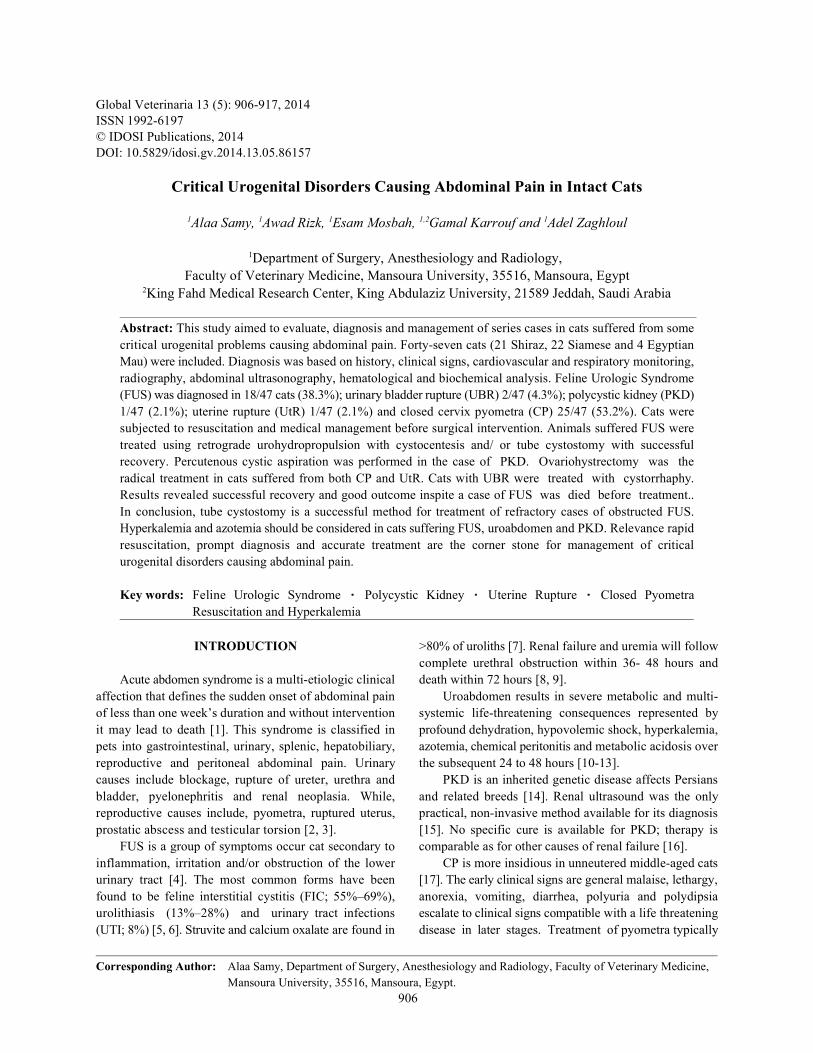
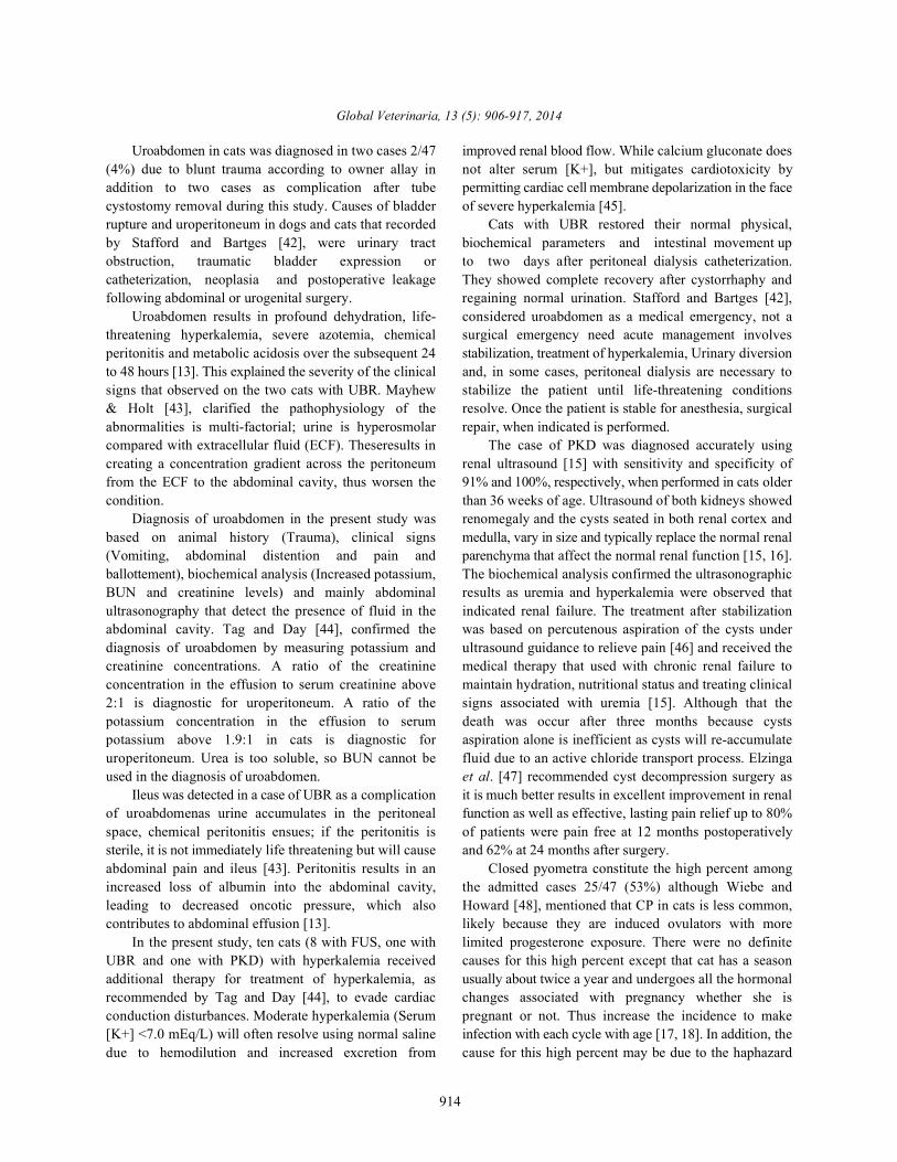
Fig. 1: (a, b) Showing ultrasonography of FUS. a)Longitudinal sonogram of the urinary bladder of a 3-year-old maleShiraz cat. There were cystic calculi. The urine is heteroechoic with shadowing mineralized dependent sediment.Irregular shaped blood clots appeared as non-shadowing medium to mildly hyperechoic that adhered to thebladder wall. b) Transverse sonogram of the left kidney in a 2-year-old male Siamese cat with a history of anuriaof 2 days duration. There is moderate hydronephrosis with uniformly dilated hypoechogenic renal pelvis(Arrows). c) Showing ultrasonography of (UBR) with presence of anechoic fluid in the peritoneal space. Ileuswas diagnosed as a distention of intestinal loops and the lumen filled with echogenic fluid (Arrow). d) Showinga transverse sonogram of the left kidney that replaced by large (White arrow) and small size (Black arrow)hypoechoic cysts. Note that these cysts are non-communicating ruling out hydronephrosis.
cats (21.3%) (FUS=8, UBR=1 and PKD=1) were severe hyperechoic irregular/amorphous shaped blood clots werehyperkalemic with hypotension and bradycardia. adhered to the bladder wall. Uniformly dilatedThey additionally received constant-rate infusion of 10% hypoechogenic renal pelvis was observed in 2/16 (12.5%)calcium gluconate (0.5–1.0 ml/kg), regular insulin (0.1–0.25 of complete obstructed cases which indicate mild toU/kg), 25% dextrose (1–2 g/U of insulin) and Sodium moderate hydronephrosis, Figure 1 (A, B).bicarbonate (1–2 mEq/kg) slowly over 20 mins. They took In cats with UBR, ultrasonography showed anechoicabout 12(n=7)-24(n=3) hours to regain their normal health fluid accumulation in the peritoneal space dissectingparameters. between organs. Ileus was diagnosed in a Shiraz cat as a
Results of Ultrasonographical Examinations: Cats with with echogenic fluid, Figure 1 (C).FUS showed fully distended urinary bladders and In PKD case, renal ultrasound showed renomegaly ofheteroechoic urine, with focal, dependent, hyperechoic, both kidneys with an irregular outline and presence ofcurvilinear echogenicity calculi which changed position varied size anechoic, smooth margined cysts in the renalas patient position changes. Shadowing mineralized tissue. These cysts eroded the normal renal architecturedependent sediment was present. Mild to medium and showed distal acoustic enhancement, Figure 1 (D).
local distention of intestinal loops and the lumen filled
Global Veterinaria, 13 (5): 906-917, 2014
910
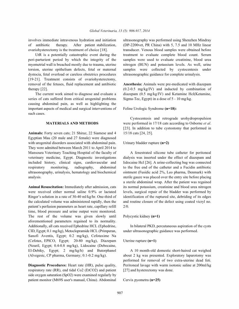
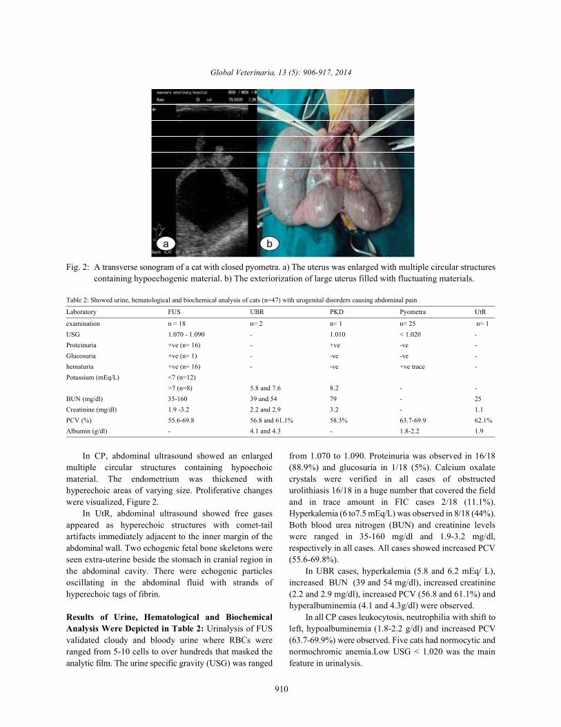
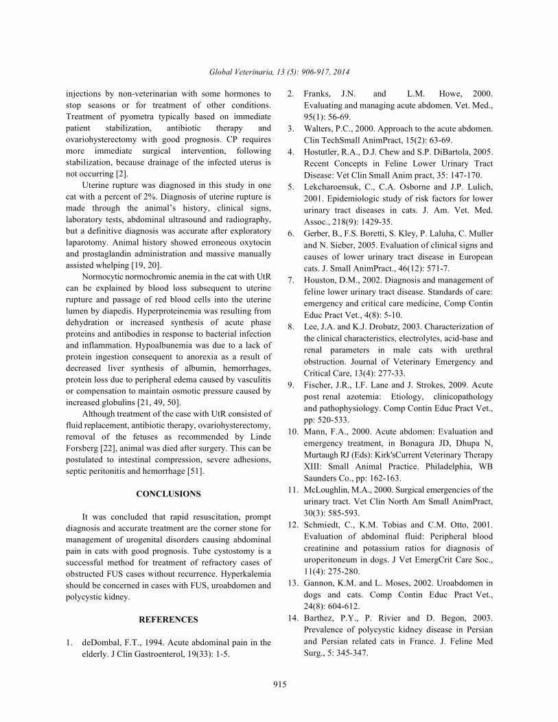
Fig. 2: A transverse sonogram of a cat with closed pyometra. a) The uterus was enlarged with multiple circular structurescontaining hypoechogenic material. b) The exteriorization of large uterus filled with fluctuating materials.
Table 2: Showed urine, hematological and biochemical analysis of cats (n=47) with urogenital disorders causing abdominal pain
Laboratory FUS UBR PKD Pyometra UtR
examination n = 18 n= 2 n= 1 n= 25 n= 1USG 1.070 - 1.090 - 1.010 < 1.020 -Proteinuria +ve (n= 16) - +ve -ve -Glucosuria +ve (n= 1) - -ve -ve -hematuria +ve (n= 16) - -ve +ve trace -Potassium (mEq/L) <7 (n=12)
>7 (n=8) 5.8 and 7.6 8.2 - -BUN (mg/dl) 35-160 39 and 54 79 - 25Creatinine (mg/dl) 1.9 -3.2 2.2 and 2.9 3.2 - 1.1PCV (%) 55.6-69.8 56.8 and 61.1% 58.3% 63.7-69.9 62.1%Albumin (g/dl) - 4.1 and 4.3 - 1.8-2.2 1.9
In CP, abdominal ultrasound showed an enlarged from 1.070 to 1.090. Proteinuria was observed in 16/18multiple circular structures containing hypoechoic (88.9%) and glucosuria in 1/18 (5%). Calcium oxalatematerial. The endometrium was thickened with crystals were verified in all cases of obstructedhyperechoic areas of varying size. Proliferative changes urolithiasis 16/18 in a huge number that covered the fieldwere visualized, Figure 2. and in trace amount in FIC cases 2/18 (11.1%).
In UtR, abdominal ultrasound showed free gases Hyperkalemia (6 to7.5 mEq/L) was observed in 8/18 (44%).appeared as hyperechoic structures with comet-tail Both blood urea nitrogen (BUN) and creatinine levelsartifacts immediately adjacent to the inner margin of the were ranged in 35-160 mg/dl and 1.9-3.2 mg/dl,abdominal wall. Two echogenic fetal bone skeletons were respectively in all cases. All cases showed increased PCVseen extra-uterine beside the stomach in cranial region in (55.6-69.8%).the abdominal cavity. There were echogenic particles In UBR cases, hyperkalemia (5.8 and 6.2 mEq/ L),oscillating in the abdominal fluid with strands of increased BUN (39 and 54 mg/dl), increased creatininehyperechoic tags of fibrin. (2.2 and 2.9 mg/dl), increased PCV (56.8 and 61.1%) and
Results of Urine, Hematological and Biochemical In all CP cases leukocytosis, neutrophilia with shift toAnalysis Were Depicted in Table 2: Urinalysis of FUS left, hypoalbuminemia (1.8-2.2 g/dl) and increased PCVvalidated cloudy and bloody urine where RBCs were (63.7-69.9%) were observed. Five cats had normocytic andranged from 5-10 cells to over hundreds that masked the normochromic anemia.Low USG < 1.020 was the mainanalytic film. The urine specific gravity (USG) was ranged feature in urinalysis.
hyperalbuminemia (4.1 and 4.3g/dl) were observed.
Global Veterinaria, 13 (5): 906-917, 2014
911
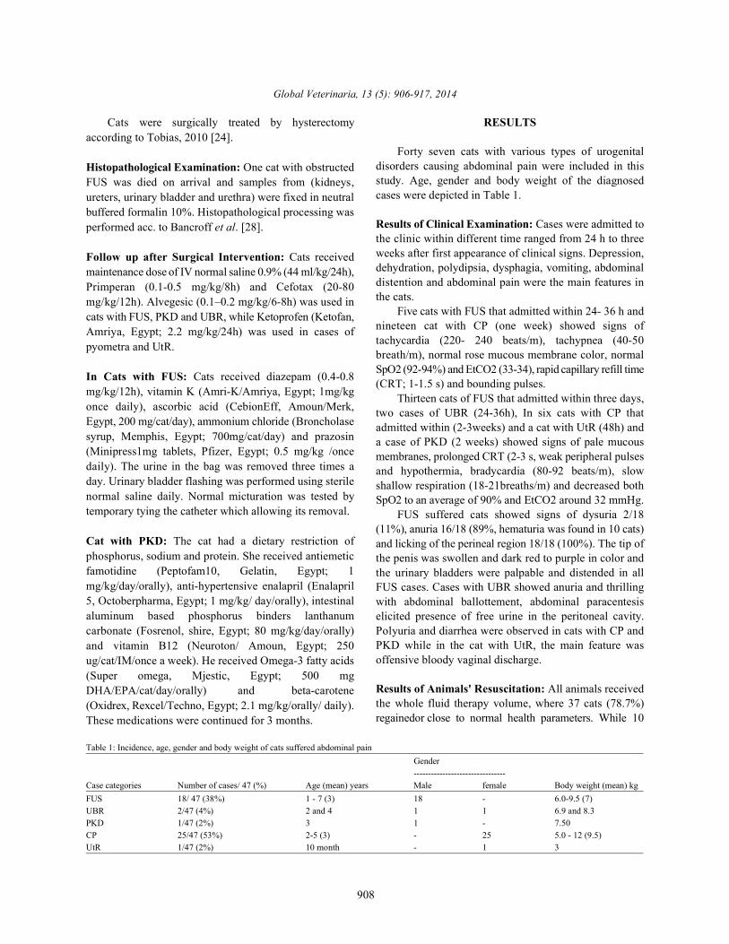
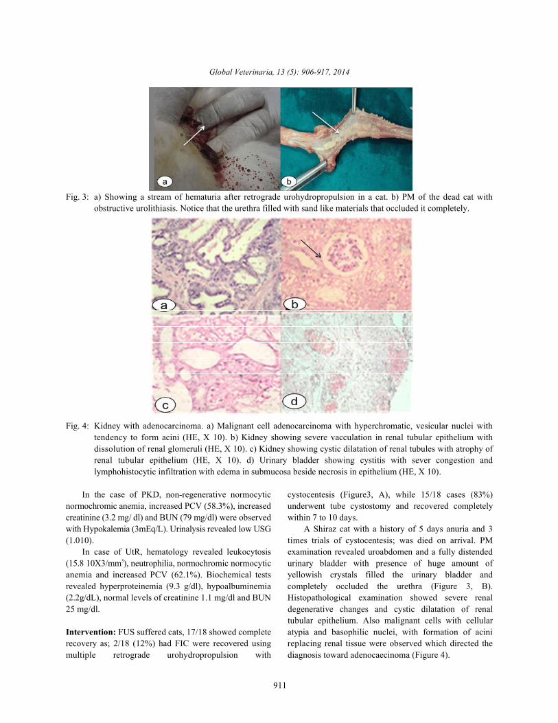
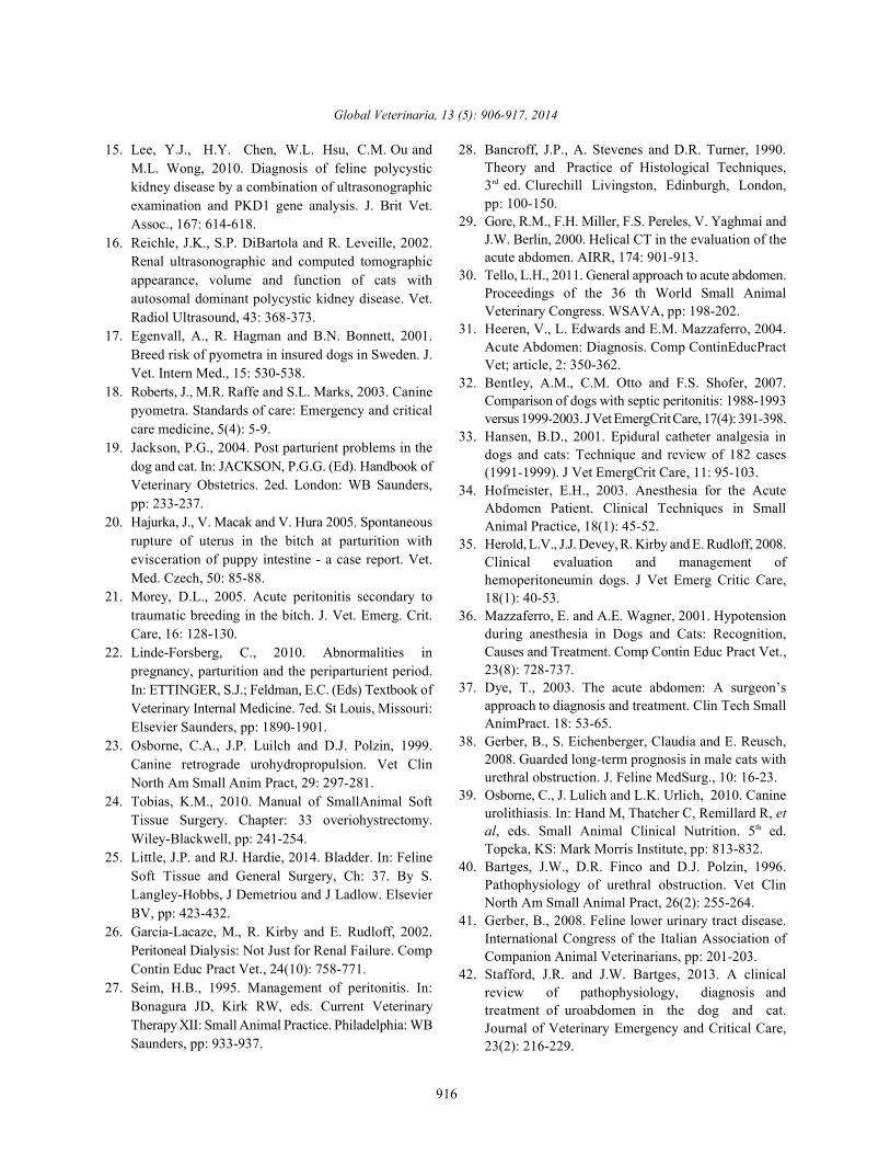
Fig. 3: a) Showing a stream of hematuria after retrograde urohydropropulsion in a cat. b) PM of the dead cat withobstructive urolithiasis. Notice that the urethra filled with sand like materials that occluded it completely.
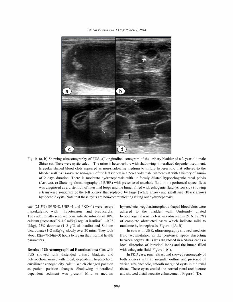
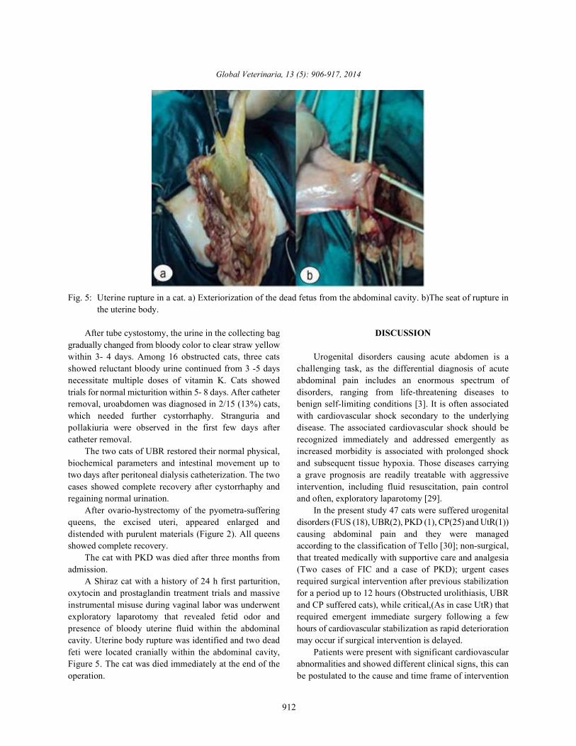
Fig. 4: Kidney with adenocarcinoma. a) Malignant cell adenocarcinoma with hyperchromatic, vesicular nuclei withtendency to form acini (HE, X 10). b) Kidney showing severe vacculation in renal tubular epithelium withdissolution of renal glomeruli (HE, X 10). c) Kidney showing cystic dilatation of renal tubules with atrophy ofrenal tubular epithelium (HE, X 10). d) Urinary bladder showing cystitis with sever congestion andlymphohistocytic infiltration with edema in submucosa beside necrosis in epithelium (HE, X 10).
In the case of PKD, non-regenerative normocytic cystocentesis (Figure3, A), while 15/18 cases (83%)normochromic anemia, increased PCV (58.3%), increased underwent tube cystostomy and recovered completelycreatinine (3.2 mg/ dl) and BUN (79 mg/dl) were observed within 7 to 10 days. with Hypokalemia (3mEq/L). Urinalysis revealed low USG A Shiraz cat with a history of 5 days anuria and 3(1.010). times trials of cystocentesis; was died on arrival. PM
In case of UtR, hematology revealed leukocytosis examination revealed uroabdomen and a fully distended(15.8 10X3/mm ), neutrophilia, normochromic normocytic urinary bladder with presence of huge amount of3
anemia and increased PCV (62.1%). Biochemical tests yellowish crystals filled the urinary bladder andrevealed hyperproteinemia (9.3 g/dl), hypoalbuminemia completely occluded the urethra (Figure 3, B).(2.2g/dL), normal levels of creatinine 1.1 mg/dl and BUN Histopathological examination showed severe renal25 mg/dl. degenerative changes and cystic dilatation of renal
Intervention: FUS suffered cats, 17/18 showed complete atypia and basophilic nuclei, with formation of acinirecovery as; 2/18 (12%) had FIC were recovered using replacing renal tissue were observed which directed themultiple retrograde urohydropropulsion with diagnosis toward adenocaecinoma (Figure 4).
tubular epithelium. Also malignant cells with cellular
Global Veterinaria, 13 (5): 906-917, 2014
912
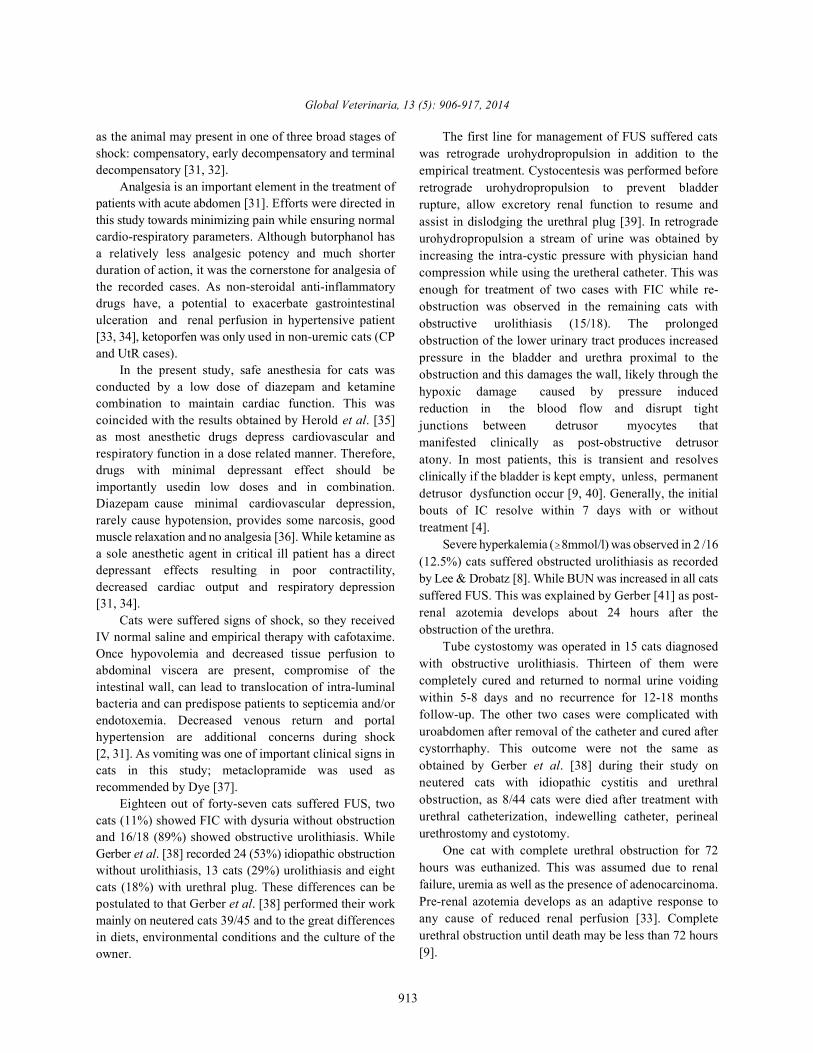
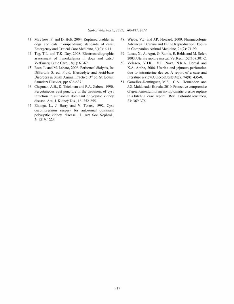
Fig. 5: Uterine rupture in a cat. a) Exteriorization of the dead fetus from the abdominal cavity. b)The seat of rupture inthe uterine body.
After tube cystostomy, the urine in the collecting bag DISCUSSIONgradually changed from bloody color to clear straw yellowwithin 3- 4 days. Among 16 obstructed cats, three cats Urogenital disorders causing acute abdomen is ashowed reluctant bloody urine continued from 3 -5 days challenging task, as the differential diagnosis of acutenecessitate multiple doses of vitamin K. Cats showed abdominal pain includes an enormous spectrum oftrials for normal micturition within 5- 8 days. After catheter disorders, ranging from life-threatening diseases toremoval, uroabdomen was diagnosed in 2/15 (13%) cats, benign self-limiting conditions [3]. It is often associatedwhich needed further cystorrhaphy. Stranguria and with cardiovascular shock secondary to the underlyingpollakiuria were observed in the first few days after disease. The associated cardiovascular shock should becatheter removal. recognized immediately and addressed emergently as
The two cats of UBR restored their normal physical, increased morbidity is associated with prolonged shockbiochemical parameters and intestinal movement up to and subsequent tissue hypoxia. Those diseases carryingtwo days after peritoneal dialysis catheterization. The two a grave prognosis are readily treatable with aggressivecases showed complete recovery after cystorrhaphy and intervention, including fluid resuscitation, pain controlregaining normal urination. and often, exploratory laparotomy [29].
After ovario-hystrectomy of the pyometra-suffering In the present study 47 cats were suffered urogenitalqueens, the excised uteri, appeared enlarged and disorders (FUS (18), UBR(2), PKD (1), CP(25) and UtR(1))distended with purulent materials (Figure 2). All queens causing abdominal pain and they were managedshowed complete recovery. according to the classification of Tello [30]; non-surgical,
The cat with PKD was died after three months from that treated medically with supportive care and analgesiaadmission. (Two cases of FIC and a case of PKD); urgent cases
A Shiraz cat with a history of 24 h first parturition, required surgical intervention after previous stabilizationoxytocin and prostaglandin treatment trials and massive for a period up to 12 hours (Obstructed urolithiasis, UBRinstrumental misuse during vaginal labor was underwent and CP suffered cats), while critical,(As in case UtR) thatexploratory laparotomy that revealed fetid odor and required emergent immediate surgery following a fewpresence of bloody uterine fluid within the abdominal hours of cardiovascular stabilization as rapid deteriorationcavity. Uterine body rupture was identified and two dead may occur if surgical intervention is delayed.feti were located cranially within the abdominal cavity, Patients were present with significant cardiovascularFigure 5. The cat was died immediately at the end of the abnormalities and showed different clinical signs, this canoperation. be postulated to the cause and time frame of intervention
Global Veterinaria, 13 (5): 906-917, 2014
913
as the animal may present in one of three broad stages of The first line for management of FUS suffered catsshock: compensatory, early decompensatory and terminaldecompensatory [31, 32].
Analgesia is an important element in the treatment ofpatients with acute abdomen [31]. Efforts were directed inthis study towards minimizing pain while ensuring normalcardio-respiratory parameters. Although butorphanol hasa relatively less analgesic potency and much shorterduration of action, it was the cornerstone for analgesia ofthe recorded cases. As non-steroidal anti-inflammatorydrugs have, a potential to exacerbate gastrointestinalulceration and renal perfusion in hypertensive patient[33, 34], ketoporfen was only used in non-uremic cats (CPand UtR cases).
In the present study, safe anesthesia for cats wasconducted by a low dose of diazepam and ketaminecombination to maintain cardiac function. This wascoincided with the results obtained by Herold et al. [35]as most anesthetic drugs depress cardiovascular andrespiratory function in a dose related manner. Therefore,drugs with minimal depressant effect should beimportantly usedin low doses and in combination.Diazepam cause minimal cardiovascular depression,rarely cause hypotension, provides some narcosis, goodmuscle relaxation and no analgesia [36]. While ketamine asa sole anesthetic agent in critical ill patient has a directdepressant effects resulting in poor contractility,decreased cardiac output and respiratory depression[31, 34].
Cats were suffered signs of shock, so they receivedIV normal saline and empirical therapy with cafotaxime.Once hypovolemia and decreased tissue perfusion toabdominal viscera are present, compromise of theintestinal wall, can lead to translocation of intra-luminalbacteria and can predispose patients to septicemia and/orendotoxemia. Decreased venous return and portalhypertension are additional concerns during shock[2, 31]. As vomiting was one of important clinical signs incats in this study; metaclopramide was used asrecommended by Dye [37].
Eighteen out of forty-seven cats suffered FUS, twocats (11%) showed FIC with dysuria without obstructionand 16/18 (89%) showed obstructive urolithiasis. WhileGerber et al. [38] recorded 24 (53%) idiopathic obstructionwithout urolithiasis, 13 cats (29%) urolithiasis and eightcats (18%) with urethral plug. These differences can bepostulated to that Gerber et al. [38] performed their workmainly on neutered cats 39/45 and to the great differencesin diets, environmental conditions and the culture of theowner.
was retrograde urohydropropulsion in addition to theempirical treatment. Cystocentesis was performed beforeretrograde urohydropropulsion to prevent bladderrupture, allow excretory renal function to resume andassist in dislodging the urethral plug [39]. In retrogradeurohydropropulsion a stream of urine was obtained byincreasing the intra-cystic pressure with physician handcompression while using the uretheral catheter. This wasenough for treatment of two cases with FIC while re-obstruction was observed in the remaining cats withobstructive urolithiasis (15/18). The prolongedobstruction of the lower urinary tract produces increasedpressure in the bladder and urethra proximal to theobstruction and this damages the wall, likely through thehypoxic damage caused by pressure inducedreduction in the blood flow and disrupt tightjunctions between detrusor myocytes thatmanifested clinically as post-obstructive detrusoratony. In most patients, this is transient and resolvesclinically if the bladder is kept empty, unless, permanentdetrusor dysfunction occur [9, 40]. Generally, the initialbouts of IC resolve within 7 days with or withouttreatment [4].
Severe hyperkalemia ( 8mmol/l) was observed in 2 /16(12.5%) cats suffered obstructed urolithiasis as recordedby Lee & Drobatz [8]. While BUN was increased in all catssuffered FUS. This was explained by Gerber [41] as post-renal azotemia develops about 24 hours after theobstruction of the urethra.
Tube cystostomy was operated in 15 cats diagnosedwith obstructive urolithiasis. Thirteen of them werecompletely cured and returned to normal urine voidingwithin 5-8 days and no recurrence for 12-18 monthsfollow-up. The other two cases were complicated withuroabdomen after removal of the catheter and cured aftercystorrhaphy. This outcome were not the same asobtained by Gerber et al. [38] during their study onneutered cats with idiopathic cystitis and urethralobstruction, as 8/44 cats were died after treatment withurethral catheterization, indewelling catheter, perinealurethrostomy and cystotomy.
One cat with complete urethral obstruction for 72hours was euthanized. This was assumed due to renalfailure, uremia as well as the presence of adenocarcinoma.Pre-renal azotemia develops as an adaptive response toany cause of reduced renal perfusion [33]. Completeurethral obstruction until death may be less than 72 hours[9].
Global Veterinaria, 13 (5): 906-917, 2014
914
Uroabdomen in cats was diagnosed in two cases 2/47 improved renal blood flow. While calcium gluconate does(4%) due to blunt trauma according to owner allay in not alter serum [K+], but mitigates cardiotoxicity byaddition to two cases as complication after tube permitting cardiac cell membrane depolarization in the facecystostomy removal during this study. Causes of bladder of severe hyperkalemia [45].rupture and uroperitoneum in dogs and cats that recorded Cats with UBR restored their normal physical,by Stafford and Bartges [42], were urinary tract biochemical parameters and intestinal movement upobstruction, traumatic bladder expression or to two days after peritoneal dialysis catheterization.catheterization, neoplasia and postoperative leakage They showed complete recovery after cystorrhaphy andfollowing abdominal or urogenital surgery. regaining normal urination. Stafford and Bartges [42],
Uroabdomen results in profound dehydration, life- considered uroabdomen as a medical emergency, not athreatening hyperkalemia, severe azotemia, chemical surgical emergency need acute management involvesperitonitis and metabolic acidosis over the subsequent 24 stabilization, treatment of hyperkalemia, Urinary diversionto 48 hours [13]. This explained the severity of the clinical and, in some cases, peritoneal dialysis are necessary tosigns that observed on the two cats with UBR. Mayhew stabilize the patient until life-threatening conditions& Holt [43], clarified the pathophysiology of the resolve. Once the patient is stable for anesthesia, surgicalabnormalities is multi-factorial; urine is hyperosmolar repair, when indicated is performed.compared with extracellular fluid (ECF). Theseresults in The case of PKD was diagnosed accurately usingcreating a concentration gradient across the peritoneum renal ultrasound [15] with sensitivity and specificity offrom the ECF to the abdominal cavity, thus worsen the 91% and 100%, respectively, when performed in cats oldercondition. than 36 weeks of age. Ultrasound of both kidneys showed
Diagnosis of uroabdomen in the present study was renomegaly and the cysts seated in both renal cortex andbased on animal history (Trauma), clinical signs medulla, vary in size and typically replace the normal renal(Vomiting, abdominal distention and pain and parenchyma that affect the normal renal function [15, 16].ballottement), biochemical analysis (Increased potassium, The biochemical analysis confirmed the ultrasonographicBUN and creatinine levels) and mainly abdominal results as uremia and hyperkalemia were observed thatultrasonography that detect the presence of fluid in the indicated renal failure. The treatment after stabilizationabdominal cavity. Tag and Day [44], confirmed the was based on percutenous aspiration of the cysts underdiagnosis of uroabdomen by measuring potassium and ultrasound guidance to relieve pain [46] and received thecreatinine concentrations. A ratio of the creatinine medical therapy that used with chronic renal failure toconcentration in the effusion to serum creatinine above maintain hydration, nutritional status and treating clinical2:1 is diagnostic for uroperitoneum. A ratio of the signs associated with uremia [15]. Although that thepotassium concentration in the effusion to serum death was occur after three months because cystspotassium above 1.9:1 in cats is diagnostic for aspiration alone is inefficient as cysts will re-accumulateuroperitoneum. Urea is too soluble, so BUN cannot be fluid due to an active chloride transport process. Elzingaused in the diagnosis of uroabdomen. et al. [47] recommended cyst decompression surgery as
Ileus was detected in a case of UBR as a complication it is much better results in excellent improvement in renalof uroabdomenas urine accumulates in the peritoneal function as well as effective, lasting pain relief up to 80%space, chemical peritonitis ensues; if the peritonitis is of patients were pain free at 12 months postoperativelysterile, it is not immediately life threatening but will cause and 62% at 24 months after surgery.abdominal pain and ileus [43]. Peritonitis results in an Closed pyometra constitute the high percent amongincreased loss of albumin into the abdominal cavity, the admitted cases 25/47 (53%) although Wiebe andleading to decreased oncotic pressure, which also Howard [48], mentioned that CP in cats is less common,contributes to abdominal effusion [13]. likely because they are induced ovulators with more
In the present study, ten cats (8 with FUS, one with limited progesterone exposure. There were no definiteUBR and one with PKD) with hyperkalemia received causes for this high percent except that cat has a seasonadditional therapy for treatment of hyperkalemia, as usually about twice a year and undergoes all the hormonalrecommended by Tag and Day [44], to evade cardiac changes associated with pregnancy whether she isconduction disturbances. Moderate hyperkalemia (Serum pregnant or not. Thus increase the incidence to make[K+] <7.0 mEq/L) will often resolve using normal saline infection with each cycle with age [17, 18]. In addition, thedue to hemodilution and increased excretion from cause for this high percent may be due to the haphazard
Global Veterinaria, 13 (5): 906-917, 2014
915
injections by non-veterinarian with some hormones to 2. Franks, J.N. and L.M. Howe, 2000.stop seasons or for treatment of other conditions.Treatment of pyometra typically based on immediatepatient stabilization, antibiotic therapy andovariohysterectomy with good prognosis. CP requiresmore immediate surgical intervention, followingstabilization, because drainage of the infected uterus isnot occurring [2].
Uterine rupture was diagnosed in this study in onecat with a percent of 2%. Diagnosis of uterine rupture ismade through the animal’s history, clinical signs,laboratory tests, abdominal ultrasound and radiography,but a definitive diagnosis was accurate after exploratorylaparotomy. Animal history showed erroneous oxytocinand prostaglandin administration and massive manuallyassisted whelping [19, 20].
Normocytic normochromic anemia in the cat with UtRcan be explained by blood loss subsequent to uterinerupture and passage of red blood cells into the uterinelumen by diapedis. Hyperproteinemia was resulting fromdehydration or increased synthesis of acute phaseproteins and antibodies in response to bacterial infectionand inflammation. Hypoalbunemia was due to a lack ofprotein ingestion consequent to anorexia as a result ofdecreased liver synthesis of albumin, hemorrhages,protein loss due to peripheral edema caused by vasculitisor compensation to maintain osmotic pressure caused byincreased globulins [21, 49, 50].
Although treatment of the case with UtR consisted offluid replacement, antibiotic therapy, ovariohysterectomy,removal of the fetuses as recommended by LindeForsberg [22], animal was died after surgery. This can bepostulated to intestinal compression, severe adhesions,septic peritonitis and hemorrhage [51].
CONCLUSIONS
It was concluded that rapid resuscitation, promptdiagnosis and accurate treatment are the corner stone formanagement of urogenital disorders causing abdominalpain in cats with good prognosis. Tube cystostomy is asuccessful method for treatment of refractory cases ofobstructed FUS cases without recurrence. Hyperkalemiashould be concerned in cases with FUS, uroabdomen andpolycystic kidney.
REFERENCES
1. deDombal, F.T., 1994. Acute abdominal pain in theelderly. J Clin Gastroenterol, 19(33): 1-5.
Evaluating and managing acute abdomen. Vet. Med.,95(1): 56-69.
3. Walters, P.C., 2000. Approach to the acute abdomen.Clin TechSmall AnimPract, 15(2): 63-69.
4. Hostutler, R.A., D.J. Chew and S.P. DiBartola, 2005.Recent Concepts in Feline Lower Urinary TractDisease: Vet Clin Small Anim pract, 35: 147-170.
5. Lekcharoensuk, C., C.A. Osborne and J.P. Lulich,2001. Epidemiologic study of risk factors for lowerurinary tract diseases in cats. J. Am. Vet. Med.Assoc., 218(9): 1429-35.
6. Gerber, B., F.S. Boretti, S. Kley, P. Laluha, C. Mullerand N. Sieber, 2005. Evaluation of clinical signs andcauses of lower urinary tract disease in Europeancats. J. Small AnimPract., 46(12): 571-7.
7. Houston, D.M., 2002. Diagnosis and management offeline lower urinary tract disease. Standards of care:emergency and critical care medicine, Comp ContinEduc Pract Vet., 4(8): 5-10.
8. Lee, J.A. and K.J. Drobatz, 2003. Characterization ofthe clinical characteristics, electrolytes, acid-base andrenal parameters in male cats with urethralobstruction. Journal of Veterinary Emergency andCritical Care, 13(4): 277-33.
9. Fischer, J.R., I.F. Lane and J. Strokes, 2009. Acutepost renal azotemia: Etiology, clinicopathologyand pathophysiology. Comp Contin Educ Pract Vet.,pp: 520-533.
10. Mann, F.A., 2000. Acute abdomen: Evaluation andemergency treatment, in Bonagura JD, Dhupa N,Murtaugh RJ (Eds): Kirk'sCurrent Veterinary TherapyXIII: Small Animal Practice. Philadelphia, WBSaunders Co., pp: 162-163.
11. McLoughlin, M.A., 2000. Surgical emergencies of theurinary tract. Vet Clin North Am Small AnimPract,30(3): 585-593.
12. Schmiedt, C., K.M. Tobias and C.M. Otto, 2001.Evaluation of abdominal fluid: Peripheral bloodcreatinine and potassium ratios for diagnosis ofuroperitoneum in dogs. J Vet EmergCrit Care Soc.,11(4): 275-280.
13. Gannon, K.M. and L. Moses, 2002. Uroabdomen indogs and cats. Comp Contin Educ Pract Vet.,24(8): 604-612.
14. Barthez, P.Y., P. Rivier and D. Begon, 2003.Prevalence of polycystic kidney disease in Persianand Persian related cats in France. J. Feline MedSurg., 5: 345-347.
Global Veterinaria, 13 (5): 906-917, 2014
916
15. Lee, Y.J., H.Y. Chen, W.L. Hsu, C.M. Ou and 28. Bancroff, J.P., A. Stevenes and D.R. Turner, 1990.M.L. Wong, 2010. Diagnosis of feline polycystickidney disease by a combination of ultrasonographicexamination and PKD1 gene analysis. J. Brit Vet.Assoc., 167: 614-618.
16. Reichle, J.K., S.P. DiBartola and R. Leveille, 2002.Renal ultrasonographic and computed tomographicappearance, volume and function of cats withautosomal dominant polycystic kidney disease. Vet.Radiol Ultrasound, 43: 368-373.
17. Egenvall, A., R. Hagman and B.N. Bonnett, 2001.Breed risk of pyometra in insured dogs in Sweden. J.Vet. Intern Med., 15: 530-538.
18. Roberts, J., M.R. Raffe and S.L. Marks, 2003. Caninepyometra. Standards of care: Emergency and criticalcare medicine, 5(4): 5-9.
19. Jackson, P.G., 2004. Post parturient problems in thedog and cat. In: JACKSON, P.G.G. (Ed). Handbook ofVeterinary Obstetrics. 2ed. London: WB Saunders,pp: 233-237.
20. Hajurka, J., V. Macak and V. Hura 2005. Spontaneousrupture of uterus in the bitch at parturition withevisceration of puppy intestine - a case report. Vet.Med. Czech, 50: 85-88.
21. Morey, D.L., 2005. Acute peritonitis secondary totraumatic breeding in the bitch. J. Vet. Emerg. Crit.Care, 16: 128-130.
22. Linde-Forsberg, C., 2010. Abnormalities inpregnancy, parturition and the periparturient period.In: ETTINGER, S.J.; Feldman, E.C. (Eds) Textbook ofVeterinary Internal Medicine. 7ed. St Louis, Missouri:Elsevier Saunders, pp: 1890-1901.
23. Osborne, C.A., J.P. Luilch and D.J. Polzin, 1999.Canine retrograde urohydropropulsion. Vet ClinNorth Am Small Anim Pract, 29: 297-281.
24. Tobias, K.M., 2010. Manual of SmallAnimal SoftTissue Surgery. Chapter: 33 overiohystrectomy.Wiley-Blackwell, pp: 241-254.
25. Little, J.P. and RJ. Hardie, 2014. Bladder. In: FelineSoft Tissue and General Surgery, Ch: 37. By S.Langley-Hobbs, J Demetriou and J Ladlow. ElsevierBV, pp: 423-432.
26. Garcia-Lacaze, M., R. Kirby and E. Rudloff, 2002.Peritoneal Dialysis: Not Just for Renal Failure. CompContin Educ Pract Vet., 24(10): 758-771.
27. Seim, H.B., 1995. Management of peritonitis. In:Bonagura JD, Kirk RW, eds. Current VeterinaryTherapy XII: Small Animal Practice. Philadelphia: WBSaunders, pp: 933-937.
Theory and Practice of Histological Techniques,3 ed. Clurechill Livingston, Edinburgh, London,rd
pp: 100-150.29. Gore, R.M., F.H. Miller, F.S. Pereles, V. Yaghmai and
J.W. Berlin, 2000. Helical CT in the evaluation of theacute abdomen. AIRR, 174: 901-913.
30. Tello, L.H., 2011. General approach to acute abdomen.Proceedings of the 36 th World Small AnimalVeterinary Congress. WSAVA, pp: 198-202.
31. Heeren, V., L. Edwards and E.M. Mazzaferro, 2004.Acute Abdomen: Diagnosis. Comp ContinEducPractVet; article, 2: 350-362.
32. Bentley, A.M., C.M. Otto and F.S. Shofer, 2007.Comparison of dogs with septic peritonitis: 1988-1993versus 1999-2003. J Vet EmergCrit Care, 17(4): 391-398.
33. Hansen, B.D., 2001. Epidural catheter analgesia indogs and cats: Technique and review of 182 cases(1991-1999). J Vet EmergCrit Care, 11: 95-103.
34. Hofmeister, E.H., 2003. Anesthesia for the AcuteAbdomen Patient. Clinical Techniques in SmallAnimal Practice, 18(1): 45-52.
35. Herold, L.V., J.J. Devey, R. Kirby and E. Rudloff, 2008.Clinical evaluation and management ofhemoperitoneumin dogs. J Vet Emerg Critic Care,18(1): 40-53.
36. Mazzaferro, E. and A.E. Wagner, 2001. Hypotensionduring anesthesia in Dogs and Cats: Recognition,Causes and Treatment. Comp Contin Educ Pract Vet.,23(8): 728-737.
37. Dye, T., 2003. The acute abdomen: A surgeon’sapproach to diagnosis and treatment. Clin Tech SmallAnimPract. 18: 53-65.
38. Gerber, B., S. Eichenberger, Claudia and E. Reusch,2008. Guarded long-term prognosis in male cats withurethral obstruction. J. Feline MedSurg., 10: 16-23.
39. Osborne, C., J. Lulich and L.K. Urlich, 2010. Canineurolithiasis. In: Hand M, Thatcher C, Remillard R, etal, eds. Small Animal Clinical Nutrition. 5 ed.th
Topeka, KS: Mark Morris Institute, pp: 813-832.40. Bartges, J.W., D.R. Finco and D.J. Polzin, 1996.
Pathophysiology of urethral obstruction. Vet ClinNorth Am Small Animal Pract, 26(2): 255-264.
41. Gerber, B., 2008. Feline lower urinary tract disease.International Congress of the Italian Association ofCompanion Animal Veterinarians, pp: 201-203.
42. Stafford, J.R. and J.W. Bartges, 2013. A clinicalreview of pathophysiology, diagnosis andtreatment of uroabdomen in the dog and cat.Journal of Veterinary Emergency and Critical Care,23(2): 216-229.
Global Veterinaria, 13 (5): 906-917, 2014
917
43. May hew, P. and D. Holt, 2004. Ruptured bladder in 48. Wiebe, V.J. and J.P. Howard, 2009. Pharmacologicdogs and cats. Compendium; standards of care: Advances in Canine and Feline Reproduction: TopicsEmergency and Critical Care Medicine, 6(10): 6-11. in Companion Animal Medicine, 24(2): 71-99.
44. Tag, T.L. and T.K. Day, 2008. Electrocardiographic 49. Lucas, X., A. Agut, G. Ramis, E. Belda and M. Soler,assessment of hyperkalemia in dogs and cats,J 2003. Uterine rupture in a cat. Vet Rec., 152(10): 301-2.VetEmerg Critic Care, 18(1): 61-67. 50. Velasco, V.J.R., V.P. Nava, N.R.A. Bernal and
45. Ross, L. and M. Labato, 2006. Peritoneal dialysis, In: K.A. Ambe, 2006. Uterine and jejunum perforationDiBartola S. ed. Fluid, Electrolyte and Acid-base due to intrauterine device. A report of a case andDisorders in Small Animal Practice, 3 ed. St. Louis: literature review.GinecolObstetMex, 74(8): 435-8.rd
Saunders Elsevier, pp: 636-637. 51. González-Domínguez, M.S., C.A. Hernández and46. Chapman, A.B., D. Thickman and P.A. Gabow, 1990. J.G. Maldonado-Estrada, 2010. Protective compromise
Percutaneous cyst puncture in the treatment of cyst of great omentum in an asymptomatic uterine ruptureinfection in autosomal dominant polycystic kidney in a bitch: a case report. Rev. ColombCiencPecu,disease. Am. J. Kidney Dis., 16: 252-255. 23: 369-376.
47. Elzinga, L., J. Barry and V. Torres, 1992. Cystdecompression surgery for autosomal dominantpolycystic kidney disease. J. Am Soc. Nephrol.,2: 1219-1226.