Embed Size (px)
Citation preview

CroniconO P E N A C C E S S EC DENTAL SCIENCE
Research Article
Prosthetic Rehabilitation and Cephalometric Evaluation of Gapo Syndrome: 29 Years Follow Up
Mehmet Dalkiz1*, Davut Gul2 and Ahmed Suat Dalkiz3
1Professor, Prosthodontist; Private Practice 1, Boulevard Sylvain Dupuis 235-10, Brussels, Belgium2Professor, Medical Genetics; Private Practice, Intergen Genetics Center, Kavaklidere, Ankara, Turkey3Private Practice, Boulevard Sylvain Dupuis 235-10, Brussels, Belgium
*Corresponding Author: Mehmet Dalkiz, Professor, Prosthodontist; Private Practice 1, Boulevard Sylvain Dupuis 235-10, Brussels, Belgium.
Citation: Mehmet Dalkiz., et al. “Prosthetic Rehabilitation and Cephalometric Evaluation of Gapo Syndrome: 29 Years Follow Up”. EC Dental Science 18.2 (2019): 252-269.
Received: September 06, 2018; Published: January 24, 2019
Abstract
Keywords: GAPO Syndrome; Prosthetic Application; Cephalometric Evaluations; Pseudoprognathia
Introduction
Objective: GAPO is the acronymic designation for a syndrome of growth retardation, alopecia, pseudo-anodontia (failure of tooth eruption) and optic atrophy. This paper reports the dental findings in a three Turkish patients with GAPO syndrome who presented as a peculiar dental finding unerupted primary and permanent dentitions, which resembled complete anodontia on clinical examina-tion. A cephalometric analysis was performed to investigate the alterations in facial bone development and diagnosed and prosthetic rehabilitation and 29 years fallow up with GAPO syndrome.Patients and Methods: This is a very rare GAPO syndrome incident caused by close relative marriages. All the teeth (permanent and deciduous) are present but interrupted. All the body hairs disappear as the person becomes mature. A Clinical, laboratory (routine blood and urine analysis, and radiological analysis), prosthetic applications, and the effect of prosthetic restoration on the relation-ship between the jaw and face, and cephalometric evaluations in GAPO syndrome are discussed. The female patient refused to ap-plication of prosthetic restoration. The male patients were applied removable complete denture and cephalometric measurements conducted. The cephalometric analysis was performed because none of the previous reports have done so, probably because there has not been too much interest in investigating the maxillofacial manifestations of GAPO syndrome. Cephalometric analysis should be conducted in further GAPO syndrome cases in order to address new characteristic of this disorder.Results: The cephalometric evaluation showed that middle part of the face and jaws size was normal. This result proves to be on opposition to the findings of the previous literatures. We are of the opinion that the findings of other authors were caused by the fact that their findings were based on inspection.Conclusion: We did not surgically remove the impacted teeth because the risk of maxillomandibular jaw bone loss was too high. The patients have been followed up by physicians from different medical specialties for management of the complications arising from GAPO syndrome for 29 years. This report three additional relative patients with GAPO syndrome and review the literature on this rare disorder. It suggest that all deficiency should be closely monitored and treated in these patients.
GAPO syndrome is an acronymic assignment of growth retardation, alopecia, pseudoanodontia (failure of tooth eruption) and optic atrophy complex. GAPO syndrome is a very rare an inherited disease that is recessively autosomal. It seems to have an autosomal reces-sive pattern of inheritance but its etiology is not yet well understood [46]. This condition was first described by Andersen and Pindborg [2] in 1947, but it was named GAPO syndrome only in 1984 by Tripton and Gorlin [55]. It consists of pseudoanodontia, growth retarda-tion, alopecia, and eye anomalies. Tipton and Gorlin [55] called the disease GAPO syndrome by using the first letters of the words Growth retardation (G), Alopecia (A), Pseudoanodontia (P), and Optic atrophy (O) in 1984. It has been reported worldwide about 50 cases until now [1-60].
Patients who suffer from GAPO syndrome may have several abnormalities including wide anterior fontanel, bossed forehead, promi-nent scalp veins, increased intracranial pressure, micrognathia, protruding lips and auricles, saddle and depressed nasal bridge, reduced perspiration, altered ability to sweat, skin redundancy, and absent eyebrows, eyelashes, and scalp hair; hyper extensible joints, hypercon-vex nails, umbilical hernia, delayed bone maturation, hepatomegaly, hypoplasia of the genitalia and mammary glands, oligoastenospermia and irregular gonadal function. Other features have been described as prominent scalp veins, large craniofacial vascular malformation,

253
Prosthetic Rehabilitation and Cephalometric Evaluation of Gapo Syndrome: 29 Years Follow Up
Citation: Mehmet Dalkiz., et al. “Prosthetic Rehabilitation and Cephalometric Evaluation of Gapo Syndrome: 29 Years Follow Up”. EC Dental Science 18.2 (2019): 252-269.
Ophthalmological findings include progressive optic atrophy, strabismus, megalocornea, bilateral keratoconus, nystagmus, ptosis, glaucoma, and abnormal pattern visual evoked response. Some cases of GAPO with severe unilateral corneal opacity secondary to end stage congenital glaucoma and severe bilateral interstitial keratitis and ocular inflammation have been reported [2,3,21,25,28,46-49].
The teeth are present although they fail to erupt. Primary failure of eruption is a very rare anomaly and in the truest sense an eruption defect which manifests as a complete failure of eruption without a distinct local or systemic etiology. Diagnosing the condition accurately by following a methodical approach and rehabilitation of the patient through surgical, orthodontic or by prosthodontic measures is the present accepted approach the condition [58].
The pseudoanodontia that resulted from the unerupted primary and permanent teeth caused an increase in bone ridge volume in a buccolingual direction, as shown in the occlusal radiograph. This condition hindered the installation of dentures. Surgical removal of the teeth, even if partial, was deemed too complicated because the teeth were ankylosed [11,27,45,50].
Silveira., et al. [13] reported that a cephalometric analysis was performed because none of the previous reports have done so, prob-ably because there has not been too much interest in investigating the orodental manifestations of this syndrome. The this researchers have suggested that further cephalometric analysis should be performed in cases with GAPO syndrome to address the new feature of this disorder.
Some authors [2,13,18,46-48,60] reported that micrognathia, and prognathia, atrophic alveolar bone in the maxillae and mandible, and the hypoplasia of the middle part of the face were found in reported cases. Steiner [50,51] and McNamara [31], have claimed that the determination of normal dimensions after linear measurements would reflect the original craniofacial sizes of patients.
Whereas this study is devoted to the examination of prosthetic restoration and the effect of that restoration to the relationship be-tween jaw and face in two cases. Further this study evaluates whether the hypoplasia of the middle part of the face and micrognathia presents or not by means of cephalometric methods in three patients with GAPO syndrome. The relative patients have been followed up by physicians from different medical specialties for management of the complications arising from GAPO syndrome for 29 years. Purpose of this article reports the prosthetic applications, cephalometric analysis and dentofacial findings three cases of Turkish patients diag-nosed with GAPO syndrome who presented as a peculiar dental finding unerupted primary and permanent dentitions, which resembled complete anodontia on clinical examination. A cephalometric analysis was performed to investigate the alterations in maxillofacial bones development.
suspected of increased intracranial pressure, skin redundancy, hyper extensible joints, gonadal function abnormalities with irregular pe-riods, amenorrhea. Several singular associations like dilated cardiomyopathy, pulmonary hypertension were also recently reported. There is a distinctive facies with a premature aged appearance [5-7,11,16,17,27,36,38,44,45, 50,54-66].
Patients evaluationPatients and Methods
This study was carried out on three related three Turkish patients, two male and one female with GAPO Syndrome. A female and a male patients of these three related patients were siblings, and related to each other in the other male patient were their cousins (Figure 1 and 2). They were born in Samsun, Turkey. The relative patients’ bodies, systemic and local maxillofacial and orodental examinations were evaluated by different medical specialist doctors and diagnosed as GAPO syndrome. The study was approved by institutional review boards, and the investigations were performed according to the Declaration of Helsinki principles. Adults provided informed consent, and the affected child provided assent with parental consent. Consents to publish clinical.
Figure 1: Pedigree shows that GAPO syndrome incident caused by close relative marriages. photographs in scientific journals were also obtained.

254
Prosthetic Rehabilitation and Cephalometric Evaluation of Gapo Syndrome: 29 Years Follow Up
Citation: Mehmet Dalkiz., et al. “Prosthetic Rehabilitation and Cephalometric Evaluation of Gapo Syndrome: 29 Years Follow Up”. EC Dental Science 18.2 (2019): 252-269.
Figure 2: These programs show that the relationship of the relative (brother and cousin).
All patient’s psychomotor development was satisfactory. Their hair was described as abundant on her scalp until the end of her second year when the loss started; there was complete alopecia at four years of age.
Of the complementary exams, serum levels of urea, creatinine, uric acid, electrolyte, bilirubin, protein, albumin, calcium, phosphor, magnesium, ferrum, and ferrum binding capacity, alkaline phosphates, lactic dehydrogenates, aspartic transaminase, creatinine phospho-kinase activities were normal. Chromosomes, T3, T4, TSH, LH, FSH and cortisol levels in the serum were also normal, routine blood and urine tests were also normal, routine blood and urine tests were also found in normal limits. The hydroxyproline levels in 24-hour urine collection after a 3-day diet without meat and containing no gelatin were measured. Total urine hydroxyproline levels were found below the normal limits in all three patients (Table 1).
Measurements Normal ValuesCases (Mg/24 Hours) (Mg/24 Hours)Case-1 (Male) 17.5 20 - 55 (18 - 25 Ages)Case-2 (Female) 34.1 35 - 49 (6 - 10 Ages)Case-1 (Male) 38.3 63 - 180 (11 - 18 Ages)
Table 1: Total urine hydroxyproline levels of the patients.
Of the complementary exams, serum levels of urea, creatinine, uric acid, electrolyte, bilirubin, protein, albumin, calcium, phosphor, magnesium, ferrum, and ferrum binding capacity, alkaline phosphates, lactic dehydrogenates, aspartic transaminase, creatinine phospho-kinase activities were normal. Chromosomes, T3, T4, TSH, LH, FSH and cortisol levels in the serum were also normal, routine blood and urine tests were also normal, routine blood and urine tests were also found in normal limits. The hydroxyproline levels in 24-hour urine collection after a 3-day diet without meat and containing no gelatin were measured. Total urine hydroxyproline levels were found below the normal limits in all three patients (Table 1).
Facial abnormalities included prominent supraorbital ridges, low nasal bridge, anteverted nostrils, long philtrum, and thick lips. In addition to, one of patients short stature, alopecia, and a premature aged appearance, physical examination revealed specific ocular anomalies including thickened and prominent upper eyelids, rarefaction of the eyebrows and eyelashes, and optic atrophy. The upper lip was larger than normal, and the lower lip was prominent.
Intraoral examination showed that the upper and lower alveolar ridges were thickened in a buccolingual direction and lined with normal mucosa. The dental findings observed in these patients were quite interesting. Deciduous and permanent dentition were delayed and unerupted; in fact, almost none of the teeth had erupted although they could be seen on the panoramic radiograms, characterizing the pseudoanodontia (Figure 3, 13, 18 and 21).

255
Prosthetic Rehabilitation and Cephalometric Evaluation of Gapo Syndrome: 29 Years Follow Up
Citation: Mehmet Dalkiz., et al. “Prosthetic Rehabilitation and Cephalometric Evaluation of Gapo Syndrome: 29 Years Follow Up”. EC Dental Science 18.2 (2019): 252-269.
Figure 3: Panoramic radiographs revealed that all primary and permanent teeth were retained. (a). Panoramic radiograms of the first case, (b). Panoramic radiograms of the second case, (c). Panoramic radiograms of the third case.
Cephalometric evaluations
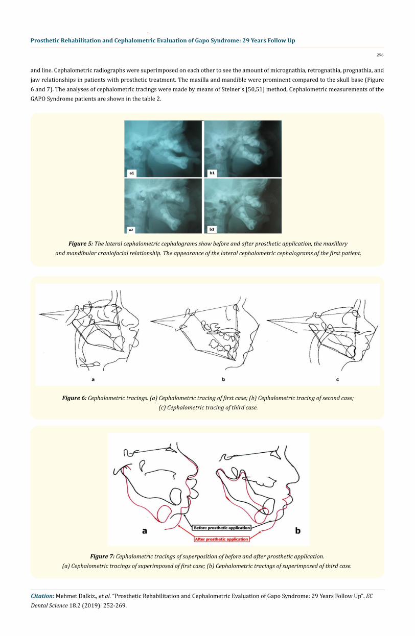
Lateral cephalograms were taken in order to evaluate the maxillomandibular and craniofacial relations and the effect of prosthetic restorations on the maxillofacial relations (Figure 4).
Result
Figure 4: Lateral cephalometric cephalograms show maxillomandibular and craniofacial relationship. (a) Lateral cephalometric cephalograms of the first case; (b) Lateral cephalometric cephalograms of the second case;
(c) Lateral cephalometric cephalograms of the third case.
Before (a1) and after prosthetic application (a2), The appearance of the lateral cephalometric cephalograms of the third patient before (b1) and after (b2) prosthetic application.
The female patient refused prosthetic restorations. Therefore prosthetic restorations were only applied on male patients. In order to evaluate the effects of prosthetic restorations on the maxillofacial relationship, lateral cephalograms were taken while complete pros-thetic dentures were in occlusal contact (Figure 4). The cephalometric tracings were taken before and after prosthetic application (Figure 5) and they were superimposed. Cephalometric analyses were performed to investigate the relation of the facial bones with the skull base and evaluate the syndrome related alterations. These cephalometric measurements were made with the selected cephalometric points
256
Prosthetic Rehabilitation and Cephalometric Evaluation of Gapo Syndrome: 29 Years Follow Up
Citation: Mehmet Dalkiz., et al. “Prosthetic Rehabilitation and Cephalometric Evaluation of Gapo Syndrome: 29 Years Follow Up”. EC Dental Science 18.2 (2019): 252-269.
Figure 5: The lateral cephalometric cephalograms show before and after prosthetic application, the maxillary and mandibular craniofacial relationship. The appearance of the lateral cephalometric cephalograms of the first patient.
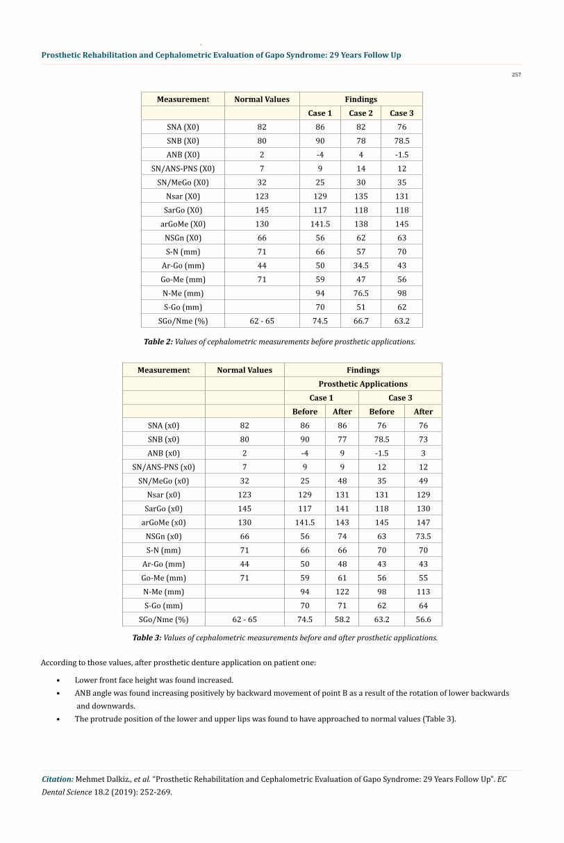
and line. Cephalometric radiographs were superimposed on each other to see the amount of micrognathia, retrognathia, prognathia, and jaw relationships in patients with prosthetic treatment. The maxilla and mandible were prominent compared to the skull base (Figure 6 and 7). The analyses of cephalometric tracings were made by means of Steiner’s [50,51] method, Cephalometric measurements of the GAPO Syndrome patients are shown in the table 2.
Figure 6: Cephalometric tracings. (a) Cephalometric tracing of first case; (b) Cephalometric tracing of second case; (c) Cephalometric tracing of third case.
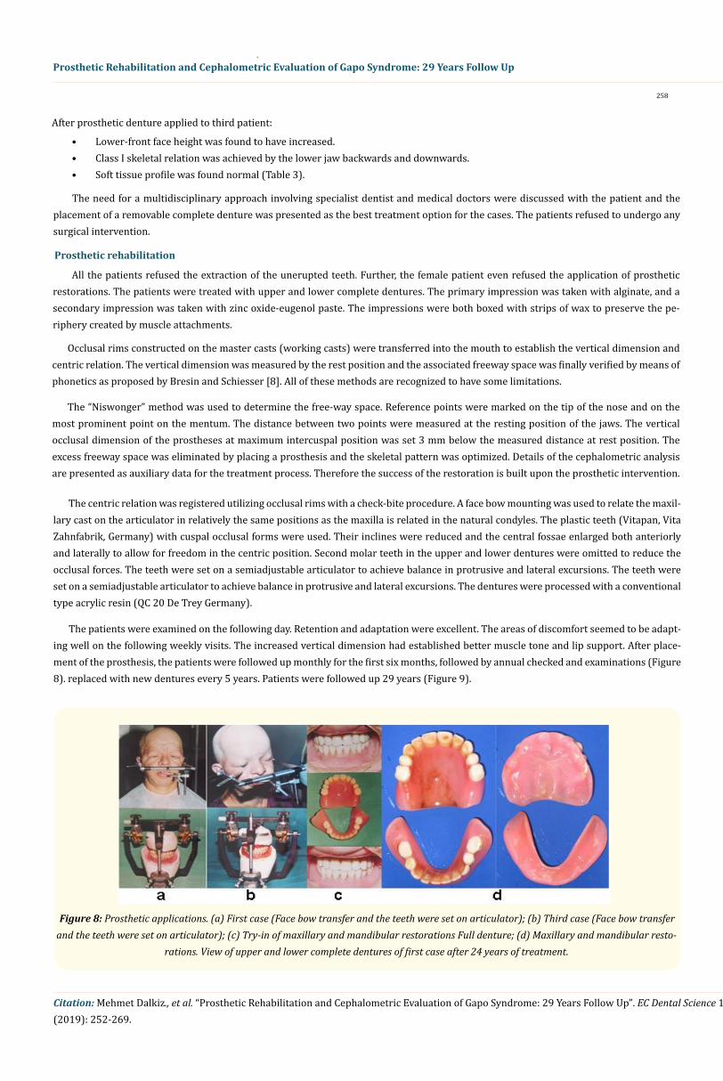
Figure 7: Cephalometric tracings of superposition of before and after prosthetic application. (a) Cephalometric tracings of superimposed of first case; (b) Cephalometric tracings of superimposed of third case.
257
Prosthetic Rehabilitation and Cephalometric Evaluation of Gapo Syndrome: 29 Years Follow Up
Citation: Mehmet Dalkiz., et al. “Prosthetic Rehabilitation and Cephalometric Evaluation of Gapo Syndrome: 29 Years Follow Up”. EC Dental Science 18.2 (2019): 252-269.
According to those values, after prosthetic denture application on patient one:
Measurement Normal Values FindingsCase 1 Case 2 Case 3
SNA (X0) 82 86 82 76SNB (X0) 80 90 78 78.5ANB (X0) 2 -4 4 -1.5
SN/ANS-PNS (X0) 7 9 14 12SN/MeGo (X0) 32 25 30 35
Nsar (X0) 123 129 135 131SarGo (X0) 145 117 118 118
arGoMe (X0) 130 141.5 138 145NSGn (X0) 66 56 62 63S-N (mm) 71 66 57 70
Ar-Go (mm) 44 50 34.5 43Go-Me (mm) 71 59 47 56N-Me (mm) 94 76.5 98S-Go (mm) 70 51 62
SGo/Nme (%) 62 - 65 74.5 66.7 63.2
Table 2: Values of cephalometric measurements before prosthetic applications.
Measurement Normal Values FindingsProsthetic Applications
Case 1 Case 3Before After Before After
SNA (x0) 82 86 86 76 76SNB (x0) 80 90 77 78.5 73ANB (x0) 2 -4 9 -1.5 3
SN/ANS-PNS (x0) 7 9 9 12 12SN/MeGo (x0) 32 25 48 35 49
Nsar (x0) 123 129 131 131 129SarGo (x0) 145 117 141 118 130
arGoMe (x0) 130 141.5 143 145 147NSGn (x0) 66 56 74 63 73.5S-N (mm) 71 66 66 70 70
Ar-Go (mm) 44 50 48 43 43Go-Me (mm) 71 59 61 56 55N-Me (mm) 94 122 98 113S-Go (mm) 70 71 62 64
SGo/Nme (%) 62 - 65 74.5 58.2 63.2 56.6
Table 3: Values of cephalometric measurements before and after prosthetic applications.
• Lower front face height was found increased.• ANB angle was found increasing positively by backward movement of point B as a result of the rotation of lower backwards
and downwards.• The protrude position of the lower and upper lips was found to have approached to normal values (Table 3).
258
Prosthetic Rehabilitation and Cephalometric Evaluation of Gapo Syndrome: 29 Years Follow Up
Citation: Mehmet Dalkiz., et al. “Prosthetic Rehabilitation and Cephalometric Evaluation of Gapo Syndrome: 29 Years Follow Up”. EC Dental Science 18.2 (2019): 252-269.
Occlusal rims constructed on the master casts (working casts) were transferred into the mouth to establish the vertical dimension and centric relation. The vertical dimension was measured by the rest position and the associated freeway space was finally verified by means of phonetics as proposed by Bresin and Schiesser [8]. All of these methods are recognized to have some limitations.
After prosthetic denture applied to third patient:
• Lower-front face height was found to have increased.• Class I skeletal relation was achieved by the lower jaw backwards and downwards.• Soft tissue profile was found normal (Table 3).
The need for a multidisciplinary approach involving specialist dentist and medical doctors were discussed with the patient and the placement of a removable complete denture was presented as the best treatment option for the cases. The patients refused to undergo any surgical intervention.
Prosthetic rehabilitation
All the patients refused the extraction of the unerupted teeth. Further, the female patient even refused the application of prosthetic restorations. The patients were treated with upper and lower complete dentures. The primary impression was taken with alginate, and a secondary impression was taken with zinc oxide-eugenol paste. The impressions were both boxed with strips of wax to preserve the pe-riphery created by muscle attachments.
The “Niswonger” method was used to determine the free-way space. Reference points were marked on the tip of the nose and on the most prominent point on the mentum. The distance between two points were measured at the resting position of the jaws. The vertical occlusal dimension of the prostheses at maximum intercuspal position was set 3 mm below the measured distance at rest position. The excess freeway space was eliminated by placing a prosthesis and the skeletal pattern was optimized. Details of the cephalometric analysis are presented as auxiliary data for the treatment process. Therefore the success of the restoration is built upon the prosthetic intervention.
The centric relation was registered utilizing occlusal rims with a check-bite procedure. A face bow mounting was used to relate the maxil-lary cast on the articulator in relatively the same positions as the maxilla is related in the natural condyles. The plastic teeth (Vitapan, Vita Zahnfabrik, Germany) with cuspal occlusal forms were used. Their inclines were reduced and the central fossae enlarged both anteriorly and laterally to allow for freedom in the centric position. Second molar teeth in the upper and lower dentures were omitted to reduce the occlusal forces. The teeth were set on a semiadjustable articulator to achieve balance in protrusive and lateral excursions. The teeth were set on a semiadjustable articulator to achieve balance in protrusive and lateral excursions. The dentures were processed with a conventional type acrylic resin (QC 20 De Trey Germany).
The patients were examined on the following day. Retention and adaptation were excellent. The areas of discomfort seemed to be adapt-ing well on the following weekly visits. The increased vertical dimension had established better muscle tone and lip support. After place-ment of the prosthesis, the patients were followed up monthly for the first six months, followed by annual checked and examinations (Figure 8). replaced with new dentures every 5 years. Patients were followed up 29 years (Figure 9).
Figure 8: Prosthetic applications. (a) First case (Face bow transfer and the teeth were set on articulator); (b) Third case (Face bow transfer and the teeth were set on articulator); (c) Try-in of maxillary and mandibular restorations Full denture; (d) Maxillary and mandibular resto-
rations. View of upper and lower complete dentures of first case after 24 years of treatment.

259
Prosthetic Rehabilitation and Cephalometric Evaluation of Gapo Syndrome: 29 Years Follow Up
Citation: Mehmet Dalkiz., et al. “Prosthetic Rehabilitation and Cephalometric Evaluation of Gapo Syndrome: 29 Years Follow Up”. EC Dental Science 18.2 (2019): 252-269.
Figure 9: After 13 years of case1 pictures shows intraoral tissues(a), upper and lower complete dentures (b), new complete dentures in the articulator (c) and mouth (d).
Patients
Case 1: In first visit 21 year-old male (born in 1971), 1.44 m tall and weighs 47 kg. He was a carpenter and artist(painter and movie actor); he is the second child of the family. It was found that he had no hair, no beard and must ache, no eyebrows and eyelashes, normal body hair. The suture lines of the cranium were significant. There was a hemangioma on the occipital region. The forehead was protruded forward. The wrinkles on the forehead were deep and significant. The earlobes were large. The eyelids were eudematic. The philtrum was broad (Figure 10-12). The psychomental development was normal.
Figure 10: The clinical photographs are showing frontal view the first patient at 3 years (a) of age and at 6 years (b) of age. Patient had less hair appearance at the age of 3 year (b). By the age of 5 years, she completely lost her hair.
Figure 11: Frontal and lateral views of the patient showing alopecia, bilateral puffiness of upper eyelids, sparse eyebrows, depressed nasal bridge, anteverted wide nostrils, thick everted upper and lower lips, long philtrum and large ears, bossed forehead.
260
Prosthetic Rehabilitation and Cephalometric Evaluation of Gapo Syndrome: 29 Years Follow Up
Citation: Mehmet Dalkiz., et al. “Prosthetic Rehabilitation and Cephalometric Evaluation of Gapo Syndrome: 29 Years Follow Up”. EC Dental Science 18.2 (2019): 252-269.
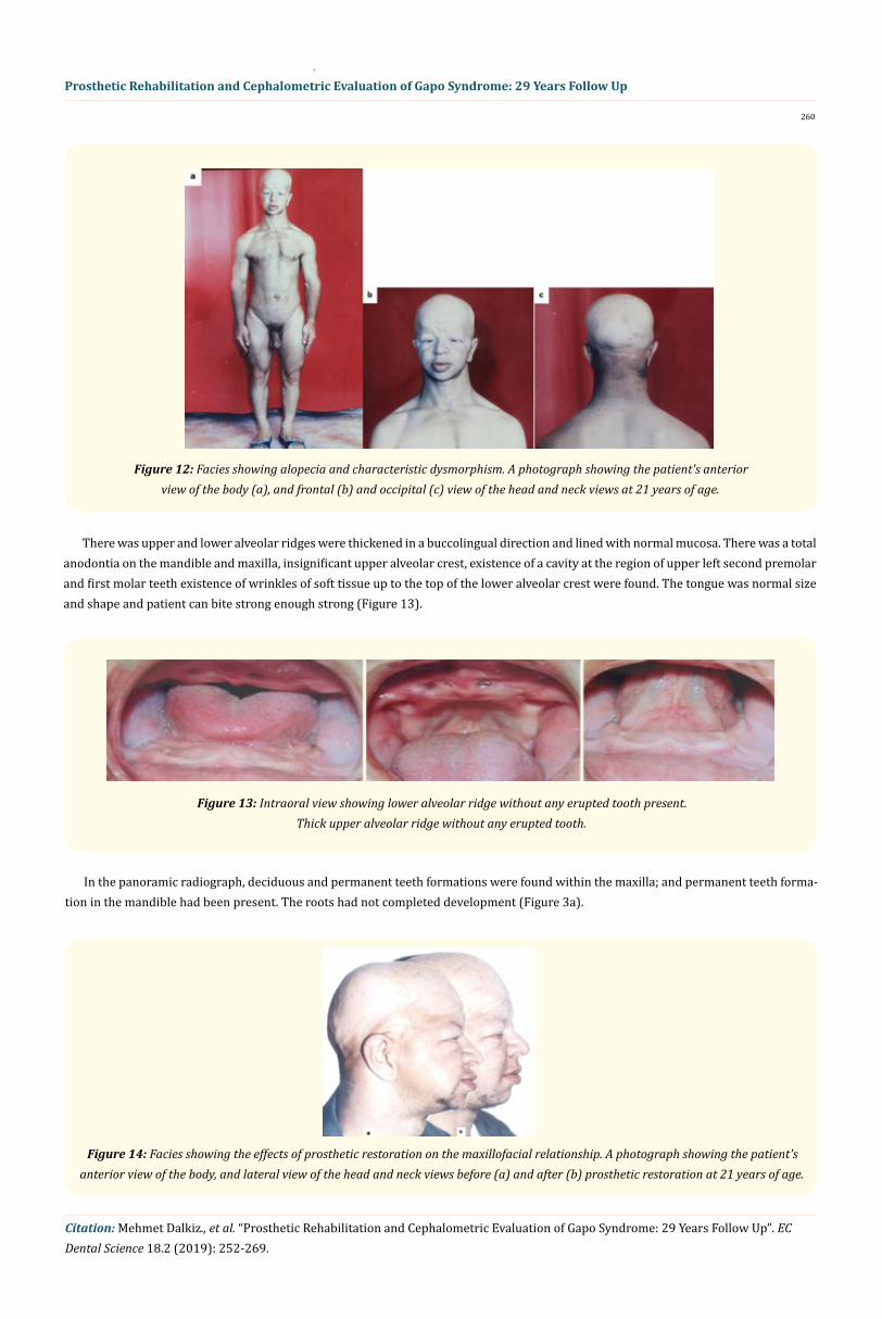
There was upper and lower alveolar ridges were thickened in a buccolingual direction and lined with normal mucosa. There was a total anodontia on the mandible and maxilla, insignificant upper alveolar crest, existence of a cavity at the region of upper left second premolar and first molar teeth existence of wrinkles of soft tissue up to the top of the lower alveolar crest were found. The tongue was normal size and shape and patient can bite strong enough strong (Figure 13).
Figure 12: Facies showing alopecia and characteristic dysmorphism. A photograph showing the patient's anterior view of the body (a), and frontal (b) and occipital (c) view of the head and neck views at 21 years of age.
Figure 13: Intraoral view showing lower alveolar ridge without any erupted tooth present. Thick upper alveolar ridge without any erupted tooth.
In the panoramic radiograph, deciduous and permanent teeth formations were found within the maxilla; and permanent teeth forma-tion in the mandible had been present. The roots had not completed development (Figure 3a).
Figure 14: Facies showing the effects of prosthetic restoration on the maxillofacial relationship. A photograph showing the patient's anterior view of the body, and lateral view of the head and neck views before (a) and after (b) prosthetic restoration at 21 years of age.

261
Prosthetic Rehabilitation and Cephalometric Evaluation of Gapo Syndrome: 29 Years Follow Up
Citation: Mehmet Dalkiz., et al. “Prosthetic Rehabilitation and Cephalometric Evaluation of Gapo Syndrome: 29 Years Follow Up”. EC Dental Science 18.2 (2019): 252-269.
Lateral cephalograms were taken in order to evaluate the maxillomandibular and craniofacial relations and the effect of prosthetic restorations on the maxillofacial relations (Figure 4a). The evaluate the effects of prosthetic restorations on the maxillofacial relationship, lateral cephalograms were taken while complete prosthetic dentures were in occlusal contact (Figure 5a1, a2). The cephalometric mea-surements were made with the selected cephalometric points and line. The maxilla and mandible were prominent compared to the skull base. The analyses of cephalometric tracings were made by means of Steiner’s [50,51] method, Cephalometric measurements of the GAPO Syndrome patients are shown in the table 2. This patient was treated with upper and lower complete dentures. Cephalometric measure-ments before and after prosthetic applications are shown in the table 3. This patient's prostheses were replaced with new dentures every 5 years (Figure 9 and 14). The patient was followed up 29 years (Figure 15).
Figure 15: Facies showing characteristic dysmorphism. A photograph showing the patient's frontal, lateral and occipital view of the head and neck views at 45 years of age in 2016.
Figure 16: The clinical photographs are showing frontal view the second patient at 9 (a) and 13 (b) months of age and at 2 years(c) of age. Patient had hair appearance at the age of from 1 month to 4 year (A). By the age of 5 years, she starts completely lost her hair.
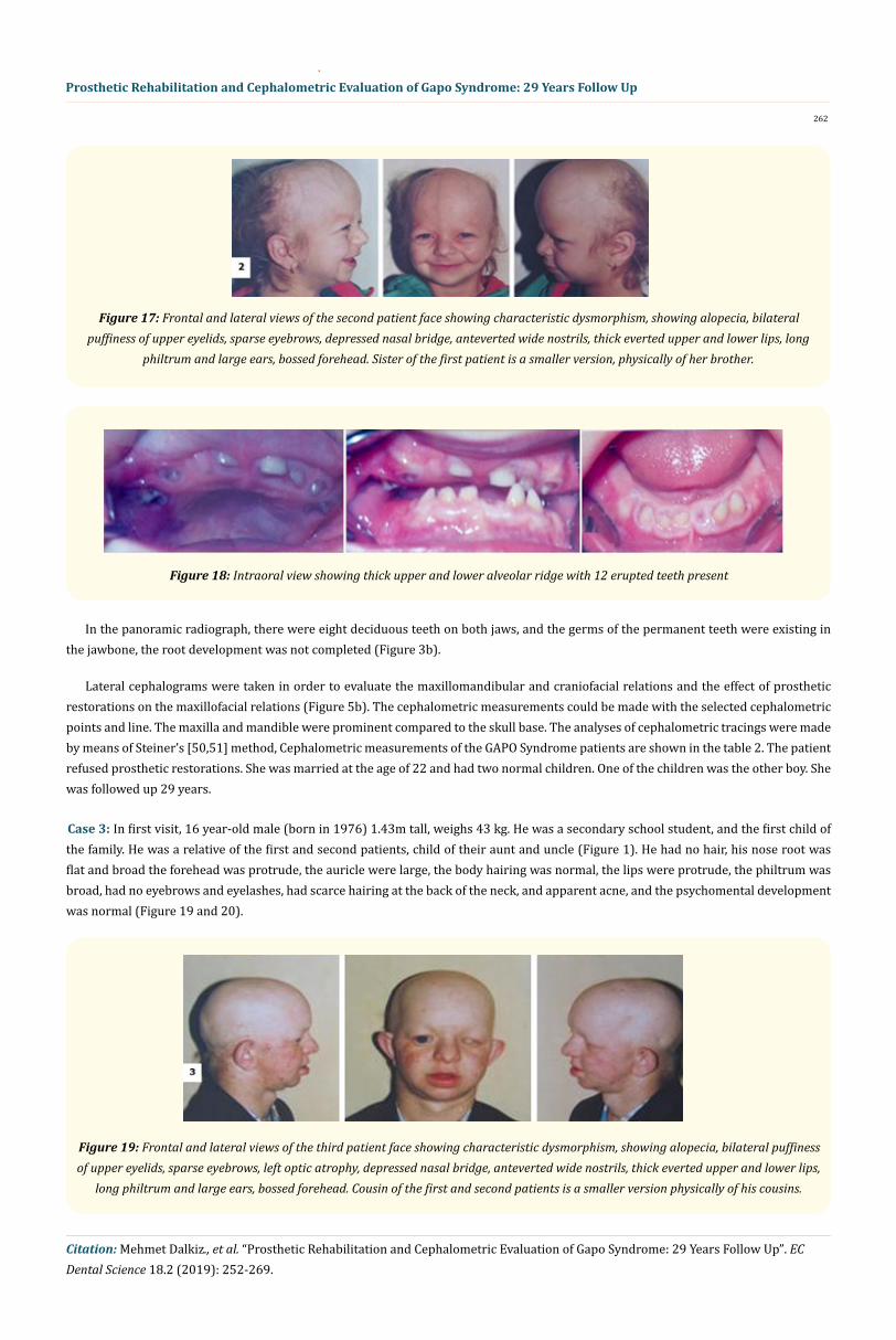
Case 2: In first visit, 9- years- old female (born in 1983), 1.05m tall, weighs 25 kg. She was an elementary school student and the sixth and the youngest child of the family. She has been losing her hair since childhood and her hair was mostly at the parietal, temporal and occipital regions, the forehead was protrude forward, had no eyebrows and eyelashes, eyelids were protrude, protrude, protrusions of zygomatic bones were significant, auricle were large, nose root was flat and broad. The philtrum was broad, and the psychomental devel-opment was normal (Figure 16 and 17).
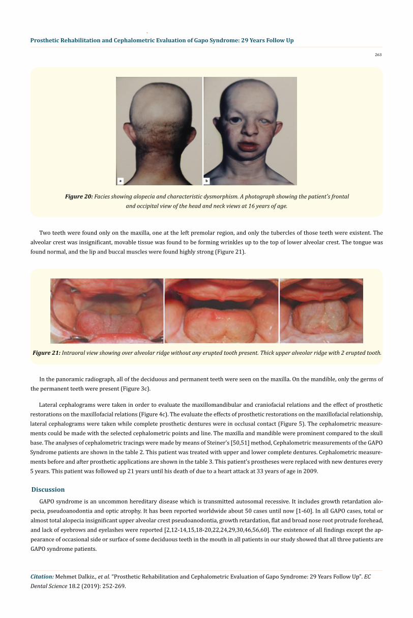
Total twelve teeth, seven on the maxilla and five on the mandible were found. Clinically crown lengths of the teeth fairly short, and there was large diastema between them. The tongue was normal and the alveolar crests were insignificant. The lips and buccal muscles were found highly powerful (Figure 18).
262
Prosthetic Rehabilitation and Cephalometric Evaluation of Gapo Syndrome: 29 Years Follow Up
Citation: Mehmet Dalkiz., et al. “Prosthetic Rehabilitation and Cephalometric Evaluation of Gapo Syndrome: 29 Years Follow Up”. EC Dental Science 18.2 (2019): 252-269.
Figure 17: Frontal and lateral views of the second patient face showing characteristic dysmorphism, showing alopecia, bilateral puffiness of upper eyelids, sparse eyebrows, depressed nasal bridge, anteverted wide nostrils, thick everted upper and lower lips, long
philtrum and large ears, bossed forehead. Sister of the first patient is a smaller version, physically of her brother.
Figure 18: Intraoral view showing thick upper and lower alveolar ridge with 12 erupted teeth present
In the panoramic radiograph, there were eight deciduous teeth on both jaws, and the germs of the permanent teeth were existing in the jawbone, the root development was not completed (Figure 3b).
Lateral cephalograms were taken in order to evaluate the maxillomandibular and craniofacial relations and the effect of prosthetic restorations on the maxillofacial relations (Figure 5b). The cephalometric measurements could be made with the selected cephalometric points and line. The maxilla and mandible were prominent compared to the skull base. The analyses of cephalometric tracings were made by means of Steiner’s [50,51] method, Cephalometric measurements of the GAPO Syndrome patients are shown in the table 2. The patient refused prosthetic restorations. She was married at the age of 22 and had two normal children. One of the children was the other boy. She was followed up 29 years.
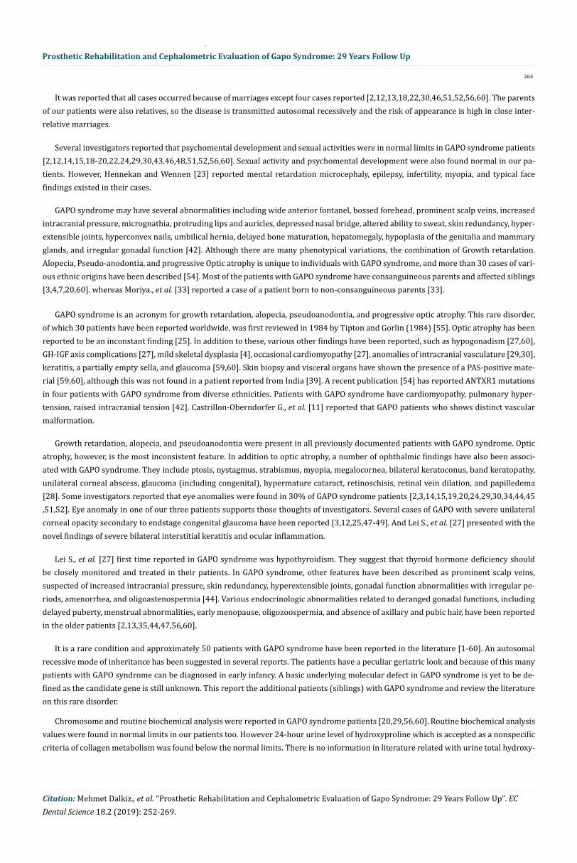
Case 3: In first visit, 16 year-old male (born in 1976) 1.43m tall, weighs 43 kg. He was a secondary school student, and the first child of the family. He was a relative of the first and second patients, child of their aunt and uncle (Figure 1). He had no hair, his nose root was flat and broad the forehead was protrude, the auricle were large, the body hairing was normal, the lips were protrude, the philtrum was broad, had no eyebrows and eyelashes, had scarce hairing at the back of the neck, and apparent acne, and the psychomental development was normal (Figure 19 and 20).
Figure 19: Frontal and lateral views of the third patient face showing characteristic dysmorphism, showing alopecia, bilateral puffiness of upper eyelids, sparse eyebrows, left optic atrophy, depressed nasal bridge, anteverted wide nostrils, thick everted upper and lower lips,
long philtrum and large ears, bossed forehead. Cousin of the first and second patients is a smaller version physically of his cousins.
263
Prosthetic Rehabilitation and Cephalometric Evaluation of Gapo Syndrome: 29 Years Follow Up
Citation: Mehmet Dalkiz., et al. “Prosthetic Rehabilitation and Cephalometric Evaluation of Gapo Syndrome: 29 Years Follow Up”. EC Dental Science 18.2 (2019): 252-269.
Figure 20: Facies showing alopecia and characteristic dysmorphism. A photograph showing the patient's frontal and occipital view of the head and neck views at 16 years of age.
Two teeth were found only on the maxilla, one at the left premolar region, and only the tubercles of those teeth were existent. The alveolar crest was insignificant, movable tissue was found to be forming wrinkles up to the top of lower alveolar crest. The tongue was found normal, and the lip and buccal muscles were found highly strong (Figure 21).
Figure 21: Intraoral view showing over alveolar ridge without any erupted tooth present. Thick upper alveolar ridge with 2 erupted tooth.
In the panoramic radiograph, all of the deciduous and permanent teeth were seen on the maxilla. On the mandible, only the germs of the permanent teeth were present (Figure 3c).
Lateral cephalograms were taken in order to evaluate the maxillomandibular and craniofacial relations and the effect of prosthetic restorations on the maxillofacial relations (Figure 4c). The evaluate the effects of prosthetic restorations on the maxillofacial relationship, lateral cephalograms were taken while complete prosthetic dentures were in occlusal contact (Figure 5). The cephalometric measure-ments could be made with the selected cephalometric points and line. The maxilla and mandible were prominent compared to the skull base. The analyses of cephalometric tracings were made by means of Steiner’s [50,51] method, Cephalometric measurements of the GAPO Syndrome patients are shown in the table 2. This patient was treated with upper and lower complete dentures. Cephalometric measure-ments before and after prosthetic applications are shown in the table 3. This patient's prostheses were replaced with new dentures every 5 years. This patient was followed up 21 years until his death of due to a heart attack at 33 years of age in 2009.
Discussion
GAPO syndrome is an uncommon hereditary disease which is transmitted autosomal recessive. It includes growth retardation alo-pecia, pseudoanodontia and optic atrophy. It has been reported worldwide about 50 cases until now [1-60]. In all GAPO cases, total or almost total alopecia insignificant upper alveolar crest pseudoanodontia, growth retardation, flat and broad nose root protrude forehead, and lack of eyebrows and eyelashes were reported [2,12-14,15,18-20,22,24,29,30,46,56,60]. The existence of all findings except the ap-pearance of occasional side or surface of some deciduous teeth in the mouth in all patients in our study showed that all three patients are GAPO syndrome patients.
264
Prosthetic Rehabilitation and Cephalometric Evaluation of Gapo Syndrome: 29 Years Follow Up
Citation: Mehmet Dalkiz., et al. “Prosthetic Rehabilitation and Cephalometric Evaluation of Gapo Syndrome: 29 Years Follow Up”. EC Dental Science 18.2 (2019): 252-269.
It was reported that all cases occurred because of marriages except four cases reported [2,12,13,18,22,30,46,51,52,56,60]. The parents of our patients were also relatives, so the disease is transmitted autosomal recessively and the risk of appearance is high in close inter-relative marriages.
Several investigators reported that psychomental development and sexual activities were in normal limits in GAPO syndrome patients [2,12,14,15,18-20,22,24,29,30,43,46,48,51,52,56,60]. Sexual activity and psychomental development were also found normal in our pa-tients. However, Hennekan and Wennen [23] reported mental retardation microcephaly, epilepsy, infertility, myopia, and typical face findings existed in their cases.
GAPO syndrome may have several abnormalities including wide anterior fontanel, bossed forehead, prominent scalp veins, increased intracranial pressure, micrognathia, protruding lips and auricles, depressed nasal bridge, altered ability to sweat, skin redundancy, hyper-extensible joints, hyperconvex nails, umbilical hernia, delayed bone maturation, hepatomegaly, hypoplasia of the genitalia and mammary glands, and irregular gonadal function [42]. Although there are many phenotypical variations, the combination of Growth retardation. Alopecia, Pseudo-anodontia, and progressive Optic atrophy is unique to individuals with GAPO syndrome, and more than 30 cases of vari-ous ethnic origins have been described [54]. Most of the patients with GAPO syndrome have consanguineous parents and affected siblings [3,4,7,20,60]. whereas Moriya., et al. [33] reported a case of a patient born to non-consanguineous parents [33].
GAPO syndrome is an acronym for growth retardation, alopecia, pseudoanodontia, and progressive optic atrophy. This rare disorder, of which 30 patients have been reported worldwide, was first reviewed in 1984 by Tipton and Gorlin (1984) [55]. Optic atrophy has been reported to be an inconstant finding [25]. In addition to these, various other findings have been reported, such as hypogonadism [27,60], GH-IGF axis complications [27], mild skeletal dysplasia [4], occasional cardiomyopathy [27], anomalies of intracranial vasculature [29,30], keratitis, a partially empty sella, and glaucoma [59,60]. Skin biopsy and visceral organs have shown the presence of a PAS-positive mate-rial [59,60], although this was not found in a patient reported from India [39]. A recent publication [54] has reported ANTXR1 mutations in four patients with GAPO syndrome from diverse ethnicities. Patients with GAPO syndrome have cardiomyopathy, pulmonary hyper-tension, raised intracranial tension [42]. Castrillon-Oberndorfer G., et al. [11] reported that GAPO patients who shows distinct vascular malformation.
Growth retardation, alopecia, and pseudoanodontia were present in all previously documented patients with GAPO syndrome. Optic atrophy, however, is the most inconsistent feature. In addition to optic atrophy, a number of ophthalmic findings have also been associ-ated with GAPO syndrome. They include ptosis, nystagmus, strabismus, myopia, megalocornea, bilateral keratoconus, band keratopathy, unilateral corneal abscess, glaucoma (including congenital), hypermature cataract, retinoschisis, retinal vein dilation, and papilledema [28]. Some investigators reported that eye anomalies were found in 30% of GAPO syndrome patients [2,3,14,15,19,20,24,29,30,34,44,45,51,52]. Eye anomaly in one of our three patients supports those thoughts of investigators. Several cases of GAPO with severe unilateral corneal opacity secondary to endstage congenital glaucoma have been reported [3,12,25,47-49]. And Lei S., et al. [27] presented with the novel findings of severe bilateral interstitial keratitis and ocular inflammation.
Lei S., et al. [27] first time reported in GAPO syndrome was hypothyroidism. They suggest that thyroid hormone deficiency should be closely monitored and treated in their patients. In GAPO syndrome, other features have been described as prominent scalp veins, suspected of increased intracranial pressure, skin redundancy, hyperextensible joints, gonadal function abnormalities with irregular pe-riods, amenorrhea, and oligoastenospermia [44]. Various endocrinologic abnormalities related to deranged gonadal functions, including delayed puberty, menstrual abnormalities, early menopause, oligozoospermia, and absence of axillary and pubic hair, have been reported in the older patients [2,13,35,44,47,56,60].
It is a rare condition and approximately 50 patients with GAPO syndrome have been reported in the literature [1-60]. An autosomal recessive mode of inheritance has been suggested in several reports. The patients have a peculiar geriatric look and because of this many patients with GAPO syndrome can be diagnosed in early infancy. A basic underlying molecular defect in GAPO syndrome is yet to be de-fined as the candidate gene is still unknown. This report the additional patients (siblings) with GAPO syndrome and review the literature on this rare disorder.
Chromosome and routine biochemical analysis were reported in GAPO syndrome patients [20,29,56,60]. Routine biochemical analysis values were found in normal limits in our patients too. However 24-hour urine level of hydroxyproline which is accepted as a nonspecific criteria of collagen metabolism was found below the normal limits. There is no information in literature related with urine total hydroxy-

265
Prosthetic Rehabilitation and Cephalometric Evaluation of Gapo Syndrome: 29 Years Follow Up
Citation: Mehmet Dalkiz., et al. “Prosthetic Rehabilitation and Cephalometric Evaluation of Gapo Syndrome: 29 Years Follow Up”. EC Dental Science 18.2 (2019): 252-269.
proline levels that may be related with growth retardation in our patients. Bone development was retarded in those patients [26]. In ad-dition, it was reported that hydroxyproline levels were directly associated with growth in children and hydroxyproline in urine was low in children with growth retardation [10].
Meguid., et al. [32] reported that electron microscopic examination of gingival biopsy showed excessive collagen fibers and endothe-lial vascularization, suggesting involvement of extracellular pathological collagenosis, Wajntal., et al. [58,59] and Phadka., et al. [48] also reported that GAPO syndrome lacked PAS positive hyaline material in the skin biopsy from thigh and scalp.
Sandgren [45] reported that GAPO syndrome could be attributed to either ectodermal dysplasia or perhaps and accumulation of extra-cellular connective tissue matrix. Andersen and Pindborg [2], Freire-Maia and Pinherio [17], Saylı and Gül [46], Silva [12], Wanjntal., et al. [58,59] reported GAPO syndrome patients with a Progeria or Progeroid syndrome due to common signs of shortness of length, alopecia normal psychomental development, micrognathia, and the hypoplasia of the middle part of the face. However, Yo and Zeng [60] reported additional findings such as growth anomalies of deciduous and permanent teeth, malpositional teeth, (tendency of being tired easily) and nonexistence of hereditary transmission in Progeria syndrome patients.
Tooth eruption is the axial or occlusal movement from its site of development within the alveolus to its functional position in occlu-sion [38]. Teeth eruption and shedding are continuous process, which replaces the exfoliated primary teeth with permanent teeth. Some-times Impaired tooth eruption takes place in the form of delayed or complete absence of eruption, giving rise to impacted, embedded permanent teeth or retained deciduous teeth [36]. Epidemiologically 25 - 50% of the population shows multiple impactions. Although impaction of teeth is widespread, multiple impacted succedaneum teeth along with numerous retained primary teeth by itself is a seldom condition. An abnormal eruption pattern creates a clinical situation that is challenging to diagnose and treat. The clinical sequence of abnormal tooth eruption includes both syndromic and non-syndromic problems [5,17,54,55].
However, according to numerous reports and literature suggests various syndromes and metabolic conditions to be associated with multiple impacted permanent or supernumerary teeth. Few of them are cleidocranial dysostosis and Gardner’s syndrome, Down syn-drome, Aarskog syndrome, Yunis-Varon syndrome, Zimmerman-Laband syndrome and Noonan’s syndrome along with hormonal distur-bances such as hypothyroidism, hypopituitarism\and hypoparathyroidism [6].
GAPO is the acronymic designation for a syndrome of growth retardation, alopecia, pseudo-anodontia (failure of tooth eruption) and optic atrophy. Patients who suffer from GAPO syndrome may have several abnormalities including broad anterior fontanel, bossed fore-head, micrognathia, pouting lips and auricles, saddle nasal bridge, reduced perspiration [12,13,16,45]. da Silveira., et al. [12,13] reports the case of a young female adult with GAPO syndrome who presented as a peculiar dental finding unerupted primary and permanent dentitions, which resembled total anodontia on clinical examination. A cephalometric analysis was performed to investigate the altera-tions in facial bone development. A cephalometric analysis was performed because none of the previous reports have done so, probably because there has not been too much interest in investigating the orodental manifestations of this syndrome. Cephalometric analysis should be conducted in further GAPO syndrome cases in order to address new characteristic of this disorder. A cephalometric analysis was performed because none of the previous reports have done so, probably because there has not been too much interest in investigat-ing the orodental manifestations of this syndrome. Cephalometric analysis should be conducted in further GAPO syndrome cases in order to address new characteristic of this disorder [45,50].
Anderson and Pindborg [2], Dellac., et al. [14], Epps., et al. [9], Fuks., et al. [18], Gagliardi., et al. [19], Manouvrier-Hanu., et al. [28,29], Shapira., et al. [49] told that the midfacial hypoplasia and micrognathia was found in 60% of reported GAPO syndrome patients. Finding the vertical plane face heights and the mandibular dimensions in normal limits by means of cephalometric examination in three different age patients diagnosed as GAPO syndrome, our findings differ from the findings of those investigators. By reviewing the related literature it is understood that their findings depend on only clinical evaluations. Steiner [50,51], Ricketts [42] and McNamara [31], have claimed that the determination of normal dimensions after linear measurements would reflect the original craniofacial sizes of patients. Refer-ence angles SNA and SNB used in Steiner analysis are prone to be influenced from many factors. However, these reference angles are still frequently used in the literature of orthodontics. Additionally, measurement of the distance between the perpendicular line traced from point S to Frankfurt’s horizontal plane and point A and pogonion, is still frequently used to determine the position of the mandible and the maxilla against the base of the cranium [31].

266
Prosthetic Rehabilitation and Cephalometric Evaluation of Gapo Syndrome: 29 Years Follow Up
Citation: Mehmet Dalkiz., et al. “Prosthetic Rehabilitation and Cephalometric Evaluation of Gapo Syndrome: 29 Years Follow Up”. EC Dental Science 18.2 (2019): 252-269.
Since our findings depend on cephalometric analysis, our results are objective based upon objective data. The improvement from class III skeletal relation to class I skeletal relation according to cephalometric analysis after prosthetic denture application in two patients may be because of pseudoprognathia. Primary failure of eruption is a very rare anomaly and in the truest sense an eruption defect which manifests as a complete failure of eruption without a distinct local or systemic etiology. Diagnosing the condition accurately by follow-ing a methodical approach and rehabilitation of the patient through surgical, orthodontic or by prosthodontic measures is the present accepted approach the condition [58]. While no study to improve the mastication and phonation functions in reported cases was seen, prosthetic denture was applied to improve the mastication and speech in our two male patients. The present study did not evaluate the facial growth. Overlap problems arising from GAPO Syndrome and from teeth absences were prosthetically compensated. Thus, pre- and post-prosthetic measurements of ANS-Me to determine the inferior vertical facial dimension can be useful to demonstrate the compensa-tion of the overlap.
The effects of the prosthetic denture on to vertical dimension and face contours were measured cephalometrically besides clinical evaluation of its effect on mastication and phonation improvement. After the insertion of the dentures, the patients, and their parents reported positive changes in their emotional reactions. The dentures improved aesthetic appearance, by increasing the vertical dimen-sion and supporting the lips. They appeared to be happy, talked spontaneously, and seemed more vigorous. Their speech mastication and nutritional status showed notable improvements with dentures.
We are of the opinion that as a result of this report on three cases with the comments on clinical and laboratory findings contributions have been made to the literature related to GAPO syndrome. While alopecia, growth retardation and pseudoanodontia have been seen in all cases. Optic atrophy has been found only in one male case. Hypoplasia of the middle part of the face and micrognathia have not been found in any of the cases. Finally, it is found that all the cases have normal psychomotor functions. The cephalometric measurements in these three cases with GAPO syndrome revealed that their middle face and jaws were normal, contrary to the previously reports in litera-ture. Hypoplasia of the middle part of the face and micrognathia have not been found in any of the cases. The phenotype of these patients are quite similar to the previously reported cases. Regarding the prosthetic treatments, patients with GAPO syndrome can be well treated by complete removable prosthesis. Prosthetic application like implant supported denture can also be used to treated these patients.
Conclusion
Bibliography
1. Aggarwal S., et al. “GAPO syndrome with deafness: new feature or incidental finding?” Clinical Dysmorphology 22.4 (2013): 161-163.
2. Andersen TH and Pindborg JJ. Et tilfaelde at total “Pseudoanodanty”, 1 Farbindelse med Kranie Deformited, Dvaergvaekst ag Ekto-dermal Dysplasia Odontal Tilster 55 (1947): 484-493.
3. Arti Nanda., et al. “GAPO syndrome: a report of two siblings and a review of literature”. Pediatric Dermatology 27.2 (2010): 156-161.
4. Bacon W., et al. “GAPO syndrome: a new case of this rare syndrome and a review of the relative importance of different phenotypic features in diagnosis”. Journal of Craniofacial Genetics and Developmental Biology 19.4 (1999): 189-200.
5. Bayar GR., et al. “Multiple impacted teeth: Report of 3 cases”. European Journal of Dentistry 2 (2008): 73-78.
6. Bhatia V., et al. “Multiple deciduous and succedaneous teeth in 25 year adult - A rare case”. Indian Journal of Dental Advancements 4.3 (2012): 952-955.
7. Bozkurt B., et al. “GAPO syndrome: four new patients with congenital glaucoma and myelinated retinal nerve fiber layer”. American Journal of Medical Genetics Part A 161A.4 (2013): 829-834.
8. Bresin VE and Schiesser EJ. “The neutral zone in complete dentures”. Principles and technique, First ed, The CV Mosby Comp., St. Lois (1973).
9. Bryan KE and Rubenstein PA. “Allele-specific effects of human deafness gamma-actin mutations (DFNA20/26) on the actin/cofilin interaction”. Journal of Biological Chemistry 284.27 (2009): 18260-18269.

267
Prosthetic Rehabilitation and Cephalometric Evaluation of Gapo Syndrome: 29 Years Follow Up
Citation: Mehmet Dalkiz., et al. “Prosthetic Rehabilitation and Cephalometric Evaluation of Gapo Syndrome: 29 Years Follow Up”. EC Dental Science 18.2 (2019): 252-269.
10. Cabacungan NB., et al. “Hydroxyproline excretion and nutritional status of children”. American Journal of Clinical Nutrition 26.2 (1973): 173-176.
11. Castrillon-Oberndorfer G., et al. “GAPO syndrome associated with craniofacial vascular malformation”. American Journal of Medical Genetics Part A 152A.1 (2010): 225-227.
12. Da Silva EO. “Dwarfism, alopecia, pseudoanodontia and other anomalies. Report of a case”. Brazilian Journal of Genetics 7.4 (1984): 743-747.
13. da Silveira HE., et al. “Dental findings in GAPO syndrome: Case report”. Brazilian Dental Journal 17.3 (2006): 259-262.
14. Dellac N., et al. “Anomalies Ophtalmologiques du Synndrome GAPO (Retard de Croissance, Alopecie, Pseudo-Anodontie, Atrophie Optique A Propos d’un Cas”. Journal Français D’Ophtalmologie 13 (1990): 547-550.
15. Frank CA. “Treatment options for impacted teeth”. Journal of the American Dental Association 131.5 (2000): 623-632.
16. Frazier-Bowers SA., et al. “The etiology of eruption disorders - further evidence of a ‘genetic paradigm’”. Seminars in Orthodontics 16.3 (2010): 180-185.
17. Freire-Mania N and Pinherio M. “Ectodermal dysplasias a clinical and genetic study”. New York: Alan R Liss (1984): 98-100.
18. Fuks A., et al. “Pseudoanodantia, cranial deformity, blindness, alopecia, and dwarfism: a new syndrome”. Journal of Dentistry for Children 45.2 (1978): 155-157.
19. Gagliardi ART., et al. “GAPO Syndrome: report of three affected brothers”. American Journal of Medical Genetics 19.2 (1984): 217-223.
20. Goloni-Bertollo EM., et al. “GAPO syndrome: three new Brazilian cases, additional osseous manifestations, and review of the litera-ture”. American Journal of Medical Genetics Part A 146A.12 (2008): 1523-1529.
21. Gorlin RJ., et al. “Pseudoanodontia, growth retardation and alopecia: a syndrome”. Paper presented at Annual Meeting of American Academy of Oral Pathology Fort Lauderdale, FL (1978).
22. Goucha S., et al. “Le syndrome GAPO”. Annales de Dermatologie et de Vénéréologie 127.5 (2000): 501-504.
23. Hennekam RCM and Reckens-Wennen LGCM. “Acquired alopecia mental retardation, short suture, microcephaly and optic atrophy”. Journal of Medical Genetics 27.10 (1990): 635-636.
24. Ilker SS., et al. “Ophthalmic findings in GAPO syndrome”. Japanese Journal of Ophthalmology 43.1 (1999): 48-52.
25. Kılınç C., et al. “Determination of 24-hour period hydroxproline levels with kloromin-T oxidation”. Optimal Medicinal Journal 4 (1991): 217-223.
26. Kocabay G and Mert M. “GAPO syndrome associated with dilated cardiomyopathy: an unreported association [letter]”. American Journal of Medical Genetics Part A 149A (2009): 415-416.
27. Lei S., et al. “GAPO syndrome: a case associated with bilateral interstitial keratitis and hypothyroidism”. Clinical Dysmorphology 19.2 (2010): 79-81.
28. Manovrier-Hanu S., et al. “Brief clinical report: The GAPO syndrome”. American Journal of Medical Genetics 26 (1987): 683-688.
29. Manovrier-Hanu S., et al. “Le Syndrome GAPO. A proopos d’un Nouveau”. Journal of Human Genetics 36 (1988): 373-378.
30. Mc Kusick VA. “Mendelian inheritance in man catalogs of autosomal dominant, autosomal recessive and X-linked phenotypes 8th edition”. Baltimore the Johns Hopkins University Press (1988): 951.
31. McNamara JA and Mc Namara JA. “A method of cephalometric evaluation”. American Journal of Orthodontics and Dentofacial Ortho-pedics 86.6 (1984): 449-469.

268
Prosthetic Rehabilitation and Cephalometric Evaluation of Gapo Syndrome: 29 Years Follow Up
Citation: Mehmet Dalkiz., et al. “Prosthetic Rehabilitation and Cephalometric Evaluation of Gapo Syndrome: 29 Years Follow Up”. EC Dental Science 18.2 (2019): 252-269.
32. Meguid NA., et al. “GAPO syndrome: first Egyptian case with ultrastructural changes in the gingiva”. Clinical Genetics 52.2 (1997): 110-115.
33. Moriya N., et al. “GAPO syndrome report on the first case in Japan”. American Journal of Medical Genetics 58.3 (1995): 257-261.
34. Mullaney PR., et al. “Growth retardation, alopecia, pseudoanodontia, and optic atrophy (GAPO syndrome) with congenital glau-coma”. Archives of Ophthalmology 115.7 (1997): 940-941.
35. Noffke CE., et al. “Impaired tooth eruption: A review”. South African Dental Journal 60.10 (2005): 422, 424-425.
36. Orbak Z., et al. “GAPO syndrome: first patient with partially empty sella”. Journal of Pediatric Endocrinology and Metabolism 15.6 (2002): 865-868.
37. Patil N., et al. “Single Family Members with Multiple Impacted Teeth: A Rare Case Report”. IJSS Case Reports and Reviews 1.9 (2015): 1-4.
38. Phadke SR., et al. “GAPO syndrome in a child without dermal hyaline deposit”. American Journal of Medical Genetics 51.3 (1994): 191-194.
39. Procaccio V., et al. “A mutation of beta-actin that alters depolymerization dynamics is associated with autosomal dominant develop-mental malformations, deafness, and dystonia”. American Journal of Human Genetics 78.6 (2006): 947-960.
40. Qu C., et al. “The role of the cytoskeleton in the formation of gap junctions by Connexin 30”. Experimental Cell Research 315.10 (2009): 1683-1692.
41. Ramaswamy AH., et al. “Anesthetic Management of A Case of GAPO Syindrome for Debridement of Mandibular Osteomyelitis”. Inter-national Journal of Recent Scientific Research 5.8 (2014): 1422-1424.
42. Ricketts RM. “The influence of orthodontic treatment on facial growth and development”. Angle Orthodontist 30.3 (1960): 103.
43. Rim PHH and Marques-de-Faria AP. “Ophthalmic Aspects of GAPO Syndrome: Case Report and Review”. Ophthalmic Genetics 26.3 (2005): 143-147.
44. Russell LJ., et al. “The GAPO syndrome: a disorder of glycosaminoglycan metabolism”. American Journal of Human Genetics 51 (1992): A107.
45. Sandgren G. “GAPO syndrome: a new case”. American Journal of Medical Genetics 58.1 (1995): 87-90.
46. Saylı BS and Gül D. “A progeroid with the extension of connective tissue fibers of subepidermal tissue a new entity”. The 24th Annual Meeting of the European Society of Human Genetics, El Sinore, Denmark (1992).
47. Saylı BS and Gül D. “GAPO syndrome in three relatives in a Turkish kindred”. American Journal of Medical Genetics 47.3 (1993): 342-345.
48. Saylı BS and Gül D. “GAPO syndrome in Türkiye”. American Journal of Medical Genetics 65.3 (1996): 252-253.
49. Shapira Y., et al. “Growth retardation, alopecia, pseudoanodontia, and optic atrophy”. Synd Ident, Case report 85.8 (1992): 14-16.
50. Steiner CC. “Cephalometrics for you and me”. American Journal of Orthodontics 39.10 (1953): 729-755.
51. Steiner CC. “Cephalometrics in clinical practice”. American Journal of Orthodontics 29.1 (1959): 8-29.
52. Stranecky´ V., et al. “Mutations in ANTXR1 cause GAPO syndrome”. American Journal of Human Genetics 92.5 (2013): 792-799.
53. Sujatha G., et al. “Idiopathic multiple impacted unerupted teeth: Case report and discussion”. Journal of Oral and Maxillofacial Pathol-ogy 16.1 (2012): 125-127.

269
Prosthetic Rehabilitation and Cephalometric Evaluation of Gapo Syndrome: 29 Years Follow Up
Citation: Mehmet Dalkiz., et al. “Prosthetic Rehabilitation and Cephalometric Evaluation of Gapo Syndrome: 29 Years Follow Up”. EC Dental Science 18.2 (2019): 252-269.
Volume 18 Issue 2 February 2019© All rights reserved by Mehmet Dalkiz., et al.
54. Suri L., et sl. “Delayed tooth eruption: Pathogenesis, diagnosis, and treatment. A literature review”. American Journal of Orthodontics and Dentofacial Orthopedics 126.4 (2004): 432-445.
55. Tipton RE and Gorlin RJ. “Growth retardation, Alopecia, Pseudoanodontia, and Optic atrophy-the GAPO syndrome”. American Jour-nal of Medical Genetics 19.2 (1984): 209-217.
56. Touzri RA., et al. “Ocular manifestations in GAPO syndrome. Report of the first Tunisian case”. Journal Français D’Ophtalmologie 26.10 (2003): 1067-1070.
57. Vijesh Kamath P., et al. “Primary eruption failure: A review”. International Journal of Applied Dental Sciences 1.4 (2015): 149-151.
58. Wajntal A., et al. “Novasindrome de displasia ectodermia: Nanismo, Alopecia, Anodontiae, Cutix Laxa”. Cienc Cult 34 (1982): 705.
59. Wajntal A., et al. “GAPO syndrome: a connective tissue disorder: report on two affected sibs and on the pathologic findings in the older”. American Journal of Medical Genetics 37.2 (1990): 213-223.
60. Yo OX and Zeng LH. “Progeria: report of a case and review of the literature”. Journal of Oral Pathology and Medicine 20.2 (1991): 86-88.

![Autosomal recessive ichthyosis with limb reduction defect ... · including autosomal dominant, autosomal recessive and X-linked inheritance [1,2]. Associated cutaneous and extracutaneous](https://img.pdfslide.net/doc/110x75/5ec8c9b91adfdf12ab3e663c/autosomal-recessive-ichthyosis-with-limb-reduction-defect-including-autosomal.jpg)