Embed Size (px)
Citation preview
CroniconO P E N A C C E S S PHARMACOLOGY AND TOXICOLOGY
Research Article
Sonu Mishra* and Virendra S Gomase
Department of Biotechnology, Mewar University, India
Received: February 09, 2016; Published: February 25, 2016
*Corresponding Author: Sonu Mishra, Department of Biotechnology, Mewar University, Chittorgarh, India.
Prediction of MHC Binding peptides and Antigenic peptides of Hsp70 from GWD
Abstract
The parasitic nematode disease (Dracunculus medinensis) which is directly related to the environmental declension and contamination are major concern of the public health, not only by the morbidity and mortality that these waterborne pathogens cause, but mostly by the higher cost represents their prevention and treatment. Despite the continued efforts to maintain water safety, waterborne outbreaks are still reported globally and its systematic treatment is the major issue. In this assay, we predicted the binding peptides of the MHC class I and MHC class II by PSSM and SVM algorithms. Antigenicity, Solvent accessibility, polar and nonpolar residue of protein is also analyzed. The regions that are likely exposed on the surface of proteins which are potentially antigenic that allows potential drug targets to identify active sites against infection as well as to design effective drug to treat the diseases or infections. In this assay, we predicted the binding affinity of Hsp70 having 647 amino acids, which shows 639 nonamers. In this study, we found the SVM based MHCII-I_Ab peptide regions, 371-AYGAAVQAA,603-VCNPIITKM,429-FTTYSDNQP,460- FELSGIPPA(optimal score is35.632); MHCII-I_Ad peptide re-gions, 237-RMVNHFVAE,33-QGNRTTPSY,533-QKDRIAAKN,374-AAVQAAILS(optimal score is 53.145);MHC-II I_Ag7 peptide regions 519-MVQEAEKYK,188-KKGHGERNV,181-AIAYGLDKK,229-LGGEDFDNR, 365-NPDEAVAYG (optimal score is 40.873) which represented the predicted binders from Hsp70 protein. The method integrates prediction of peptide MHC class I binding; proteosomal C terminal cleav-age and TAP transport efficiency of the protein of GWD. Thus a small fragment of antigen can stimulate immune responses against whole antigen. This theme can be implemented in designing subunit and synthetic peptide vaccines.
Keywords: Dracunculiasis; Epitopes; Antigenic peptides; MHC-Binders; Tappred; PSSM; SVM; Nonamers; heat shock protein 70
Citation: Sonu Mishra and Virendra S Gomase. “Prediction of MHC Binding peptides and Antigenic peptides of Hsp70 from GWD”. EC Pharmacology and Toxicology 2.1 (2016): 36-53.
AbbreviationsMHC I: Major Histocompatibility Complex-Class IMHC I: Major Histocompatibility Complex-Class IIPSSM: Position Specific Scoring MatricesSVM: Support Vector MachineGWD: Guinea worm disease UniProt: The Universal Protein ResourceNCBI: National Center for Biotechnology InformationTAP: Transporter Associated with Antigen Processing HPLC: High Performance Liquid ChromatographyTapPred: TAPPred is an on-line service for predicting binding affinity of peptides toward the TAP transporter. The prediction of TAP bind-ing peptides is important in order to identify the MHC class-1 restricted T cell epitopes. The Prediction is based on cascade SVM, using sequence and properties of the amino acids. The correlation coefficient of 0.88 was obtained by using jack-knife validation test.Rankpep: This server predicts peptide binders to MHCI and MHCII molecules from protein sequence/s or sequence alignments using Position Specific Scoring Matrices (PSSMs). In addition, it predicts those MHCI ligands whose C-terminal end is likely to be the result of proteasomal cleavage.
Prediction of MHC Binding peptides and Antigenic peptides of Hsp70 from GWD37
Citation: Sonu Mishra and Virendra S Gomase. “Prediction of MHC Binding peptides and Antigenic peptides of Hsp70 from GWD”. EC Pharmacology and Toxicology 2.1 (2016): 36-53.
The mild and non-lethal heat shock protects cells of various origins induced by a subsequent severe heat shock as well as lethal stimuli [1-5] against cell death , which has been noticed by the several individual group s of the investigators. Later on ,its cleared that the enhanced cell survival was intimately linked to the induction and accumulation of heat-inducible proteins and especially to that of a 70kD protein that was designated as heat shock protein 70(Hsp70) [6-9] Hsp70 found to be the most conserved protein in evolution [10-12] and the Hsp70 protein family is highly homologous protein with overlapping and distinct function [13]. Study reveals that the membrane-bound or extracellularly located HSPs act as danger signals and elicit immune responses mediated by the adaptive or innate immune system.
Heat shock proteins (HSP) belong to the protein family where cell produces the response with respect to the exposure to stressful conditions such as cold [14], UV light [15] and during wound healing or tissue remodeling [16].There are group of the HSP protein which function as the chaperone like by stabilizing new proteins in order to ensure correct folding or by helping to refold proteins which are undergone any damaged due to cell stress [17]. The HSP’s virtual abundance has been noticed from bacteria to humans i.e almost all liv-ing organisms. HSP-70 protein is named according to its molecular weight. Hsp 70s (70-kDa) proteins provide assistance in wide range of folding processes , which includes the folding of protein, assembly of newly synthesized proteins, refolding of misfolded protein and aggregated proteins, membrane translocation of organellar and secretory proteins, and control of the activity of regulatory proteins[18-24]. This protein has also perform housekeeping functions in the cell in which they are built-in components of folding and signal trans-duction pathways, and in quality control functions this protein proofread the structure of proteins and repair misfolded conformers. This functional activity of this protein is based on the attribute of Hsp70 to interact with hydrophobic peptide segments of proteins in an ATP-controlled fashion. The two most distinct functional regions of HSP 70 are (1) peptide binding domain (PBD) and (2) the amino-terminal ATPase domain (ABD). Peptide binding domain holds a groove with an affinity for neutral, hydrophobic amino acid residues. Whereas C-terminal /amino terminal domain – rich in alpha helical structure acts as a ‘lid’ for the substrate binding domain. When the HSP70 is ATP bounded ,it open up the lid for peptide binding and release relatively rapidly, whereas, when this protein is ADP bounded, it usually shut down the lid, and peptides are closely bound to the substrate binding domain. In the malignant melanoma the over expressed Hsp 70 protein has been found [25] and under expression in renal cell cancer [26]. The extracellular HSPs act as powerful way of sending a “Risk signal” to the immune system, to generate response against infection or disease. The predicted antigen protein from GWD might play an important role in the new paradigms of synthetic vaccine development and target validation. By considering the importance of the HSP protein, we have used this protein to study the antigenicity of protein, its solvent accessibility, polar and nonpolar residue to analyse the regions that are likely exposed on the surface of proteins which could be the potentially antigenic that allows potential drug targets to identify active sites against infection as well as to design effective drug to treat Dracunculiasis. D.medinesis (a little dragon from Medina) is the causative agent and it is the only species from the 12 species of Dracunculus [27-30] which infects humans, com-monly known as “Guinea worm disease (GWD)”. The other Dracunculus species generally resides in the internal tissues and body cavities of non-human mammals and reptiles (snake and turtles) [31]. This little dragon undergo a very unusual life cycle of six developmental stages with incubation period last for 1 to one an half years approximately [32]. This is one of the most neglected tropicalparasites which bears clinical importance and needs to be eradicated after small pox [33]. After reaching to the maturation stage, these worms copulate and an adult female produces millions of eggs in its uterus whereas male dies. Later on stage, this female worms release the larvae which trigger a painful blister of diameter of 1 to 6cm, unremarkably and predominantly localized on the skin of lower limbs (80-90%) in most of the reported cases). The infected person develops slight fever, localskinredness, swelling and severe pruritus around the blister. Other symptoms includes: diarrhea, nausea, vomiting and dizziness. The blister burst within three days and female worms one or more slowly comes out from the wounds which causes an excoriating burning sensation and pain [34]. Immersing or pouring water over the blister provides pain relief. But this is only the moment that adult female is exposed to the external environment [35]. The uniqueness of this worm can be observed when the patients emerge the limbs in open water sources, this worm smartly recognizes the temperature dif-ference and start releasing milky white liquid in the water which holds millions of immature larvae, when larvae released in water are ingested by copepods where they mount twice and become infective larvae within two weeks [36].
Introduction
Prediction of MHC Binding peptides and Antigenic peptides of Hsp70 from GWD38
Citation: Sonu Mishra and Virendra S Gomase. “Prediction of MHC Binding peptides and Antigenic peptides of Hsp70 from GWD”. EC Pharmacology and Toxicology 2.1 (2016): 36-53.
It has been seen that, the single epitope can accelerate and generate the immune response in large population. This approach is usu-ally based on the phenomenon of cross-protection. The World Health Assembly called in the 1986 in order to dracunculiasis elimination. The global Guinea Worm Eradication Program, supported by The Carter Center, World Health Organization (WHO), UNICEF, CDC, and other partners. In 1986, an estimated 3.5 million cases occurred each year in 20 countries in Africa and Asia. Dracunculiasis remains endemic in four countries in 2014 (South Sudan, Chad, Mali, and Ethiopia), but the number of overall reported incidence is decreases in 2013 by 73%and in 2014 by 71% compared with 2012. The failures in surveillance and containment is due to lack of clean drinking water, insecurity in Mali and parts of South Sudan, and an unusual epidemiologic pattern in Chad are the main remaining challenges to dracunculiasis eradication [37]. A case of Onchocerca volvulus has been reported in the Cameroon which is mimicking Dracunculus-medinensis [38]. More than two decades after the International Drinking Water Supply and Sanitation Decade (IDWSSD) implemented by the United Nations(1981-1990) [39], the disease still lingers, underscoring the daunting challenge of disease control, as has been the case of the failure of previous attempts to eradicate diseases like malaria, hookworm and yaws [40]. Till date there is no accurate and efficient curative drug of vaccine is available against Dracunculiasis [41]. The investigation suggests that the immunity is not developed by the infected individual [42,43].The specific antibodies (total, IgG1 and IgG4) of GWD has been noticed significantly higher than the levels measured in the same individuals eight months later during the time of patency [44].It was observed that the mean level of specific IgG1 and IgG4 is higher during the month of potency of the infected individual where as variation in the IgE value is relatively negotiable and constant before, during the infection and after the recovery. There is possibility that variation in antibody production is regulated by infected larvae (i.e by transmission) and / or by adult worms (i.e by patency) is still need to be clear out. It is possibility that increased production of IgG1, IgG4 during the time of patency plays a role in blocking or protecting immune responses otherwise it could have killed ingested infected larvae [45]. The antigenic peptides from GWD can be the most desirable segment for the development of peptide vaccine [46]. In this study we have study the MHC binding peptide. MHC molecules are cell surface protein that binds to the peptides derived from host or antigenic proteins and present them to cell surface for recognition by T-cells. T cell recognition is animportant mechanism of the adaptive immune system by which the host identifies and responds to foreign antigens [46,47]. The MHC molecule ex-ists in two polymorphic form i.e MHC I & MHC II. The peptide presented by MHC class I molecules from proteins that synthesized within the cell, whereas, MHC class II molecule present peptides derived from endocytosed extracellular proteins. MHC molecules have been well characterized due to their role in immune reactions and they take active part in host immune reactions and involvement of MHC class molecule in response to almost all antigens and it give impacts on specific sites. The participation of MHC I molecule in response to approximately all antigens makes the study more interesting. They bind to some of the peptide fragments generated after proteolytic cleavage of antigen [48]. Identification of MHC-binding peptides and T-cell epitopes helps improve our understanding of specificity of immune responses [49-52]. Antigenic peptides are most suitable for peptide vaccine development because single epitope can generate large the immune response [53-55].
The antigenic protein sequence of Hsp70 from Dracunculus medinensis was retrieved from www.ncbi.nlm.nih.gov, UniProt databases are initially the most important [56-58].
Prediction of antigenicity program predicts those segments from Antigen Hsp70 protein that are likely to be antigenic by eliciting an antibody response. In this research work antigenic epitopes of Dracunculus medinensis antigen Hsp70 are determined by using the Hopp and Woods, Welling, Parker, Bepipred ,Kolaskar and Tongaonkar antigenicity methods [59-64].
Methodology
Database Searching
Prediction of Antigenicity
The major histocompatibility complex (MHC) peptide binding of Dracunculus medinensis is predicted using neural networks trained on C terminals of known epitopes. Rankpep predicting tool predicts peptide binders to MHC-I ligands using PSSMs, whose C-terminal end is likely to be the result of proteosomal cleavage. The sequence similarity is observed to the peptides that bind to a given MHC
Prediction of Mhc Binding Peptide
Prediction of MHC Binding peptides and Antigenic peptides of Hsp70 from GWD39
Citation: Sonu Mishra and Virendra S Gomase. “Prediction of MHC Binding peptides and Antigenic peptides of Hsp70 from GWD”. EC Pharmacology and Toxicology 2.1 (2016): 36-53.
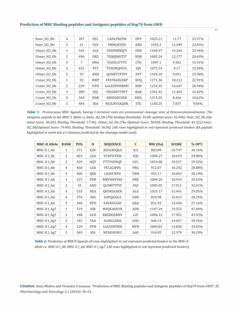
In the present study, we predicted the cascade SVM based several TAP binders which was based on the sequence and the features of amino acids [70]. We found the MHCI binding regions (Table- 3), the binding affinity of Dracunculus medinensis.
Prediction of Antigenic Peptides by Cascade SVM based TAPPred method
molecule. Traditionally, the sequence patterns used for the prediction of peptides binding to MHC molecules. Such sequence patterns, however, have proven to be too simple, as the complexity of the binding motif cannot be precisely represented by the few residues pres-ent in the pattern [65]. RANKPEP uses “Position Specific Scoring Matrices (PSSMs) or profiles” from set of aligned peptides known to bind to a given MHC molecule as the predictor of MHC-peptide binding and overcome the complexity of the binding motif limitation. RANKPEP web server is a variability masking feature to focus on the prediction of conserved epitopes, which could thus help to avoid immune evasion resulting from mutation. Support Vector Machine (SVM) based method for prediction of promiscuous MHC class II binding peptides from protein sequence; SVM has been trained on the binary input of single amino acid sequence [66-69].
We also analyzed the solvent accessible regions of proteins having highest probability that a given protein region lies on the surface of a protein Surface Accessibility, backbone or chain flexibility by Emini., et al. [71] and Karplus and Schulz [72]. The different scales were used to predict the hydrophobic and hydrophilic characteristics of amino acids which is rich in charged and polar residues. The methods used are Sweet., et al. (1983), Kyte& Doolittle (1982), Abraham & Leo(19987), Bull and Breese (1974), Guy (1985), Miyazawa, ., et al. (1985), Roseman (1988), Wolfenden., et al. (1981), Wilson., et al. (1981), Cowan (1990),Chothia (1976) [73-82].
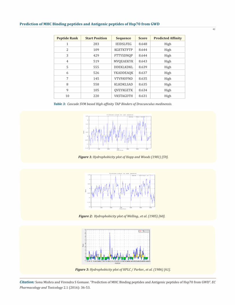
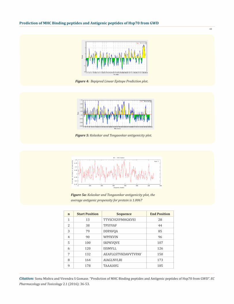
In this prediction, we investigated the area of greatest local Hydrophilicity through antigenic determinants . In the Hopp-Woods scale Hydrophilicity Prediction Result Data found high in Position: 570, Score: 2.467 (max) i.e., 567-DRKKIED-573 in a protein, as-suming that the antigenic determinants would be exposed on the surface of the protein and thus would be located in hydrophilic regions (Figure 1). Welling & al antigenicity plot provides value as the log of the quotient between percentage in average proteins and percentage in a sample of known antigenic regions. The prediction result found highest in Position: 251 Score: 1.453 (max) i.e., 248-RKHKKDL-554 (Figure 2). We also study Hydrophobicity plot of HPLC / Parker Hydrophilicity Prediction Result Data found in Position: 531, Score: 6.600 (max) 528-ADD EAQK-534 (Figure 3), BepiPred predicts the location of linear B-cell epitopes Result found in posi-tion: 630(Residue: A) max score: 2.548 i.e., 627-MYQSAGG-633 (Figure 4), Kolaskar and Tongaonkar antigenicity methods (Figure 5) Predicted peptides result found i.e
Solvent Accessible Regions
Prediction of Antigenic Peptides
Results The Dracunculusmedinensis antigen Hsp70, contain a long residue of 647 amino acids with 639 nonamers.MAKHNAVGIDLGTTYSCVGVFMHGKVEIIANDQGNRTTPSYVAFTDTERLIGDAAKNQVAMNPNNTVFDAKRLIGRRFDDPAVQADMKHWP-FKVINAEGSKPKVQVEYKGETKTFTPEEISSMVLLKMKETAEAFLGSTVKDAVVTVPAYFNDSQRQATKDAGAIAGLNVLRIINEPTAAAIAYGLDK-KGHGERNVLIFDLGGGTFDVSILTIEDGIFEVKSTAGDTHLGGEDFDNRMVNHFVAEFKRKHKKDLSTNPRALRRLRTACERAKRTLSSSSQASIE-IDSLFEGIDFYTNITRARFEELCADLFRSTMDPVEKALRDAKMDKSQMHDIVLVGGSTRIPKVQKLLSDFFSGKELNKSINPDEAVAYGAAVQAAILS-GDKSEAVQDLLLLDVAPLSLGIETAGGVMTALIKRNTTIPTKTAQTFTTYSDNQPGVLIQVFEGERAMTKDNNLLGKFELSGIPPAPRGVPQIEVTF-DIDANGILNVSAQDKSTGKQNKITITNDKGRLSKDEIERMVQEAEKYKADDEAQKDRIAAKNALESYAFNMKQTIDDEKLKDKLSADDRKKIEDKC-DEIIKWLDRNQTAEKDEFEHHQKELEAVCNPIITKMYQSAGGMPGNPGGFPGGGAPGGGHQGGGGPTIEEVD
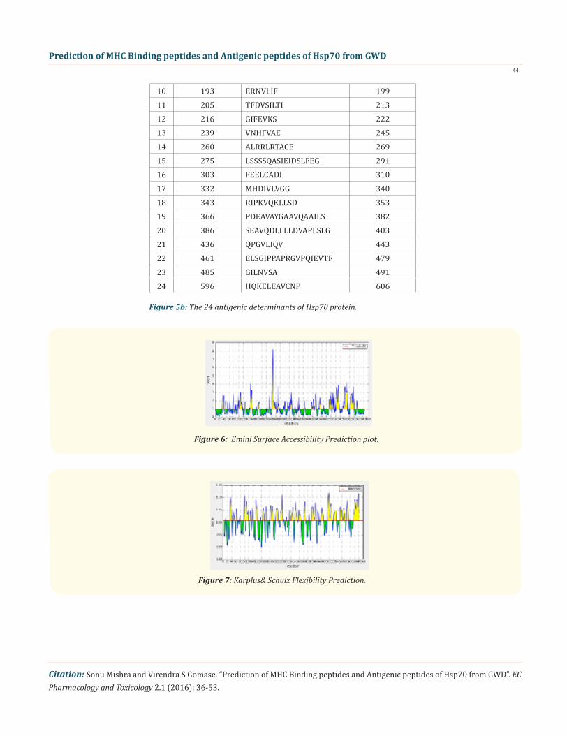
13-TTYSCVGVFMHGKVEI-28,38-TPSYVAF-44,79-DDPAVQA-85,90-WPFKVIN-96,100-SKPKVQVE-107,120-ISSMVLL-126,132-AEAFLGSTVKDAVVTVPAY150,164-AIAGLNVLRI-173,178-TAAAIAYG-185,193-ERNVLIF-199,205-TFDVSILTI-213,216-GIFEVKS-222,239-VNHFVAE-245,260-ALRRLRTACE269,275-LSSSSQASIEIDSLFEG-291,303-FEELCADL-310,332-MHDIVLVGG-340,343-RIP-KVQKLLSD-353,366-PDEAVAYGAAVQAAILS-382,386-SEAVQDLLLLDVAPLSLG-403,436-QPGVLIQV-443,461-ELSGIPPAPRGVPQIEVTF-479, 485-GILNVSA-491,596-HQKELEAVCNP-606 and the predicted antigenic fragments can bind to MHC molecule is the first bottlenecks in vaccine design.
Prediction of MHC Binding peptides and Antigenic peptides of Hsp70 from GWD40
Citation: Sonu Mishra and Virendra S Gomase. “Prediction of MHC Binding peptides and Antigenic peptides of Hsp70 from GWD”. EC Pharmacology and Toxicology 2.1 (2016): 36-53.
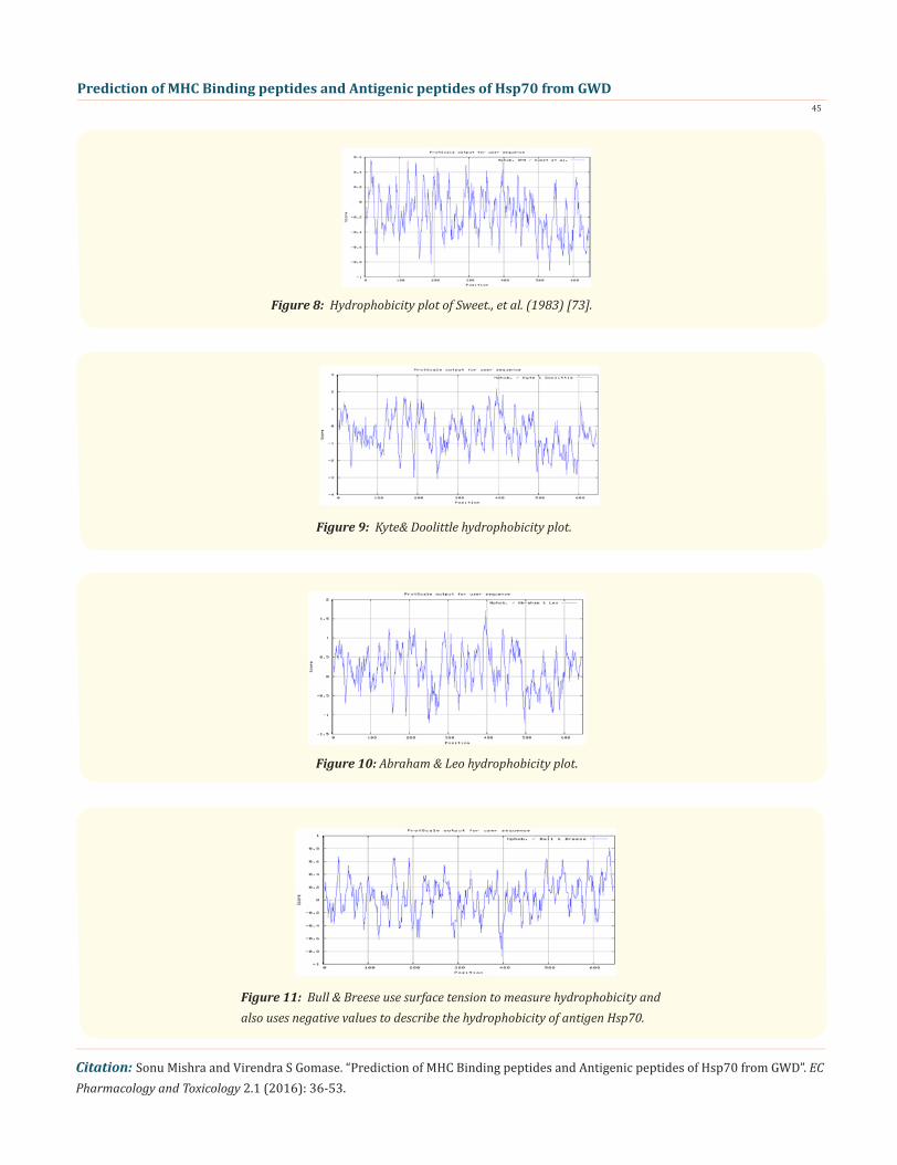
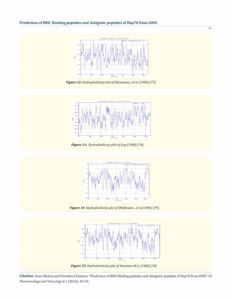
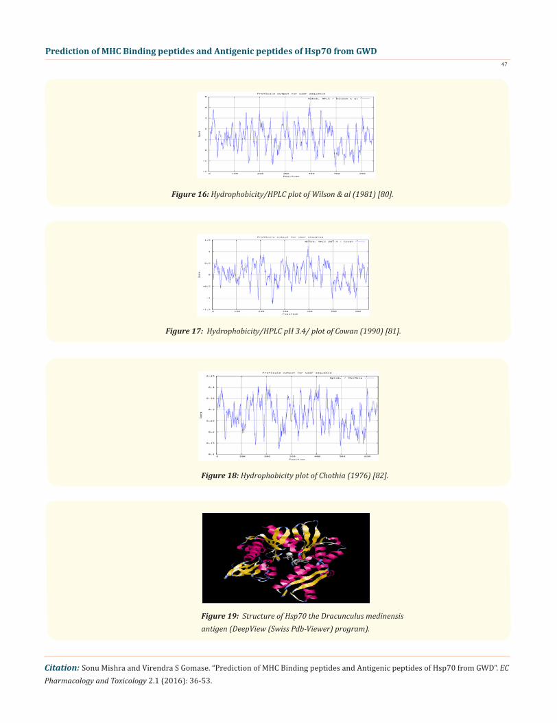
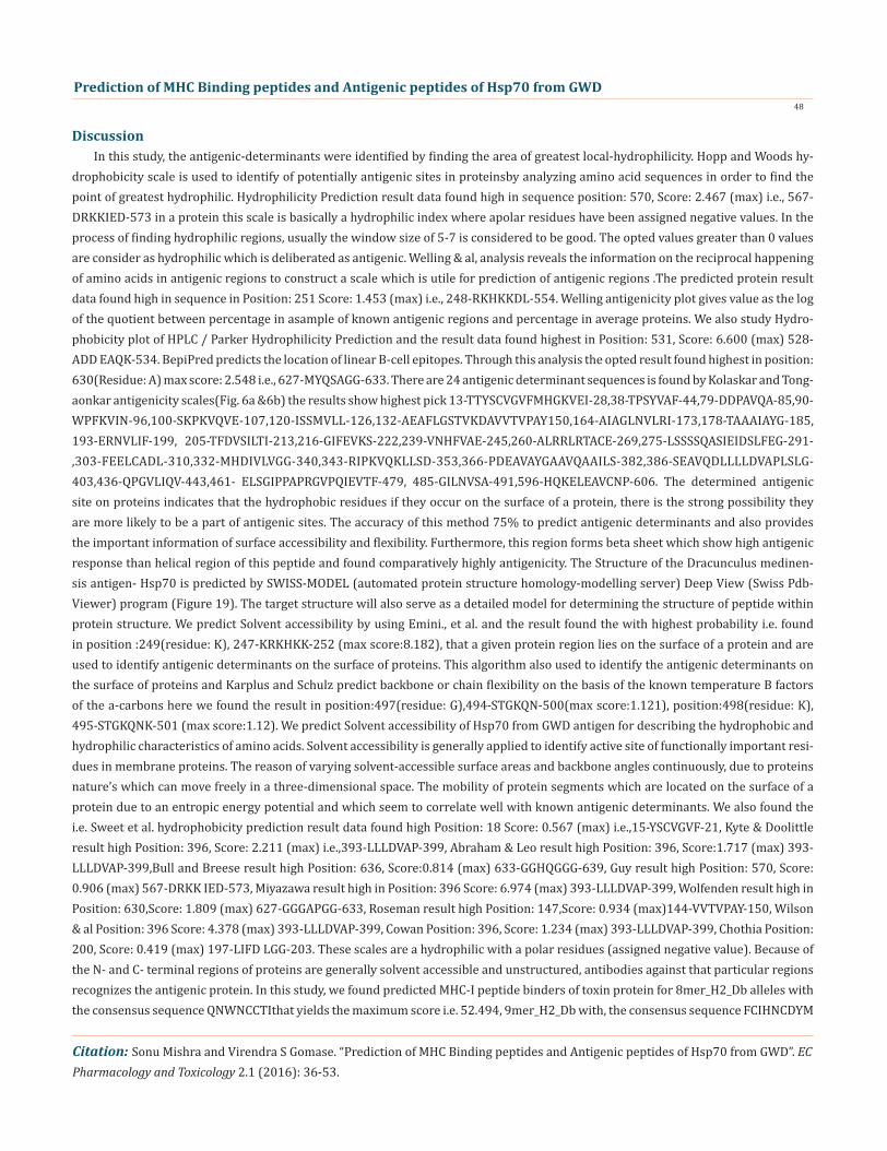
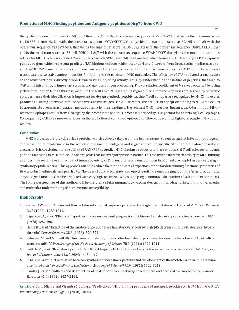
We also predict solvent accessible regions in proteins; different measurement was performed for the prediction of antigenic activ-ity, surface region of peptides. Emini., et al. (Figure 6) predicts the highest probability in position: 249(residue: K), 247-KRKHKK-252-(max score: 8.182), that a given protein region lies on the surface of a protein and are used to identify antigenic determinants on the surface of proteins. Karplus and Schulz (Figure 7) High score is found i.e. found in position: 497(residue: G), 494-STGKQN-500(max score: 1.121), position: 498(residue: K), 495-STGKQNK-501 (max score: 1.12). The hydrophobicity and hydrophilic characteristics of amino acids is determined by several other scales i.e. Sweet., et al. hydrophobicity prediction result data found high in position: 18 Score: 0.567 (max) i.e.,15-YSCVGVF-21 (Figure 8), Kyte & Doolittle result high in position: 396, Score: 2.211 (max)i.e.,393-LLLDVAP-399 (Figure 9), Abraham & Leo result high Position: 396, Score:1.717(max)393-LLLDVAP-399 (Figure 10),Bull & Breese use surface tension to measure in Position: 636, Score:0.814 (max) 633-GGHQGGG-639 (Figure 11), Miyazawa result high in Position: 396 Score: 6.974 (max) 393-LLLDVAP-399 (Figure 12),Guy result high in Position: 570 Score: 0.906 (max) 567-DRKK IED-573 (Figure 13), Wolfenden result high in Position: 630, Score: 1.809 (max) 627-GGGAPGG-633 (Figure 14), Roseman result high in Position: 147, Score: 0.934 (max)144-VVTVPAY-150 (Figure 15),Wilson & al Position: 396 Score: 4.378 (max) 393-LLLDVAP-399 (Figure 16), Cowan Position: 396, Score: 1.234 (max) 393-LLLDVAP-399 Figure 17), Chothia Position: 200, Score: 0.419 (max) 197-LIFD LGG-203 (Figure 18).
13-TTYSCVGVFMHGKVEI-28,38-TPSYVAF-44,79-DDPAVQA-85,90-WPFKVIN-96,100-SKPKVQVE-107,120-ISSMVLL-126,132-AEAFLGSTVKDAVVTVPAY150,164-AIAGLNVLRI-173,178-TAAAIAYG-185,193-ERNVLIF-199,205-TFDVSILTI-213,216-GIFEVKS-222,239-VNHFVAE-245,260-ALRRLRTACE269,275-LSSSSQASIEIDSLFEG-291,303-FEELCADL-310,332-MHDIVLVGG-340,343-RIP-KVQKLLSD-353,366-PDEAVAYGAAVQAAILS-382,386-SEAVQDLLLLDVAPLSLG-403,436-QPGVLIQV-443,461-ELSGIPPAPRGVPQIEVTF-479, 485-GILNVSA-491,596-HQKELEAVCNP-606 and the predicted antigenic fragments can bind to MHC molecule is the first bottlenecks in vaccine design.
Prediction of Solvent Accessible Regions
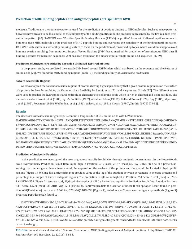
We found the binding of peptides to a number of different alleles using Position Specific Scoring Matrix. Hsp70 of Dracunculus medinensis antigen, with sequence 647 amino acid residues long, is having 639 nonamers. MHC molecules actively participate in host immune reactions and these are the cell surface proteins. We have predicted MHC-I peptide binders of Hsp70 from Dracunculus medi-nensis was tested with on a set of 4 different alleles i.e. H2-Db (mouse) 8mer, H2-Db (mouse) 9mer, H2-Db (mouse) 10mer and H2-Db (mouse) 11mer (Table-1) and MHC-II peptide binders for I_Ab.p, I_Ad.p,I_Ag7.p alleles highlighted in red represent predicted binders (Table-2). Here RANKPEP outcome by PSSM-specific binding threshold is obtained by scoring all the antigenic peptide sequences in-cluded in the alignment from which a profile is derived, and it is defined as the score value that includes 85% of the peptides within the set. Peptides whose score is above the binding threshold will appear in red whereas, peptides highlighted in violet is produced by the cleavage prediction model.The cascade SVM based ‘TAPPred’ method has been used ,where we found more than 80 High affinity TAP Transporter peptide regions, which represents the predicted TAP binders residues which occur at N and C termini of protein from GWD (Table-3).
Prediction of MHC Binding Peptide
MHC-I Allele RANK POS. N SEQUENCE C MW (Da) SCORE % OPT.8mer_H2_Db 1 636 GGH QGGGGPTI EEV 667.71 23.449 44.67%8mer_H2_Db 2 497 KST GKQNKITI TND 883.04 21.607 41.16%8mer_H2_Db 3 88 ADM KHWPFKVI NAE 1013.28 19.048 36.29%8mer_H2_Db 4 434 TYS DNQPGVLI QVF 836.94 16.851 32.10%8mer_H2_Db 5 34 NDQ GNRTTPSY VAF 876.92 14.346 27.33%9mer_H2_Db 1 60 NQV AMNPNNTVF DAK 989.1 17.155 34.06%9mer_H2_Db 2 1 MAKHNAVGI DLG 922.1 15.959 31.69%9mer_H2_Db 3 53 LIG DAAKNQVAM NPN 929.05 14.653 29.09%
Prediction of MHC Binding peptides and Antigenic peptides of Hsp70 from GWD41
Citation: Sonu Mishra and Virendra S Gomase. “Prediction of MHC Binding peptides and Antigenic peptides of Hsp70 from GWD”. EC Pharmacology and Toxicology 2.1 (2016): 36-53.
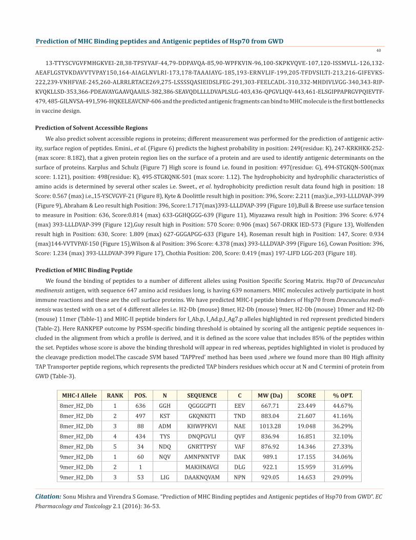
Table 1: Promiscuous MHC ligands, having C-terminal ends are proteosomal cleavage sites of Dracunculusmedinensis. The antigenic peptide to the MHC-1 Allele i.e. 8mer_H2_Db (The binding thresholds: 33.04, optimal score: 52.494), 9mer_H2_Db (Op-timal Score: 50.365, Binding Threshold: 17.96), 10mer_H2_Db (The Optimal Score: 58.858, Binding Threshold: 41.32),11mer_H2_Db(Optimal Score: 79.495, Binding Threshold: 56.96). (All rows highlighted in red represent predicted binders &A peptide highlighted in violet has a C-teminus predicted by the cleavage model used).
9mer_H2_Db 4 307 EEL CADLFRSTM DPV 1025.21 11.77 23.37%9mer_H2_Db 5 21 VGV FMHGKVEII AND 1055.3 11.499 22.83%10mer_H2_Db 1 545 ALE SYAFNMKQTI DDE 1184.37 15.269 25.94%10mer_H2_Db 2 496 DKS TGKQNKITIT NDK 1085.24 12.177 20.69%10mer_H2_Db 3 7 HNA VGIDLGTTYS CVG 1007.1 9.362 15.91%10mer_H2_Db 4 431 TFT TYSDNQPGVL IQV 1075.14 9.17 15.58%10mer_H2_Db 5 33 AND QGNRTTPSYV AFT 1104.18 9.051 15.38%11mer_H2_Db 1 92 HWP FKVINAEGSKP KVQ 1171.36 18.212 22.91%11mer_H2_Db 2 229 DTH LGGEDFDNRMV NHF 1234.35 16.647 20.94%11mer_H2_Db 3 289 DSL FEGIDFYTNIT RAR 1301.42 11.465 14.42%11mer_H2_Db 4 569 DDR KKIEDKCDEII KWL 1315.55 8.446 10.62%11mer_H2_Db 5 484 IDA NGILNVSAQDK STG 1140.25 7.837 9.86%
MHC-II Allele RANK POS. N SEQUENCE C MW (Da) SCORE % OPT.MHC-II I_Ab 1 371 EAV AYGAAVQAA ILS 802.89 10.747 30.16%MHC-II I_Ab 2 603 LEA VCNPIITKM YQS 1000.27 10.619 29.80%MHC-II I_Ab 3 429 AQT FTTYSDNQP GVL 1054.08 10.517 29.52%MHC-II I_Ab 4 460 LGK FELSGIPPA PRG 912.07 10.292 28.88%MHC-II I_Ab 5 600 QKE LEAVCNPII TKM 953.17 10.043 28.19%MHC-II I_Ad 1 237 FDN RMVNHFVAE FKR 1084.26 18.933 35.63%MHC-II I_Ad 2 33 AND QGNRTTPSY VAF 1005.05 17.011 32.01%MHC-II I_Ad 3 533 DEA QKDRIAAKN ALE 1025.17 15.441 29.05%MHC-II I_Ad 4 374 AYG AAVQAAILS GDK 824.98 15.013 28.25%MHC-II I_Ad 5 368 NPD EAVAYGAAV QAA 831.93 14.436 27.16%MHC-II I_Ag7 1 519 IER MVQEAEKYK ADD 1107.29 19.553 47.84%MHC-II I_Ag7 2 188 GLD KKGHGERNV LIF 1006.12 17.951 43.92%MHC-II I_Ag7 3 181 TAA AIAYGLDKK GHG 960.14 14.027 34.32%MHC-II I_Ag7 4 229 DTH LGGEDFDNR MVN 1004.03 13.828 33.83%MHC-II I_Ag7 5 365 KSI NPDEAVAYG AAV 916.95 12.379 30.29%
Table 2: Prediction of MHCII ligands all rows highlighted in red represent predicted binders to the MHC-II Allele i.e. MHC-II I_Ab, MHC-II I_Ad, MHC-II I_Ag7. (All rows highlighted in red represent predicted binders).
Prediction of MHC Binding peptides and Antigenic peptides of Hsp70 from GWD42
Citation: Sonu Mishra and Virendra S Gomase. “Prediction of MHC Binding peptides and Antigenic peptides of Hsp70 from GWD”. EC Pharmacology and Toxicology 2.1 (2016): 36-53.
Peptide Rank Start Position Sequence Score Predicted Affinity1 283 IEIDSLFEG 8.648 High2 109 KGETKTFTP 8.644 High3 429 FTTYSDNQP 8.644 High4 519 MVQEAEKYK 8.643 High5 555 DDEKLKDKL 8.639 High6 526 YKADDEAQK 8.637 High7 145 VTVPAYFND 8.635 High8 558 KLKDKLSAD 8.635 High9 105 QVEYKGETK 8.634 High
10 220 VKSTAGDTH 8.631 High
Table 3: Cascade SVM based High affinity TAP Binders of Dracunculus medinensis.
Figure 1: Hydrophobicity plot of Hopp and Woods (1981) [59].
Figure 2: Hydrophobicity plot of Welling., et al. (1985) [60].
Figure 3: Hydrophobicity plot of HPLC / Parker., et al. (1986) [61].
Prediction of MHC Binding peptides and Antigenic peptides of Hsp70 from GWD43
Citation: Sonu Mishra and Virendra S Gomase. “Prediction of MHC Binding peptides and Antigenic peptides of Hsp70 from GWD”. EC Pharmacology and Toxicology 2.1 (2016): 36-53.
Figure 4: Bepipred Linear Epitope Prediction plot.
Figure 5: Kolaskar and Tongaonkar antigenicity plot.
Figure 5a: Kolaskar and Tongaonkar antigenicity plot, the average antigenic propensity for protein is 1.0067
n Start Position Sequence End Position1 13 TTYSCVGVFMHGKVEI 282 38 TPSYVAF 443 79 DDPAVQA 854 90 WPFKVIN 965 100 SKPKVQVE 1076 120 ISSMVLL 1267 132 AEAFLGSTVKDAVVTVPAY 1508 164 AIAGLNVLRI 1739 178 TAAAIAYG 185
Prediction of MHC Binding peptides and Antigenic peptides of Hsp70 from GWD44
Citation: Sonu Mishra and Virendra S Gomase. “Prediction of MHC Binding peptides and Antigenic peptides of Hsp70 from GWD”. EC Pharmacology and Toxicology 2.1 (2016): 36-53.
Figure 5b: The 24 antigenic determinants of Hsp70 protein.
10 193 ERNVLIF 19911 205 TFDVSILTI 21312 216 GIFEVKS 22213 239 VNHFVAE 24514 260 ALRRLRTACE 26915 275 LSSSSQASIEIDSLFEG 29116 303 FEELCADL 31017 332 MHDIVLVGG 34018 343 RIPKVQKLLSD 35319 366 PDEAVAYGAAVQAAILS 38220 386 SEAVQDLLLLDVAPLSLG 40321 436 QPGVLIQV 44322 461 ELSGIPPAPRGVPQIEVTF 47923 485 GILNVSA 49124 596 HQKELEAVCNP 606
Figure 6: Emini Surface Accessibility Prediction plot.
Figure 7: Karplus& Schulz Flexibility Prediction.
Prediction of MHC Binding peptides and Antigenic peptides of Hsp70 from GWD45
Citation: Sonu Mishra and Virendra S Gomase. “Prediction of MHC Binding peptides and Antigenic peptides of Hsp70 from GWD”. EC Pharmacology and Toxicology 2.1 (2016): 36-53.
Figure 8: Hydrophobicity plot of Sweet., et al. (1983) [73].
Figure 9: Kyte& Doolittle hydrophobicity plot.
Figure 10: Abraham & Leo hydrophobicity plot.
Figure 11: Bull & Breese use surface tension to measure hydrophobicity and also uses negative values to describe the hydrophobicity of antigen Hsp70.
Prediction of MHC Binding peptides and Antigenic peptides of Hsp70 from GWD46
Citation: Sonu Mishra and Virendra S Gomase. “Prediction of MHC Binding peptides and Antigenic peptides of Hsp70 from GWD”. EC Pharmacology and Toxicology 2.1 (2016): 36-53.
Figure 12: Hydrophobicity plot of Miyazawa., et al. (1985) [77].
Figure 13: Hydrophobicity plot of Guy (1988) [76].
Figure 14: Hydrophobicity plot of Wolfenden., et al.(1981) [79].
Figure 15: Hydrophobicity plot of Roseman M.A. (1988) [78].
Prediction of MHC Binding peptides and Antigenic peptides of Hsp70 from GWD47
Citation: Sonu Mishra and Virendra S Gomase. “Prediction of MHC Binding peptides and Antigenic peptides of Hsp70 from GWD”. EC Pharmacology and Toxicology 2.1 (2016): 36-53.
Figure 16: Hydrophobicity/HPLC plot of Wilson & al (1981) [80].
Figure 17: Hydrophobicity/HPLC pH 3.4/ plot of Cowan (1990) [81].
Figure 18: Hydrophobicity plot of Chothia (1976) [82].
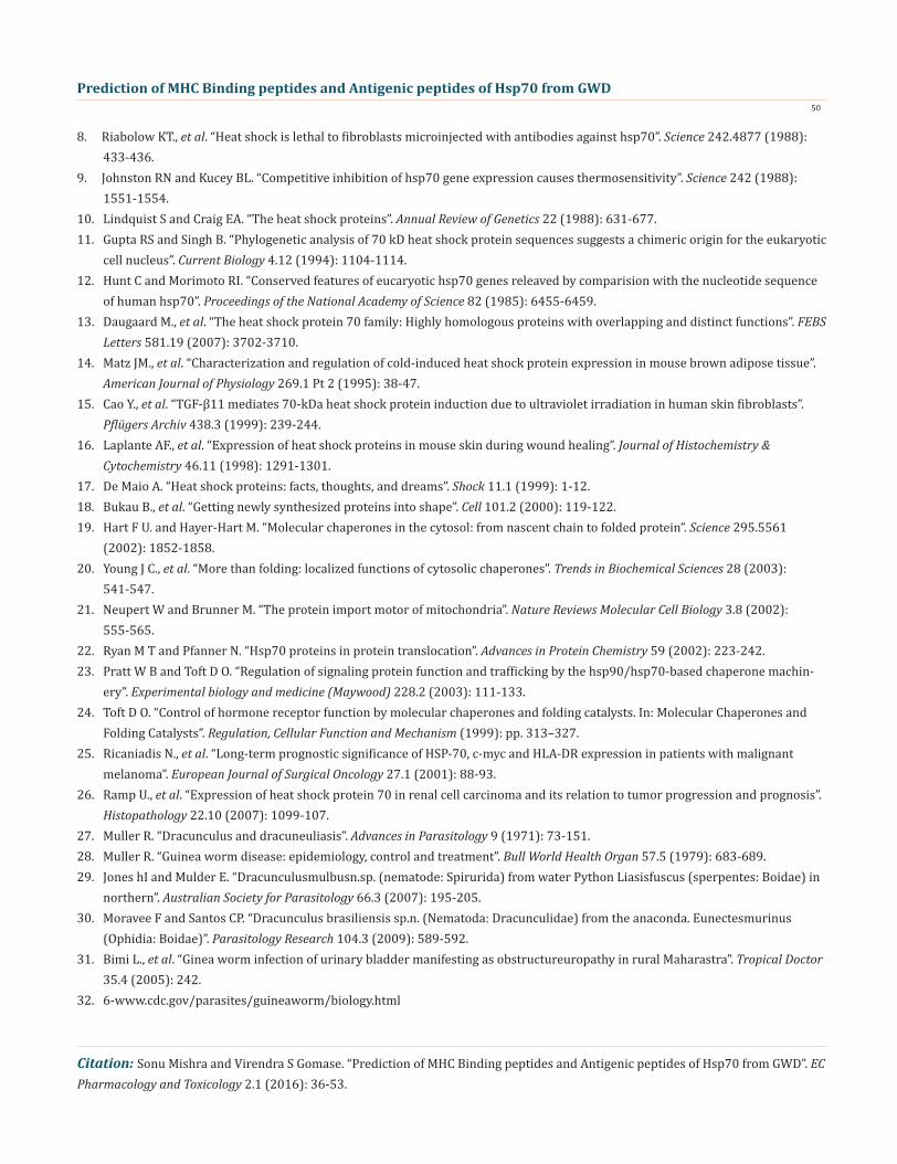
Figure 19: Structure of Hsp70 the Dracunculus medinensis antigen (DeepView (Swiss Pdb-Viewer) program).
Prediction of MHC Binding peptides and Antigenic peptides of Hsp70 from GWD48
Citation: Sonu Mishra and Virendra S Gomase. “Prediction of MHC Binding peptides and Antigenic peptides of Hsp70 from GWD”. EC Pharmacology and Toxicology 2.1 (2016): 36-53.
In this study, the antigenic-determinants were identified by finding the area of greatest local-hydrophilicity. Hopp and Woods hy-drophobicity scale is used to identify of potentially antigenic sites in proteinsby analyzing amino acid sequences in order to find the point of greatest hydrophilic. Hydrophilicity Prediction result data found high in sequence position: 570, Score: 2.467 (max) i.e., 567-DRKKIED-573 in a protein this scale is basically a hydrophilic index where apolar residues have been assigned negative values. In the process of finding hydrophilic regions, usually the window size of 5-7 is considered to be good. The opted values greater than 0 values are consider as hydrophilic which is deliberated as antigenic. Welling & al, analysis reveals the information on the reciprocal happening of amino acids in antigenic regions to construct a scale which is utile for prediction of antigenic regions .The predicted protein result data found high in sequence in Position: 251 Score: 1.453 (max) i.e., 248-RKHKKDL-554. Welling antigenicity plot gives value as the log of the quotient between percentage in asample of known antigenic regions and percentage in average proteins. We also study Hydro-phobicity plot of HPLC / Parker Hydrophilicity Prediction and the result data found highest in Position: 531, Score: 6.600 (max) 528-ADD EAQK-534. BepiPred predicts the location of linear B-cell epitopes. Through this analysis the opted result found highest in position: 630(Residue: A) max score: 2.548 i.e., 627-MYQSAGG-633. There are 24 antigenic determinant sequences is found by Kolaskar and Tong-aonkar antigenicity scales(Fig. 6a &6b) the results show highest pick 13-TTYSCVGVFMHGKVEI-28,38-TPSYVAF-44,79-DDPAVQA-85,90-WPFKVIN-96,100-SKPKVQVE-107,120-ISSMVLL-126,132-AEAFLGSTVKDAVVTVPAY150,164-AIAGLNVLRI-173,178-TAAAIAYG-185, 193-ERNVLIF-199, 205-TFDVSILTI-213,216-GIFEVKS-222,239-VNHFVAE-245,260-ALRRLRTACE-269,275-LSSSSQASIEIDSLFEG-291-,303-FEELCADL-310,332-MHDIVLVGG-340,343-RIPKVQKLLSD-353,366-PDEAVAYGAAVQAAILS-382,386-SEAVQDLLLLDVAPLSLG-403,436-QPGVLIQV-443,461- ELSGIPPAPRGVPQIEVTF-479, 485-GILNVSA-491,596-HQKELEAVCNP-606. The determined antigenic site on proteins indicates that the hydrophobic residues if they occur on the surface of a protein, there is the strong possibility they are more likely to be a part of antigenic sites. The accuracy of this method 75% to predict antigenic determinants and also provides the important information of surface accessibility and flexibility. Furthermore, this region forms beta sheet which show high antigenic response than helical region of this peptide and found comparatively highly antigenicity. The Structure of the Dracunculus medinen-sis antigen- Hsp70 is predicted by SWISS-MODEL (automated protein structure homology-modelling server) Deep View (Swiss Pdb-Viewer) program (Figure 19). The target structure will also serve as a detailed model for determining the structure of peptide within protein structure. We predict Solvent accessibility by using Emini., et al. and the result found the with highest probability i.e. found in position :249(residue: K), 247-KRKHKK-252 (max score:8.182), that a given protein region lies on the surface of a protein and are used to identify antigenic determinants on the surface of proteins. This algorithm also used to identify the antigenic determinants on the surface of proteins and Karplus and Schulz predict backbone or chain flexibility on the basis of the known temperature B factors of the a-carbons here we found the result in position:497(residue: G),494-STGKQN-500(max score:1.121), position:498(residue: K), 495-STGKQNK-501 (max score:1.12). We predict Solvent accessibility of Hsp70 from GWD antigen for describing the hydrophobic and hydrophilic characteristics of amino acids. Solvent accessibility is generally applied to identify active site of functionally important resi-dues in membrane proteins. The reason of varying solvent-accessible surface areas and backbone angles continuously, due to proteins nature’s which can move freely in a three-dimensional space. The mobility of protein segments which are located on the surface of a protein due to an entropic energy potential and which seem to correlate well with known antigenic determinants. We also found the i.e. Sweet et al. hydrophobicity prediction result data found high Position: 18 Score: 0.567 (max) i.e.,15-YSCVGVF-21, Kyte & Doolittle result high Position: 396, Score: 2.211 (max) i.e.,393-LLLDVAP-399, Abraham & Leo result high Position: 396, Score:1.717 (max) 393-LLLDVAP-399,Bull and Breese result high Position: 636, Score:0.814 (max) 633-GGHQGGG-639, Guy result high Position: 570, Score: 0.906 (max) 567-DRKK IED-573, Miyazawa result high in Position: 396 Score: 6.974 (max) 393-LLLDVAP-399, Wolfenden result high in Position: 630,Score: 1.809 (max) 627-GGGAPGG-633, Roseman result high Position: 147,Score: 0.934 (max)144-VVTVPAY-150, Wilson & al Position: 396 Score: 4.378 (max) 393-LLLDVAP-399, Cowan Position: 396, Score: 1.234 (max) 393-LLLDVAP-399, Chothia Position: 200, Score: 0.419 (max) 197-LIFD LGG-203. These scales are a hydrophilic with a polar residues (assigned negative value). Because of the N- and C- terminal regions of proteins are generally solvent accessible and unstructured, antibodies against that particular regions recognizes the antigenic protein. In this study, we found predicted MHC-I peptide binders of toxin protein for 8mer_H2_Db alleles with the consensus sequence QNWNCCTIthat yields the maximum score i.e. 52.494, 9mer_H2_Db with, the consensus sequence FCIHNCDYM
Discussion
Prediction of MHC Binding peptides and Antigenic peptides of Hsp70 from GWD49
Citation: Sonu Mishra and Virendra S Gomase. “Prediction of MHC Binding peptides and Antigenic peptides of Hsp70 from GWD”. EC Pharmacology and Toxicology 2.1 (2016): 36-53.
ConclusionMHC molecules are the cell surface proteins, which actively take part in the host immune responses against infection (pathogens)
and reason of its involvement in the response to almost all antigens and it gives effects on specific sites. From the above result and discussion it is concluded that the ability of RANKPEP to predict MHC binding peptides, and thereby potential T-cell epitopes, antigenic peptide that binds to MHC molecule are antigenic that means hydrophilic in nature. This means the increase in affinity of MHC binding peptides may result in enhancement of immunogenicity of Dracunculus medinensis antigen Hsp70 and are helpful in the designing of synthetic peptide vaccine. This approach can help reduce the time and cost of experimentation for determining functional properties of Dracunculus medinensis antigen Hsp70. The Overall conducted study and opted results are encouraging. Both the ‘sites of action’ and ‘physiological functions’ can be predicted with very high accuracies which is helping to minimize the number of validation experiments. The future perspectives of this method will be useful in cellular immunology, vaccine design, immunodiagnostics, immunotherapeutic and molecular understanding of autoimmune susceptibility.
that yields the maximum score i.e. 50.365, 10mer_H2_Db with, the consensus sequence SGYYNFFWCL that yields the maximum score i.e. 58.858, 11mer_H2_Db with, the consensus sequence CGVYNFYYCCY that yields the maximum score i.e. 79.495 and I_Ab with the consensus sequence YYAPWCNNA that yields the maximum score i.e. 35.632,I_Ad with the consensus sequence QMVHAAHAE that yields the maximum score i.e. 53.145, MHC-II I_Ag7 with the consensus sequence WYAHAFKYV that yields the maximum score i.e. 40.873 for MHC II allele was tasted. We also use a cascade SVM based TAPPred method which found 160 High affinity. TAP Transporter peptide regions which represent predicted TAP binders residues which occur at N and C termini from Dracunculus medinensis anti-gen Hsp70. TAP is one of the important conveyor, which allow antigenic peptides to move from cytosol to ER. TAP directs binds and translocate the selective antigen peptides for binding to the particular MHC molecules. The efficiency of TAP-mediated translocation of antigenic peptides is directly proportional to its TAP binding affinity. Thus, by understanding the nature of peptides, that bind to TAP with high affinity, is important steps in endogenous antigen processing. The correlation coefficient of 0.88 was obtained by using jackknife validation test. In this test, we found the MHCI and MHCII binding regions. T cell immune responses are derived by antigenic epitopes hence their identification is important for design synthetic peptide vaccine. T cell epitopes are recognized by MHCI molecules producing a strong defensive immune response against antigen Hsp70. Therefore, the prediction of peptide binding to MHCI molecules by appropriate processing of antigen peptides occurs by their binding to the relevant MHC molecules. Because, the C-terminus of MHCI-restricted epitopes results from cleavage by the proteasome and thus, proteasome specifity is important for determing T-cell epitopes. Consequently, RANKPEP moreover, focus on the prediction of conserved epitopes and the sequences highlighted in purple in the output results.
Bibliography
1. Gerner EW., et al. “A transient thermotolerant survival response produced by single thermal doses in HeLa cells”. Cancer Research 36.3 (1976): 1035-1040.2. Sapareto SA., et al. “Effects of hyperthermia on survival and progression of Chinese hamster ovary cells”. Cancer Research 38.2 (1978): 393-400.3. Henle KJ., et al. “Induction of thermotolerance in Chinese hamster ovary cells by high (45 degrees) or low (40 degrees) hyper- thermia”. Cancer Research 38.3 (1978): 570-574.4. Petersen NS and Mitchell HK. “Recovery of protein synthesis after heat shock: prior heat treatment affects the ability of cells to translate mRNA”. Proceedings of the National Academy of Science 78.3 (1981): 1708-1711.5. Jäättelä M., et al. “Heat shock protects WEHI-164 target cells from the cytolysis by tumor necrosis factors a and beta”. European Journal of Immunology 19.8 (1989): 1413-1417.6. Li GC and Werb Z. “Correlation between synthesis of heat shock proteins and development of thermotolerance in Chinese ham- ster fibroblasts”. Proceedings of the National Academy of Science 79.10 (1982): 3122-3218.7. Landry J., et al. “Synthesis and degradation of heat shock proteins during development and decay of thermotolerance”. Cancer Research 42.6 (1982): 2457-2461.
Prediction of MHC Binding peptides and Antigenic peptides of Hsp70 from GWD50
Citation: Sonu Mishra and Virendra S Gomase. “Prediction of MHC Binding peptides and Antigenic peptides of Hsp70 from GWD”. EC Pharmacology and Toxicology 2.1 (2016): 36-53.
8. Riabolow KT., et al. “Heat shock is lethal to fibroblasts microinjected with antibodies against hsp70”. Science 242.4877 (1988): 433-436.9. Johnston RN and Kucey BL. “Competitive inhibition of hsp70 gene expression causes thermosensitivity”. Science 242 (1988): 1551-1554.10. Lindquist S and Craig EA. “The heat shock proteins”. Annual Review of Genetics 22 (1988): 631-677.11. Gupta RS and Singh B. “Phylogenetic analysis of 70 kD heat shock protein sequences suggests a chimeric origin for the eukaryotic cell nucleus”. Current Biology 4.12 (1994): 1104-1114.12. Hunt C and Morimoto RI. “Conserved features of eucaryotic hsp70 genes releaved by comparision with the nucleotide sequence of human hsp70”. Proceedings of the National Academy of Science 82 (1985): 6455-6459.13. Daugaard M., et al. “The heat shock protein 70 family: Highly homologous proteins with overlapping and distinct functions”. FEBS Letters 581.19 (2007): 3702-3710.14. Matz JM., et al. “Characterization and regulation of cold-induced heat shock protein expression in mouse brown adipose tissue”. American Journal of Physiology 269.1 Pt 2 (1995): 38-47.15. Cao Y., et al. “TGF-β11 mediates 70-kDa heat shock protein induction due to ultraviolet irradiation in human skin fibroblasts”. Pflügers Archiv 438.3 (1999): 239-244.16. Laplante AF., et al. “Expression of heat shock proteins in mouse skin during wound healing”. Journal of Histochemistry & Cytochemistry 46.11 (1998): 1291-1301.17. De Maio A. “Heat shock proteins: facts, thoughts, and dreams”. Shock 11.1 (1999): 1-12.18. Bukau B., et al. “Getting newly synthesized proteins into shape”. Cell 101.2 (2000): 119-122.19. Hart F U. and Hayer-Hart M. “Molecular chaperones in the cytosol: from nascent chain to folded protein”. Science 295.5561 (2002): 1852-1858.20. Young J C., et al. “More than folding: localized functions of cytosolic chaperones”. Trends in Biochemical Sciences 28 (2003): 541-547.21. Neupert W and Brunner M. “The protein import motor of mitochondria”. Nature Reviews Molecular Cell Biology 3.8 (2002): 555-565.22. Ryan M T and Pfanner N. “Hsp70 proteins in protein translocation”. Advances in Protein Chemistry 59 (2002): 223-242.23. Pratt W B and Toft D O. “Regulation of signaling protein function and trafficking by the hsp90/hsp70-based chaperone machin- ery”. Experimental biology and medicine (Maywood) 228.2 (2003): 111-133.24. Toft D O. “Control of hormone receptor function by molecular chaperones and folding catalysts. In: Molecular Chaperones and Folding Catalysts”. Regulation, Cellular Function and Mechanism (1999): pp. 313–327.25. Ricaniadis N., et al. “Long-term prognostic significance of HSP-70, c-myc and HLA-DR expression in patients with malignant melanoma”. European Journal of Surgical Oncology 27.1 (2001): 88-93.26. Ramp U., et al. “Expression of heat shock protein 70 in renal cell carcinoma and its relation to tumor progression and prognosis”. Histopathology 22.10 (2007): 1099-107.27. Muller R. “Dracunculus and dracuneuliasis”. Advances in Parasitology 9 (1971): 73-151.28. Muller R. “Guinea worm disease: epidemiology, control and treatment”. Bull World Health Organ 57.5 (1979): 683-689.29. Jones hI and Mulder E. “Dracunculusmulbusn.sp. (nematode: Spirurida) from water Python Liasisfuscus (sperpentes: Boidae) in northern”. Australian Society for Parasitology 66.3 (2007): 195-205.30. Moravee F and Santos CP. “Dracunculus brasiliensis sp.n. (Nematoda: Dracunculidae) from the anaconda. Eunectesmurinus (Ophidia: Boidae)”. Parasitology Research 104.3 (2009): 589-592. 31. Bimi L., et al. “Ginea worm infection of urinary bladder manifesting as obstructureuropathy in rural Maharastra”. Tropical Doctor 35.4 (2005): 242.32. 6-www.cdc.gov/parasites/guineaworm/biology.html
Prediction of MHC Binding peptides and Antigenic peptides of Hsp70 from GWD51
Citation: Sonu Mishra and Virendra S Gomase. “Prediction of MHC Binding peptides and Antigenic peptides of Hsp70 from GWD”. EC Pharmacology and Toxicology 2.1 (2016): 36-53.
33. Greenaway C. “Dracunculiasis (Guinea worm disease)”. Canadian Medical Association Journal 170.9 (2004): 495-500.34. Miillner A., et al. “Chemistry and pharmacology of neglected helminthic disease”. Current Medicinal Chemistry 18.5 (2011): 767-789.35. Ruiz-Tiben E and Hopkins DR. “Dracunculiasis (Guinea worm disease) eradication”. Advances in Parasitology 61 (2006): 275-309.36. IriemenamNC., et al. “Dracunculiasis–The saddle is virtually ended”. Parasitology Research 102.3 (2008): 343-347.37. Hopkins DR., et al. “Progress toward global eradication of dracunculiasis--January 2013-June 2014”. Centers for Disease Control and Prevention 63.46 (2014): 1050-1054.38. Eta Ngole Mbong., et al. “Not every worm wrapped around a stick is a guinea worm: a case of Onchocerca volvulus mimicking Dracunculusmedinensis”. Parasit Vectors 8 (2015): 374.39. Richards FO and Ruiz-TibenEand Hopkins DR. “Dracunculiasis eradication and the legacy of the smallpox campaign: what’s new and innovative? What’s old and principled?” Vaccine 29.4 (2011): 86-90.40. Hopkins DR. “Disease eradication”. The New England Journal of Medicine 368 (2013): 54-63.41. Muller R. “Life cycle of Dracunculus Medinesis”. In workshop on opportunities for control of dracunculiasis: contaminated pa- pers, washinton,DC:National Academy Press] (1985).42. Cairncross S., et al. “Dracunculiasis (Guinea worm disease) and the eradication initiative”. Clinical Microbiology Reviews 15 (2002): 223-246.43. Issaka –Tinogah A., et al. “Lack of effect of ivermectin on prepatent guinea – worm: a single- blind, placebo-controlled trial”. Transactions of the Royal Society of Tropical Medicine 88 (1994): 346-348.44. Bloch P., et al. “The antibody response to Dracunculusmedinensis in an endemic human population of northern Ghana”. Helmint- hology 67 (1993): 37-48.45. BlochP and Simonsen E PAUL. “Immunoepidemiology of Dracunculusmedinensis infections II.Variation in antibody respomses in relation to transmission season and patency”. The American Journal of Tropical Medicine and Hygiene 59.6 (1998): 985-990.46. Flower DR. “Vaccines: how they work”. In Bioinformatics for Vaccinology Wiley-Blackwell, Oxford, UK. (2008): 73-112 .47. Batalia Michael and Collins EJ. “Peptide Binding by Class 1 and Class II MHC Molecules”. Biopoly 43.4 (1997): 281-302.48. Marrack P., et al. “Review Evolutionarily conserved amino acids that control TCR-MHC interaction”. Annual Review of Immunology 26 (2008): 171-203.49. Chapman HA. “Endosomal proteolysis and MHC class II function”. Current Opinion in Immunology 10.1 (1998): 93-102.50. Watts C. “The exogenous pathway for antigen presentation on major histocompatibility complex class II and CD1 molecules”. Nature Immunology 5.7 (2004): 685-92.51. Neefjes J., et al. “Review towards a systems understanding of MHC class I and MHC class II antigen presentation”. Nature Reviews Immunology 11.12 (2011): 823-836.52. Kumar M., et al. “Identification of DNA-binding proteins using support vector machines and evolutionary profiles”. BMC Bioinfor- matics 8 (2007): 463.53. Gomase VS and Chitlange NR. “Prediction of MHC Class Antigen Peptides from Echinococcus Multilocularis: Application of Com- puter Intelligence”. Scientific Reports 1.3 (2012): 191.54. Gomase VS., et al. “Prediction of MHC Binding Peptides and Epitopes from Alfalfa mosaic virus”. Current Drug Discovery Technolo- gies 4.2 (2007): 117-215.55. Sherkhane AS., et al. “Prediction of Major Histocompatibility Complex Binding Peptides and Epitopes from Najanaja Cardiotoxin (CTX)”. Drug Invention Today 4.8 (2012): 435-438.56. http://www.ncbi.nlm.nih.gov57. Sayers E W., et al. “Database resources of the National Center for Biotechnology Information”. Nucleic Acids Research 40.Database issue (2012): D13-25. 58. Bairoch A., et al. “The Universal Protein Resource (UniProt)”. Nucleic Acids Research 33 (2005): D154-159.
Prediction of MHC Binding peptides and Antigenic peptides of Hsp70 from GWD52
Citation: Sonu Mishra and Virendra S Gomase. “Prediction of MHC Binding peptides and Antigenic peptides of Hsp70 from GWD”. EC Pharmacology and Toxicology 2.1 (2016): 36-53.
59. Hoop TP and Woods KR. “Prediction of protein antigenic determinants from amino acid sequences”. Proceedings of the National
Academy of Sciences of the USA 78.6 (1978): 3824-3828.60. Welling GW., et al. “Prediction of sequential antigenic regions in proteins”. FEBS Letters 188.2 (1985): 215-218.61. Parker KC., et al. “Scheme for ranking potential HLA-A2 binding peptides based on independent binding of individual peptide side-chains”. The Journal of Immunology 152.1 (1994): 163-175.62. Jens Erik., et al. “Improved method for predicting linear B-cell epitopes”. Immunome Research 2 (2006): 2.63. Kolaskar AS and Tongaonkar PC. “A semi-empirical method for prediction of antigenic determinants on protein antigens”. FEBS
Letters 276.1-2 (1990): 172-174.64. Mishra Sonu and Virendra S Gomase. “Prediction of antigenic epitope from D. medinensis: new paradigm of synthetic vaccine development”. ICRRDESH (2015).65. Ruppert J., et al. “Prominent role of secondary anchor residues in peptide binding to HLA-A2.1 molecules”. Cell 74.5 (1993): 929-937.66. Reche PA., et al. “Prediction of MHC Class I Binding Peptides Using Profile Motifs”. Human Immunology 63.9 (2002): 701-709.67. RecheP A and Reinherz EL. “Sequence variability analysis of human class I and class II MHC molecules: functional and structural correlates of amino acid polymorphisms”. Journal of Molecular Biology 331.3 (2003): 623-641.68. Craiu A., et al. “Two distinct proteolytic processes in the generation of a major histocompatibility complex class I-presented pep- tide”. Proceedings of the National Academy of Sciences USA 94.20 (1997): 10850-10855.69. Pieters J. “MHC class II-restricted antigen processing and presentation”. Advances in Immunology 75 (2000): 159-208.70. Bhasin M and Raghava GPS. “Analysis and prediction of affinity of TAP binding peptides using cascade SVM”. Protein Science 13.3 (2004): 596-607.71. Emini EA., et al. “Induction of hepatitis a virus-neutralizing antibody by a virus-specific synthetic peptide”. Journal of Virology
55.3 (1985): 836-839. 72. Karplus PA and Schulz GE. “Prediction of chain flexibility in proteins: a tool for the selection of peptide antigen”. Naturwissen-
schaften 72.4 (1985): 212-213. 73. Sweet RM and Eisenberg D. “Correlation of sequence hydrophobicities measures similarity in three-dimensional protein struc- ture”. Journal of Molecular Biology 171.4 (1983): 479-488.74. Kyte J and Doolittle RF. “A Simple Method for Displaying the Hydropathic Character of a Protein”. Journal of Molecular Biology
157.1 (1982): 105-132.75. Abraham DJ and Leo AJ. “Extension of the fragment method to calculate amino acid zwitterions and side chain partition coef- ficients”. Proteins 2.2 (1987): 130-152.76. Bull HB and Breese K. “Surface tension of amino acid solutions: A hydrophobicity scale of the amino acid residues”. Archives of
Biochemistry and Biophysics 161 (1974): 665-670.77. Miyazawa S and Jernigen RL. “Estimation of Effective Interresidue Contact Energies from Protein Crystal Structures: Quasi-Chem- ical Approximation”. Macromolecules 18.3 (1985): 534-552.78. Roseman MA. “Hydrophilicity of polar amino acid side-chains is markedly reduced by flanking peptide bonds”. Journal of Molecu-
lar Biology 200.3 (1988): 513-522.79. Wolfenden RV., et al. “Affinities of amino-acid side-chains for solvent water”. Biochemistry 20.4 (1985): 849-855.80. Wilson J., et al. “The behaviour of peptides on reverse-phase K.supports during high-pressure liquid chromatography”. Biochemi-
cal Journal 199 (1981): 31-41.81. Cowan R and Whittaker RG. “Hydrophobicity indices at ph 3.4 determined by HPLC”. Peptide Research 3 (1990): 75-80. 82. Chothia C. “The nature of accessible and buried surfaces in proteins”. Journal of Molecular Biology 105.1 (1976): 1-12.
Prediction of MHC Binding peptides and Antigenic peptides of Hsp70 from GWD53
Citation: Sonu Mishra and Virendra S Gomase. “Prediction of MHC Binding peptides and Antigenic peptides of Hsp70 from GWD”. EC Pharmacology and Toxicology 2.1 (2016): 36-53.
Volume 2 Issue 1 February 2016© All rights reserved by Sonu Mishra and Virendra S Gomase.