Embed Size (px)
Citation preview
Cross-interference of plant developmentand plant–microbe interactionsEdouard Evangelisti, Thomas Rey and Sebastian Schornack
Available online at www.sciencedirect.com
ScienceDirect
Plant roots are host to a multitude of filamentous
microorganisms. Among these, arbuscular mycorrhizal fungi
provide benefits to plants, while pathogens trigger diseases
resulting in significant crop yield losses. It is therefore
imperative to study processes which allow plants to
discriminate detrimental and beneficial interactions in order to
protect crops from diseases while retaining the ability for
sustainable bio-fertilisation strategies. Accumulating evidence
suggests that some symbiosis processes also affect plant–
pathogen interactions. A large part of this overlap likely
constitutes plant developmental processes. Moreover,
microbes utilise effector proteins to interfere with plant
development. Here we list relevant recent findings on how
plant–microbe interactions intersect with plant development
and highlight future research leads.
Addresses
Sainsbury Laboratory, University of Cambridge, Cambridge CB2 1LR,
UK
Corresponding author: Schornack, Sebastian
Current Opinion in Plant Biology 2014, 20:118–126
This review comes from a themed issue on Biotic interactions
Edited by Makoto Hayashi and Martin Parniske
For a complete overview see the Issue and the Editorial
Available online 10th June 2014
http://dx.doi.org/10.1016/j.pbi.2014.05.014
1369-5266/# 2014 Elsevier Ltd. All rights reserved.
IntroductionPlants’ success in conquering land can in part be attrib-
uted to their ability to team up with filamentous micro-
organisms. The oldest land plant fossils from the Rhynie
chert give evidence of fungal structures inside plant cells
[1] and more than 70% of all existing higher plants are
colonised by arbuscular mycorrhizal (AM) fungi [2]. The
fungal partner provides mineral nutrients such as phos-
phorus. Conversely, plants provide carbohydrates gener-
ated through photosynthesis. Plant carbohydrates are also
attractive to root-infecting filamentous pathogens such as
fungi and oomycetes. Pathogenic oomycetes such as
Phytophthora palmivora and beneficial fungi represent
extreme opposites but nevertheless share common root
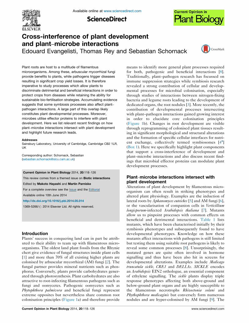
colonisation principles (Figure 1a) and therefore provide
Current Opinion in Plant Biology 2014, 20:118–126
means to identify more general plant processes required
for both, pathogenic and beneficial interactions [8].
Traditionally, plant–pathogen research has focussed on
immune suppression strategies while symbiosis research
revealed a strong contribution of cellular and develop-
mental processes for microbial colonisation, especially
through studies of interactions between nitrogen-fixing
bacteria and legume roots leading to the development of
dedicated organs, the root nodules [3]. More recently, the
contribution of developmental processes intersecting
with plant–pathogen interactions gained growing interest
in order to elucidate core colonisation principles
(Figure 1b). Changes in root development are visible
through reprogramming of colonised plant tissues result-
ing in significant morphological and structural alterations
and the formation of specific cellular interfaces for nutri-
ent exchange, collectively termed symbiosomes [4�](Box 1). Here we specifically highlight plant components
that support a cross-interference of development and
plant–microbe interactions and also discuss recent find-
ings that microbial effector proteins can modulate plant
development processes.
Plant–microbe interactions intersect withplant developmentAlterations of plant development by filamentous micro-
organism can often result in striking phenotypes and
altered plant physiology. Examples are the induction of
lateral roots by Aphanomyces euteiches [5] and AM fungi [6],
or the vascularisation of companion cells in Verticilliumlongisporum-infected Arabidopsis thaliana [7]. Mutants
allow us to pinpoint processes with common effects on
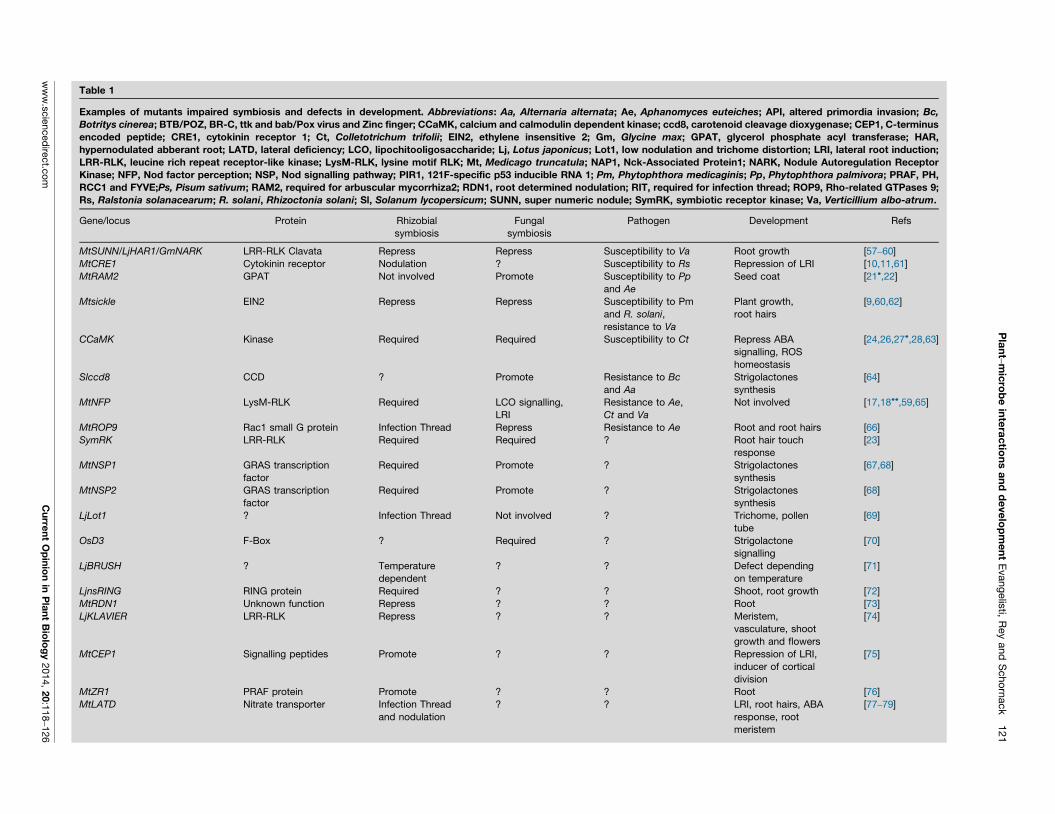
beneficial and detrimental interactions. Table 1 lists
mutants, which have been characterised initially by their
symbiosis phenotypes and subsequently found to have
developmental phenotypes. Knowledge on how these
mutants affect interactions with pathogens is still limited
but testing them using suitable root pathogens is likely to
reveal some common processes [8]. Unsurprisingly, the
mutated genes are quite often involved in hormone
signalling and thus have been also hit in screens for
developmental alterations. Examples include Medicagotruncatula sickle, CRE1 and DELLAs. SICKLE encodes
an Arabidopsis EIN2 orthologue, an essential component
of ethylene signalling. The sickle plants display triple
response phenotypes affecting both above-ground and
below-ground plant organs and are highly susceptible to
the filamentous necrotrophs Rhizoctonia solani and
Phythophthora medicaginis but conversely form numerous
nodules and are hyper-colonised by AM fungi [9]. The
www.sciencedirect.com
Plant–microbe interactions and development Evangelisti, Rey and Schornack 119
Figure 1
Inner root cortex cells form highly branched fungal arbuscules
The vasculature is not colonised by arbuscular
mycorrhiza fungiFilamentous pathogens can enter the central cylinder
Phytophthora palmivora forms haustoria inside root cells
Pathogen infection often results in death of colonised tissue
Plant physiology
Plant immunity
Plant Development
SymbiosisResearch
Plant-PathogenResearch
corecolonisationprinciples
(a)
(b)
Colonised root cotex stays alive
Arbuscular mycorrhiza fungiattach and penetrate throughhyphopodiaExtensive fungal
mycelium connects soiland plant root
Pathogens often penetratethrough appressoria
Current Opinion in Plant Biology
Commonalities and contrasts of pathogenic and symbiotic microbe interactions with plant roots. (a) Structural similarities and differences in Medicago
truncatula root colonisation between a filamentous oomycete pathogen (left) and arbuscular mycorrhiza fungi (right). (b) Growing research interest
(indicated by stars) focusses on core colonisation principles requiring the integrated study of plant physiology, plant immunity and plant development
and of developmental aspects of plant–microbe interactions, a traditional topic of symbiosis research.
cytokinin receptor MtCRE1 promotes invasion by both
pathogenic and symbiotic bacteria [10,11] but its import-
ance for filamentous microbes has not been assessed yet.
The use of della mutants recently enabled identification
www.sciencedirect.com
of gibberellic acid (GA3) as a repressor of accommodation
structure formation in M. truncatula and Pisum sativum[12��,13�]. Strikingly, a dominant negative DELLA
protein rescues cyclops, a common symbiosis pathway
Current Opinion in Plant Biology 2014, 20:118–126
120 Biotic interactions
Box 1 Symbiosomes, different or all the same?
Symbiosomes are specialised intracellular interfaces formed by
filamentous microorganisms inside plant cells [4�]. Their core
structure is a plant cell wall-piercing microbial hypha engulfed by the
plant protoplast. Symbiosomes have been termed haustoria of fungi
and oomycetes, invading hyphae of fungi such as Colletotrichum
sp. and Magnaporthe oryzae, coils of Piriformospora indica and
other fungi and arbuscules of AM fungi. Symbiosome shape varies
greatly and factors influencing it are not fully understood. Never-
theless, they are all assumed to have two main functions: nutrient
transfer and microbial effector delivery. Extensive branching of
symbiosomes is likely attributable to efficient nutrient and informa-
tion exchange and often assigned to beneficial symbiosis. Whether
fossil symbiosome-like structures serve mutual nutrient exchange or
are extensive one-way pathogenic haustoria will remain unresolved.
Plant and microbe are separated by a specialised membrane termed
extrahaustorial membrane (EHM), periarbuscular membrane (PAM)
or extrainvasive hyphae membrane (EIHM) depending on the
microorganism. PAMs in rice and legumes harbour phosphate
transporters which are absent from the remaining plasma membrane
[51,52]. Conversely, EHMs of pathogenic fungi and oomycetes are
lacking numerous transmembrane proteins including immune re-
ceptors. Notably, membrane adhering proteins are still present
[53,54]. Differences in symbiosome membrane protein composition
[53] compared to the plasma membrane often are attributed to
presence of a sealing neckband structure, found in many obligate
biotrophs. Absence of a neckband at the PAM and EHMs of
Phytophthora species highlights the need for further research into
membrane protein separation mechanisms. Mechanisms resulting in
formation and decoration of symbiosome membranes largely remain
elusive. Exclusive PAM integration of the MtPt4 phosphate trans-
porter has been attributed to repolarisation of secretion timed with
MtPt4 promoter activation during arbuscule formation [55]. Another
open question is the point of new membrane material deployment.
The neck, the oldest part of a symbiosome, shows accumulation of
plant endomembrane compartments and callose deposition. How-
ever, candidate vesicle-fusion sites can be traced all over haustoria
and the fine branches of arbuscules [53,56]. Future work using
photo-convertible fluorescent probes will shed light on temporal and
spatial changes in symbiosome membrane processes.
mutant thus bypassing symbiosis signalling and support-
ing the hypothesis of GA signalling repression by this
pathway. Notably, DELLA proteins are also known to
bind JAZ proteins [14], repressors of the jasmonate path-
way. Hence contribution of jasmonate-related defence
responses depending on GA and dominant negative
DELLA proteins might provide further clues about the
role of hormonal balance in regulation of mycorrhizal
symbiosis. Since hormonal pathways link development
to immunity, it remains to be untangled whether specific
microbes interfere with them to suppress immunity or to
alter development.
Plant–microbe interactions utilise similarchemical signaturesChitin-derived microbial signals are triggers of plant
symbiotic responses [15�]. Interestingly, similar but not
identical chitin-derived signals are also perceived by
peripheral plant immune receptors. Chitin-binding LysM
domain-containing receptor-like kinases are key players
Current Opinion in Plant Biology 2014, 20:118–126
in both symbiosis and defence. Numerous activities rang-
ing from immune suppression upon perception of sym-
biotic signatures in Arabidopsis, cell death induction upon
ectopic expression in Nicotiana benthamiana leaves [16]
and involvement in symbiotic [17] to pathogenic inter-
action with filamentous microbes [18��] have been
assigned to them. The finding that a LysM receptor of
the symbiosis-incapable Arabidopsis perceives symbiotic
Nod-factors to suppress immunity [19] shows that speci-
ficity of signal integration from LysM receptors and their
downstream targets are not fully resolved. It is therefore
possible that chitin-derived signals of plant origin may
also play a role in developmental processes.
Cutin is a structural component of above-ground organs.
However, plant cutin monomers have been shown to be a
crucial signal for infection structure formation by filamen-
tous pathogens [20]. Recently this was extended to
pathogenic oomycetes (P. palmivora and A. euteiches)and beneficial AM fungi [21�,22]. A mutant of M. trunca-tula RAM2, a glycerol-3-phosphate acyltransferase, failed
to display appressorium formation by the filamentous
plant pathogen P. palmivora as well as arbuscule devel-
opment by beneficial AM fungi. The altered seed coat of
ram2 mutants points to its involvement in development
[22].
Essential components of specific plant–microbe interactions gain additional rolesStudies of core symbiosis players, the receptor kinase
SymRK and the Calcium and calmodulin dependent
kinase CCaMK revealed their unexpected involvement
in responses to pathogen and mechanical cues. SymRK is
important for root hair resistance to mechanical stresses
[23]. CCaMK was proposed to cope with stress triggered
by penetration events of mycorrhizal fungus and the
pathogenic fungus Colletotrichum trifolii [24]. CCaMK is
presumed to be the main sensor of the nuclear calcium
spiking triggered specifically by endosymbionts [25].
However, this protein is also a major regulator of bacterial
communities associated with rice roots in natural environ-
ments [26] suggesting sources other than endosymbionts
may be producers of CCaMK-read calcium signatures. A
possible mechanism underlying fine-tuning of root micro-
biome by CCaMK is its role in abscisic acid (ABA)
signalling and reactive oxygen species homeostasis
recently demonstrated in rice leaves [27�]. Overexpres-
sion of wheat CCaMK in Arabidopsis resulted in plants
which were less susceptible to ABA during germination
and seedling growth [28]. Thus, CCaMK although
initially implied only in symbiosis might have additional
functions. This is supported by the presence of CCaMK/
DMI3 in Charophyta, since AM fungal mycorrhiza has not
been reported from these green algae [29].
Another link between microbial accommodation and de-
velopment is provided by MLO proteins. MLO has been
www.sciencedirect.com
Pla
nt–m
icro
be
inte
rac
tion
s a
nd
de
ve
lop
me
nt
Evang
elis
ti, R
ey
and
Scho
rnack
121
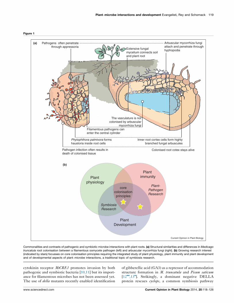
Table 1
Examples of mutants impaired symbiosis and defects in development. Abbreviations: Aa, Alternaria alternata; Ae, Aphanomyces euteiches; API, altered primordia invasion; Bc,
Botritys cinerea; BTB/POZ, BR-C, ttk and bab/Pox virus and Zinc finger; CCaMK, calcium and calmodulin dependent kinase; ccd8, carotenoid cleavage dioxygenase; CEP1, C-terminus
encoded peptide; CRE1, cytokinin receptor 1; Ct, Colletotrichum trifolii; EIN2, ethylene insensitive 2; Gm, Glycine max; GPAT, glycerol phosphate acyl transferase; HAR,
hypernodulated abberant root; LATD, lateral deficiency; LCO, lipochitooligosaccharide; Lj, Lotus japonicus; Lot1, low nodulation and trichome distortion; LRI, lateral root induction;
LRR-RLK, leucine rich repeat receptor-like kinase; LysM-RLK, lysine motif RLK; Mt, Medicago truncatula; NAP1, Nck-Associated Protein1; NARK, Nodule Autoregulation Receptor
Kinase; NFP, Nod factor perception; NSP, Nod signalling pathway; PIR1, 121F-specific p53 inducible RNA 1; Pm, Phytophthora medicaginis; Pp, Phytophthora palmivora; PRAF, PH,
RCC1 and FYVE;Ps, Pisum sativum; RAM2, required for arbuscular mycorrhiza2; RDN1, root determined nodulation; RIT, required for infection thread; ROP9, Rho-related GTPases 9;
Rs, Ralstonia solanacearum; R. solani, Rhizoctonia solani; Sl, Solanum lycopersicum; SUNN, super numeric nodule; SymRK, symbiotic receptor kinase; Va, Verticillium albo-atrum.
Gene/locus Protein Rhizobial
symbiosis
Fungal
symbiosis
Pathogen Development Refs
MtSUNN/LjHAR1/GmNARK LRR-RLK Clavata Repress Repress Susceptibility to Va Root growth [57–60]
MtCRE1 Cytokinin receptor Nodulation ? Susceptibility to Rs Repression of LRI [10,11,61]
MtRAM2 GPAT Not involved Promote Susceptibility to Pp
and Ae
Seed coat [21�,22]
Mtsickle EIN2 Repress Repress Susceptibility to Pm
and R. solani,
resistance to Va
Plant growth,
root hairs
[9,60,62]
CCaMK Kinase Required Required Susceptibility to Ct Repress ABA
signalling, ROS
homeostasis
[24,26,27�,28,63]
Slccd8 CCD ? Promote Resistance to Bc
and Aa
Strigolactones
synthesis
[64]
MtNFP LysM-RLK Required LCO signalling,
LRI
Resistance to Ae,
Ct and Va
Not involved [17,18��,59,65]
MtROP9 Rac1 small G protein Infection Thread Repress Resistance to Ae Root and root hairs [66]
SymRK LRR-RLK Required Required ? Root hair touch
response
[23]
MtNSP1 GRAS transcription
factor
Required Promote ? Strigolactones
synthesis
[67,68]
MtNSP2 GRAS transcription
factor
Required Promote ? Strigolactones
synthesis
[68]
LjLot1 ? Infection Thread Not involved ? Trichome, pollen
tube
[69]
OsD3 F-Box ? Required ? Strigolactone
signalling
[70]
LjBRUSH ? Temperature
dependent
? ? Defect depending
on temperature
[71]
LjnsRING RING protein Required ? ? Shoot, root growth [72]
MtRDN1 Unknown function Repress ? ? Root [73]
LjKLAVIER LRR-RLK Repress ? ? Meristem,
vasculature, shoot
growth and flowers
[74]
MtCEP1 Signalling peptides Promote ? ? Repression of LRI,
inducer of cortical
division
[75]
MtZR1 PRAF protein Promote ? ? Root [76]
MtLATD Nitrate transporter Infection Thread
and nodulation
? ? LRI, root hairs, ABA
response, root
meristem
[77–79]
ww
w.s
cie
nced
irect.c
om
C
urre
nt
Op
inio
n in
Pla
nt
Bio
log
y 2
014,
20:1
18
–126
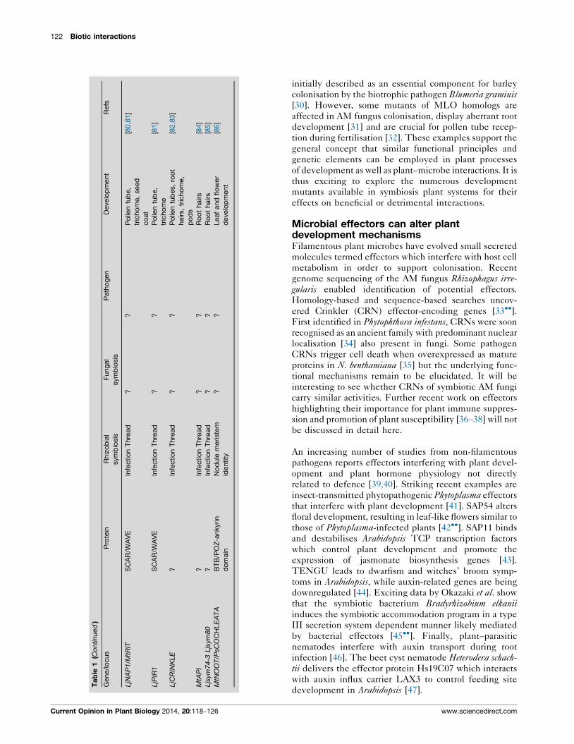
122 Biotic interactions
Ta
ble
1(C
ontinued
)
Gene/lo
cus
Pro
tein
Rhiz
ob
ial
sym
bio
sis
Fung
al
sym
bio
sis
Path
og
en
Develo
pm
ent
Refs
LjN
AP
1/M
tRIT
SC
AR
/WA
VE
Infe
ctio
nT
hre
ad
??
Po
llen
tub
e,
tric
ho
me,
seed
co
at
[80,8
1]
LjP
IR1
SC
AR
/WA
VE
Infe
ctio
nT
hre
ad
??
Po
llen
tub
e,
tric
ho
me
[81]
LjC
RIN
KLE
?In
fectio
nT
hre
ad
??
Po
llen
tub
es,
roo
t
hairs,
tric
ho
me,
po
ds
[82,8
3]
MtA
PI
?In
fectio
nT
hre
ad
??
Ro
ot
hairs
[84]
Ljs
ym74-3
Ljs
ym80
?In
fectio
nT
hre
ad
??
Ro
ot
hairs
[85]
MtN
OO
T/P
sCO
CH
LE
AT
AB
TB
/PO
Z-a
nkyrin
do
main
No
dule
meriste
m
identity
??
Leaf
and
flo
wer
develo
pm
ent
[86]
Current Opinion in Plant Biology 2014, 20:118–126
initially described as an essential component for barley
colonisation by the biotrophic pathogen Blumeria graminis[30]. However, some mutants of MLO homologs are
affected in AM fungus colonisation, display aberrant root
development [31] and are crucial for pollen tube recep-
tion during fertilisation [32]. These examples support the
general concept that similar functional principles and
genetic elements can be employed in plant processes
of development as well as plant–microbe interactions. It is
thus exciting to explore the numerous development
mutants available in symbiosis plant systems for their
effects on beneficial or detrimental interactions.
Microbial effectors can alter plantdevelopment mechanismsFilamentous plant microbes have evolved small secreted
molecules termed effectors which interfere with host cell
metabolism in order to support colonisation. Recent
genome sequencing of the AM fungus Rhizophagus irre-gularis enabled identification of potential effectors.
Homology-based and sequence-based searches uncov-
ered Crinkler (CRN) effector-encoding genes [33��].First identified in Phytophthora infestans, CRNs were soon
recognised as an ancient family with predominant nuclear
localisation [34] also present in fungi. Some pathogen
CRNs trigger cell death when overexpressed as mature
proteins in N. benthamiana [35] but the underlying func-
tional mechanisms remain to be elucidated. It will be
interesting to see whether CRNs of symbiotic AM fungi
carry similar activities. Further recent work on effectors
highlighting their importance for plant immune suppres-
sion and promotion of plant susceptibility [36–38] will not
be discussed in detail here.
An increasing number of studies from non-filamentous
pathogens reports effectors interfering with plant devel-
opment and plant hormone physiology not directly
related to defence [39,40]. Striking recent examples are
insect-transmitted phytopathogenic Phytoplasma effectors
that interfere with plant development [41]. SAP54 alters
floral development, resulting in leaf-like flowers similar to
those of Phytoplasma-infected plants [42��]. SAP11 binds
and destabilises Arabidopsis TCP transcription factors
which control plant development and promote the
expression of jasmonate biosynthesis genes [43].
TENGU leads to dwarfism and witches’ broom symp-
toms in Arabidopsis, while auxin-related genes are being
downregulated [44]. Exciting data by Okazaki et al. show
that the symbiotic bacterium Bradyrhizobium elkaniiinduces the symbiotic accommodation program in a type
III secretion system dependent manner likely mediated
by bacterial effectors [45��]. Finally, plant–parasitic
nematodes interfere with auxin transport during root
infection [46]. The beet cyst nematode Heterodera schach-tii delivers the effector protein Hs19C07 which interacts
with auxin influx carrier LAX3 to control feeding site
development in Arabidopsis [47].
www.sciencedirect.com
Plant–microbe interactions and development Evangelisti, Rey and Schornack 123
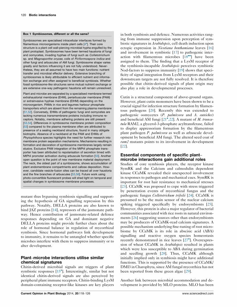
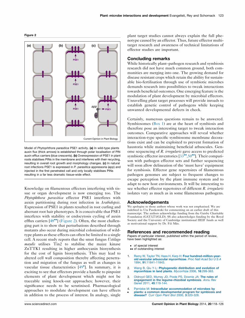
Figure 2
appPSE1 PSE1
P
PSE1
PSE1
(a) (b) (c)
Current Opinion in Plant Biology
Model of Phytophthora parasitica PSE1 activity. (a) In wild-type plants
auxin flux (thick arrows) is established through polar localisation of PIN
auxin efflux carriers (blue crescents). (b) Overexpression of PSE1 in plant
roots stabilises PINs in the membrane and interferes with their recycling,
resulting in overall root growth and morphology changes. (c) In natural
root infections PSE1 is expressed in P. parasitica appressoria (app) and
injected in the first penetrated cell and only locally stabilises PINs
resulting in a far less dramatic tissue-wide effect.
Knowledge on filamentous effectors interfering with tis-
sue or organ development is now emerging too. The
Phytophthora parasitica effector PSE1 interferes with
auxin partitioning during root infection in Arabidopsis.Expression of PSE1 in plants resulted in root curling and
aberrant root hair phenotypes. It is conceivable that PSE1
interferes with stability or endocytosis cycling of auxin
efflux carriers [48��] (Figure 2). Often, the most challen-
ging part is to show that perturbations described through
mutants also occur during microbial colonisation of wild-
type plants as these effects can often be limited to a single
cell. A recent study reports that the smut fungus Ustilagomaydis utilises Tin2 to stabilise the maize kinase
ZnTTK1 resulting in higher anthocyanin biosynthesis
for the cost of lignin biosynthesis. This may lead to
altered cell wall composition thereby affecting penetra-
tion and migration of the fungus as well as changes in
vascular tissue characteristics [49�]. In summary, it is
exciting to see that effectors provide a handle to pinpoint
elements of plant development which might not be
traceable using knock-out approaches; however, their
significance needs to be scrutinised. Pharmacological
approaches to modulate development can have effects
in addition to the process of interest. In analogy, single
www.sciencedirect.com
plant target studies cannot always explain the full phe-
notype caused by an effector. Thus, future effector multi-
target research and awareness of technical limitations of
effector studies are important.
Concluding remarksWhile historically plant–pathogen research and symbiosis
research did not have much common ground, both com-
munities are merging into one. The growing demand for
disease resistant crops which retain the ability for sustain-
able bio-fertilisation through use of symbiotic microbes
demands research into possibilities to tweak interactions
towards beneficial outcomes. One emerging feature is the
modulation of plant development by microbial effectors.
Unravelling plant target processes will provide inroads to
establish genetic control of pathogens while keeping
unwanted developmental defects in check.
Certainly, numerous questions remain to be answered.
Symbiosomes (Box 1) are at the heart of symbiosis and
therefore pose an interesting target to tweak interaction
outcomes. Comparative approaches will reveal whether
interaction-type specific symbiosome membrane decora-
tions exist and can be exploited to prevent formation of
haustoria while maintaining beneficial arbuscules. Gen-
ome sequencing of R. irregularis gave access to predicted
symbiotic effector inventories [33��,50��]. Their compari-
son with pathogen effector sets and further sequencing
will soon allow delineation of the ‘must have’ equipment
for symbiosis. Effector gene repertoires of filamentous
pathogen genomes are subject to frequent changes to
escape perception by the plant immune system and to
adapt to new host environments. It will be interesting to
see whether effector repertoires of different R. irregularisisolates vary as much as in some filamentous pathogens.
AcknowledgementsWe apologise to those authors whose work was not emphasised. We areindebted to Uta Paszkowski for commenting on an earlier draft of thismanuscript. The authors acknowledge funding from the Gatsby CharitableFoundation (GAT3273/GLD). SS also acknowledges funding by the RoyalSociety and the University of Cambridge institutional HEIF funds as wellas motivational support by Dr. Fei Mao.
References and recommended readingPapers of particular interest, published within the period of review,have been highlighted as:
� of special interest�� of outstanding interest
1. Remy W, Taylor TN, Hass H, Kerp H: Four hundred-million-year-old vesicular arbuscular mycorrhizae. Proc Natl Acad Sci U S A1994, 91:11841-11843.
2. Wang B, Qiu Y-L: Phylogenetic distribution and evolution ofmycorrhizas in land plants. Mycorrhiza 2006, 16:299-363.
3. Oldroyd GED, Murray JD, Poole PS, Downie JA: The rules ofengagement in the legume-rhizobial symbiosis. Annu RevGenet 2011, 45:119-144.
4.�
Parniske M: Intracellular accommodation of microbes byplants: a common developmental program for symbiosis anddisease? Curr Opin Plant Biol 2000, 3:320-328.
Current Opinion in Plant Biology 2014, 20:118–126
124 Biotic interactions
This pioneering review suggests common and contrasting principles inmicrobial accommodation in plants.
5. Djebali N, Jauneau A, Ameline-Torregrosa C, Chardon F,Jaulneau V, Mathe C, Bottin A, Cazaux M, Pilet-Nayel M-L,Baranger A et al.: Partial resistance of Medicago truncatula toAphanomyces euteiches is associated with protection of theroot stele and is controlled by a major QTL rich in proteasome-related genes. Mol Plant Microbe Interact 2009, 22:1043-1055.
6. Olah B, Briere C, Becard G, Denarie J, Gough C: Nod factors anda diffusible factor from arbuscular mycorrhizal fungi stimulatelateral root formation in Medicago truncatula via the DMI1/DMI2 signalling pathway. Plant J 2005, 44:195-207.
7. Reusche M, Thole K, Janz D, Truskina J, Rindfleisch S, Drubert C,Polle A, Lipka V, Teichmann T: Verticillium infection triggersVASCULAR-RELATED NAC DOMAIN7-dependent de novoxylem formation and enhances drought tolerance inArabidopsis. Plant Cell Online 2012, 24:3823-3837.
8. Rey T, Schornack S: Interactions of beneficial and detrimentalroot-colonizing filamentous microbes with plant hosts.Genome Biol 2013, 14:121.
9. Penmetsa RV, Uribe P, Anderson J, Lichtenzveig J, Gish J-C,Nam YW, Engstrom E, Xu K, Sckisel G, Pereira M et al.: TheMedicago truncatula ortholog of Arabidopsis EIN2, sickle, is anegative regulator of symbiotic and pathogenic microbialassociations. Plant J 2008, 55:580-595.
10. Gonzalez-Rizzo S, Crespi M, Frugier F: The Medicago truncatulaCRE1 cytokinin receptor regulates lateral root developmentand early symbiotic interaction with Sinorhizobium meliloti.Plant Cell 2006, 18:2680-2693.
11. Moreau S, Fromentin J, Vailleau F, Vernie T, Huguet S,Balzergue S, Frugier F, Gamas P, Jardinaud MF: The symbiotictranscription factor MtEFD and cytokinins are positivelyacting in the Medicago truncatula and Ralstoniasolanacearum pathogenic interaction. New Phytol 2014,201:1343-1357.
12.��
Floss DS, Levy JG, Levesque-Tremblay V, Pumplin N,Harrison MJ: DELLA proteins regulate arbuscule formation inarbuscular mycorrhizal symbiosis. Proc Natl Acad Sci U S A2013, 110:E5025-E5034.
The authors demonstrate that GA signalling is crucial for the formation ofintracellular accommodation structures by AM fungi in both dicots andmonocots. In addition, DELLA activity in the vascular tissue and endo-dermis is sufficient to enable arbuscule formation in the inner cortex.
13.�
Yu N, Luo D, Zhang X, Liu J, Wang W, Jin Y, Dong W, Liu J, Liu H,Yang W et al.: A DELLA protein complex controls thearbuscular mycorrhizal symbiosis in plants. Cell Res 2014,24:130-133.
This manuscript describes a possible link between GA signalling andsymbiosis by reporting the possible presence of the DELLA protein SLR1in complexes containing GRAS-type transcription factors DIP1 andRAM1, the latter of which previously has been implicated in symbiosissignalling.
14. Hou X, Lee LYC, Xia K, Yan Y, Yu H: DELLAs modulatejasmonate signaling via competitive binding to JAZs. Dev Cell2010, 19:884-894.
15.�
Genre A, Chabaud M, Balzergue C, Puech-Pages V, Novero M,Rey T, Fournier J, Rochange S, Becard G, Bonfante P et al.: Short-chain chitin oligomers from arbuscular mycorrhizal fungitrigger nuclear Ca2+ spiking in Medicago truncatula roots andtheir production is enhanced by strigolactone. New Phytol2013, 198:190-202.
This manuscript reports short chain chitin oligomers (CO4/5) as inducersof calcium spiking in root organ cultures. Calcium spiking was induced ina symbiosis-pathway dependent manner, but independent of the recep-tor of lipochito-oligosaccharidic Nod factors.
16. Pietraszewska-Bogiel A, Lefebvre B, Koini MA, Klaus-Heisen D,Takken FL, Geurts R, Cullimore JV, Gadella TW: Interaction ofMedicago truncatula lysin motif receptor-like kinases, NFPand LYK3, produced in Nicotiana benthamiana inducesdefence-like responses. PLoS One 2013, 8:e65055.
17. Czaja LF, Hogekamp C, Lamm P, Maillet F, Martinez EA, Samain E,Denarie J, Kuster H, Hohnjec N: Transcriptional responses
Current Opinion in Plant Biology 2014, 20:118–126
toward diffusible signals from symbiotic microbes revealMtNFP- and MtDMI3-dependent reprogramming of host geneexpression by arbuscular mycorrhizal fungallipochitooligosaccharides. Plant Physiol 2012, 159:1671-1685.
18.��
Rey T, Nars A, Bonhomme M, Bottin A, Huguet S, Balzergue S,Jardinaud MF, Bono JJ, Cullimore J, Dumas B et al.: NFP, a LysMprotein controlling Nod factor perception, also intervenes inMedicago truncatula resistance to pathogens. New Phytol2013, 198:875-886.
The authors show that a receptor assumed to be exclusively involved inroot nodule symbiosis also contributes to disease severity by the root-pathogenic oomycete A. euteiches.
19. Liang Y, Cao Y, Tanaka K, Thibivilliers S, Wan J, Choi J, Kang C Ho,Qiu J, Stacey G: Nonlegumes respond to rhizobial Nod factorsby suppressing the innate immune response. Science 2013,341:1384-1387.
20. DeZwaan TM, Carroll AM, Valent B, Sweigard JA: Magnaporthegrisea pth11p is a novel plasma membrane protein thatmediates appressorium differentiation in response toinductive substrate cues. Plant Cell 1999, 11:2013-2030.
21.�
Wang E, Schornack S, Marsh JF, Gobbato E, Schwessinger B,Eastmond P, Schultze M, Kamoun S, Oldroyd GED: A commonsignaling process that promotes mycorrhizal and oomycetecolonization of plants. Curr Biol 2012, 22:2242-2246.
This manuscript describes a common role for cutin monomers in pene-tration of M. truncatula root tissue by beneficial and detrimental filamen-tous microorganisms.
22. Gobbato E, Wang E, Higgins G, Bano SA, Henry C, Schultze M,Oldroyd GED: RAM1 and RAM2 function and expression duringarbuscular mycorrhizal symbiosis and Aphanomyceseuteiches colonization. Plant Signal Behav 2013 http://dx.doi.org/10.4161/psb.26049.
23. Esseling JJ, Lhuissier FG, Emons AM: A nonsymbiotic root hairtip growth phenotype in NORK-mutated legumes: implicationsfor nodulation factor-induced signaling and formation of amultifaceted root hair pocket for bacteria. Plant Cell 2004,16:933-944.
24. Genre A, Ortu G, Bertoldo C, Martino E, Bonfante P: Biotic andabiotic stimulation of root epidermal cells reveals commonand specific responses to arbuscular mycorrhizal fungi. PlantPhysiol 2009, 149:1424-1434.
25. Miller JB, Pratap A, Miyahara A, Zhou L, Bornemann S, Morris RJ,Oldroyd GED: Calcium/calmodulin-dependent protein kinaseis negatively and positively regulated by calcium, providing amechanism for decoding calcium responses during symbiosissignaling. Plant Cell Online 2013 http://dx.doi.org/10.1105/tpc.113.116921.
26. Ikeda S, Okubo T, Takeda N, Banba M, Sasaki K, Imaizumi-Anraku H, Fujihara S, Ohwaki Y, Ohshima K, Fukuta Y et al.: Thegenotype of the calcium/calmodulin-dependent proteinkinase gene (CCaMK) determines bacterial communitydiversity in rice roots under paddy and upland field conditions.Appl Environ Microbiol 2011, 77:4399-4405.
27.�
Shi B, Ni L, Zhang A, Cao J, Zhang H, Qin T, Tan M, Zhang J,Jiang M: OsDMI3 is a novel component of abscisic acidsignaling in the induction of antioxidant defense in leaves ofrice. Mol Plant 2012, 5:1359-1374.
Here, DMI3, a classical common symbiosis pathway component isreported to have additional functions in ABA signalling and reactiveoxygen species homeostasis in leaves.
28. Yang C, Li A, Zhao Y, Zhang Z, Zhu Y, Tan X, Geng S, Guo H,Zhang X, Kang Z et al.: Overexpression of a wheat CCaMK genereduces ABA sensitivity of Arabidopsis thaliana during seedgermination and seedling growth. Plant Mol Biol Rep 2010,29:681-692.
29. Delaux PM, Sejalon-Delmas N, Becard G, Ane J-M: Evolution ofthe plant–microbe symbiotic ‘toolkit’. Trends Plant Sci 2013,6:298-304.
30. Buschges R, Hollricher K, Panstruga R, Simons G, Wolter M,Frijters A, van Daelen R, van der Lee T, Diergaarde P,Groenendijk J et al.: The barley Mlo gene: a novel controlelement of plant pathogen resistance. Cell 1997, 88:695-705.
www.sciencedirect.com
Plant–microbe interactions and development Evangelisti, Rey and Schornack 125
31. Chen Z, Noir S, Kwaaitaal M, Hartmann HA, Wu M-J, Mudgil Y,Sukumar P, Muday G, Panstruga R, Jones AM: Two seven-transmembrane domain MILDEW RESISTANCE LOCUS Oproteins cofunction in Arabidopsis rootthigmomorphogenesis. Plant Cell 2009, 21:1972-1991.
32. Kessler SA, Shimosato-Asano H, Keinath NF, Wuest SE, Ingram G,Panstruga R, Grossniklaus U: Conserved molecularcomponents for pollen tube reception and fungal invasion.Science 2010, 330:968-971.
33.��
Lin K, Limpens E, Zhang Z, Ivanov S, Saunders DGO, Mu D,Pang E, Cao H, Cha H, Lin T et al.: Single nucleus genomesequencing reveals high similarity among nuclei of anendomycorrhizal fungus. PLoS Genet 2014, 10:e1004078.
This manuscript demystifies the long lasting hypothesis that nuclei of asingle AM fungus isolate are markedly different in their genetic setup. Theauthors also report CRN proteins as potential effector candidates of R.irregularis. Overall the repertoire of effectors seems to be small comparedto filamentous pathogens.
34. Schornack S, Damme M van, Bozkurt TO, Cano LM, Smoker M,Thines M, Gaulin E, Kamoun S, Huitema E: Ancient class oftranslocated oomycete effectors targets the host nucleus.Proc Natl Acad Sci U S A 2010, 107:17421-17426.
35. Stam R, Howden AJM, Delgado-Cerezo M, Amaro MM,Motion TM, Pham GB, Huitema EJ: Characterization of celldeath inducing Phytophthora capsici CRN effectors suggestsdiverse activities in the host nucleus. Front Plant Sci 2013,4:387.
36. McLellan H, Boevink PC, Armstrong MR, Pritchard L, Gomez S,Morales J, Whisson SC, Beynon JL, Birch PRJ: An RxLR effectorfrom Phytophthora infestans prevents re-localisation of twoplant NAC transcription factors from the endoplasmicreticulum to the nucleus. PLoS Pathog 2013, 9:e1003670.
37. Caillaud M-C, Asai S, Rallapalli G, Piquerez S, Fabro G,Jones JDG: A downy mildew effector attenuates salicylic acid–triggered immunity in Arabidopsis by interacting with the hostmediator complex. PLoS Biol 2013, 11:e1001732.
38. Kloppholz S, Kuhn H, Requena N: A secreted fungal effector ofGlomus intraradices promotes symbiotic biotrophy. Curr Biol2011, 21:1204-1209.
39. Kay S, Hahn S, Marois E, Wieduwild R, Bonas U: Detailedanalysis of the DNA recognition motifs of the Xanthomonastype III effectors AvrBs3 and AvrBs3Drep16. Plant J 2009,59:859-871.
40. Block A, Guo M, Li G, Elowsky C, Clemente TE, Alfano JR: ThePseudomonas syringae type III effector HopG1 targetsmitochondria, alters plant development and suppresses plantinnate immunity. Cell Microbiol 2010, 12:318-330.
41. Sugio A, MacLean AM, Kingdom HN, Grieve VM, Manimekalai R,Hogenhout SA: Diverse targets of phytoplasma effectors: fromplant development to defense against insects. Annu RevPhytopathol 2011, 49:175-195.
42.��
MacLean AM, Sugio A, Makarova OV, Findlay KC, Grieve VM,Toth R, Nicolaisen M, Hogenhout SA: Phytoplasma effectorSAP54 induces indeterminate leaf-like flower development inArabidopsis plants. Plant Physiol 2011, 157:831-841.
This is one of the publications from the Hogenhout lab which demonstratethe exciting interference of Phytoplasma effectors with plant develop-ment processes resulting in the production of leaf-like flowers that aresimilar to those produced by Phytoplasma-infected plants.
43. Sugio A, Kingdom HN, MacLean AM, Grieve VM, Hogenhout SA:Phytoplasma protein effector SAP11 enhances insect vectorreproduction by manipulating plant development and defensehormone biosynthesis. Proc Natl Acad Sci U S A 2011, 108:1254-1263.
44. Hoshi A, Oshima K, Kakizawa S, Ishii Y, Ozeki J, Hashimoto M,Komatsu K, Kagiwada S, Yamaji Y, Namba S: A unique virulencefactor for proliferation and dwarfism in plants identified from aphytopathogenic bacterium. Proc Natl Acad Sci U S A 2009,106:6416-6421.
45.��
Okazaki S, Kaneko T, Sato S, Saeki K: Hijacking of leguminousnodulation signaling by the rhizobial type III secretion system.Proc Natl Acad Sci U S A 2013, 110:17131-17136.
www.sciencedirect.com
The authors report that the root nodule forming bacterium B. elkanii haslikely adopted type III delivered effectors to activate host symbiosissignalling. Nod-factor deficient B. elkanii still induced nodules unlesstheir type III secretion systems were mutated. The absence of root haircurling and infection threads suggests that B. elkanii utilises other formsof colonisation.
46. Grunewald W, Noorden G van, Isterdael GV, Beeckman T,Gheysen G, Mathesius U: Manipulation of auxin transport inplant roots during Rhizobium symbiosis and nematodeparasitism. Plant Cell Online 2009, 21:2553-2562.
47. Lee C, Chronis D, Kenning C, Peret B, Hewezi T, Davis EL,Baum TJ, Hussey R, Bennett M, Mitchum MG: The novel cystnematode effector protein 19C07 interacts with theArabidopsis auxin influx transporter LAX3 to control feedingsite development. Plant Physiol 2011, 155:866-880.
48.��
Evangelisti E, Govetto B, Minet-Kebdani N, Kuhn M-L, Attard A,Ponchet M, Panabieres F, Gourgues M: The Phytophthoraparasitica RXLR effector Penetration-Specific Effector 1favours Arabidopsis thaliana infection by interfering withauxin physiology. New Phytol 2013, 199:476-489.
This manuscript describes a filamentous pathogen effector which altersroot morphology by interfering with auxin efflux carrier distribution pat-terns.
49.�
Tanaka S, Brefort T, Neidig N, Djamei A, Kahnt J, Vermerris W,Koenig S, Feussner K, Feussner I, Kahmann R: A secretedUstilago maydis effector promotes virulence by targetinganthocyanin biosynthesis in maize. Elife 2014, 3:e01355.
The authors report a role for the effector Tin2 in rerouting metabolicpathways to reduce lignin biosynthesis thereby presumably allowingbetter access of Ustilago to vascular tissues.
50.��
Tisserant E, Malbreil M, Kuo A, Kohler A, Symeonidi A, Balestrini R,Charron P, Duensing N, Frey NF dit, Gianinazzi-Pearson V et al.:Genome of an arbuscular mycorrhizal fungus provides insightinto the oldest plant symbiosis. Proc Natl Acad Sci U S A 2013,110:20117-20122.
Sequencing of R. irregularis did not give evidence for cell wall degradingenzymes. It remains to be elucidated what mechanisms arbuscularmycorrhizal fungi use to penetrate root cortex cell walls. No orthologuesof bacterial genes coding for enzymes involved in symbiotic lipochito-oligosaccharide factors have been identified, which contradicts the long-standing assumption that rhizobia acquired them from AM fungi.
51. Pumplin N, Zhang X, Noar RD, Harrison MJ: Polar localization ofa symbiosis-specific phosphate transporter is mediated by atransient reorientation of secretion. Proc Natl Acad Sci U S A2012, 109:E665-E672.
52. Kobae Y, Hata S: Dynamics of periarbuscular membranesvisualized with a fluorescent phosphate transporter inarbuscular mycorrhizal roots of rice. Plant Cell Physiol 2010,51:341-353.
53. Lu Y-J, Schornack S, Spallek T, Geldner N, Chory J, Schellmann S,Schumacher K, Kamoun S, Robatzek S: Patterns of plantsubcellular responses to successful oomycete infectionsreveal differences in host cell reprogramming and endocytictrafficking. Cell Microbiol 2012, 14:682-697.
54. Haney CH, Long SR: Plant flotillins are required for infection bynitrogen-fixing bacteria. Proc Natl Acad Sci U S A 2010,107:478-483.
55. Pumplin N, Harrison MJ: Live-cell imaging revealsperiarbuscular membrane domains and organelle location inMedicago truncatula roots during arbuscular mycorrhizalsymbiosis. Plant Physiol 2009, 151:809-819.
56. Ivanov S, Fedorova E, Bisseling T: Intracellular plantmicrobe associations: secretory pathways and the formationof perimicrobial compartments. Curr Opin Plant Biol 2010,13:372-377.
57. Amiour N, Recorbet G, Robert F, Gianinazzi S, Dumas-Gaudot E:Mutations in DMI3 and SUNN modify the appressorium-responsive root proteome in arbuscular mycorrhiza. Mol PlantMicrobe Interact 2006, 19:988-997.
58. Nishimura R, Hayashi M, Wu GJ, Kouchi H, Imaizumi-Anraku H,Murakami Y, Kawasaki S, Akao S, Ohmori M, Nagasawa M et al.:
Current Opinion in Plant Biology 2014, 20:118–126
126 Biotic interactions
HAR1 mediates systemic regulation of symbiotic organdevelopment. Nature 2002, 420:426-429.
59. Schaarschmidt S, Gresshoff PM, Hause B: Analyzing thesoybean transcriptome during autoregulation ofmycorrhization identifies the transcription factors GmNF-YA1a/b as positive regulators of arbuscular mycorrhization.Genome Biol 2013, 14:R62.
60. Ben C, Toueni M, Montanari S, Tardin MC, Fervel M, Negahi A,Saint-Pierre L, Mathieu G, Gras MC, Noel D et al.: Naturaldiversity in the model legume Medicago truncatula allowsidentifying distinct genetic mechanisms conferring partialresistance to Verticillium wilt. J Exp Bot 2013, 64:317-332.
61. Ariel F, Brault-Hernandez M, Laffont C, Huault E, Brault M, Plet J,Moison M, Blanchet S, Ichante JL, Chabaud M et al.: Two directtargets of cytokinin signaling regulate symbiotic nodulation inMedicago truncatula. Plant Cell 2012, 24:3838-3852.
62. Sun J, Cardoza V, Mitchell DM, Bright L, Oldroyd G, Harris JM:Crosstalk between jasmonic acid, ethylene and Nod factorsignaling allows integration of diverse inputs for regulation ofnodulation. Plant J 2006, 46:961-970.
63. Shi B, Ni L, Liu Y, Zhang A, Tan M, Jiang M: OsDMI3-mediatedactivation of OsMPK1 regulates the activities of antioxidantenzymes in abscisic acid signalling in rice. Plant Cell Environ2014, 37:341-352.
64. Torres-Vera R, Garcıa JM, Pozo MJ, Lopez-Raez JA: Dostrigolactones contribute to plant defence? Mol Plant Pathol2014, 15:211-216.
65. Maillet F, Poinsot V, Andre O, Puech-Pages V, Haouy A,Gueunier M, Cromer L, Giraudet D, Formey D, Niebel A et al.:Fungal lipochitooligosaccharide symbiotic signals inarbuscular mycorrhiza. Nature 2011, 469:58-63.
66. Kiirika LM, Bergmann HF, Schikowsky C, Wimmer D, Korte J,Schmitz U, Niehaus K, Colditz F: Silencing of the Rac1 GTPaseMtROP9 in Medicago truncatula stimulates early mycorrhizaland oomycete root colonizations but negatively affectsrhizobial infection. Plant Physiol 2012, 159:501-516.
67. Delaux PM, Becard G, Combier JP: NSP1 is a component of theMyc signaling pathway. New Phytol 2013, 199:59-65.
68. Liu W, Kohlen W, Lillo A, Op den Camp R, Ivanov S, Hartog M,Limpens E, Jamil M, Smaczniak C, Kaufmann K et al.:Strigolactone biosynthesis in Medicago truncatula and ricerequires the symbiotic GRAS-type transcription factors NSP1and NSP2. Plant Cell 2011, 23:3853-3865.
69. Ooki Y, Banba M, Yano K, Maruya J, Sato S, Tabata S, Saeki K,Hayashi M, Kawaguchi M, Izui K et al.: Characterization of theLotus japonicus symbiotic mutant lot1 that shows a reducednodule number and distorted trichomes. Plant Physiol 2005,137:1261-1271.
70. Yoshida S, Kameoka H, Tempo M, Akiyama K, Umehara M,Yamaguchi S, Hayashi H, Kyozuka J, Shirasu K: The D3 F-boxprotein is a key component in host strigolactone responsesessential for arbuscular mycorrhizal symbiosis. New Phytol2012, 196:1208-1216.
71. Maekawa-Yoshikawa M, Muller J, Takeda N, Maekawa T, Sato S,Tabata S, Perry J, Wang TL, Groth M, Brachmann A et al.: Thetemperature-sensitive brush mutant of the legume Lotusjaponicus reveals a link between root development and noduleinfection by rhizobia. Plant Physiol 2009, 149:1785-1796.
72. Shimomura K, Nomura M, Tajima S, Kouchi H: LjnsRING, a novelRING finger protein, is required for symbiotic interactionsbetween Mesorhizobium loti and Lotus japonicus. Plant CellPhysiol. 2006, 47:1572-1581.
Current Opinion in Plant Biology 2014, 20:118–126
73. Schnabel EL, Kassaw TK, Smith LS, Marsh JF, Oldroyd GE,Long SR, Frugoli JA: The root determined nodulation1 generegulates nodule number in roots of Medicago truncatula anddefines a highly conserved, uncharacterized plant gene family.Plant Physiol 2011, 157:328-340.
74. Miyazawa H, Oka-Kira E, Sato N, Takahashi H, Wu GJ, Sato S,Hayashi M, Betsuyaku S, Nakazono M, Tabata S et al.: Thereceptor-like kinase KLAVIER mediates systemic regulation ofnodulation and non-symbiotic shoot development in Lotusjaponicus. Development 2010, 137:4317-4325.
75. Imin N, Mohd-Radzman NA, Ogilvie HA, Djordjevic MA: Thepeptide-encoding CEP1 gene modulates lateral root andnodule numbers in Medicago truncatula. J Exp Bot 2013,64:5395-5409.
76. Hopkins J, Pierre O, Kazmierczak T, Gruber V, Frugier F,Clement M, Frendo P, Herouart D, Boncompagni E: MtZR1, aPRAF protein, is involved in the development of roots andsymbiotic root nodules in Medicago truncatula. Plant CellEnviron 2013, 37:658-669.
77. Bagchi R, Salehin M, Adeyemo OS, Salazar C, Shulaev V,Sherrier DJ, Dickstein R: Functional assessment of theMedicago truncatula NIP/LATD protein demonstratesthat it is a high-affinity nitrate transporter. Plant Physiol 2012,160:906-916.
78. Bright LJ, Liang Y, Mitchell DM, Harris JM: The LATD gene ofMedicago truncatula is required for both nodule and rootdevelopment. Mol Plant Microbe Interact 2005, 18:521-532.
79. Liang Y, Mitchell DM, Harris JM: Abscisic acid rescues the rootmeristem defects of the Medicago truncatula latd mutant. DevBiol 2007, 304:297-307.
80. Miyahara A, Richens J, Starker C, Morieri G, Smith L, Long S,Downie JA, Oldroyd GE: Conservation in function of a SCAR/WAVE component during infection thread and root hair growthin Medicago truncatula. Mol Plant Microbe Interact 2010,23:1553-1562.
81. Yokota K, Fukai E, Madsen LH, Jurkiewicz A, Rueda P, Radutoiu S,Held M, Hossain MS, Szczyglowski K, Morieri G et al.:Rearrangement of actin cytoskeleton mediates invasion ofLotus japonicus roots by Mesorhizobium loti. Plant Cell 2009,21:267-284.
82. Tansengco ML, Imaizumi-Anraku H, Yoshikawa M, Takagi S,Kawaguchi M, Hayashi M, Murooka Y: Pollen development andtube growth are affected in the symbiotic mutant of Lotusjaponicus, crinkle. Plant Cell Physiol 2004, 45:511-520.
83. Tansengco ML, Hayashi M, Kawaguchi M, Imaizumi-Anraku H,Murooka Y: Crinkle, a novel symbiotic mutant that affects theinfection thread growth and alters the root hair, trichome, andseed development in Lotus japonicus. Plant Physiol 2003,131:1054-1063.
84. Teillet A, Garcia J, de Billy F, Gherardi M, Huguet T, Barker DG, deCarvalho-Niebel F, Journet EP: api, A novel Medicago truncatulasymbiotic mutant impaired in nodule primordium invasion. MolPlant Microbe Interact 2008, 21:535-546.
85. Yano K, Tansengco ML, Hio T, Higashi K, Murooka Y, Imaizumi-Anraku H, Kawaguchi M, Hayashi M: New nodulation mutantsresponsible for infection thread development in Lotusjaponicus. Mol Plant Microbe Interact 2006, 19:801-810.
86. Couzigou J-M, Zhukov V, Mondy S, Abu el Heba G, Cosson V,Ellis THN, Ambrose M, Wen J, Tadege M, Tikhonovich I et al.:Nodule Root and cochleata maintain nodule development andare legume orthologs of Arabidopsis blade-on-petiole genes.Plant Cell 2012, 24:4498-4510.
www.sciencedirect.com