Embed Size (px)
Citation preview

CROSS SECTIONAL ANATOMY OF SPINE
By Vivek jr 115/5/12


C7 – Prom. Spinous Process
T3- Level with Medial Scapular Spine
T7 – Inferior angle of scapula
L2 – Lowest Rib
L4 – Iliac Crest

Flexibility and Mobility
Flexion (forward bending) Extension (backward bending) Side bending (left and right) Rotation (left and right) Combination of above
MUSCLES ASSOCIATED WITH


Cervical vs Thoracic vs Lumbar
Cervical vertebrae
Smallest
Foramen in each transverse process

ATLAS
C1 –Atlas
Forms the joint connecting the skull and spine
Has no body

AXIS (Second cervical vertebra)
(C2) of the spine is named the axis.
It forms the pivot upon which the Atlas- rotates
strong odontoid process which rises perpendicularly from the upper surface of the body.


Thoracic Vertebra
Intermediate in size Increase in size as one
proceeds down the spineThey are distinguished by
the presence of facets on the sides of the bodies
Facets on the transverse processes of all, except the eleventh and twelfth,

Lumbar Vertebra
The lumbar vertebrae are the largest segments of the movable part of the vertebral column
Absence of a foramen in the transverse process,
Absence of facets on the sides of the body.

The cervical curve, convex forward, C1/2 – C7
The thoracic curve, is concave
T2 – T12
The lumbar T12 – L4, It is convex anteriorly.
The pelvic curve L4/5 - Coccyx

The spinal cord• Gross anatomy
– 3 layers of meninges– Epidural space (fat & vessels)– CSF – subarachnoid space– Terminates at L1/2 vertebral level
(conus medullaris)• Dura extends to S2 vertebral
level– Connects via filum terminale &
denticulate ligaments (pia)– 31 pairs of spinal nerves (mixed)
• cauda equina– Cervical & lumbar enlargements

Spinal Cord Anatomy
• Conus medullaris – terminal portion of the spinal cord• Filum terminale – fibrous extension of the pia mater; anchors
the spinal cord to the coccyx• Denticulate ligaments – delicate shelves of pia mater; attach
the spinal cord to the vertebrae• Spinal nerves – 31 pairs attach to the cord by paired roots
– Cervical nerves are named for inferior vertebra – All other nerves are named for superior vertebra
• Cervical and lumbar enlargements – sites where nerves serving the upper and lower limbs emerge
• Cauda equina – collection of nerve roots at the inferior end of the vertebral canal

The 3 Meningeal Layers• Dura mater:
– outer layer of spinal cord– subdural space:
• between arachnoid mater and dura mater
• Arachnoid mater:– middle meningeal layer– subarachnoid space:
• between arachnoid mater and pia mater
• filled with cerebrospinal fluid (CSF)
• Pia mater:– inner meningeal layer

The Meninges

Structures of the Spinal Cord
• Paired denticulate ligaments:– extend from pia mater to
dura mater– stabilize side-to-side
movement
• Blood vessels:– along surface of spinal pia
mater– within subarachnoid space

Cross-sectional anatomy of the spinal cord
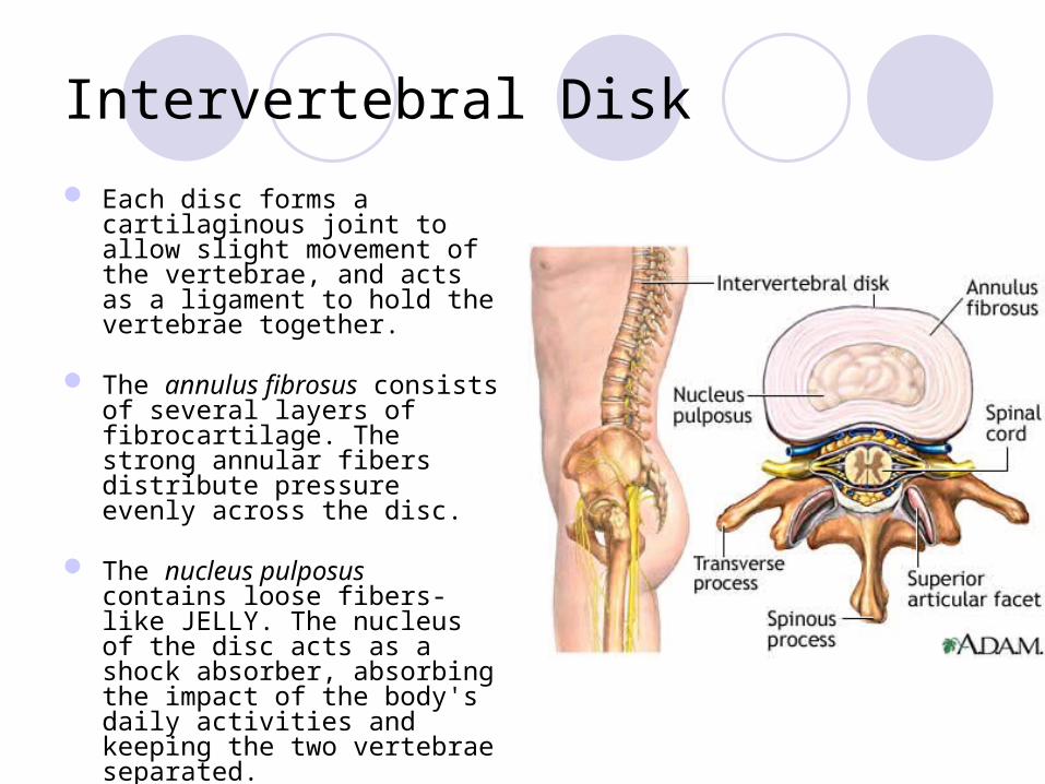
Intervertebral Disk
Each disc forms a cartilaginous joint to allow slight movement of the vertebrae, and acts as a ligament to hold the vertebrae together.
The annulus fibrosus consists of several layers of fibrocartilage. The strong annular fibers distribute pressure evenly across the disc.
The nucleus pulposus contains loose fibers- like JELLY. The nucleus of the disc acts as a shock absorber, absorbing the impact of the body's daily activities and keeping the two vertebrae separated.


Spinal Nerves
Figure 13.6

• The cervical enlargement corresponds with the attachments of the large nerves which supply the upper limbs.
• It extends from about the C-3 to T-2, its maximum circumference (about 38 mm.) being on a level with the attachment of the sixth pair of cervical nerves.
• The reason behind the enlargement of the cervical region is because of the increased neural input and output to the upper limbs.

• The lumbar enlargement (or lumbosacral enlargement) gives attachment to the nerves which supply the lower limbs.
• It commences about the level of T11, and reaches its maximum circumference, of about 33 mm., at L1 (lumbar vertebra), below which it tapers rapidly into the conus medull

Nerve Plexuses• All ventral rami except T2-T12 form interlacing nerve
networks called plexuses• Plexuses are found in the cervical, brachial, lumbar, and
sacral regions• Each resulting branch of a plexus contains fibers from
several spinal nerves• Fibers travel to the periphery via several different routes• Each muscle receives a nerve supply from more than
one spinal nerve• Damage to one spinal segment cannot completely
paralyze a muscle

The 4 Major Plexuses of Ventral Rami
1. Cervical plexus2. Brachial plexus3. Lumbar plexus4. Sacral plexus

Cervical Plexus
Figure 13.8
C1-C4 (c5-PHRENIC N.)

Brachial Plexus
Figure 13.9aC5-C8+T1 – FORM THE BRACHIAL PLEXUS

Lumbar Plexus
Figure 13.10
Arises from (T12) L1-L4 and innervates the thigh, abdominal wall, and psoas muscle

Sacral Plexus
Figure 13.11
-Arises from L4-S4 and serves the buttock, lower limb, pelvic structures, and the perineum.

Figure 13.7b
SPINAL NERVE INNERVATION: BACK, ANTEROLATERAL THORAX, AND ABDOMINAL WALL
Spinalis muscleLongismus thoracis
Iliocostalis thoracis

THANK YOU