Embed Size (px)
Citation preview
Therapeutics, Targets, and Chemical Biology
Crosstalk between KIT and FGFR3 PromotesGastrointestinal Stromal Tumor Cell Growth andDrug ResistanceNathalie Javidi-Sharifi1,2, Elie Traer1,2, Jacqueline Martinez1, Anu Gupta3,Takehiro Taguchi4, Jennifer Dunlap1,5, Michael C. Heinrich1,2,6,Christopher L. Corless1,5, Brian P. Rubin3,7,8, Brian J. Druker1,2,9, andJeffrey W. Tyner1,10
Abstract
Kinase inhibitors such as imatinib have dramaticallyimproved outcomes for patients with gastrointestinal stromaltumor (GIST), but many patients develop resistance to thesetreatments. Although in some patients this event correspondswith mutations in the GIST driver oncogenic kinase KIT, otherpatients develop resistance without KIT mutations. In thisstudy, we address this patient subset in reporting a functionaldependence of GIST on the FGF receptor FGFR3 and its cross-talk with KIT in GIST cells. Addition of the FGFR3 ligandFGF2 to GIST cells restored KIT phosphorylation during ima-tinib treatment, allowing sensitive cells to proliferate in thepresence of the drug. FGF2 expression was increased in imati-nib-resistant GIST cells, the growth of which was blocked by
RNAi-mediated silencing of FGFR3. Moreover, combining KITand FGFR3 inhibitors synergized to block the growth of ima-tinib-resistant cells. Signaling crosstalk between KIT and FGFR3activated the MAPK pathway to promote resistance to imatinib.Clinically, an IHC analysis of tumor specimens from imatinib-resistant GIST patients revealed a relative increase in FGF2levels, with a trend toward increased expression in imatinib-na€�ve samples consistent with possible involvement in drugresistance. Our findings provide a mechanistic rationale toevaluate existing FGFR inhibitors and multikinase inhibitorsthat target FGFR3 as promising strategies to improve treatmentof patients with GIST with de novo or acquired resistance toimatinib. Cancer Res; 75(5); 880–91. �2014 AACR.
IntroductionGastrointestinal stromal tumors (GIST) are the most com-
mon mesenchymal neoplasms of the gastrointestinal tractwith 5,000 to 6,000 new cases in the United States each year(1). The receptor tyrosine kinase (RTK) KIT is highly expressedand carries activating mutations in most GISTs (2). The major-ity of GISTs with wild-type KIT have activating mutations in
the RTK platelet-derived growth factor receptor a (PDGFRA;refs. 3, 4). Activation of the PI3K pathway downstream of mut-ant KIT/PDGFRA is essential for GIST cell growth and survival(5). In addition, MAPK pathway signaling is activated down-stream of KIT, and plays a pivotal role in tumorigenesis throughthe stabilization of the transcription factor ETV1 and activationof an oncogenic transcriptional program (6). The introductionof targeted tyrosine kinase inhibitor (TKI) therapy has revolu-tionized the clinical management of GIST and exemplifiesthe success of targeted therapy in solid tumors, where 80%to 90% of patients with GIST with unresectable or disseminateddisease initially attain at least disease stabilization, or completeor partial response to imatinib mesylate (7). However, nearly50% of GIST cases treated with imatinib develop second-ary resistance in the first 2 years (8). Most frequently, secondaryresistance is due to acquisition of additional mutations inKIT or PDGFRA that decrease the binding affinity for imatinib(9). However, another mechanism that is likely to account foracquired resistance in a subset of GISTs is activation of path-ways other than KIT and PDGFRA, thereby bypassing theinhibitory effects of KIT/PDGFRA-targeted small molecules.
RTKs are tightly regulated in normal cells, but frequentlyacquire transforming functions due to mutation(s), overexpres-sion, and autocrine paracrine stimulation in human cancers.Selective TKIs can block this activity and constitute a promisingapproach for molecularly guided therapeutics. For example, theFGF signaling network is deregulated in several human cancers,including breast, bladder, prostate, endometrial, and non–small
1Knight Cancer Institute, Oregon Health and Science University,Portland, Oregon. 2Division of Hematology and Medical Oncology,Oregon Health and Science University, Portland, Oregon. 3Depart-ment of Molecular Genetics, Lerner Research Institute, Cleveland,Ohio. 4Division of Human Health and Medical Science, GraduateSchool of Kuroshio Science, Kochi University, Nankoku, Kochi, Japan.5Department of Anatomic Pathology, Oregon Health and ScienceUniversity, Portland, Oregon. 6Portland VA Medical Center, Portland,Oregon. 7Taussig Cancer Center, Cleveland Clinic, Cleveland, Ohio.8Department of Anatomic Pathology, Cleveland Clinic, Cleveland,Ohio. 9Howard Hughes Medical Institute, Portland, Oregon. 10Depart-ment of Cell, Developmental and Cancer Biology, Oregon Health andScience University, Portland, Oregon.
Note: Supplementary data for this article are available at Cancer ResearchOnline (http://cancerres.aacrjournals.org/).
Corresponding Author: JeffreyW. Tyner, Oregon Health and Science UniversityKnight Cancer Institute, 3181 Southwest Sam Jackson Park Road, Mailcode L592,Portland, OR 97239. Phone: 503 346-0603; Fax: 503 494-3688; E-mail:[email protected]
doi: 10.1158/0008-5472.CAN-14-0573
�2014 American Association for Cancer Research.
CancerResearch
Cancer Res; 75(5) March 1, 2015880
on February 25, 2016. © 2015 American Association for Cancer Research. cancerres.aacrjournals.org Downloaded from
Published OnlineFirst November 28, 2014; DOI: 10.1158/0008-5472.CAN-14-0573
cell lung cancer (NSCLC; ref. 10). Receptors may be aberrantlyactivated through mutations (11, 12), amplifications (13), orfusions (14). The ligands for FGF receptors have also shownaberrant activity in a variety of cancers. High expression of FGF3,FGF8, and FGF10 has been reported in breast cancer (15), andcorrelates withmalignant behavior. In prostate cancer (16), FGF2expressed by stromal cells promotes tumor progression (17).Activation of the FGF signaling axis by FGF8, FGF9, and FGF10overexpression is also associated with an aggressive clinical phe-notype (18). In addition, FGF2 has recently been shown tomediate resistance to chemotherapy, and, as laid out in this paper,may also provide intrinsic protection of tumor cells in the pres-ence of small-molecule kinase inhibitors.
Materials and MethodsCell culture and reagents
GIST T1 cells were treated with 10,000 nmol/L imatinib for 2months andGIST10Rgrewout as a resistant colony. The cellswereexpanded and sequenced for KIT (exon 11–21) and PDGFRA andPDGFRB (exon 12, 14, and 18). No secondary mutations werefound in any of these RTKs. GIST T1, GIST 10R, andGIST 882 cellswere cultured in RPMI medium supplemented with 15% FBS(Atlanta Biologicals), penicillin/streptomycin (Invitrogen), andFungizone. GIST T1, GIST 10R, and GIST 882 cells were Sangersequenced to verify reported activating mutations in KIT. Inaddition, comparative RNA sequencing of GIST T1 and GIST10R cells revealed equivalent gene expression and mutationalprofiles, confirming that these cell lines were derived from thesame original clone.
siRNA and kinase inhibitorsThe RAPID siRNA library has been previously described
(19–21). All siRNAs were from Thermo Fisher Scientific Dhar-macon RNAi Technologies and cumulatively target the entiretyrosine kinase gene family as well as NRAS and KRAS (93 genestargeted total). Each well contained a pool of four siRNAs.Cells were aliquoted at 66 mL per well in a 96-well plate and34 mL of siRNA/OptiMEM/siRNA mixture was added to eachwell. Oligofectamine and siRNA were used at a ratio of 1:6. Forassessment of cell viability and proliferation, cells were sub-jected to the MTS assay after 96 hours. PD173074, AZD-6244,and PI-103 were purchased from Selleck; imatinib and CHIR-258 were purchased from LC Labs.
ImmunoblottingAll immunoblotting was performed using standard protocols.
Data were analyzed with ImageJ.
GIST tissue samplesAll patient specimens were obtained with informed consent of
the patients on protocols approved by the Institutional ReviewBoard of Oregon Health and Science University (Portland, OR).To prepare fresh-frozen GIST tissue samples for immunoblotting,tissue was dissected on dry ice using a razor blade. Four shavingsper sample were sonicated three times for 3 seconds in 2� CellSignaling Lysis Buffer.
Real time RT-PCRRNA was isolated from cells using the RNeasy Mini kit
(Qiagen). RNA was transcribed to cDNA using Superscript III
reverse transcriptase (Life Technologies), using undiluted RNA.Subsequently, cDNA was diluted 1:4 and quantified via real-timeRT-PCR following the TaqMan Gene Expression Assay protocol(Life Technologies). Samples were plated in triplicate, and everyassay included a water control.
IHCAnalysis of GI stromal tumor samples was performed by IHC
on paraffin-embedded tissue sections. Antigen retrieval wasconducted using boiling citrate buffer (10 mmol/L sodiumcitrate, pH 6.0) for 30 minutes. Please see SupplementaryMaterials for a complete description of staining and imaging.
Statistical analysesFor cell proliferation assays, a Student t test was carried out
for each treatment condition compared with untreated cells orappropriate controls. The P values for the t tests are indicated byasterisks: �, 0.01 � P < 0.05; ��, 0.001 � P < 0.01; ���, P < 0.001.To determine the significance of combination indices to indi-cate synergy, upper and lower confidence limits were calculat-ed. Data points for combinations with upper confidence limitsbelow 1 were considered synergistic.
For further experimental details, see Supplementary Materialsand Methods.
ResultssiRNA-mediated knockdown identifies FGFR3 dependence inGIST cell lines
GIST T1 cells harbor a heterozygous deletion of KIT exon11 and consequently exhibit high sensitivity to imatinib withpotent suppression of cell proliferation at concentrations rang-ing from 100 to 1,000 nmol/L. The GIST 10R cell line grewout as a colony after 2 months treatment of GIST T1 cells with10,000 nmol/L imatinib. Consequently, GIST 10R cells exhibitno IC50 at concentrations of imatinib up to 10,000 nmol/L,although an IC25 is still apparent at 100 nmol/L (Supplemen-tary Fig. S1A). Interestingly, GIST 10R cells do exhibit reducedphosphorylation of KIT and its downstream signaling mole-cules after exposure to 1,000 nmol/L imatinib (SupplementaryFig. S1B), and no secondary mutations were found in KIT,indicating that drug resistance in GIST 10R cells must be dueto alternative mechanisms. The fact that inhibition is equalat equal concentrations of imatinib is likely due to the factthat GIST 10R does not carry additional mutations in KIT thatshould render this primary drug target less susceptible toinhibition. In addition, comparative RNA sequencing betweenGIST T1 and 10R revealed no point mutations or remarkablechanges in gene expression that would explain drug resistancein these cells. To investigate possible alternative therapeutictargets in these cells, we transfected GIST T1 and 10R cells witha panel of siRNAs that collectively target the entire tyrosinekinase gene family in addition to NRAS and KRAS (93 genestotal; refs. 19, 20). As expected, siRNA against KIT significantlyreduced the relative number of proliferating GIST T1 cells(Fig. 1A). We also observed a significant reduction in prolifer-ation after silencing of colony stimulating factor 1 receptor,tyrosine kinase 2 (TYK2), and FGFR3. Polo-like kinase 1 plays acritical role in mitosis in all cells and was used as a positivecontrol for effective siRNA-mediated silencing. Interestingly,GIST 10R cells retained residual sensitivity to KIT silencing
FGF Signaling in Imatinib-Resistant GIST
www.aacrjournals.org Cancer Res; 75(5) March 1, 2015 881
on February 25, 2016. © 2015 American Association for Cancer Research. cancerres.aacrjournals.org Downloaded from
Published OnlineFirst November 28, 2014; DOI: 10.1158/0008-5472.CAN-14-0573
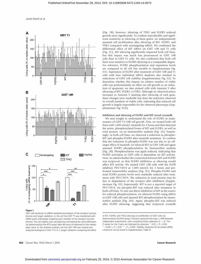
(Fig. 1B); however, silencing of TYK2 and FGFR3 reducedgrowth more significantly. To confirm reproducible and signif-icant sensitivity to silencing of these genes, we independentlyassessed cell proliferation after silencing of KIT, FGFR3, andTYK2 compared with nontargeting siRNA. We confirmed thedifferential effect of KIT siRNA on GIST 10R and T1 cells(Fig. 1C). KIT silencing significantly impacted both cell lines,but this impact was much less pronounced in GIST 10Rcells than in GIST T1 cells. We also confirmed that both celllines were sensitive to FGFR3 silencing to a comparable degree.For reference, FGFR3 phosphorylation and expression levelsare compared in all cell line models in Supplementary Fig.S1C. Expression of FGFR3 after treatment of GIST 10R and T1cells with four individual siRNA duplexes also resulted inreductions of GIST cell viability (Supplementary Fig. S2). Todetermine whether this impact on relative number of viablecells was predominantly an effect on cell growth or an induc-tion of apoptosis, we also stained cells with Annexin V aftersilencing of KIT, FGFR3, or TYK2. Although we observed minorincreases in Annexin V staining after silencing of each gene,these changes were markedly less than the reduction observedin overall numbers of viable cells, indicating that reduced cellgrowth is largely responsible for the observed phenotype (Sup-plementary Fig. S1D).
Inhibition and silencing of FGFR3 and KIT reveal crosstalkWe next sought to understand the role of FGFR3 in main-
tenance of GIST T1/10R cell growth. First, we treated both celllines with 1,000 nmol/L imatinib for 2 hours and then detectedthe active, phosphorylated forms of KIT and FGFR3, as well astotal protein, on an immunoblot analysis (Fig. 2A). Surpris-ingly, in both cell lines, we observed a reduction in phospho-KIT and phospho-FGFR3 after imatinib treatment. To confirmthat the reduction in phospho-FGFR3 was not due to an off-target effect of imatinib, we silenced KIT in GIST 10R and againassessed FGFR3 phosphorylation by immunoblot analysis(Fig. 2B). Phosphorylation was again reduced, indicating thatFGFR3 activation in GIST cells is dependent on KIT activity.Next, we asked whether the connection between KIT and FGFR3was reciprocal, so that FGFR3 inhibition or silencing wouldaffect KIT activity. We treated GIST 10R cells with the FGFRinhibitor PD173074 at 1,000 nmol/L for 2 hours and per-formed immunoblot analyses (Fig. 2C). Phospho-FGFR3 andtotal FGFR3 protein levels were markedly reduced after treat-ment with PD173074. The reduction in total protein may bedue to degradation of the receptor after inhibition (Supple-mentary Fig. S3). Importantly, KIT is not a reported target ofPD173074, yet phospho-KIT was reduced after treatment inboth cell lines. To rule out direct inhibition of KIT as the sourcefor reduced phosphorylation, we silenced FGFR3 using siRNAin GIST 10R cells and assessed KIT phosphorylation by immu-noblot analysis (Fig. 2D). Again, phospho-KIT was reducedafter FGFR3 silencing, suggesting that reciprocal crosstalk
Figure 1.GIST cell sensitivity to siRNA-mediated knockdown of the receptor tyrosinekinome and target validation. A, the cell line GIST T1 was transfected withsiRNA pools individually targeting each member of the receptor tyrosinekinome. The cell viability was calculated by normalizing the cell proliferation(as determined by the MTS assay) after 96 hours of treatment to the medianplate value. B, the imatinib-resistant cell line GIST 10R was treated andanalyzed analogous to GIST T1 in A. C, target validation comparing the effect
of KIT, FGFR3, and TYK2 silencing on proliferation of GIST cells (asdetermined by the MTS assay). The bars represent the mean� SEM betweenindependent experiments, each containing three replicates (n ¼ 3). TheP values for the t tests are indicated by asterisks: �, 0.01 � P < 0.05;�� , 0.001 � P < 0.01; ��� , P < 0.001. Viability measures for all tested siRNAconstructs can be found in Supplementary Table S1.
Javidi-Sharifi et al.
Cancer Res; 75(5) March 1, 2015 Cancer Research882
on February 25, 2016. © 2015 American Association for Cancer Research. cancerres.aacrjournals.org Downloaded from
Published OnlineFirst November 28, 2014; DOI: 10.1158/0008-5472.CAN-14-0573
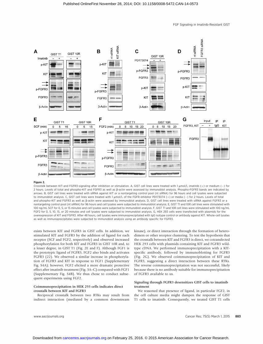
exists between KIT and FGFR3 in GIST cells. In addition, westimulated KIT and FGFR3 by the addition of ligand for eachreceptor (SCF and FGF2, respectively) and observed increasedphosphorylation for both KIT and FGFR3 in GIST 10R and, toa lesser degree, in GIST T1 (Fig. 2E and F). Although FGF1 isthe prototypic ligand of FGFR3, FGF2 also binds and activatesFGFR3 (22). We observed a similar increase in phosphoryla-tion of FGFR3 and KIT in response to FGF1 (SupplementaryFig. S4A); however, FGF2 elicited a more dramatic protectiveeffect after imatinib treatment (Fig. 3A–C) compared with FGF1(Supplementary Fig. S4B). We thus chose to conduct subse-quent experiments using FGF2.
Coimmunoprecipitation in HEK 293 cells indicates directcrosstalk between KIT and FGFR3
Reciprocal crosstalk between two RTKs may result fromindirect interaction (mediated by a common downstream
kinase), or direct interaction through the formation of hetero-dimers or other receptor clustering. To test the hypothesis thatthe crosstalk between KIT and FGFR3 is direct, we cotransfectedHEK 293 cells with plasmids containing KIT and FGFR3 wild-type cDNA. We performed immunoprecipitation with a KIT-specific antibody, followed by immunoblotting for FGFR3(Fig. 2G). We observed coimmunoprecipitation of KIT andFGFR3, suggesting a direct interaction between these RTKs.The reverse coimmunoprecipitation was not successful, likelybecause there is no antibody suitable for immunoprecipitationof FGFR3 available to us.
Signaling through FGFR3 desensitizes GIST cells to imatinibtreatment
We reasoned that presence of ligand, in particular FGF2, inthe cell culture media might dampen the response of GISTT1 cells to imatinib. Consequently, we treated GIST T1 cells
Figure 2.Crosstalk between KIT-and FGFR3-signaling after inhibition or stimulation. A, GIST cell lines were treated with 1 mmol/L imatinib (þ) or medium (�) for2 hours. Levels of total and phospho-KIT and FGFR3 as well as b-actin were assessed by immunoblot analysis. Phospho-FGFR3 bands are indicated byarrows. B, GIST cell lines were treated with siRNA against KIT or a nontargeting control pool (nt siRNA) for 96 hours and cell lysates were subjectedto immunoblot analysis. C, GIST cell lines were treated with 1 mmol/L of the FGFR-inhibitor PD173074 (þ) or media (�) for 2 hours. Levels of totaland phospho-KIT and FGFR3 as well as b-actin were assessed by immunoblot analysis. D, GIST cell lines were treated with siRNA against FGFR3 or anontargeting control pool (nt siRNA) for 96 hours and cell lysates were subjected to immunoblot analysis. E, GIST T1 and 10R cell lines were stimulated with100 ng/mL SCF for 0, 5, or 15 minutes and cell lysates were subjected to immunoblot analysis. F, GIST T1 and 10R cell lines were stimulated with 100 ng/mLFGF2 for 0, 5, 10, 15, or 20 minutes and cell lysates were subjected to immunoblot analysis. G, HEK 293 cells were transfected with plasmids for theoverexpression of KIT and FGFR3. After 48 hours, cell lysates were immunoprecipitated with IgG isotype control or antibody against KIT. Whole-cell lysatesas well as immunoprecipitates were subjected to immunoblot analysis using an antibody specific for FGFR3.
FGF Signaling in Imatinib-Resistant GIST
www.aacrjournals.org Cancer Res; 75(5) March 1, 2015 883
on February 25, 2016. © 2015 American Association for Cancer Research. cancerres.aacrjournals.org Downloaded from
Published OnlineFirst November 28, 2014; DOI: 10.1158/0008-5472.CAN-14-0573
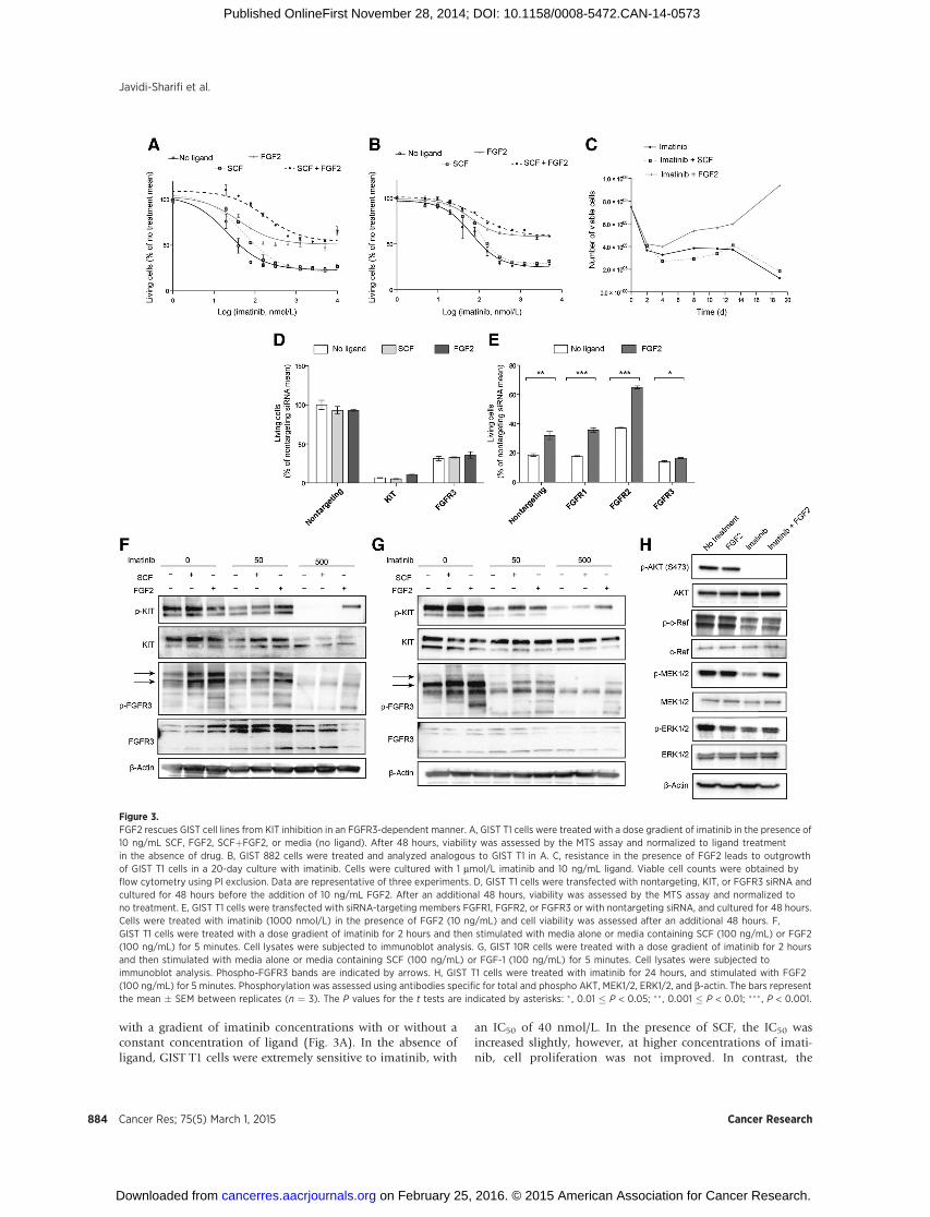
with a gradient of imatinib concentrations with or without aconstant concentration of ligand (Fig. 3A). In the absence ofligand, GIST T1 cells were extremely sensitive to imatinib, with
an IC50 of 40 nmol/L. In the presence of SCF, the IC50 wasincreased slightly, however, at higher concentrations of imati-nib, cell proliferation was not improved. In contrast, the
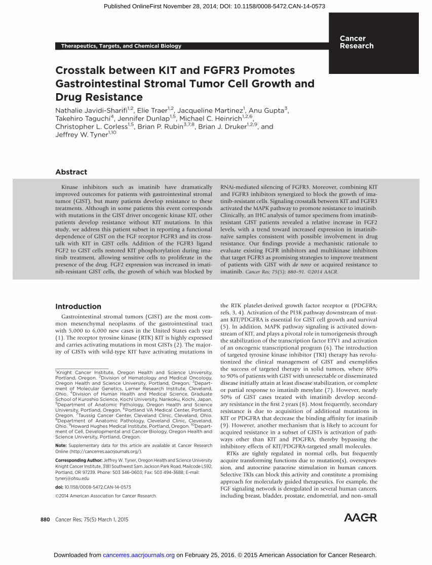
Figure 3.FGF2 rescues GIST cell lines from KIT inhibition in an FGFR3-dependent manner. A, GIST T1 cells were treated with a dose gradient of imatinib in the presence of10 ng/mL SCF, FGF2, SCFþFGF2, or media (no ligand). After 48 hours, viability was assessed by the MTS assay and normalized to ligand treatmentin the absence of drug. B, GIST 882 cells were treated and analyzed analogous to GIST T1 in A. C, resistance in the presence of FGF2 leads to outgrowthof GIST T1 cells in a 20-day culture with imatinib. Cells were cultured with 1 mmol/L imatinib and 10 ng/mL ligand. Viable cell counts were obtained byflow cytometry using PI exclusion. Data are representative of three experiments. D, GIST T1 cells were transfected with nontargeting, KIT, or FGFR3 siRNA andcultured for 48 hours before the addition of 10 ng/mL FGF2. After an additional 48 hours, viability was assessed by the MTS assay and normalized tono treatment. E, GIST T1 cells were transfected with siRNA-targeting members FGFR1, FGFR2, or FGFR3 or with nontargeting siRNA, and cultured for 48 hours.Cells were treated with imatinib (1000 nmol/L) in the presence of FGF2 (10 ng/mL) and cell viability was assessed after an additional 48 hours. F,GIST T1 cells were treated with a dose gradient of imatinib for 2 hours and then stimulated with media alone or media containing SCF (100 ng/mL) or FGF2(100 ng/mL) for 5 minutes. Cell lysates were subjected to immunoblot analysis. G, GIST 10R cells were treated with a dose gradient of imatinib for 2 hoursand then stimulated with media alone or media containing SCF (100 ng/mL) or FGF-1 (100 ng/mL) for 5 minutes. Cell lysates were subjected toimmunoblot analysis. Phospho-FGFR3 bands are indicated by arrows. H, GIST T1 cells were treated with imatinib for 24 hours, and stimulated with FGF2(100 ng/mL) for 5 minutes. Phosphorylation was assessed using antibodies specific for total and phospho AKT, MEK1/2, ERK1/2, and b-actin. The bars representthe mean � SEM between replicates (n ¼ 3). The P values for the t tests are indicated by asterisks: � , 0.01 � P < 0.05; ��, 0.001 � P < 0.01; ��� , P < 0.001.
Javidi-Sharifi et al.
Cancer Res; 75(5) March 1, 2015 Cancer Research884
on February 25, 2016. © 2015 American Association for Cancer Research. cancerres.aacrjournals.org Downloaded from
Published OnlineFirst November 28, 2014; DOI: 10.1158/0008-5472.CAN-14-0573
addition of FGF2 increased cell proliferation in the presence ofimatinib dramatically, with no IC50 even at concentrations ashigh as 10,000 nmol/L. Of note, the combination of FGF2 andSCF conferred a further increase in viability at low concentra-tions of imatinib. Similar results were observed using a different,KIT-sensitive GIST-derived cell line, GIST 882 (Fig. 3B). We alsocultured GIST T1 cells with 1,000 nmol/L imatinib in thepresence or absence of 10 ng/mL SCF or FGF2 (Fig. 3C). Viablecells were counted every 2 to 3 days for 19 days. As expected,culture with SCF did not confer any growth advantage over cellscultured with imatinib and a vehicle control. The addition ofFGF2, however, increased the number of viable cells starting atday 4. Importantly, cells not only persisted in the culture, but,after a lag phase, continued to divide and exceeded the numberinitially plated on day 0. We hypothesized that the desensiti-zation of GIST cells to imatinib is indeed mediated through theinteraction between KIT and FGFR3, and not the result of analternative survival pathway replacing KIT signaling. To this end,we measured GIST T1 cell growth after KIT silencing in thepresence or absence of FGF2 with the hypothesis that presenceof KIT protein would be required for FGF2 rescue (Fig. 3D). Aspredicted, rescue of cell growth by FGF2 was ineffective after KITknockdown, indicating that FGF2 rescue requires presence ofboth KIT and FGFR3. We also showed that inhibition of FGFR3by PD173074, which inhibits GIST T1 cell proliferation with anIC50 of 300 nmol/L, can be partially reversed by the addition ofSCF (Supplementary Fig. S5) and that SCF rescue is ineffectiveafter silencing of FGFR3 (Fig. 3D). To test whether desensitiza-tion to imatinib is, indeed, mediated by FGFR3, we performedsiRNA knockdown of FGFR1, FGFR2, and FGFR3 in GIST T1,and subsequently treated with imatinib and FGF2 (Fig. 3E).Knockdown of FGF receptors was confirmed via real-timeRT-PCR (Supplementary Fig. S6). FGF2 rescue of imatinibsensitivity remained effective after FGFR1 and FGFR2 silencing;however, FGFR3 silencing ablated the response to FGF2, impli-cating FGFR3 but not FGFR1 or FGFR2 in FGF2-mediateddrug resistance. We next treated GIST T1 cells with 0, 50, or500 nmol/L imatinib and stimulated cells with SCF or FGF2. Atbaseline, KIT phosphorylation was responsive to both SCF andFGF2 stimulation. The response to SCF was conserved in thecontext of 50 nmol/L imatinib treatment, but was markedlydecreased after treatment with 500 nmol/L imatinib. In contrast,FGF2 still restored KIT phosphorylation at 500 nmol/L (Fig. 3F).To ensure that this observation was not specific to the cell lineGIST T1, we repeated this experiment in the cell line GIST 882(Fig. 3G). Again, we observed that KIT phosphorylation in GIST882 was completely ablated at 500 nmol/L imatinib withoutFGF2 stimulation, but could be partially restored by the addi-tion of FGF2. We subsequently sought to determine the effectsof FGF2 stimulation on downstream signaling in the setting ofimatinib treatment (Fig. 3H). Analysis of AKT and MAPKpathway activation revealed that both pathways are inhibitedafter imatinib treatment. However, although AKT phosphory-lation remained inhibited after the addition of FGF2, MEK1/2and ERK1/2 phosphorylation was restored.
Combined inhibition of KIT and FGFR3 is highly synergistic inimatinib-resistant GIST cells
We hypothesized that combination treatment using imati-nib and the selective FGFR inhibitor PD173074 may restoreimatinib sensitivity in resistant GIST cells. Accordingly, we
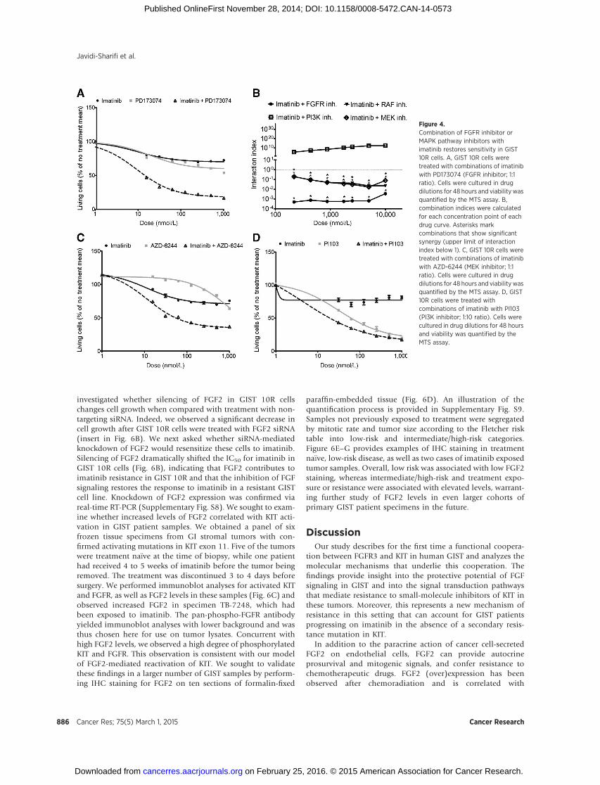
performed dose–response curves in the imatinib-resistant GIST10R cells using imatinib and PD173074 alone as well as aconstant, equimolar ratio combination of the two drugs(Fig. 4A). We then determined the combination index (CI) foreach dose point using the Chou–Talalay method to quantifysynergy. Figure 4B shows CI values plotted against the log ofdrug dose. Over the entire dosing curve, the CI values rangedfrom 0.0005 to 0.004, indicating an extremely high degree ofsynergy between the two drugs.
We next wanted to determine whether signaling pathwaysactivated downstream of FGFR3 might also exhibit synergywith KIT inhibition. To this end, we treated GIST 10R cellswith combinations of imatinib and PLX-4720, a B-Raf inhib-itor (Supplementary Fig. S7), AZD-6244, a MAPK inhibitor(Fig. 4C), and PI103, a PI3K inhibitor (Fig. 4D). Combinationof imatinib with inhibitors of the RAF/MAPK pathway exhib-ited significant synergy at all concentrations tested. In contrast,combination of imatinib with inhibitors of the PI3K-AKTpathway did not result in synergy with imatinib. Combinedtreatment with imatinib and AZD-6244, in particular, led to adecrease in cell growth similar to that observed after imatiniband PD173074 treatment, suggesting that the MAPK pathwayis a key mediator of imatinib resistance in GIST 10R cells.
The MAPK signaling pathway is activated in GIST 10R cells inresponse to imatinib
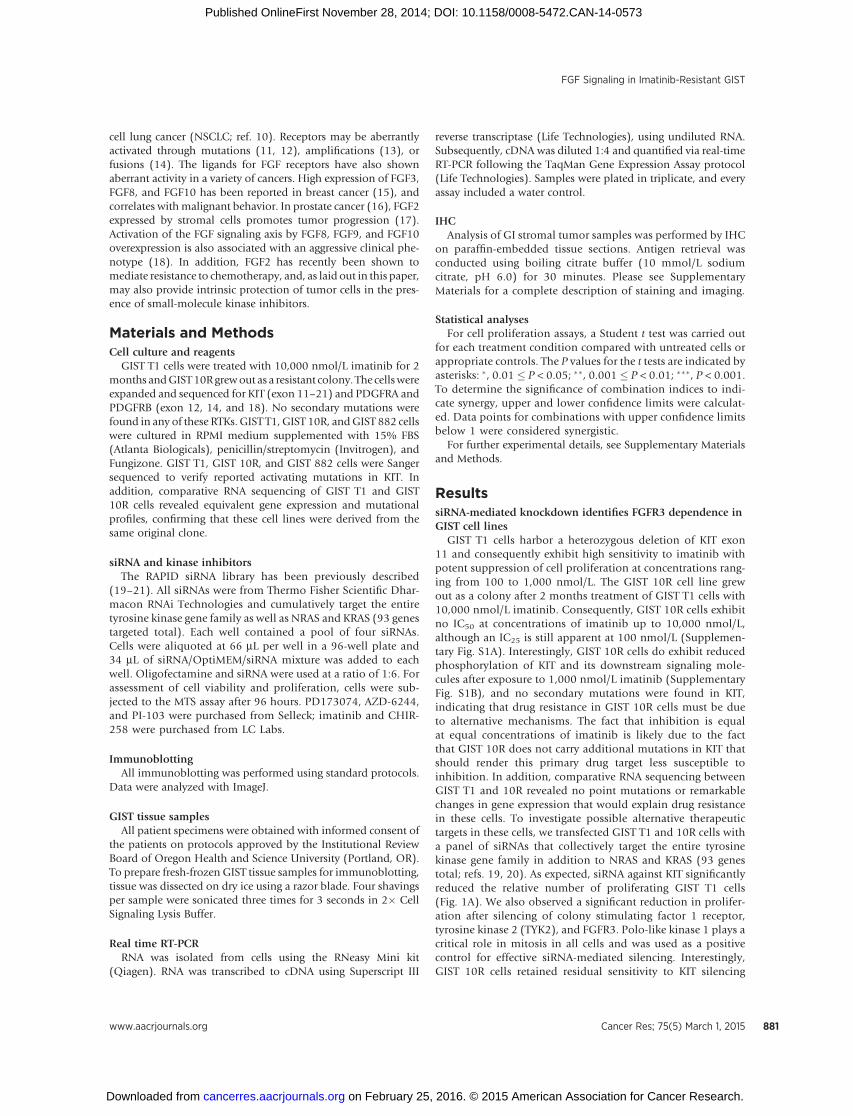
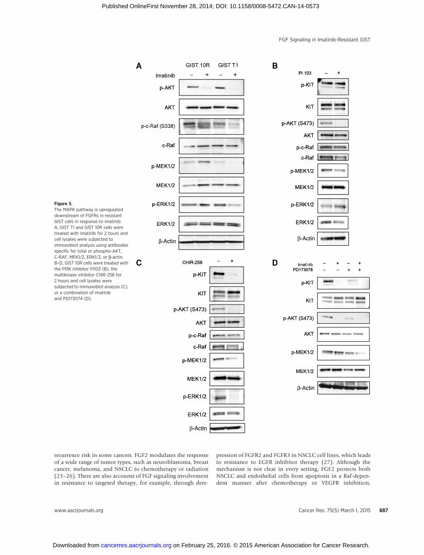
We performed immunoblot analysis to assess phosphory-lation of AKT, c-Raf, MEK, and ERK in GIST 10R and T1 cellsafter 2-hour treatment with 1,000 nmol/L imatinib (Fig. 5A). Inboth cell lines, AKT phosphorylation was equivalently reducedafter treatment. However, we observed divergent effects onMAPK pathway activation. Phosphorylation of c-Raf wasreduced less markedly in GIST 10R compared with GIST T1cells. Even more strikingly, phosphorylation of MEK and ERKwas abolished in GIST T1 after imatinib treatment, butincreased in GIST 10R. To determine whether this feedbackmechanism could be solely regulated at the receptor level, orwhether it might also be regulated after direct inactivation ofthe downstream PI3K signaling, we treated GIST 10R cells withthe PI3K inhibitor PI103 and asked whether the same increasedphosphorylation of MAPK signaling resulted (Fig. 5B). Noincrease in c-Raf, MEK, or ERK phosphorylation was observedafter inhibition of PI3K. To assess whether FGF receptors areinvolved in mediating this MAPK feedback mechanism, weinhibited KIT and FGFRs, either by the dual inhibitor CHIR-258 (dovitinib; Fig. 5C), or by a combination of imatinib andPD173074 (Fig. 5D). We found that MAPK activation wasabrogated in both cases, providing a mechanistic basis for thesynergy observed in Fig. 4.
FGF2 is overexpressed in GIST 10R cells and is increased intumor tissue after imatinib treatment
Because our data suggest that FGF2 can promote imatinibresistance in GIST cells, we wanted to determine whether FGF2levels are higher in GIST 10R than GIST T1 cells and whetherthis may underlie the resistance of GIST 10R cells to imatinib. Itis well recognized that FGF2 associates with heparan sulfate inthe extracellular matrix. We were thus unable to detect it insupernatant but found it present in cell lysate. Evaluation ofFGF2 protein levels did reveal increased levels of FGF2 in GIST10R cells compared with GIST T1 (Fig. 6A). Subsequently, we
FGF Signaling in Imatinib-Resistant GIST
www.aacrjournals.org Cancer Res; 75(5) March 1, 2015 885
on February 25, 2016. © 2015 American Association for Cancer Research. cancerres.aacrjournals.org Downloaded from
Published OnlineFirst November 28, 2014; DOI: 10.1158/0008-5472.CAN-14-0573
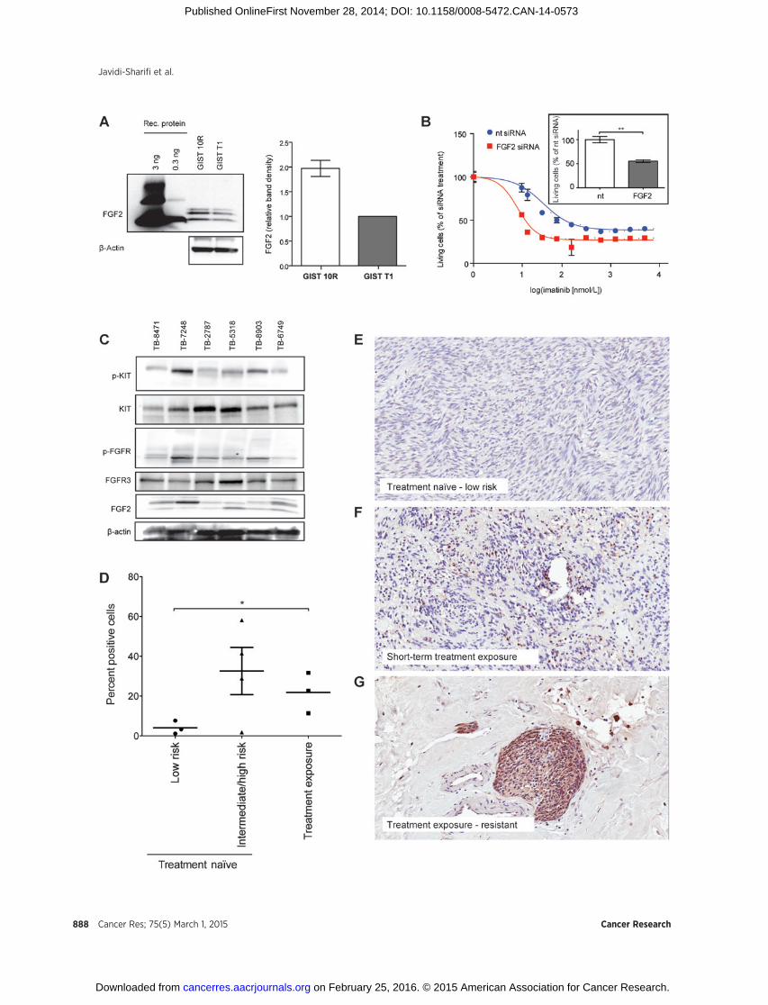
investigated whether silencing of FGF2 in GIST 10R cellschanges cell growth when compared with treatment with non-targeting siRNA. Indeed, we observed a significant decrease incell growth after GIST 10R cells were treated with FGF2 siRNA(insert in Fig. 6B). We next asked whether siRNA-mediatedknockdown of FGF2 would resensitize these cells to imatinib.Silencing of FGF2 dramatically shifted the IC50 for imatinib inGIST 10R cells (Fig. 6B), indicating that FGF2 contributes toimatinib resistance in GIST 10R and that the inhibition of FGFsignaling restores the response to imatinib in a resistant GISTcell line. Knockdown of FGF2 expression was confirmed viareal-time RT-PCR (Supplementary Fig. S8). We sought to exam-ine whether increased levels of FGF2 correlated with KIT acti-vation in GIST patient samples. We obtained a panel of sixfrozen tissue specimens from GI stromal tumors with con-firmed activating mutations in KIT exon 11. Five of the tumorswere treatment na€�ve at the time of biopsy, while one patienthad received 4 to 5 weeks of imatinib before the tumor beingremoved. The treatment was discontinued 3 to 4 days beforesurgery. We performed immunoblot analyses for activated KITand FGFR, as well as FGF2 levels in these samples (Fig. 6C) andobserved increased FGF2 in specimen TB-7248, which hadbeen exposed to imatinib. The pan-phospho-FGFR antibodyyielded immunoblot analyses with lower background and wasthus chosen here for use on tumor lysates. Concurrent withhigh FGF2 levels, we observed a high degree of phosphorylatedKIT and FGFR. This observation is consistent with our modelof FGF2-mediated reactivation of KIT. We sought to validatethese findings in a larger number of GIST samples by perform-ing IHC staining for FGF2 on ten sections of formalin-fixed
paraffin-embedded tissue (Fig. 6D). An illustration of thequantification process is provided in Supplementary Fig. S9.Samples not previously exposed to treatment were segregatedby mitotic rate and tumor size according to the Fletcher risktable into low-risk and intermediate/high-risk categories.Figure 6E–G provides examples of IHC staining in treatmentna€�ve, low-risk disease, as well as two cases of imatinib exposedtumor samples. Overall, low risk was associated with low FGF2staining, whereas intermediate/high-risk and treatment expo-sure or resistance were associated with elevated levels, warrant-ing further study of FGF2 levels in even larger cohorts ofprimary GIST patient specimens in the future.
DiscussionOur study describes for the first time a functional coopera-
tion between FGFR3 and KIT in human GIST and analyzes themolecular mechanisms that underlie this cooperation. Thefindings provide insight into the protective potential of FGFsignaling in GIST and into the signal transduction pathwaysthat mediate resistance to small-molecule inhibitors of KIT inthese tumors. Moreover, this represents a new mechanism ofresistance in this setting that can account for GIST patientsprogressing on imatinib in the absence of a secondary resis-tance mutation in KIT.
In addition to the paracrine action of cancer cell-secretedFGF2 on endothelial cells, FGF2 can provide autocrineprosurvival and mitogenic signals, and confer resistance tochemotherapeutic drugs. FGF2 (over)expression has beenobserved after chemoradiation and is correlated with
Figure 4.Combination of FGFR inhibitor orMAPK pathway inhibitors withimatinib restores sensitivity in GIST10R cells. A, GIST 10R cells weretreated with combinations of imatinibwith PD173074 (FGFR inhibitor; 1:1ratio). Cells were cultured in drugdilutions for 48 hours and viability wasquantified by the MTS assay. B,combination indices were calculatedfor each concentration point of eachdrug curve. Asterisks markcombinations that show significantsynergy (upper limit of interactionindex below 1). C, GIST 10R cells weretreated with combinations of imatinibwith AZD-6244 (MEK inhibitor; 1:1ratio). Cells were cultured in drugdilutions for 48 hours and viability wasquantified by the MTS assay. D, GIST10R cells were treated withcombinations of imatinib with PI103(PI3K inhibitor; 1:10 ratio). Cells werecultured in drug dilutions for 48 hoursand viability was quantified by theMTS assay.
Javidi-Sharifi et al.
Cancer Res; 75(5) March 1, 2015 Cancer Research886
on February 25, 2016. © 2015 American Association for Cancer Research. cancerres.aacrjournals.org Downloaded from
Published OnlineFirst November 28, 2014; DOI: 10.1158/0008-5472.CAN-14-0573
recurrence risk in some cancers. FGF2 modulates the responseof a wide range of tumor types, such as neuroblastoma, breastcancer, melanoma, and NSCLC to chemotherapy or radiation(23–26). There are also accounts of FGF signaling involvementin resistance to targeted therapy, for example, through dere-
pression of FGFR2 and FGFR3 in NSCLC cell lines, which leadsto resistance to EGFR inhibitor therapy (27). Although themechanism is not clear in every setting, FGF2 protects bothNSCLC and endothelial cells from apoptosis in a Raf-depen-dent manner after chemotherapy or VEGFR inhibition,
Figure 5.The MAPK pathway is upregulateddownstream of FGFRs in resistantGIST cells in response to imatinib.A, GIST T1 and GIST 10R cells weretreated with imatinib for 2 hours andcell lysates were subjected toimmunoblot analysis using antibodiesspecific for total or phospho-AKT,C-RAF, MEK1/2, ERK1/2, or b-actin.B–D, GIST 10R cells were treated withthe PI3K inhibitor PI103 (B), themultikinase inhibitor CHIR-258 for2 hours and cell lysates weresubjected to immunoblot analysis (C),or a combination of imatiniband PD173074 (D).
FGF Signaling in Imatinib-Resistant GIST
www.aacrjournals.org Cancer Res; 75(5) March 1, 2015 887
on February 25, 2016. © 2015 American Association for Cancer Research. cancerres.aacrjournals.org Downloaded from
Published OnlineFirst November 28, 2014; DOI: 10.1158/0008-5472.CAN-14-0573
Javidi-Sharifi et al.
Cancer Res; 75(5) March 1, 2015 Cancer Research888
on February 25, 2016. © 2015 American Association for Cancer Research. cancerres.aacrjournals.org Downloaded from
Published OnlineFirst November 28, 2014; DOI: 10.1158/0008-5472.CAN-14-0573
respectively (28, 29). FGF2 overexpression is not only asso-ciated with resistance to chemo- and radiotherapy, but alsocorrelates with an increased risk of recurrence and reducedoverall survival. The array of tumor types and treatments towhich FGF2 is connected suggests a global protective role forthis ligand, which is in line with FGF2's role in normal tissuesduring injury and inflammation.
Here, we present one of the first accounts of FGF2-mediatingresistance to targeted therapy. Recently, activation of FGFR3 andthe downstream MEK/ERK pathway was also implicated in resis-tance to a targeted agent, the B-RAF inhibitor vemurafenib, inmelanoma (25). The protective effect of FGF2 in this setting ismediated by the MAPK pathway and downstream activation ofribosomal protein S6 kinase 2 (30, 31). Similarly, an autocrinesignaling loop was identified in NSCLC, where FGFRs and theirligands were coexpressed and provided an alternative pathway toEGFR signaling in cells treated with gefitinib (32). This correlateswell with our finding that MAPK pathway members are prefer-entially activated after FGF2 stimulation in the presence of ima-tinib in GIST T1 cells. Similarly, this pathway remained active inGIST 10R cells during imatinib treatment, and these cells could beresensitized by combined imatinib and MAPK inhibitor treat-ment. The Raf/MEK/ERK signaling pathway is already recognizedas an important driver of cell proliferation, survival, and angio-genesis in GIST, as evidenced by an ongoing phase II multicentertrial of the Raf inhibitor sorafenib in imatinib- and sunitinib-resistant GIST. Selective inhibition of this pathway also inhibitedproliferation and induced apoptosis and cell-cycle arrest inpatient-derived GIST xenograft lines (33). Taken together withour observations, this underscores the potential of the Raf/MEK/ERK signaling pathway as potential future targets of moleculartherapy in GIST.
We propose that, in addition to preferential activation ofthe MAPK pathway, FGFR3 activation partially restores KITactivity. In the setting of overexpression of both receptors, wedemonstrated an interaction between the two receptors.Although these data are supportive of a direct interactionbetween KIT and FGFR3, this is only one mechanism poten-tially underlying the crosstalk observed in GIST cells, andother possibilities such as the involvement of downstreammediators should not be discarded. Receptor crosstalk andheterodimerization to circumvent targeted therapy have beenexplored extensively in the setting of EGFR inhibition. EGFRcan interact with other RTKs such as MET, ERBB2, and IGF-1R[38]. These mechanisms were identified at the clinical andpreclinical level in NSCLC and breast cancer. Overexpressionand activation of EGFR can promote transphosphorylation ofMET in lung and epidermal carcinoma cell lines. Activation of
MET occurs in an EGFR-ligand dependent manner in thesetting of EGFR overexpression, or independently of ligandin glioblastomas expressing a constitutively active EGFR var-iant. Xenograft models of glioblastoma require targeting ofboth EGFR and MET to achieve growth inhibition (34). Thecrosstalk between KIT and FGFR3 we present in this paperinvolves two hitherto unconnected signaling pathways, whichare highly relevant to human cancers. Our findings are con-sistent with the biology reported for crosstalk in other sys-tems. Although no previous reports exist of transactivationbetween KIT and FGFR3, there is evidence that FGFRs cancrosstalk with other RTKs. For example, the cytoplasmicdomains of FGF receptors and EphA4 can interact and trans-phosphorylate each other (35).
In light of the propensity of FGF2 to desensitize GISTcells to the short- and long-term effects of imatinib, we sug-gest that FGF2 expression in treated tumors provides thebasis for the development of resistance. Future studies shoulddetermine the mechanism of FGF2 upregulation and exa-mine the dynamics of FGF2 expression with imatinib treat-ment. This would provide valuable information to identifypatient populations who may be at risk of FGF-mediatedresistance, either by constitutive overexpression or by sus-tained upregulation in response to therapy. Our observationsare an added incentive to pursue targeted treatment thatcombines inhibition of KIT with antagonism of protectivesignaling from autocrine loops or the tumor stroma. Thisstrategy could be especially powerful with screening to iden-tify patients at risk for microenvironment-induced resistance.
In summary, we show for the first time that the FGFR3 pathwaycrosstalks with KIT, and that FGF2 mediates survival and out-growth of GIST cells during imatinib treatment. We furtherelucidate the molecular mechanisms of FGF2-mediated drugrescue. Our data suggest that incorporation of FGFR3 inhibitorsto combination therapeutic regimens may be beneficial in over-coming clinical resistance to targeted therapies for some patientswith GIST.
Disclosure of Potential Conflicts of InterestM.C. Heinrich is a consultant in Novartis, Ariad, MolecularMD, and
Pfizer, reports receiving commercial research support from AROG, hasreceived honoraria from speakers' bureau from Novartis, and has ownershipinterest (including patents) in Novartis. B.P. Rubin received honorariafrom speakers bureau from Novartis. B.J. Druker is a scientific advisoryboard member for Lorus Therapeutics and received commercial researchsupport from Oncotide Pharmaceuticals subcontract from NIH STTR, Novar-tis clinical trial funding, Bristol-Myers Squibb clinical trial funding, ARIADclinical trial funding, has ownership interest in OHSU invention #843,OHSU invention #0996, OHSU invention #0606, MolecularMD, Blueprint
Figure 6.FGF2 is overexpressed in GIST 10R cells and a pretreated patient sample. A, cell lysates from untreated GIST 10R and GIST T1 cells were subjected toimmunoblot analysis using antibodies specific for FGF2 or b-actin. Recombinant FGF2 is included for comparison. Sample blot is shown. FGF levels onimmunoblot analyses were quantified as bioluminescence units relative to actin and normalized to levels in GIST T1 cells for comparison. B, GIST 10R cellswere transfected with nontargeting or FGF2 siRNA and cultured for 48 hours. An imatinib gradient was added to cells and viability was assessed afteranother 48 hours. For comparison, viability was normalized to the effect of the respective siRNA alone. The effect of siRNA alone on viable cell number isshown in the inset. The bars represent the mean � SEM between replicates (n ¼ 3). GIST 10R cells were transfected with nontargeting or FGF2 siRNAand cultured for 96 hours. Viability was determined by the MTS assay. The P value is indicated: ��, 0.001 � P < 0.01. C, frozen GIST tissue samples wereprepared for immunoblot analysis and probed with antibodies specific for total and phospho KIT, FGFR, FGF2, or b-actin. D, GIST tumor FFPE sampleswere subjected to IHC staining for FGF2. Staining was quantified using ImageScope software. � , 0.01 � P < 0.05. E, example of IHC staining for FGF2on a treatment na€�ve, low-risk tumor sample. F, IHC staining for FGF2 on a tumor sample after short-term exposure to imatinib. G, example of IHC staining forFGF2 on an imatinib exposed, resistant tumor sample.
www.aacrjournals.org Cancer Res; 75(5) March 1, 2015 889
FGF Signaling in Imatinib-Resistant GIST
on February 25, 2016. © 2015 American Association for Cancer Research. cancerres.aacrjournals.org Downloaded from
Published OnlineFirst November 28, 2014; DOI: 10.1158/0008-5472.CAN-14-0573
Medicines, Millipore via Dana-Farber Cancer Institute for 4G10, and is aconsultant/advisory board member for MolecularMD, Blueprint Medicines,Gilead Sciences, Cell Therapeutics, Inc., AstraZeneca, Cylene Pharmaceuti-cals, Lorus Therapeutics, and Drug Discovery and Development Center,Biomedical Sciences Institutes. No potential conflicts of interest were dis-closed by the other authors.
Authors' ContributionsConception and design: N. Javidi-Sharifi, M.C. Heinrich, C.L. Corless,B.P. Rubin, J.W. TynerDevelopment of methodology: N. Javidi-Sharifi, E. Traer, M.C. Heinrich,J.W. TynerAcquisition of data (provided animals, acquired and managed patients,provided facilities, etc.): N. Javidi-Sharifi, J. Martinez, A. Gupta, B.J. Druker,J.W. TynerAnalysis and interpretation of data (e.g., statistical analysis, biostatistics,computational analysis): N. Javidi-Sharifi, E. Traer, J. Dunlap, M.C. Heinrich,C.L. Corless, B.J. Druker, J.W. TynerWriting, review, and/or revision of the manuscript: N. Javidi-Sharifi, E. Traer,M.C. Heinrich, C.L. Corless, B.P. Rubin, B.J. Druker, J.W. Tyner
Administrative, technical, or material support (i.e., reporting or organizingdata, constructing databases): T. Taguchi, M.C. Heinrich, J.W. TynerStudy supervision: M.C. Heinrich, B.J. Druker, J.W. Tyner
Grant SupportN. Javidi-Sharifi was supported by the Oregon Clinical and Translational
Research Institute (OCTRI), grant number TL1 RR024159 from the NationalCenter for Advancing Translational Sciences (NCATS), a component of theNIH, and NIH Roadmap for Medical Research. B.J. Druker was an investi-gator of the Howard Hughes Medical Institute. J.W. Tyner was supported bygrants from the V Foundation for Cancer Research, The Leukemia andLymphoma Society, the Gabrielle's Angel Foundation for Cancer Research,and the National Cancer Institute (5R00CA151457-04; 1R01CA183974-01).B.P. Rubin and A. Gupta were supported by the Life Raft Group.
The costs of publication of this articlewere defrayed inpart by the payment ofpage charges. This article must therefore be hereby marked advertisement inaccordance with 18 U.S.C. Section 1734 solely to indicate this fact.
Received February 25, 2014; revisedOctober 14, 2014; acceptedNovember 2,2014; published OnlineFirst November 28, 2014.
References1. Miettinen M, Lasota J. Gastrointestinal stromal tumors: review on mor-
phology, molecular pathology, prognosis, and differential diagnosis. ArchPathol Lab Med 2006;130:1466–78.
2. Hirota S, Nishida T, Isozaki K, Taniguchi M, Nakamura J, Okazaki T, et al.Gain-of-function mutation at the extracellular domain of KIT in gastroin-testinal stromal tumours. J Pathol 2001;193:505–10.
3. Hirota S, Ohashi A, Nishida T, Isozaki K, Kinoshita K, Shinomura Y,et al. Gain-of-function mutations of platelet-derived growth factorreceptor a gene in gastrointestinal stromal tumors. Gastroenterology2003;125:660–7.
4. Heinrich MC, Corless CL, Duensing A, McGreevey L, Chen CJ, Joseph N,et al. PDGFRA activating mutations in gastrointestinal stromal tumors.Science 2003;299:708–10.
5. Bauer S, Duensing A, Demetri GD, Fletcher JA. KIT oncogenic signalingmechanisms in imatinib-resistant gastrointestinal stromal tumor: PI3-kinase/AKT is a crucial survival pathway. Oncogene 2007;26:7560–8.
6. Chi P, Chen Y, Zhang L, Guo X, Wongvipat J, Shamu T, et al. ETV1 is alineage survival factor that cooperates with KIT in gastrointestinal stromaltumours. Nature 2010;467:849–53.
7. Schlemmer M, Bauer S, Sch€utte R, Hartmann J, Bokemeyer C, Hosius C,et al. Activity and side effects of imatinib in patients with gastrointestinalstromal tumors: data from a German multicenter trial. Eur J Med Res2011;16:206–12.
8. BlankeCD, RankinC,Demetri GD, RyanCW, vonMehrenM, BenjaminRS,et al. Phase III randomized, intergroup trial assessing imatinib mesylate attwo dose levels in patients with unresectable or metastatic gastrointestinalstromal tumors expressing the kit receptor tyrosine kinase: S0033. J ClinOncol 2008;26:626–32.
9. Liegl B, Kepten I, Le C, Zhu M, Demetri GD, Heinrich MC, et al. Hetero-geneity of kinase inhibitor resistance mechanisms in GIST. J Pathol 2008;216:64–74.
10. Greulich H, Pollock PM. Targeting mutant fibroblast growth factor recep-tors in cancer. Trends Mol Med 2011;17:283–92.
11. Di Martino E, Tomlinson DC, Knowles MA. A decade of FGF receptorresearch in bladder cancer: past, present, and future challenges. Adv Urol2012;2012:429213.
12. Tomlinson DC, Baldo O, Harnden P, Knowles MA. FGFR3 protein expres-sion and its relationship to mutation status and prognostic variables inbladder cancer. J Pathol 2007;213:91–8.
13. Onwuazor ON, Wen X-Y, Wang D-Y, Zhuang L, Masih-Khan E, Claudio J,et al. Mutation, SNP, and isoform analysis of fibroblast growth factorreceptor 3 (FGFR3) in 150 newly diagnosed multiple myeloma patients.Blood 2003;102:772–3.
14. Li Z. Themyeloma-associated oncogene fibroblast growth factor receptor 3is transforming in hematopoietic cells. Blood 2001;97:2413–9.
15. Tomlinson DC, Knowles MA, Speirs V. Mechanisms of FGFR3 actions inendocrine resistant breast cancer. Int J Cancer 2012;130:2857–66.
16. Kwabi-Addo B, Ozen M, Ittmann M. The role of fibroblast growthfactors and their receptors in prostate cancer. Endocr Relat Cancer 2004;11:709–24.
17. Yang F, Strand DW, Rowley DR. Fibroblast growth factor-2 mediatestransforming growth factor-beta action in prostate cancer reactive stroma.Oncogene 2008;27:450–9.
18. Murphy T, Darby S, Mathers ME, Gnanapragasam VJ. Evidence for distinctalterations in the FGF axis in prostate cancer progression to an aggressiveclinical phenotype. J Pathol 2010;220:452–60.
19. Tyner JW,Walters DK,Willis SG, LuttroppM,Oost J, LoriauxM, et al. RNAiscreening of the tyrosine kinome identifies therapeutic targets in acutemyeloid leukemia. Blood 2008;111:2238–45.
20. Tyner JW, Deininger MW, Loriaux MM, Chang BH, Gotlib JR, Willis SG,et al. RNAi screen for rapid therapeutic target identification in leukemiapatients. Proc Natl Acad Sci U S A 2009;106:8695–700.
21. Bicocca VT, Chang BH, Masouleh BK, MuschenM, LoriauxMM, Druker BJ,et al. Crosstalk betweenROR1 and the Pre-B cell receptor promotes survivalof t(1;19) acute lymphoblastic leukemia. Cancer Cell 2012;22:656–67.
22. Eswarakumar VP, Lax I, Schlessinger J. Cellular signaling by fibroblastgrowth factor receptors. Cytokine Growth Factor Rev 2005;16:139–49.
23. JohnsonMD, O'Connell MJ, Pilcher W, Reeder JE. Fibroblast growth factorreceptor-3 expression inmeningiomaswith stimulation of proliferation bythe phosphoinositide 3 kinase-Akt pathway. J Neurosurg 2010;112:934–9.
24. Tomlinson D, Knowles M, Speirs V. Mechanisms of FGFR3 actions inendocrine resistant breast cancer. Int J Cancer 2012;130:2857–66.
25. Yadav V, Zhang X, Liu J, Estrem S, Li S, Gong X-Q, et al. Reactivation ofmitogen-activated protein kinase (MAPK) pathway by FGF receptor 3(FGFR3)/Ras mediates resistance to vemurafenib in human B-RAFV600E mutant melanoma. J Biol Chem 2012;287:28087–98.
26. Wesche J, Haglund K, Haugsten EM. Fibroblast growth factors and theirreceptors in cancer. Biochem J 2011;437:199–213.
27. Ware KE, Marshall ME, Heasley LR, Marek L, Hinz TK, Hercule P, et al.Rapidly acquired resistance to EGFR tyrosine kinase inhibitors in NSCLCcell lines through de-repression of FGFR2 and FGFR3 expression. PLoSONE 2010;5:e14117.
28. Semrad TJ, Mack PC. Fibroblast growth factor signaling in non–small-celllung cancer. Clin Lung Cancer 2012;13:90–5.
29. Ware KE, Hinz TK, Kleczko E, Singleton KR, Marek LA, Helfrich BA, et al. Amechanism of resistance to gefitinib mediated by cellular reprogrammingand the acquisition of an FGF2-FGFR1 autocrine growth loop. Oncogen-esis 2013;2:e39.
30. Salm F, Cwiek P, Ghosal A, Lucia Buccarello A, Largey F, Wotzkow C, et al.RNA interference screening identifies a novel role for autocrine fibroblast
Cancer Res; 75(5) March 1, 2015 Cancer Research890
Javidi-Sharifi et al.
on February 25, 2016. © 2015 American Association for Cancer Research. cancerres.aacrjournals.org Downloaded from
Published OnlineFirst November 28, 2014; DOI: 10.1158/0008-5472.CAN-14-0573
growth factor signaling in neuroblastoma chemoresistance. Oncogene2013;32:3944–53.
31. Pardo OE, Wellbrock C, Khanzada UK, Aubert M, Arozarena I, DavidsonS, et al. FGF-2 protects small cell lung cancer cells from apoptosisthrough a complex involving PKCepsilon, B-Raf and S6K2. EMBO J2006;25:3078–88.
32. Marek L, Ware KE, Fritzsche A, Hercule P, Helton WR, Smith JE, et al.Fibroblast growth factor (FGF) and FGF receptor-mediated autocrine sig-naling in non – small-cell lung cancer cells. Cell Prolif 2009;75:196–207.
33. HuynhH, Lee JWJ, Chow PKH, Ngo VC, Lew GB, Lam IWL, et al. Sorafenibinduces growth suppression in mouse models of gastrointestinal stromaltumor. Mol Cancer Ther 2009;8:152–9.
34. Karamouzis MV, Konstantinopoulos PA, Papavassiliou AG. TargetingMETas a strategy to overcome crosstalk-related resistance to EGFR inhibitors.Lancet Oncol 2009;10:709–17.
35. YokoteH, Fujita K, Jing X, SawadaT, Liang S, Yao L, et al. Trans-activationofEphA4 and FGF receptors mediated by direct interactions between theircytoplasmic domains. Proc Natl Acad Sci U S A 2005;102:18866–71.
www.aacrjournals.org Cancer Res; 75(5) March 1, 2015 891
FGF Signaling in Imatinib-Resistant GIST
on February 25, 2016. © 2015 American Association for Cancer Research. cancerres.aacrjournals.org Downloaded from
Published OnlineFirst November 28, 2014; DOI: 10.1158/0008-5472.CAN-14-0573
2015;75:880-891. Published OnlineFirst November 28, 2014.Cancer Res Nathalie Javidi-Sharifi, Elie Traer, Jacqueline Martinez, et al. Stromal Tumor Cell Growth and Drug ResistanceCrosstalk between KIT and FGFR3 Promotes Gastrointestinal
Updated version
10.1158/0008-5472.CAN-14-0573doi:
Access the most recent version of this article at:
Material
Supplementary
http://cancerres.aacrjournals.org/content/suppl/2014/11/27/0008-5472.CAN-14-0573.DC1.html
Access the most recent supplemental material at:
Cited articles
http://cancerres.aacrjournals.org/content/75/5/880.full.html#ref-list-1
This article cites 35 articles, 12 of which you can access for free at:
Citing articles
http://cancerres.aacrjournals.org/content/75/5/880.full.html#related-urls
This article has been cited by 2 HighWire-hosted articles. Access the articles at:
E-mail alerts related to this article or journal.Sign up to receive free email-alerts
Subscriptions
Reprints and
To order reprints of this article or to subscribe to the journal, contact the AACR Publications Department at
Permissions
To request permission to re-use all or part of this article, contact the AACR Publications Department at
on February 25, 2016. © 2015 American Association for Cancer Research. cancerres.aacrjournals.org Downloaded from
Published OnlineFirst November 28, 2014; DOI: 10.1158/0008-5472.CAN-14-0573