Embed Size (px)
Citation preview
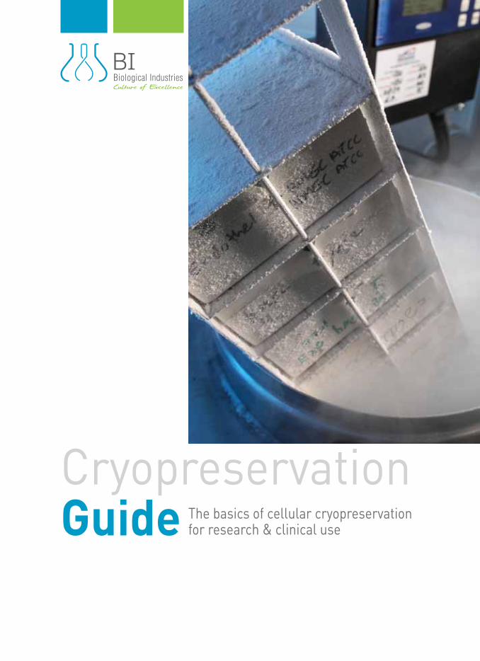
CryopreservationGuide
The basics of cellular cryopreservation for research & clinical use
23456788-10111213
IntroductionCryopreservation BasicsAdvantages of CryopreservationChallenges of CryopreservationFreezing Methods and Storage Creating a Master Cell Bank and Working Cell BankChoosing a Cryopreservation MediaCryopreservation ProtocolsSafety Tips and ConsiderationsTroubleshootingBibliography
0102030405060708091011
Contents
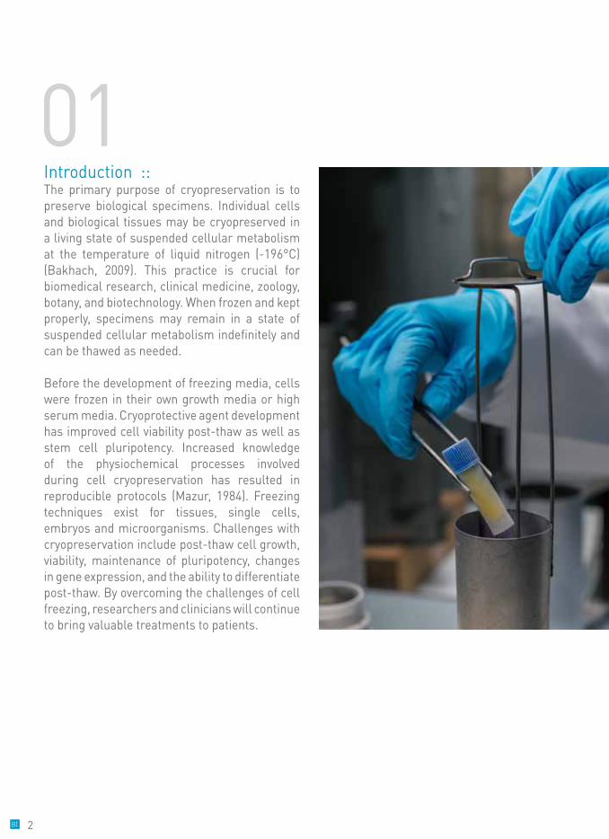
Introduction :: The primary purpose of cryopreservation is to preserve biological specimens. Individual cells and biological tissues may be cryopreserved in a living state of suspended cellular metabolism at the temperature of liquid nitrogen (-196°C) (Bakhach, 2009). This practice is crucial for biomedical research, clinical medicine, zoology, botany, and biotechnology. When frozen and kept properly, specimens may remain in a state of suspended cellular metabolism indefinitely and can be thawed as needed.
Before the development of freezing media, cells were frozen in their own growth media or high serum media. Cryoprotective agent development has improved cell viability post-thaw as well as stem cell pluripotency. Increased knowledge of the physiochemical processes involved during cell cryopreservation has resulted in reproducible protocols (Mazur, 1984). Freezing techniques exist for tissues, single cells, embryos and microorganisms. Challenges with cryopreservation include post-thaw cell growth, viability, maintenance of pluripotency, changes in gene expression, and the ability to differentiate post-thaw. By overcoming the challenges of cell freezing, researchers and clinicians will continue to bring valuable treatments to patients.
01
2
Cryopreservation Basics :: The purpose of cryopreservation is to store cells indefinitely by halting the cell’s metabolism with ultralow temperatures. The freeze-thaw process is stressful to all cells and tissues. Therefore, effective techniques were developed to prevent cell death and damage. One common cryopreservation technique involves changing cell maintenance media to culture media containing a cryopreservation agent, such as dimethyl sulfoxide (DMSO). Cells are then cooled at a rate of -1°C/min (for mammalian cells) by transferring to -80°C in a specialized cooling container. After cells have cooled to -80°C, they are transferred to ultralow temperature storage of below -135°C. The most common ultralow temperature storage is liquid nitrogen in liquid or vapor form. After freezing, cells are stored as original seed and working stock assets.
Ultralow storage temperatures work by maintaining cells below the glass transition temperature (Tg) of pure water (Miller, 1969). This suspends all molecular processes and prevents free radical generation that negatively effects cryopreserved cultures (Baust J. , 2007; Baust, Corwin, Van Buskirk, & Baust, 2015). Cells survive by cooling the cultures to
a “glassy” state slowly, so that less ice forms within the cell. Too much intracellular ice can cause mechanical damage to the cell when thawing. In addition, cells must not freeze too slowly because cells will shrink and dehydrate. Physical stress of shrinkage and dehydration results in high solute concentrations that increase toxicity (Mazur, Leibo, & Chu, 1969). Damage is also prompted by pH shifts and protein degradation that compromises the cell membrane or metabolic pathways.
Technologies for the cryopreservation of cells and tissues are constantly improving. The best cryopreservation results are obtained through research-based approaches that are adjusted for the specific purpose of cryopreservation. There are six applications of cryopreservation that include preservation of cells or organs, cryosurgery, biochemistry or molecular biology, food sciences, ecology or plant physiology, and medical applications (transfusion, bone marrow and cells transplantation, artificial insemination, and in vitro fertilization) (Jang, et al., 2017). This guide will cover the basics of cellular cryopreservation for research and clinical use.
02
3
Advantages of Cryopreservation ::There are many advantages to cell freezing. Proper cryopreservation of cells is the best way to save money, improve reproducibility of experiments, and protect important stocks of cells that may be difficult to replace. Cost savings occur because regular cell stock orders will not be necessary since cells may be split and multiplied. Expanding cells for cryopreservation will accommodate long experiments or industrial high throughput screening efforts. Keeping cell passage numbers low increases the likelihood that populations will remain homogenous between experiments because changes occur when populations are expanding. Therefore, keeping a cryopreservation master cell bank and working stocks will improve reproducibility between similar experiments. Gene expression or other cellular changes may be documented if passages are frozen down, tested, and compared.
WHY SHOULD YOU FREEZE YOUR CELLS?• Store your cells for future study.• Cell storage acts as an insurance policy in case of contamination, failure, or cell shortage. • Actively growing your cells over a long period of time may alter gene expression and differentiate cells. Continued growth and passage of cells may cause them to lose their original features. This endangers reliable research results!• Freezing and storage of cells is a crucial step to ensure long-term cell use and reproducible results.• Save time and money. Obtaining new cells is costly and time-consuming!
03In case of contamination, errors, or unintended variables occur then the original stocks can be revisited.
4
Challenges of Cryopreservation ::Government regulations of cells for medical use have increased the need for documented cell handling processes. To account for these regulations, protocols for cryopreservation must be established and adhered to by researchers and medical personnel. While optimized cryopreservation protocols exist for most areas of research and medicine, technical issues persist. These issues include low survival, altered gene expression and morphology, loss of cellular function, epigenetic changes, and differences in protein composition. (Baust J. , 2007; Van Buskirk, 2007; Baust, Corwin, Van Buskirk, & Baust, 2015; Allegrucci & Young, 2007; Stacey, 2007). If optimal conditions are not provided during cell freezing then damage and cellular apoptosis or necrosis may occur post-thaw.
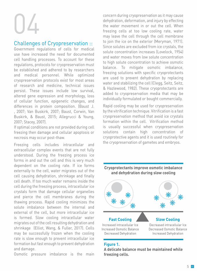
Freezing cells includes intracellular and extracellular complex events that are not fully understood. During the freezing process ice forms in and out the cell and this is very much dependent on the cooling rate. If ice forms externally to the cell, water migrates out of the cell causing dehydration, shrinkage and finally cell death. If too much water remains inside the cell during the freezing process, intracellular ice crystals form that damage cellular organelles and pierce the cell membranes during the thawing process. Rapid cooling minimizes the solute imbalance between the internal and external of the cell, but more intracellular ice is formed. Slow cooling intracellular water migrates out of the cell resulting dehydration and shrinkage (Elliot, Wang, & Fuller, 2017). Cells may be successfully frozen when the cooling rate is slow enough to prevent intracellular ice formation but fast enough to prevent dehydration and damage. Osmotic pressure imbalance is the main
concern during cryopreservation as it may cause dehydration, deformation, and injury by effecting the water movement in or out the cell. When freezing cells at too low cooling rate, water may leave the cell through the cell membrane to join the ice on the exterior (Meryman, 1971). Since solutes are excluded from ice crystals, the solute concentration increases (Lovelock, 1954) and water moves from low solute concentration to high solute concentration to achieve osmotic balance. To mitigate osmotic imbalance, freezing solutions with specific cryoprotectants are used to prevent dehydration by replacing water and stabilizing the cell (Clegg, Seitz, Seitz, & Hazlewood, 1982). These cryoprotectants are added to cryopreservation media that may be individually formulated or bought commercially.
Rapid cooling may be used for cryopreservation by the vitrification technique. Vitrification is a fast cryopreservation method that avoid ice crystals formation within the cell. Vitrification method is usually successful when cryopreservation solutions contain high concentration of cryoprotective agents and it is used routinely for the cryopreservation of gametes and embryos.
Figure 1. A delicate balance must be maintained while freezing cells.
Cryoprotectants improve osmotic imbalance and dehydration during slow cooling
Slow CoolingDecreased intracellular IceDecreased Osmotic Balance
Increased Dehydration
Fast CoolingIncreased intracellular IceIncreased Osmotic Balance
Decreased Dehydration
04
5
05SHOULD YOU STORE YOUR CRYOVIALS IN THE VAPOROUS OR LIQUID NITROGEN LAYER?
The current vogue is that the vaporous layer is better. The difference in temperature between layers is not great and the liquid phase is at greater risk for contamination.
Recommended: After 24 hours you should check one of the vials for viability and recovery.
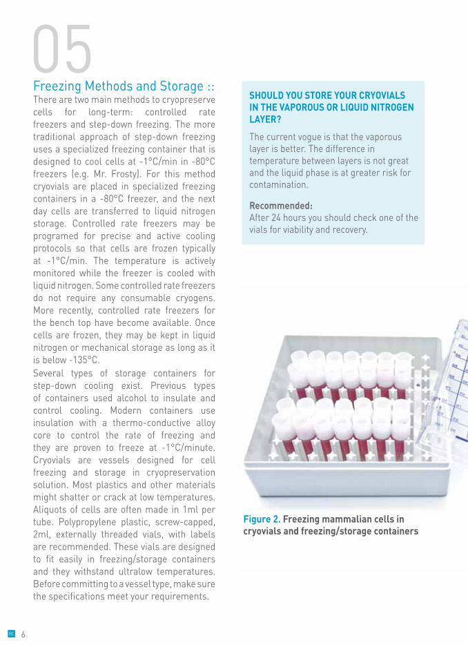
Freezing Methods and Storage ::There are two main methods to cryopreserve cells for long-term: controlled rate freezers and step-down freezing. The more traditional approach of step-down freezing uses a specialized freezing container that is designed to cool cells at -1°C/min in -80°C freezers (e.g. Mr. Frosty). For this method cryovials are placed in specialized freezing containers in a -80°C freezer, and the next day cells are transferred to liquid nitrogen storage. Controlled rate freezers may be programed for precise and active cooling protocols so that cells are frozen typically at -1°C/min. The temperature is actively monitored while the freezer is cooled with liquid nitrogen. Some controlled rate freezers do not require any consumable cryogens. More recently, controlled rate freezers for the bench top have become available. Once cells are frozen, they may be kept in liquid nitrogen or mechanical storage as long as it is below -135°C. Several types of storage containers for step-down cooling exist. Previous types of containers used alcohol to insulate and control cooling. Modern containers use insulation with a thermo-conductive alloy core to control the rate of freezing and they are proven to freeze at -1°C/minute. Cryovials are vessels designed for cell freezing and storage in cryopreservation solution. Most plastics and other materials might shatter or crack at low temperatures. Aliquots of cells are often made in 1ml per tube. Polypropylene plastic, screw-capped, 2ml, externally threaded vials, with labels are recommended. These vials are designed to fit easily in freezing/storage containers and they withstand ultralow temperatures. Before committing to a vessel type, make sure the specifications meet your requirements.
Figure 2. Freezing mammalian cells in cryovials and freezing/storage containers
6
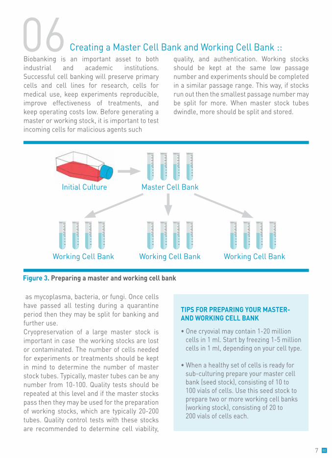
Biobanking is an important asset to both industrial and academic institutions. Successful cell banking will preserve primary cells and cell lines for research, cells for medical use, keep experiments reproducible, improve effectiveness of treatments, and keep operating costs low. Before generating a master or working stock, it is important to test incoming cells for malicious agents such
as mycoplasma, bacteria, or fungi. Once cells have passed all testing during a quarantine period then they may be split for banking and further use. Cryopreservation of a large master stock is important in case the working stocks are lost or contaminated. The number of cells needed for experiments or treatments should be kept in mind to determine the number of master stock tubes. Typically, master tubes can be any number from 10-100. Quality tests should be repeated at this level and if the master stocks pass then they may be used for the preparation of working stocks, which are typically 20-200 tubes. Quality control tests with these stocks are recommended to determine cell viability,
quality, and authentication. Working stocks should be kept at the same low passage number and experiments should be completed in a similar passage range. This way, if stocks run out then the smallest passage number may be split for more. When master stock tubes dwindle, more should be split and stored.
06 Creating a Master Cell Bank and Working Cell Bank ::
TIPS FOR PREPARING YOUR MASTER- AND WORKING CELL BANK
• One cryovial may contain 1-20 million cells in 1 ml. Start by freezing 1-5 million cells in 1 ml, depending on your cell type.
• When a healthy set of cells is ready for sub-culturing prepare your master cell bank (seed stock), consisting of 10 to 100 vials of cells. Use this seed stock to prepare two or more working cell banks (working stock), consisting of 20 to 200 vials of cells each.
7
(Cat. No.: 05-710-1)
07
08Cryopreservation Protocols ::
In order to lower the solute concentration during the freezing process, cryoprotectants must be used. Cryoprotectants are compounds that protect cells from intracellular ice formation. DMSO, glycerol, ethylene glycol, and propylene glycol are all permeating cryoprotectants. Their mechanism of action involves entering the cell freely and replacing water, lowering the amount of ice formed, and acting as a secondary solvent for salts (Lovelock, 1953; Pegg, 1984). Other cryoprotectants include high molecular weight compounds. Hydroxyethylstarch and polyvinyl pyrollidone are non-permeating cryoprotectants that are thought to stabilize the cell membrane (Anchordoguy, Rudolph, Carpenter, & Crowe, 1987). The type of cryoprotectant desired depends on the cell type, tissue, or organism being frozen. DMSO and glycerol are the most commonly used cryoprotectants, with DMSO being more common for mammalian cells. DMSO may be used at a concentration of up to 10% w/v (1.28M) and can be filtered for cell culture use (Hunt, 2017). If single addition of DMSO media causes toxicity then multi-step addition may be used to gradually increase DMSO concentration and decrease osmotic damage. Another common cryoprotectant is glycerol. Glycerol may act as a nonelectrolyte to decrease electrolyte concentration in cell freezing solutions and is most commonly used for preservation of microorganisms, red blood cells and spermatozoa (Jang, et al., 2017). While these agents protect cells during the slow freezing process they can also cause cell toxicity. The use of serum may help to protect the cells but this is not recommended for clinical or commercial uses that may require serum-free culture conditions. As an alternative to homebrew of cryopreservation media, commercially available media provide
safe and reliable cryopreservation results. Defined freezing media, such as CryoStem™ (Biological Industries, cat. no. 05-710-1), are proven effective for human embryonic stem cell and human induced pluripotent stem cell cryopreservation. Cells frozen with this medium maintain pluripotency, attachment ability, and viability (Nishishita, Muramatsu, & Kawamata, 2015). However, even defined and safe media should be removed promptly after thawing cells to prevent toxicity.
Choosing a Cryopreservation Media ::
Freezing human pluripotent stem cells (hPSCs) using CryoStemTM Freezing Medium
Notes:
• hPSC may be freezed as clumps or single cells with high viability and minimal differentiation post thaw. The single cells hPSC can be thawed onto recombinant Laminin coated culture ware without the addition of Rock inhibitors. In case of using other matrices (e.g. Matrigel™) rock inhibitor is required.
• Keep CryoStemTM freezing medium on ice at all times.
8
1. Aspirate hPSC culture medium.
2. Rinse wells with Dulbecco’s PBS w/o Ca & Mg (Cat# 02-023-1), or DMEM:F-12 (1:1) (Cat# 01-170-1).
3. Add dissociation solution as desired. Cells may be detached using the enzyme and method that the culture has been routinely passaged with. In case of using collagenase, dispase or EDTA, incubate at 37°C or at room-temperature until the edges of the colonies begin to loosen from the plate. Incubation time will vary between cell lines, colony size and detachment solution used. Begin checking the culture after 3 minutes.
4. Cells cultured on laminin may be detached using Recombinant Trypsin-EDTA Solution (Cat# 03-079-1) to yield a single cell suspension. 5. Transfer the clumps or cell suspension to a conical tissue culture centrifuge tube.
6. Centrifuge at 200 x g for 5 minutes at roomtemperature. Remove and discard supernatant. 7. Gently suspend the pellet in ice cold CryoStem™. The final volume is the number of vials desired multiplied by one ml In case of aggregates, do not break up cell clumps any more than necessary, two or three gentle pipeting motions are usually sufficient.
8. Quickly transfer 1 ml into each cryogenic vial.
9. Place the vials into a freezing container (e.g. Mr. Frosty™) and transfer to -80°C for overnight.
10. The following day, transfer vials to liquid nitrogen storage (vapor phase).
Notes:
• Keep Serum-Free Freezing Media freezing medium on ice at all times.
• The Serum-Free Cell Freezing Medium has been extensively tested on multiple cell lines, including 3T3, BGM, vero cells, MRC-5, HEK-293, HEp-2, BSC-1, BF16-F10, BHK-21, CHO, HELA, MA-10, and mesenchymal cells.
1. For freezing adherent cells, detach cells using dissociation solution according to the manual instructions. For freezing of cells in suspension skip to step 2.
2. Centrifuge to pellet the cells (200-300g, 3-5 minutes).
3. Suspend the pellet in ice-cold Serum-Free Freezing Medium at a concentration of 3-5 million cells per ml.
4. Freeze the cells gradually (1-2°C per minute) in a freezing container (e.g. Mr. Frosty)and store them in liquid nitrogen.
Cryopreservation of serum-free cultures using Serum-Free Freezing Media
(cat.no.: 05-065-1)
• Always use a reliable freezing media that is serum-free and animal-component free to insure that animal products will not influence the human cells.
• Serum is not necessary when freezing hPSCs.
• Cryoprotectants, such as DMSO or glycerol, in freezing media help to protect cells from damage caused by ice crystal formation.
9
TIPS FOR BEST RESULTS WHILE FREEZING HUMAN PLURIPOTENTSTEM CELLS• Follow institutional regulation and guidelines for all hPSC work.• Cells may be frozen as aggregates or single cells. • It is best to cryopreserve when cells are at their maximum growth rate (log phase). Cryopreservation at ~80% confluence should work best. • Mycoplasma testing is recommended before freezing.• Label your vials (cell, passage, lot #, date, your name) with a marker that will withstand alcohol and liquid N2. Printed labels also work well. Keep the records online as well as a hardcopy.• Follow your cryopreservation protocol carefully. Work quickly and step by step. Add the freezing media gradually. Do not allow freezing media to return to room temperature.• DO NOT sacrifice sterility and personal safety for speed!
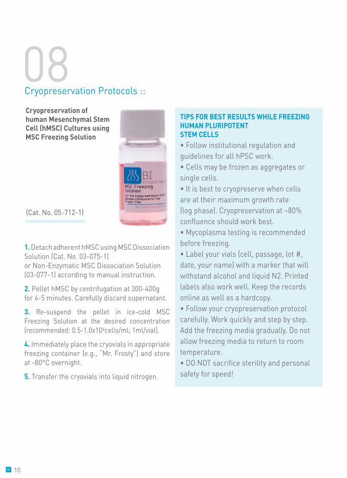
Cryopreservation of human Mesenchymal Stem Cell (hMSC) Cultures using MSC Freezing Solution
(Cat. No. 05-712-1)
1. Detach adherent hMSC using MSC Dissociation Solution (Cat. No. 03-075-1) or Non-Enzymatic MSC Dissociation Solution (03-077-1) according to manual instruction.
2. Pellet hMSC by centrifugation at 300-400g for 4-5 minutes. Carefully discard supernatant.
3. Re-suspend the pellet in ice-cold MSC Freezing Solution at the desired concentration (recommended: 0.5-1.0x10⁶cells/ml; 1ml/vial).
4. Immediately place the cryovials in appropriate freezing container (e.g., “Mr. Frosty”) and store at -80°C overnight.
5. Transfer the cryovials into liquid nitrogen.
08Cryopreservation Protocols ::
10
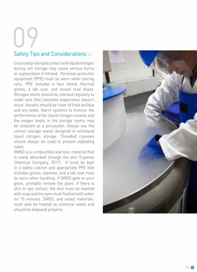
Close and prolonged contact with liquid nitrogen during cell storage may cause serious burns or asphyxiation if inhaled. Personal protective equipment (PPE) must be worn when storing cells. PPE includes a face shield, thermal gloves, a lab coat, and closed toed shoes. Nitrogen levels should be checked regularly to make sure that complete evaporation doesn’t occur. Vessels should be clear of frost buildup and any leaks. Alarm systems to monitor the performance of the liquid nitrogen vessels and the oxygen levels in the storage rooms may be installed as a precaution. Always use the correct storage vessel designed to withstand liquid nitrogen storage. Threaded cryovials should always be used to prevent exploding tubes.DMSO is a combustible and toxic material that is easily absorbed through the skin (Cayman Chemical Company, 2017). It must be kept in a safety cabinet and appropriate PPE that includes gloves, eyewear, and a lab coat must be worn when handling. If DMSO gets on your glove, promptly remove the glove. If there is skin or eye contact, the skin must be washed with soap and the eyes must flushed with water for 15 minutes. DMSO, and soiled materials, must also be treated as chemical waste and should be disposed properly.
Safety Tips and Considerations :: 09
11
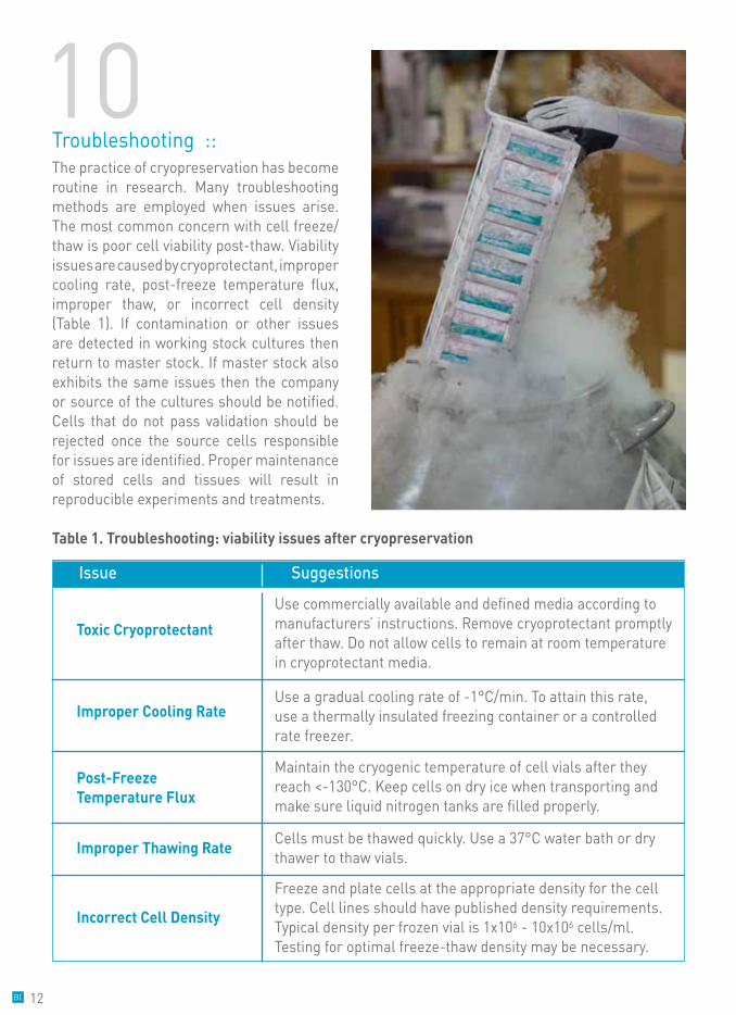
The practice of cryopreservation has become routine in research. Many troubleshooting methods are employed when issues arise. The most common concern with cell freeze/thaw is poor cell viability post-thaw. Viability issues are caused by cryoprotectant, improper cooling rate, post-freeze temperature flux, improper thaw, or incorrect cell density (Table 1). If contamination or other issues are detected in working stock cultures then return to master stock. If master stock also exhibits the same issues then the company or source of the cultures should be notified. Cells that do not pass validation should be rejected once the source cells responsible for issues are identified. Proper maintenance of stored cells and tissues will result in reproducible experiments and treatments.
Troubleshooting ::10
Table 1. Troubleshooting: viability issues after cryopreservation
Issue Suggestions
Toxic Cryoprotectant
Improper Cooling Rate
Post-Freeze Temperature Flux
Improper Thawing Rate
Incorrect Cell Density
Use commercially available and defined media according to manufacturers’ instructions. Remove cryoprotectant promptly after thaw. Do not allow cells to remain at room temperature in cryoprotectant media.
Use a gradual cooling rate of -1°C/min. To attain this rate, use a thermally insulated freezing container or a controlled rate freezer.
Maintain the cryogenic temperature of cell vials after they reach <-130°C. Keep cells on dry ice when transporting and make sure liquid nitrogen tanks are filled properly.
Freeze and plate cells at the appropriate density for the cell type. Cell lines should have published density requirements. Typical density per frozen vial is 1x10⁶ - 10x106 cells/ml. Testing for optimal freeze-thaw density may be necessary.
Cells must be thawed quickly. Use a 37°C water bath or dry thawer to thaw vials.
12
Bibliography ::Allegrucci, C., & Young, L. (2007). Differences between human embryonic stem cell lines. Hum. Reprod. Update, 13, 103-120.
Anchordoguy, T., Rudolph, A., Carpenter, J., & Crowe, J. (1987). Modes of interaction of cryoprotectants with membrane phospholipid during freezing. Cryobiology, 24, 324-331.
Bakhach, J. (2009, September). The cryopreservationof composite tissues: Principles and recent advancement on cryopreservation of different type of tissues. Organogenesis, 5(3), 119-126.
Baust, J. (2007). Properties of cells and tissues influencing preservation outcome: molecular basis of preservation-induced cell death. In J. Baust, & J. Baust (Eds.), Advances in Biopreservation (pp. 63-87). New York: CRC Press.
Cayman Chemical Company. (2017). Safety Data Sheet: DMSO Assay Reagent. Ann Arbor: Cayman Chemical Company.
Clegg, J., Seitz, P., Seitz, W., & Hazlewood, C. (1982). Cellular responses to extreme water loss: the water replacement hypothesis. Cryobiology, 306-316.
Elliot, G., Wang, S., & Fuller, B. (2017). Cryoprotectants: A review of the actions and applications of cryoprotective solutes that modulate cell recovery from ultra-low temperatures. Cryobiology, 1-18.
Hunt, C. (2017). Cryopreservation: Vitrification and Controlled Rate Cooling . In J. Crook, & T. Ludwig (Eds.), Stem Cell Banking: Concepts and Protocols, Methods in Molecular Biology (Vol. 1590, pp. 41-77). Springer Science+Business Mecia LLC.Jang, T., Park, S., Yang, J., Kim, J., Seok, J., Park, U., et al. (2017). Cryopreservation and its clinical applications. Integrative Medicine Research, 6, 12-18.
Lovelock, J. (1953). The mechanism of the protective effect of glycerol against haemolysis by freezing and thawing. Biochim Biophys Acta, 11, 28-36.
Lovelock, J. (1954). The protective action of neutral solutes against haemolysis by freezing and thawing. Biochem. J. , 56, 265-270.
Mazur, P. (1984). Freezing of live cells: mechanisms and implications. Am J Physiol, 247, 125-142.
Mazur, P., Leibo, S., & Chu, E. (1969). A two-factor hypothesis of freezing injury. Evidence from hamster tissue-culture cells. Exp. Cell Res. , 71(2), 345-355.
Meryman, H. (1971). Cryoprotective agents. Cryobiology, 8, 173-183.
Miller, A. (1969). Glass-transition temperature of water. Science, 163(3873), 1325-1326.
Nishishita, N., Muramatsu, M., & Kawamata, S. (2015). ) An effective freezing/thawing method for human pluripotent stem cells cultured in chemically-defined and feeder-free conditions. . Am J Stem Cells, 4(1), 38-49.
Pegg, D. (1984). Red cell volume in glycerol/sodium chloride/water mixtures. Cryobiology, 21, 234-331.
Stacey, G. (2007). Risk assessment of cell culture procedures. In Medicines from Animal Cell Culture (pp. 569-587). Chichester: Wiley.
Van Buskirk, R. (2007). Viability and functional assays used to assess preservation efficacy: the multiple endpoint/tier approach. In Advances in Cryopreservation (pp. 123-142). Boca Raton: CRC Press.
11
13

Biological Industries Israel Beit Haemek Ltd. Kibbutz Beit Haemek 2511500, [email protected] | www.bioind.com
T. 04-9960595 F. 04-9968896
©2017 Biological Industries. All rights reserved. The trademarks mentioned herein are the property of Biological Industries and/or its affiliates or their respective owners. E82/1 6/17