Embed Size (px)
Citation preview
ENDOLUMINAL SURGERY
Cryorecanalization: keys to success
Aydın Yılmaz • Zafer Aktas • Ibrahim Onur Alici •
Atalay Caglar • Hilal Sazak • Fatma Ulus
Received: 28 June 2011 / Accepted: 16 March 2012 / Published online: 19 May 2012
� Springer Science+Business Media, LLC 2012
Abstract
Background Symptomatic airway obstructions are com-
mon with endobronchial exophytic tumors and may result
in lethal complications. Recently, a cryorecanalization
procedure has emerged that plays a role in the immediate
management of airway obstruction. This study was con-
ducted to investigate the value of cryorecanalization for the
immediate management of endobronchial obstructive
pathology and to determine the factors that affect the
success of the procedure.
Methods We analyzed 40 patients with symptoms of
airway obstruction who were admitted to our hospital from
2006 to 2010. Patients with exophytic stenosis due to pri-
mary bronchial or metastatic neoplasms who underwent
cryorecanalization procedures were included. Patients were
excluded if they had involvement of a major artery near the
site of the intervention. The procedure was not performed
on patients with coagulation abnormalities or thrombocyte
count and aggregation problems. The data were collected
retrospectively.
Results Successful cryorecanalization was achieved in
72.5 % of patients. We found that the success rate was
mainly related to the presence of the distal involvement
and the older age of obstruction. Restenosis rate was
12.8 %. The mean survival time after the cryorecanaliza-
tion procedure was 11 ± 12.7 months. No complications
occurred in 14 patients. No severe bleeding was observed
for any patients, and moderate hemorrhaging occurred in
ten patients, which was stopped with an argon plasma
coagulator. We experienced no intraoperative mortality.
Conclusions Cryorecanalization is a successful and safe
intervention for the immediate management of endobron-
chial stenosis. Appropriate patient selection and high suc-
cess rates should be achieved after careful radiological
assessments and with early management.
Keywords Cryorecanalization � Lung cancer �Airway obstruction � Interventional bronchoscopy
Airway obstruction is commonly observed with endo-
bronchial exophytic tumors. Up to 30 % of lung cancers
cause obstructions at the level of the trachea and main
bronchi [1]. The most common manifestations of these
obstructions are dyspnea, cough, and hemoptysis [2], and
they may result in lethal complications, such as respiratory
failure, atelectasis, and postobstructive pneumonia. With
regard to treatment, many interventional bronchoscopists
prefer laser, electrocautery, argon plasma coagulation,
mechanical recanalization, and stents because of their
immediate effects [3].
Endobronchial cryotherapy is used to treat malignant
airway tumors, but conventional usage is limited to the
palliation of noncritical endobronchial exophytic lesions
and early lung cancers and for the removal of foreign
bodies and clots [4]. In addition, 8–10 days are required for
a complete therapeutic effect [5]. Clean-up bronchoscopy
is usually performed. Recently, a new technique, cryore-
canalization, has emerged for immediate debulking of
exophytic endobronchial tumors [2, 6, 7]. In this technique,
a bronchoscopist retracts frozen tissue at the tip of the
probe with a bronchoscope, resulting in immediate tumor
A. Yılmaz � Z. Aktas � I. O. Alici (&) � H. Sazak � F. Ulus
Ataturk Chest Diseases and Thoracic Surgery Education and
Research Hospital, 06280 Ankara, Turkey
e-mail: [email protected]
A. Caglar
Department of Econometrics, Pamukkale University, Denizli,
Turkey
123
Surg Endosc (2012) 26:2969–2974
DOI 10.1007/s00464-012-2260-1
and Other Interventional Techniques
removal and recanalization of the airway lumen. This
technique seems to be feasible and has many advantages,
including its use of flexible probes, immediate efficacy, and
low risk of complications. The technique is easy to learn
and has a low cost compared with other endobronchial
treatment modalities [7]. This study was conducted to
investigate the efficacy and safety of cryorecanalization for
the immediate management of endobronchial obstructive
pathology.
Materials and methods
We analyzed 40 patients with symptoms of airway
obstruction who were admitted to our hospital from 2006 to
2010. Patients with endoluminal protrusions of primary
bronchial or metastatic neoplasms were included. The data
were collected retrospectively. In all patients, computed
tomography of the thorax was performed, and virtual
bronchoscopic images were obtained (Fig. 1). Anatomic
location, endobronchial length, infiltrative characteristics
of obstructive lesions, and involvement of the distal
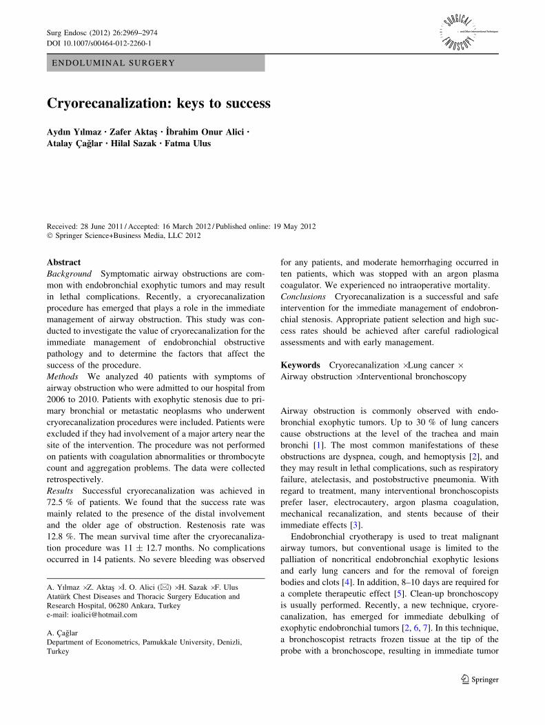
bronchial tree were assessed before the procedures. Distal
bronchial involvement was described as external com-
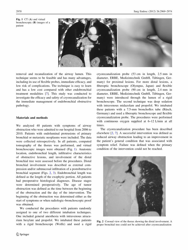
pression and/or submucosal infiltration of a postobstructive
bronchial segment (Figs. 2, 3). Endobronchial length was
defined as the length of the exophytic portion. All patients
had preoperative histological diagnoses. Disease stages
were determined preoperatively. The age of tumor
obstruction was defined as the time between the beginning
of the obstruction and the day of the intervention. The
beginning of the obstruction was determined either as the
start of symptoms or when radiologic–bronchoscopic proof
was obtained.
We conducted the procedures with patients randomly
assigned to one of two different intubation techniques.
One included general anesthesia with intravenous atracu-
rium besylate and propofol. We intubated these patients
with a rigid bronchoscope (Wolfe) and used a rigid
cryorecanalization probe (53 cm in length, 2.5 mm in
diameter, ERBE, Medizintechnik GmbH, Tubingen, Ger-
many) for proximal lesions. For more distal lesions, a
fiberoptic bronchoscope (Olympus, Japan) and flexible
cryorecanalization probe (90 cm in length, 2.4 mm in
diameter, ERBE, Medizintechnik GmbH, Tubingen, Ger-
many) were introduced through the lumen of a rigid
bronchoscope. The second technique was deep sedation
with intravenous midazolam and propofol. We intubated
these patients with a 7.5-mm bronchoflex tube (Rusch,
Germany) and used a fiberoptic bronchoscope and flexible
cryorecanalization probe. The procedures were performed
with continuous oxygen supplied at 6–12 L/min at all
times.
The cryorecanalization procedure has been described
elsewhere [2, 7]. A successful intervention was defined as
reduced airway obstruction leading to an improvement in
the patient’s general condition that was associated with
symptom relief. Failure was defined when the primary
condition of the intervention could not be reached.
Fig. 1 CT (A) and virtual
bronchoscopic (B) images of a
patient
Fig. 2 Coronal view of the thorax showing the distal involvement. A
proper bronchial tree could not be achieved after cryorecanalization
2970 Surg Endosc (2012) 26:2969–2974
123
The most common complications of interventional tech-
niques were bleeding and bronchial wall damage. We clas-
sified bleeding as mild, moderate, or severe [2]. Mild
hemorrhagia was defined as bleeding that could be controlled
with a cold 0.9 % NaCl solution (2–4 �C) or epinephrine
solution (1 mg/10 ml saline water). Moderate hemorrhagia
was defined as bleeding that could be controlled with argon
plasma coagulation. Severe hemorrhagia was defined as
bleeding that required additional treatments (transfusion of
red blood cells, fresh-frozen plasma, or coagulation factors),
vasopressor support, or a rescue operation.
Patients with significant vascular involvement adjacent
to the site of the intervention, obstructions due purely to
external compression of the surrounding tumor, and
involvement of the posterior membranous walls of the
bronchi (because they do not contain cryoresistant cartilage
tissue) were excluded. The procedure was not performed
on patients with coagulation abnormalities or thrombocyte
count and aggregation problems.
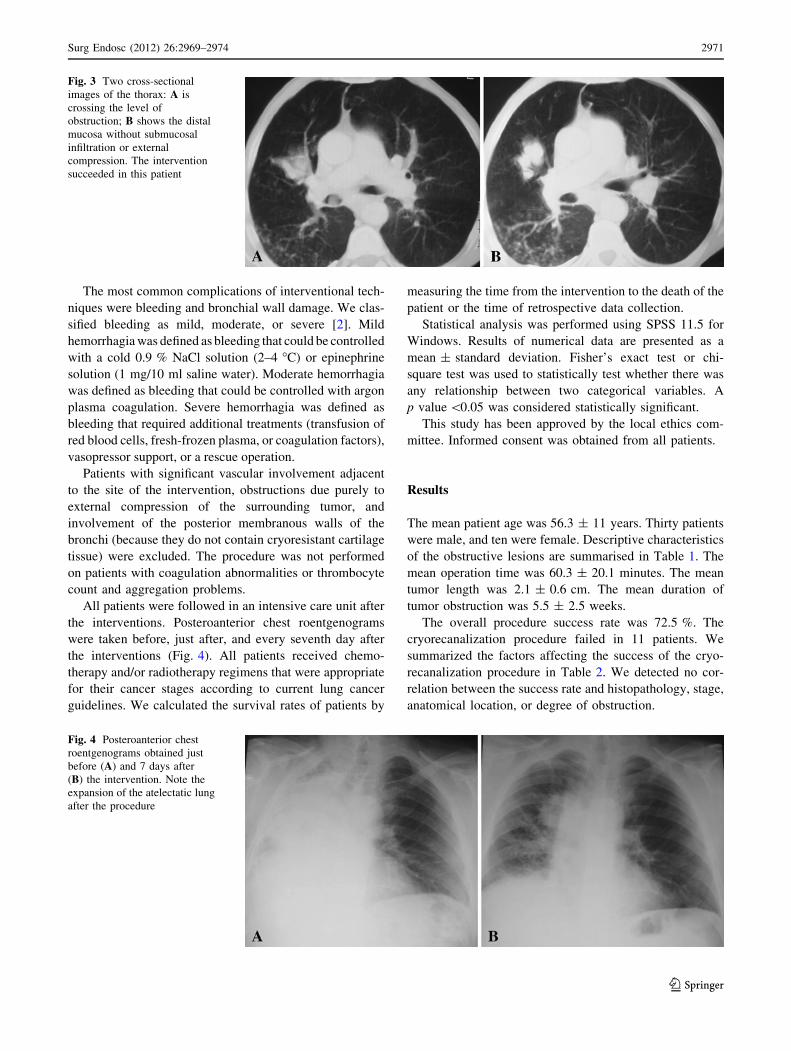
All patients were followed in an intensive care unit after
the interventions. Posteroanterior chest roentgenograms
were taken before, just after, and every seventh day after
the interventions (Fig. 4). All patients received chemo-
therapy and/or radiotherapy regimens that were appropriate
for their cancer stages according to current lung cancer
guidelines. We calculated the survival rates of patients by
measuring the time from the intervention to the death of the
patient or the time of retrospective data collection.
Statistical analysis was performed using SPSS 11.5 for
Windows. Results of numerical data are presented as a
mean ± standard deviation. Fisher’s exact test or chi-
square test was used to statistically test whether there was
any relationship between two categorical variables. A
p value \0.05 was considered statistically significant.
This study has been approved by the local ethics com-
mittee. Informed consent was obtained from all patients.
Results
The mean patient age was 56.3 ± 11 years. Thirty patients
were male, and ten were female. Descriptive characteristics
of the obstructive lesions are summarised in Table 1. The
mean operation time was 60.3 ± 20.1 minutes. The mean
tumor length was 2.1 ± 0.6 cm. The mean duration of
tumor obstruction was 5.5 ± 2.5 weeks.
The overall procedure success rate was 72.5 %. The
cryorecanalization procedure failed in 11 patients. We
summarized the factors affecting the success of the cryo-
recanalization procedure in Table 2. We detected no cor-
relation between the success rate and histopathology, stage,
anatomical location, or degree of obstruction.
Fig. 3 Two cross-sectional
images of the thorax: A is
crossing the level of
obstruction; B shows the distal
mucosa without submucosal
infiltration or external
compression. The intervention
succeeded in this patient
Fig. 4 Posteroanterior chest
roentgenograms obtained just
before (A) and 7 days after
(B) the intervention. Note the
expansion of the atelectatic lung
after the procedure
Surg Endosc (2012) 26:2969–2974 2971
123
The mean survival time after the cryorecanalization
procedure was 11 ± 12.7 months. We continue to follow
nine patients who have a mean survival time of 21.2 ±
4.4 months.
A summary of complications is listed in Table 3. No
complications occurred in 14 patients. No severe bleeding
was detected for any patient during the procedure. Mild
hemorrhagia, which was bleeding that could be relieved by
a bronchial cold water wash, was observed in 15 patients.
Moderate hemorrhagia that required management with
argon plasma coagulation occurred in ten patients, and
intraoperative hypoxia and carbon dioxide retention
occurred in one other patient. We experienced no periop-
erative mortality.
We had 12 patients with preoperative diagnoses of
unclassified non-small cell carcinoma. With the larger
tissue specimens obtained by cryorecanalization, eight
were diagnosed with squamous cell carcinoma and
two with adenocarcinoma. Two specimens remained
unclassified.
Discussion
Symptomatic airway obstructions are life-threatening com-
plications of bronchial neoplasms and require immediate
management. Obstructions are caused by endobronchial
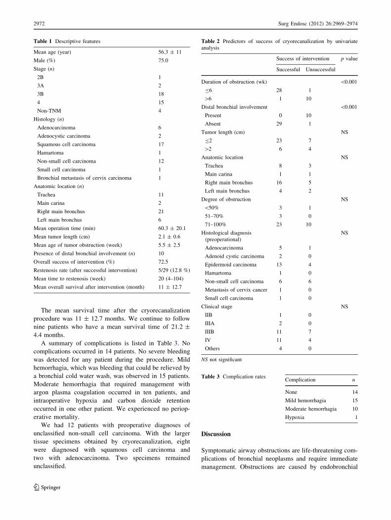
Table 1 Descriptive features
Mean age (year) 56.3 ± 11
Male (%) 75.0
Stage (n)
2B 1
3A 2
3B 18
4 15
Non-TNM 4
Histology (n)
Adenocarcinoma 6
Adenocystic carcinoma 2
Squamous cell carcinoma 17
Hamartoma 1
Non-small cell carcinoma 12
Small cell carcinoma 1
Bronchial metastasis of cervix carcinoma 1
Anatomic location (n)
Trachea 11
Main carina 2
Right main bronchus 21
Left main bronchus 6
Mean operation time (min) 60.3 ± 20.1
Mean tumor length (cm) 2.1 ± 0.6
Mean age of tumor obstruction (week) 5.5 ± 2.5
Presence of distal bronchial involvement (n) 10
Overall success of intervention (%) 72.5
Restenosis rate (after successful intervention) 5/29 (12.8 %)
Mean time to restenosis (week) 20 (4–104)
Mean overall survival after intervention (month) 11 ± 12.7
Table 2 Predictors of success of cryorecanalization by univariate
analysis
Success of intervention p value
Successful Unsuccessful
Duration of obstruction (wk) \0.001
B6 28 1
[6 1 10
Distal bronchial involvement \0.001
Present 0 10
Absent 29 1
Tumor length (cm) NS
B2 23 7
[2 6 4
Anatomic location NS
Trachea 8 3
Main carina 1 1
Right main bronchus 16 5
Left main bronchus 4 2
Degree of obstruction NS
\50% 3 1
51–70% 3 0
71–100% 23 10
Histological diagnosis
(preoperational)
NS
Adenocarcinoma 5 1
Adenoid cystic carcinoma 2 0
Epidermoid carcinoma 13 4
Hamartoma 1 0
Non-small cell carcinoma 6 6
Metastasis of cervix cancer 1 0
Small cell carcinoma 1 0
Clinical stage NS
IIB 1 0
IIIA 2 0
IIIB 11 7
IV 11 4
Others 4 0
NS not significant
Table 3 Complication ratesComplication n
None 14
Mild hemorrhagia 15
Moderate hemorrhagia 10
Hypoxia 1
2972 Surg Endosc (2012) 26:2969–2974
123
exophytic growths, external compression of the airway, or
both [3]. The immediate effects of laser, electrocautery, and
argon plasma coagulation techniques are effective, espe-
cially for exophytic tumors. Bronchial stents are preferable
for obstructions due to external compression. However,
cryotherapy, brachytherapy, and photodynamic therapy are
not considered therapeutic options for symptomatic airway
obstructions, because their actions are delayed [5]. Recently,
cryorecanalization procedures have been used to manage
obstructive endobronchial masses. We performed this tech-
nique in our patients with symptomatic airway obstructions
and present our retrospectively obtained results.
The success rates of an Nd-YAG laser are reported to be
50–80 % [1]. Reichle et al. [8] reported success rates of
67% with Argon plasma coagulator (APC). In two previous
studies [2, 7], the success rates of cryorecanalization were
found to be 83 and 91 %, respectively. Our success rate
was 72.5 % and was not related to histopathology, stage,
anatomical location, or degree of obstruction. We found
that the success rate was mainly related to the involvement
of postobstructive bronchial segments and the duration of
tumor obstruction. Intervention usually fails when tumor
closure extends, and the distal respiratory tract cannot be
reached [7]. In all successful operations in our study, distal
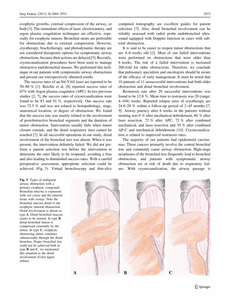
involvement of the bronchial tree was absent. When it was
present, the intervention definitely failed. We did not per-
form a patient selection test before the intervention to
determine the ones likely to be reopened, avoiding a bias
and also leading to diminished success rates. With a careful
preoperative assessment, appropriate selection could be
achieved (Fig. 5). Virtual bronchoscopy and thin-slice
computed tomography are excellent guides for patient
selection [7]. Also, distal bronchial involvement can be
reliably assessed with radial probe endobronchial ultra-
sound equipped with Doppler function in cases with sub-
total obstruction.
It is said to be easier to reopen tumor obstructions that
are 4–6 weeks old [2]. Most of our failed interventions
were performed on obstructions that were older than
6 weeks. The risk of a failed intervention is increased
280-fold for older obstructions. Therefore, we conclude
that pulmonary specialists and oncologists should be aware
of the efficacy of early management. It must be noted that
10 patients of 11 unsuccessful interventions had both older
obstruction and distal bronchial involvement.
Restenosis rate after 29 successful interventions was
found to be 12.8 %. Mean time to restenosis was 20 (range,
4–104) weeks. Reported relapse rates of cryotherapy are
24.8–28 % within a follow-up period of 2–45 months [7,
9]. Airway patency after 6 weeks in the patients without
stenting was 0 % after mechanical debridement, 60 % after
laser resection, 72 % after APC, 72 % after combined
mechanical, and laser resection and 70 % after combined
AP-C and mechanical debridement [10]. Cryorecanaliza-
tion is related to improved restenosis rates.
The majority of our patients had epidermoid carcino-
mas. These cancers primarily involve the central bronchial
tree and commonly cause airway obstruction. High-stage
neoplasms of the bronchial tree frequently lead to bronchial
obstruction, and patients with symptomatic airway
obstruction are at risk of death due to respiratory fail-
ure. With cryorecanalization, the airway passage is
Fig. 5 Types of malignant
airway obstruction with a
primary exophytic compound.
Bronchial mucosa is expressed
with red colour and the tumoral
tissue with orange. Note the
bronchial mucosa distal to the
exophytic tumoral obstruction.
Distal involvement is absent in
type A. Distal bronchial mucosa
seems to be normal. In type B,
distal bronchial lumen is
compressed externally by the
tumor. In type C, exophytic
obstructing tumor continues
submucosally through the distal
bronchus. Proper bronchial tree
could not be achieved both in
type B and C, we mentioned
this situation as the distal
involvement (Color figure
online)
Surg Endosc (2012) 26:2969–2974 2973
123
immediately restored, and early deaths are successfully
avoided. In this study, we reached a mean survival rate of
eleven months after performing cryorecanalization. Note
that cryorecanalization may provide an appropriate treat-
ment option to a patient with a life-threatening airway
obstruction and risk of early death.
Other therapeutic options that have immediate effects
(argon plasma coagulation, laser, and electrocautery) have
significant rates of severe complications, including bleed-
ing, fire, airway perforation, and stenosis [4]. The oxygen
supply must be stopped while shooting to avoid the risk of
a fire, resulting in hypoxia. Cryotherapy, however, is a
procedure that is easy to learn, relatively safe, and cheap
[4, 7]. The oxygen supply does not need to be stopped
(except the period that APC used to control bleeding), and
there is a nearly 0 % risk of bronchial wall perforation. In
addition, it is very useful for the ingrowth of stents.
Cryotherapy also increases the efficacy of chemotherapy
and radiotherapy [5, 11].We did not experience any severe
hemorrhages or bronchial wall perforations in our 40
interventions. Moderate hemorrhage was observed in ten
patients and was easily stopped with argon plasma
coagulation.
Using a cryorecanalization probe, the airway lumen
could be restored by removing a large fragment of tissue.
This removal provides a chance to obtain a proper histo-
pathological diagnosis [12, 13]. Cryoprobes have increased
diagnostic performance for endobronchial lesions com-
pared with forceps biopsies. They also can be used to
obtain larger transbronchial lung biopsies with high histo-
logical quality [14]. We had 12 patients diagnosed with
non-small cell carcinoma by using pathologic specimens
obtained with bronchoscopic forceps. After evaluating
large cryobiopsies, we obtained diagnoses of squamous cell
carcinoma in eight and adenocarcinoma in two. Recently,
because therapeutic agents with differential activities or
limited indications have emerged, the proper subtyping of
non-small cell carcinoma has gained special importance
[15].
Future large, randomized, controlled trials are needed to
compare this technique with other modalities and highlight
the definitive indications for this procedure.
Cryorecanalization is a safe and effective method for the
immediate management of airway obstruction. Recently,
this technique gained its place in the treatment of malig-
nant endobronchial obstruction both with and without
critical airway narrowing [16].
Conclusions
Appropriate patient selection and high success rates should
be achieved after careful radiological assessments and with
early management. Despite its high efficacy, low cost, and
relative safety, this technique remains underutilized.
Disclosures Dr. Aydın Yilmaz, Dr. Zafer Aktas, Dr. Ibrahim O.
Alici, Mr. Atalay Caglar, Dr. Hilal Sazak, and Dr. Fatma Ulus have
no conflict of interest or financial ties to disclose.
References
1. Cavaliere S, Venuta F, Foccoli P, Toninelli C, La Face B (1996)
Endoscopic treatment of malignant airway obstructions in 2,008
patients. Chest 110:1536–1542
2. Schumann C, Hetzel M, Babiak AJ, Hetzel J, Merk T, Wibmer T
et al (2010) Endobronchial tumour debulking with a flexible
cryoprobe for immediate treatment of malignant stenosis. J Tho-
rac Cardiovasc Surg 139:997–1000
3. Bolliger CT, Sutedja TG, Strausz J, Freitag L (2006) Therapeutic
bronchoscopy with immediate effect: laser, electrocautery, argon
plasma coagulation and stents. Eur Respir J 27:1258–1271
4. Bolliger CT, Sutedja TG, Strausz J, Freitag L (2002) ERS/ATS
statement on interventional pulmonology. Eur Respir J 19:356–373
5. Vergnon JM, Huber RM, Moghissi K (2006) Place of cryother-
apy, brachytherapy and photodynamic therapy in therapeutic
bronchoscopy of lung cancers. Eur Respir J 28:200–218
6. Schumann C, Lepper PM, Barth TFE, Moller P, Kruger S (2009)
Successful immediate cryorecanalization of a simultaneous high-
grade tracheal and bronchial stenosis as rare manifestations of
bronchial-associated lymphoid tissue lymphoma. J Thorac Car-
diovasc Surg 137:e17–e19
7. Hetzel M, Hetzel J, Schumann C, Marx N, Babiak A (2004)
Cryorecanalization: a new approach for the immediate manage-
ment of acute airway obstruction. J Thorac Cardiovasc Surg
127:1427–1431
8. Reichle G, Freitag L, Kullmann HJ, Prenzel R, Macha HN, Farin G
(2000) Argon plasma coagulation in bronchology: a new method—
alternative or complementary? Pneumologie 54:508–516
9. Deygas N, Froudarakis M, Ozenne G, Vergnon JM (2001)
Cryotherapy in early superficial bronchogenic carcinoma. Chest
120:26–31
10. Herth FJ, Eberhardt R, Becker HD, Ernst A (2005) Relief of
malignant airway obstruction: a prospective and randomised
comparison of five different endoscopic techniques. Chest
128(4):209S
11. Mathur PN, Wolf KM, Busk MF, Briete WM, Datzman M (1996)
Fiberoptic bronchoscopic cryotherapy in the management of
tracheobronchial obstruction. Chest 110:718–723
12. Hetzel J, Hetzel M, Hasel C, Moeller P, Babiak A (2008) Old
meets modern: the use of traditional cryoprobes in the age of
molecular biology. Respiration 76:193–197
13. Aktas Z, Gunay E, Taci Hoca N, Yilmaz A, Demirag F, Gunay S,
Sipit T, Kurt EB (2010) Endobronchial cryobiopsy or forceps
biopsy for lung cancer diagnosis. Ann Thorac Med 5(4):242–246.
doi:10.4103/1817-1737.69117
14. Babiak A, Hetzel J, Krishna G, Fritz P, Moeller P, Balli T, Hetzel
M (2009) Transbronchial cryobiopsy: a new tool for lung biop-
sies. Respiration 78:203–208
15. Loo PS, Thomas SC, Nicolson MC, Fyfe MN, Kerr KM (2010)
Subtyping of undifferentiated non-small cell carcinomas in
bronchial biopsy specimens. J Thorac Oncol 5:442–447
16. Du Rand IA, Barber PV, Goldring J et al (2011) British Thoracic
Society guideline for advanced diagnostic and therapeutic flexi-
ble bronchoscopy in adults. Thorax 66:iii1–iii21
2974 Surg Endosc (2012) 26:2969–2974
123