Embed Size (px)
Citation preview
Crystal Structure and Solution Species of Ce(III) and Ce(IV) Formates:From Mononuclear to Hexanuclear ComplexesChristoph Hennig,*,†,‡ Atsushi Ikeda-Ohno,†,§,∥ Werner Kraus,⊥ Stephan Weiss,† Philip Pattison,⊗
Hermann Emerich,⊗ Paula M. Abdala,⊗ and Andreas C. Scheinost†,‡
†Helmholtz-Zentrum Dresden-Rossendorf, Institute of Resource Ecology, Bautzner Landstrasse 400, D-01314 Dresden, Germany‡The Rossendorf Beamline, ESRF, BP 220, F-38043 Grenoble, France§School of Civil and Environmental Engineering, The University of New South Wales, UNSW, Sydney, NSW 2052, Australia∥Institute for Environmental Research, Australian Nuclear Science and Technology Organisation, Locked Bag 2001, Kirrawee DC,New South Wales 2232, Australia⊥BAM Federal Institute for Materials Research and Testing, Richard-Willstatter-Strasse 11, D-12489 Berlin, Germany⊗Swiss−Norwegian Beamlines, ESRF, BP 220, F-38043 Grenoble, France
*S Supporting Information
ABSTRACT: Cerium(III) and cerium(IV) both form formatecomplexes. However, their species in aqueous solution and thesolid-state structures are surprisingly different. The species inaqueous solutions were investigated with Ce K-edge EXAFSspectroscopy. Ce(III) formate shows only mononuclearcomplexes, which is in agreement with the predictedmononuclear species of Ce(HCOO)2+ and Ce(HCOO)2
+. Incontrast, Ce(IV) formate forms in aqueous solution a stableh e x a n u c l e a r c o m p l e x o f [ C e 6 ( μ 3 - O ) 4 ( μ 3 -OH)4(HCOO)x(NO3)y]
12−x−y. The structural differencesreflect the different influence of hydrolysis, which is weak forCe(III) and strong for Ce(IV). Hydrolysis of Ce(IV) ionscauses initial polymerization while complexation throughHCOO− results in 12 chelate rings stabilizing the hexanuclear Ce(IV) complex. Crystals were grown from the above-mentioned solutions. Two crystal structures of Ce(IV) formate were determined. Both form a hexanuclear complex with a[Ce6(μ3-O)4(μ3-OH)4]
12+ core in aqueous HNO3/HCOOH solution. The pH titration with NaOH resulted in a structure withthe composition [Ce6(μ3-O)4(μ3-OH)4(HCOO)10(NO3)2(H2O)3]·(H2O)9.5, while the pH adjustment with NH3 resulted in[Ce6(μ3-O)4(μ3-OH)4(HCOO)10(NO3)4]·(NO3)3(NH4)5(H2O)5. Furthermore, the crystal structure of Ce(III) formate,Ce(HCOO)3, was determined. The coordination polyhedron is a tricapped trigonal prism which is formed exclusively bynine HCOO− ligands. The hexanuclear Ce(IV) formate species from aqueous solution is widely preserved in the crystal structure,whereas the mononuclear solution species of Ce(III) formate undergoes a polymerization during the crystallization process.
1. INTRODUCTIONA characteristic feature of the lanthanide elements (Ln) inaqueous solution is the dominant formation of stable trivalentcations, Ln3+. There are only a few exceptions to this rule. Oneexample is Ce4+, which is considered as the only lanthanideforming stable tetravalent ions in aqueous solution.1 Thestability of Ce4+ ion originates in its [Xe]f0 electronconfiguration. In general, the Ce3+ ion is the thermodynamicallymost stable form in aqueous solution. The Ce4+ ion iskinetically stabilized, usually by anion complexation, showinglittle deterioration over several months.2 Ce(III) cannot beoxidized to Ce(IV) by molecular oxygen but throughelectrolysis or strong oxidizing agents such as ozone.In aqueous solution the Ce3+ ion is strongly hydrated
because of its high charge to ionic radius ratio. As one of thelighter lanthanides, Ce3+ generally forms a mononuclear
nonahydrated aqua complex, Ce(H2O)93+, whereas the heavier
lanthanides show 8-fold hydration.3,4 The hydrolysis of Ce3+ isrelatively weak. In contrast, the Ce4+ ion shows a strongtendency toward hydrolysis.5 Because of this strong hydrolysis,small oligomers occur through hydrolytic polymerization evenin highly acidic media5,6 and colloidal CeO2 nanocrystals arefurther formed at moderate acidic condition.7 The variety ofhydrates and hydrolysis species between Ce3+ and Ce4+
contributes to the wide variation of the redox potential values(E0) reported for the Ce3+/Ce4+ couples in various media.8,9
Ce3+ and Ce4+ both readily undergo complexation withcarboxylic ligands.1,10 Formate, HCOO−, the anion derivedfrom formic acid, is the simplest carboxylic ligand whose
Received: April 22, 2013Published: October 3, 2013
Article
pubs.acs.org/IC
© 2013 American Chemical Society 11734 dx.doi.org/10.1021/ic400999j | Inorg. Chem. 2013, 52, 11734−11743
chemical character is strongly determined by the carboxylicgroup.11 Formic acid is miscible with water at any ratio becauseof the polar character of the carboxylic group. Formate acts asan important intermediate of metabolism in bacteria and aspreliminary carrier of carbon dioxide.12 Formic acid isconsidered as one of the most promising materials for hydrogenstorage in fuel cells.13 CeO2 belongs to a family of catalystsemployed to decompose formic acid in such fuel cells to releasehydrogen.14 The adsorption of formic acid onto the CeO2catalyst has been reported to proceed through formation of abidentate bridge between two neighboring Ce atoms.15
One of the most important applications of the chemistry ofcerium carboxylate complexes is in the area of organic synthesis.For instance, ceric ammonium nitrate (CAN, (NH4)2Ce-(NO3)6) is one of the most frequently used reagents to producecarboxylate compounds from alcohols.16 Moreover, it is oftenemployed in carboxylic acid media for synthesizing otherorganic compounds.16 Another emerging application ofcarboxylate-related complexes of cerium is its use as an artificialsite-selective DNA cutter, which hydrolyzes single-strandedDNA at a desired site.17 These applications require afundamental knowledge of cerium carboxylate complexes tounderstand the actual chemical processes occurring in thesystems, which is indispensable to further developments. Basedon this background, formate is an appropriate ligand to studythe coordination chemistry of cerium carboxylate complexesparticularly in aqueous solutions.For Ce(III) formate in aqueous solution, two stability
constants are reported: Ce(HCOO)2+, log ß1 = 1.79 andCe(HCOO)2
+ with log ß2 = 2.97.10,18−20 However, nostructural data are reported on Ce(III) formate complexes insolution so far. Besides, there is no single-crystal X-raydiffraction data reporting the structure of the Ce(III) formate.Only one study reports neutron powder diffraction data of thedeuterated compound of Ce(DCOO)3 whose structure wasestimated in analogy to other isostructural Ln(III) formats.21
Another study reports single-crystal X-ray diffraction of a mixedcompound, Ce0.9Gd0.1(HCOO)3, which is isostructural toCe(DCOO)3.
22 To the best of our knowledge, there are nostability constants available in the literature for Ce(IV) formate.Furthermore, there are neither crystal structures of Ce(IV)formate published nor are there structural data of the relevantsolution species.
2. EXPERIMENTAL SECTION2.1. Sample Preparation. Aqueous Solutions. The liquid
samples, their pH, and the sample-ID are summarized in Table 1.Ce(III) formate. An aqueous solution of Ce(III) formate was
prepared by dissolving 50 mg of Ce(HCOO)3 in 10 mL of deionizedwater (sample A2). Ce(HCOO)3 was taken from the later describedsolid sample P1. The pH value approached 5.74 without furtheradjustment. A reference sample with the pure Ce(III) aquo species,Ce3+(aq), was prepared by dissolving 434 mg of Ce(NO3)3·6H2O(>98.5%, Merck) to achieve a solution with 0.1 M Ce(III) in 10 mL ofdeionized water (sample A1). The pH was adjusted at 2.0 with 0.1 MHNO3.Ce(IV) formate. A weighed amount of Ce(NO3)3·6H2O (99.99%,
Merck) was dissolved into aqueous HNO3 to give a 0.12 M Ce(III)solution in 0.56 M HNO3. This solution was purged by bubbling N2gas for 3 h to remove dissolved oxygen and then electrolyzed at 1.9 Vto oxidize Ce(III) to Ce(IV) by using a potentiostat (MetrohmAutolab PGSTAT12/30/302) with a three electrode system (Pt-plateworking and counter electrodes, and a Ag/AgCl reference electrode in3 M NaCl).
An 18 mL portion of the 0.12 M Ce(IV) solution in 0.56 M HNO3was mixed with 2.0 mL of 10 M HCOOH to give 0.1 M Ce(IV), 0.5 MHNO3, and 1 M HCOOH. The resultant solution (sample Init) wasemployed as a stock solution for subsequent pH titration experiments.The stock solution was sequentially titrated by a NH3 solution (25%,Merck) to prepare a series of Ce(IV) solution samples with differentpH values. At each pH, a 1 mL of the titrated solution was collectedfor XAFS samples. The pH measurement was performed by using apH meter (inoLab WTW-pH720) calibrated with four different pHbuffer solutions (pH = 1.68, 4.01, 6.86, and 9.18 at 298 K). All samplepreparation and sealing were performed in an inert glovebox filled withN2 to avoid the penetration of atmospheric oxygen or carbon dioxideinto the sample solutions. Additionally, deionized water used forsample preparation was degassed and deoxygenated by purging N2prior to its use. The electrochemically prepared Ce(IV) solutions wereemployed for single crystal preparation.
Solid Compounds. Ce(HCOO)3 (sample 1). Several attempts weremade to obtain single crystals of Ce(HCOO)3. Attempts to crystallizethe compound directly from a aqueous solution of Ce(III) formateresulted only in powder samples. Single crystals of Ce(HCOO)3 wereobtained from a solution of 1.0 M Ce(IV), 1.0 M HNO3/10.0 MHCOOH (initial pH = 0.8 adjusted with NH3) which underwent slowphotoreduction of Ce(IV) to Ce(III). The single crystals weredeposited after 6 months in the sealed vial without evaporation underambient conditions.
Ce(HCOO)3 (microcrystalline powder, sample P1). One-hundredmg of CeCl3 × 7 H2O (Laborchemie Apolda, p.a. grade) was dissolvedin 10 mL of diethylene glycol monoethyl ether (Merck, synthesisgrade). A white precipitate was formed after addition of 40 μL of 98−100% formic acid (Merck, p.a. grade) and 200 μL of 25% NH3 (Merck,p.a. grade). The solid precipitate was washed two times withdiethylene glycol monoethyl ether and one time with diethyl etherand dried at air. The powder X-ray diffraction (XRD) pattern, shownin the Supporting Information, Figure S1, is in agreement with thelattice parameters of compound 1.
[Ce6(μ3-O)4(μ3-OH)4(HCOO)10(NO3)2(H2O)3]·(H2O)9.5 (sam-ple 2). A yellow colored solution of 1.0 M Ce(IV) in 1.0 MHNO3/10.0 M HCOOH (pH = 0.2 adjusted with NaOH) wascentrifuged with 3,500 rpm for 10 min, and the supernatant solutionwas transferred into a plastic tube. Slow evaporation of the solution forseveral days under ambient conditions yielded clear yellow crystals. Apowder diffraction pattern of the sample shows only the presence ofone crystal structure (Supporting Information, Figure S2). Anal. Calcdfor C10H39Ce6N2O46.5: Ce, 47.4; C, 6.8; H, 2.2; N, 1.6; O, 42.0. Found:Ce, 48.3; C, 6.6; H, 2.1; N, 1.7; O 41.6. IR (KBr) νmax (cm
−1): 3431(b, H2O), 1583 (b), 1481 (b), 1384 (m), 1357 (b), 1280 (w), 1031(b), 811 (s), 781 (w), 748 (s), 553 (s), 428 (w), 407 (vw).
Table 1. List of Ce(III) and Ce(IV) Samples in Solution
sample ID solution composition pH
A1 0.1 M Ce(III)(NO3)3·6H2O in H2O 2.0A2 0.018 M Ce(III)(HCOO)3 in H2O 5.74Init 0.12 M Ce(IV) in 0.56 M HNO3 aF1 0.1 M Ce(IV) in 0.5 M HNO3/1.0 M HCOOH 1.60b
F2 0.1 M Ce(IV) in 0.5 M HNO3/1.0 M HCOOH 1.23F3 0.1 M Ce(IV) in 0.5 M HNO3/1.0 M HCOOH 0.88F4 0.1 M Ce(IV) in 0.5 M HNO3/1.0 M HCOOH 0.60F5 0.1 M Ce(IV) in 0.5 M HNO3/1.0 M HCOOH 0.66F6 0.1 M Ce(IV) in 0.5 M HNO3/1.0 M HCOOH 0.83F7 0.1 M Ce(IV) in 0.5 M HNO3/1.0 M HCOOH 1.16F8 0.1 M Ce(IV) in 0.5 M HNO3/1.0 M HCOOH 1.56F9 0.1 M Ce(IV) in 0.5 M HNO3/1.0 M HCOOH 2.05F10 0.1 M Ce(IV) in 0.5 M HNO3/1.0 M HCOOH 3.50
aThe pH value of this sample depends on the duration of theelectrolysis. bSample F1 results from sample Init by adding HCOOHwithout pH adjustment. The pH values of the subsequent sampleswere adjusted with NH3.
Inorganic Chemistry Article
dx.doi.org/10.1021/ic400999j | Inorg. Chem. 2013, 52, 11734−1174311735

[Ce6(μ3-O)4(μ3-OH)4(HCOO)10(NO3)4]·(NO3)3(NH4)5(H2O)5(sample 3). A yellow colored solution of 1.0 M Ce(IV) in 1.0 MHNO3/10.0 M HCOOH (pH = 0.5 adjusted with NH3) wascentrifuged with 3,500 rpm for 10 min. The supernatant solution wastransferred into a plastic tube and evaporated slowly under ambientconditions. Clear yellow crystals were deposited after several days ofthe evaporation. A powder diffraction pattern of the sample showsonly the presence of one crystal structure (Supporting Information,Figure S3). Anal. Calcd for C10H44Ce6N12O54: Ce, 41.3; C, 5.9; H, 2.2;N, 8.2; O, 42.4. Found: Ce, 37.1; C, 5.8; H, 2.3; N, 8.3; O, 41.5. IR(KBr) νmax (cm
−1): 3400 (b, H2O), 3240 (b, NH4), 1583 (b), 1469(w), 1429 (s), 1385 (s), 1358 (m), 1098 (s), 1037 (s), 831 (s), 810(s), 781 (s), 744 (s), 553 (s), 442 (w), 409 (vw).2.2. Crystal Structure and Spectroscopic Characterization. X-
ray Diffraction. The single crystal X-ray data collection was carried outon a Bruker AXS SMART diffractometer at room temperature usingMo Kα radiation (λ = 0.71073 Å) monochromatized by a graphitecrystal. Data reduction was performed by using the Bruker AXSSAINT and SADABS packages. The structures were solved by directmethods and refined by full-matrix least-squares calculation usingSHELX.23 Anisotropic thermal parameters were employed for non-hydrogen atoms. The hydrogen atoms were treated isotropically withUiso =1.2 times the Ueq value of the parent atom. Only the hydrogenatom from compound 1 was found in the difference Fourier map andfixed with restraints for bond length and angles. The hydrogen atomsincluded in the cluster were introduced in ideal positions. The protonsof H2O and NH4
+ anions could not be located in the differenceFourier map. Crystal data and refinement details are summarized inTable 2. The crystallographic data for the structures have beendeposited with the Cambridge Crystallographic Data Centre. TheCCDC numbers are listed in Table 2. Copies of the data can be
obtained, free of charge, on application to CCDC, 12 Union Road,Cambridge CB2 1EZ, U.K., fax: +44 1223 336033 or e-mail: [email protected].
EXAFS Measurements. EXAFS measurements were performed intransmission mode using a Si(111) double-crystal monochromator atthe Swiss−Norwegian Beamlines at the European SynchrotronRadiation Facility (Grenoble, France). Ce K-edge spectra werecollected using ionization chambers filled with 25% krypton and80% nitrogen before the sample (I0) and with pure krypton after thesample (I1, I2) at ambient temperature and pressure. CeO2 was usedfor energy calibration. The Ce K-edge threshold energy, Ek=0, was setto 40.447 keV at the first inflection point of the Ce absorption edge.EXAFS data were extracted from the raw absorption spectra bystandard methods including a spline approximation for the atomicbackground using the programs WINXAS24 and EXAFSPAK.25
Theoretical phase and amplitude functions were calculated withFEFF 8.2026 by using atomic parameters of the crystal structuresobtained in this study, and structures described in the SupportingInformation, Figures S11 and S13. The FT peaks are shifted to lowervalues R + Δ relative to the true near-neighbor distances R because ofthe phase shift of the electron wave in the adjacent atomic potentials.This Δ shift is considered as a variable during the shell fits. Theamplitude reduction factor, S0
2, was defined as 0.9 and fixed to thatvalue in the data fits. A series of solution samples was analyzed byiterative target transformation factor analysis using the ITFAprogram.27a The standard deviations of the relative concentrationswere determined with the algorithm from ref 27b.
XANES Measurements. Ce L3-edge XANES measurements wereperformed using a Si(111) double-crystal monochromator on theRossendorf Beamline at the European Synchrotron Radiation Facility(Grenoble, France). Higher harmonics were rejected by two Si coatedmirrors. The spectra were collected using ionization chambers filledwith nitrogen and a Ge fluorescence detector at ambient temperatureand pressure. The Ce L3 energy was calibrated against the first strongpeak at 5.725 keV in the XANES spectrum of an aqueous Ce(III)solution with 0.01 M Ce(NO3)3·6H2O.
Chemical Analysis. The concentration of Ce was measured afteracidic sample digestion by inductively coupled plasma massspectrometry (ICP/MS) with an ELAN 5000 type spectrometer(Perkin-Elmer, Germany) with an error of 5%. Elemental analysis wasperformed by a CHNO Rapid Elemental Analyzer (Heraeus, Hanau,Germany) with error limits of C ± 0.3, H ± 0.2, N ± 0.4, O ± 1.0. IRspectra were recorded with a Bruker Vertex 80/v spectrometer with adtgs detector.
3. RESULTS AND DISCUSSION
3.1. Crystal Structures. The structures of the complexeswere determined by single-crystal X-ray diffraction. Crystallo-graphic data and structure refinement parameters aresummarized in Table 2.Compound 1, Ce(HCOO)3, crystallizes in the trigonal space
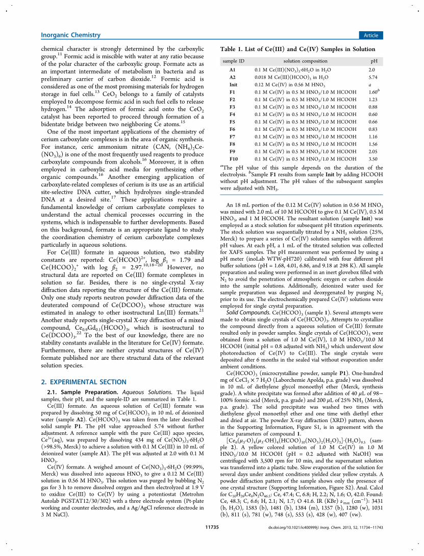
group R3m with Z = 3. The structure has thus a 3-fold rotationsymmetry. The Ce(III) atoms of Ce(HCOO)3 are 9-foldcoordinated (see Figure 1). A list of bond lengths is given in theSupporting Information, Table S1. The coordination poly-hedron of the Ce(III) cation is a tricapped trigonal prism. TheCe−O distances of the capping oxygens (O1) are 2.583(8) and2.601(8) Å, whereas the equatorial Ce−O distances are2.506(9) Å. There exists only one symmetry-independentHCOO− molecule in the structure. One oxygen atom (O1)binds to two neighboring Ce(III) atoms by forming infinitelinear chains along the [001] direction (Figure 1). The otheroxygen atom (O2) of the carboxylic group forms amonodentate bond with a Ce(III) atom on the equatorialplane of the coordination polyhedron. As a whole, the HCOO−
molecule acts as a tridentate ligand. The Ce(III) chains are
Table 2. Summary of Crystallographic Data and StructureRefinement Details for Compounds 1, 2, and 3
1 2 3
chemical formula C3H3CeO6 C10H39Ce6N2O46.5 C10H44Ce6N12O54
formula mass 275.17 1746.95 2007.05
crystal system trigonal monoclinic orthorhombic
a/Å 10.706(2) 12.3326(10) 12.3752(17)
b/Å 10.706(2) 19.5209(16) 26.152(3)
c/Å 4.1205(12) 18.9260(16) 15.698(3)
α/deg 90.00 90.00 90.00
β/deg 90.00 108.313(4) 90.00
γ/deg 120.00 90.00 90.00
unit cell volume/Å3 409.01(18) 4325.6(6) 5080.4(13)
temperature/K 296(2) 296(2) 296(2)
Space group R3m P21/c Pbcn
No. of formula unitsper unit cell, Z
3 4 4
radiation type MoKα MoKα MoKα
absorptioncoefficient,μ/mm−1
8.311 6.314 5.410
no. of reflectionsmeasured
1780 61601 5054
no. of independentreflections
264 11248 5054
Rint 0.1116 0.1197 0.0000
final R1 values (I >2σ(I))
0.0325 0.0468 0.0347
final wR(F2) values (I> 2σ(I))
0.0741 0.1190 0.1049
final R1 values (alldata)
0.0325 0.0604 0.0427
final wR(F2) values(all data)
0.0741 0.1245 0.1116
goodness of fit on F2 1.176 1.035 1.291
CCDC no. 925639 925637 925638
Inorganic Chemistry Article
dx.doi.org/10.1021/ic400999j | Inorg. Chem. 2013, 52, 11734−1174311736
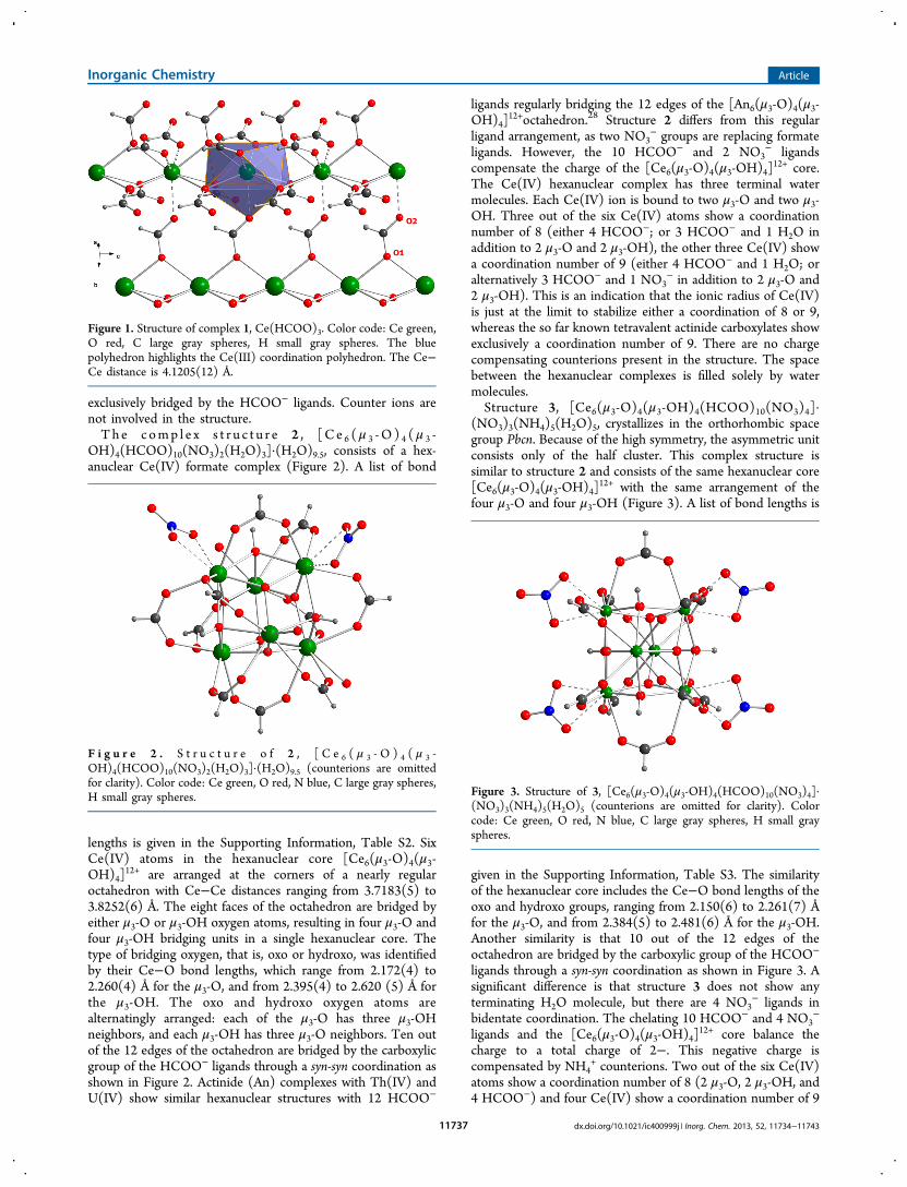
exclusively bridged by the HCOO− ligands. Counter ions arenot involved in the structure.Th e c omp l e x s t r u c t u r e 2 , [C e 6 (μ 3 -O ) 4 (μ 3 -
OH)4(HCOO)10(NO3)2(H2O)3]·(H2O)9.5, consists of a hex-anuclear Ce(IV) formate complex (Figure 2). A list of bond
lengths is given in the Supporting Information, Table S2. SixCe(IV) atoms in the hexanuclear core [Ce6(μ3-O)4(μ3-OH)4]
12+ are arranged at the corners of a nearly regularoctahedron with Ce−Ce distances ranging from 3.7183(5) to3.8252(6) Å. The eight faces of the octahedron are bridged byeither μ3-O or μ3-OH oxygen atoms, resulting in four μ3-O andfour μ3-OH bridging units in a single hexanuclear core. Thetype of bridging oxygen, that is, oxo or hydroxo, was identifiedby their Ce−O bond lengths, which range from 2.172(4) to2.260(4) Å for the μ3-O, and from 2.395(4) to 2.620 (5) Å forthe μ3-OH. The oxo and hydroxo oxygen atoms arealternatingly arranged: each of the μ3-O has three μ3-OHneighbors, and each μ3-OH has three μ3-O neighbors. Ten outof the 12 edges of the octahedron are bridged by the carboxylicgroup of the HCOO− ligands through a syn-syn coordination asshown in Figure 2. Actinide (An) complexes with Th(IV) andU(IV) show similar hexanuclear structures with 12 HCOO−
ligands regularly bridging the 12 edges of the [An6(μ3-O)4(μ3-OH)4]
12+octahedron.28 Structure 2 differs from this regularligand arrangement, as two NO3
− groups are replacing formateligands. However, the 10 HCOO− and 2 NO3
− ligandscompensate the charge of the [Ce6(μ3-O)4(μ3-OH)4]
12+ core.The Ce(IV) hexanuclear complex has three terminal watermolecules. Each Ce(IV) ion is bound to two μ3-O and two μ3-OH. Three out of the six Ce(IV) atoms show a coordinationnumber of 8 (either 4 HCOO−; or 3 HCOO− and 1 H2O inaddition to 2 μ3-O and 2 μ3-OH), the other three Ce(IV) showa coordination number of 9 (either 4 HCOO− and 1 H2O; oralternatively 3 HCOO− and 1 NO3
− in addition to 2 μ3-O and2 μ3-OH). This is an indication that the ionic radius of Ce(IV)is just at the limit to stabilize either a coordination of 8 or 9,whereas the so far known tetravalent actinide carboxylates showexclusively a coordination number of 9. There are no chargecompensating counterions present in the structure. The spacebetween the hexanuclear complexes is filled solely by watermolecules.Structure 3, [Ce6(μ3-O)4(μ3-OH)4(HCOO)10(NO3)4]·
(NO3)3(NH4)5(H2O)5, crystallizes in the orthorhombic spacegroup Pbcn. Because of the high symmetry, the asymmetric unitconsists only of the half cluster. This complex structure issimilar to structure 2 and consists of the same hexanuclear core[Ce6(μ3-O)4(μ3-OH)4]
12+ with the same arrangement of thefour μ3-O and four μ3-OH (Figure 3). A list of bond lengths is
given in the Supporting Information, Table S3. The similarityof the hexanuclear core includes the Ce−O bond lengths of theoxo and hydroxo groups, ranging from 2.150(6) to 2.261(7) Åfor the μ3-O, and from 2.384(5) to 2.481(6) Å for the μ3-OH.Another similarity is that 10 out of the 12 edges of theoctahedron are bridged by the carboxylic group of the HCOO−
ligands through a syn-syn coordination as shown in Figure 3. Asignificant difference is that structure 3 does not show anyterminating H2O molecule, but there are 4 NO3
− ligands inbidentate coordination. The chelating 10 HCOO− and 4 NO3
−
ligands and the [Ce6(μ3-O)4(μ3-OH)4]12+ core balance the
charge to a total charge of 2−. This negative charge iscompensated by NH4
+ counterions. Two out of the six Ce(IV)atoms show a coordination number of 8 (2 μ3-O, 2 μ3-OH, and4 HCOO−) and four Ce(IV) show a coordination number of 9
Figure 1. Structure of complex 1, Ce(HCOO)3. Color code: Ce green,O red, C large gray spheres, H small gray spheres. The bluepolyhedron highlights the Ce(III) coordination polyhedron. The Ce−Ce distance is 4.1205(12) Å.
F i g u r e 2 . S t r u c t u r e o f 2 , [ C e 6 ( μ 3 - O ) 4 ( μ 3 -OH)4(HCOO)10(NO3)2(H2O)3]·(H2O)9.5 (counterions are omittedfor clarity). Color code: Ce green, O red, N blue, C large gray spheres,H small gray spheres. Figure 3. Structure of 3, [Ce6(μ3-O)4(μ3-OH)4(HCOO)10(NO3)4]·
(NO3)3(NH4)5(H2O)5 (counterions are omitted for clarity). Colorcode: Ce green, O red, N blue, C large gray spheres, H small grayspheres.
Inorganic Chemistry Article
dx.doi.org/10.1021/ic400999j | Inorg. Chem. 2013, 52, 11734−1174311737
(2 μ3-O, 2 μ3-OH, 3 HCOO−, and 2 oxygen atoms from abidentate NO3
−). In the space between the hexanuclearcomplexes are, furthermore, 2 NO3
− groups arranged whosecharge is compensated by NH4
+. Short distances related withpotential hydrogen bridges are shown in SupportingInformation, Figure S6.The hydrogen atoms in this crystal structure could not be
located experimentally. The charge balance would be uncertainif both the hexanuclear cluster and the counterions containhydrogen atoms which could not be identified experimentally.The charge of the hexanuclear cluster is neutralized by NH4
+.However, it is difficult to distinguish the electron density of thenitrogen atoms in NH4
+ explicitly from that of the oxygenatoms in H2O. Although precaution has taken to keep Ce(IV)stable, evidence is required that the synthesis did not result in aCe(III)/Ce(IV) mixed-valent crystal structure. The oxidationstate of cerium can be verified by analyzing the Ce L3-edgeXANES (Figure 4). The Ce L3-edge spectrum of Ce(III)
consists of a single peak just above the absorption thresholdassociated with the electron transition 2p64f1(5d,6s)3→2p54f1(5d,6s)4. The Ce L3-edge spectrum of Ce(IV) showstwo resonances whose origin is interpreted as a result of mixedstate between f0 and f1L29 or alternatively as an almost pure f0
state30,31 with a peak at higher energy following the transition2p64f0(5d,6s)4→2p54f0(5d,6s)5. The energy difference betweenthe resonances and the characteristic transition intensities isoften used to determine the valence of Ce or the ratio ofCe(III)/Ce(IV) in mixed-valent systems.32−35 An aqueoussolution of 0.01 M Ce(III) nitrate was used as reference forCe(III), while CeO2 was used as reference for Ce(IV). The L3-edge XANES spectrum of sample 3 shows essentially the samespectral features as those observed for the Ce(IV) reference.The presence of Ce(III) can be therefore excluded in sample 3.The formation of the hexanuclear Ce(IV) formate complexes
corresponds with the Ce(IV) hydrolysis and the deprotonationof HCOOH in the same pH region. The Ce(IV) hydrolysisresults in an oligomerization through oxo and hydroxo bonds,whereas the carboxylic function of formate introduces chelatingligands, stabilizing the hexanuclear oligomer.
Despite several attempts we were so far not successful inobtaining the Ce(IV) formate complex without nitratecoordination. Nitrate was used in the synthesis because itsupports the stability of Ce(IV) in aqueous solution most likelythrough complex formation.36,37 As a matter of fact, the crystalpreparation of Ce(IV) formate complexes in perchlorate andchloride media only results in the reduction of Ce(IV) toCe(III) just a few hours after the preparation of mother Ce(IV)solutions. The hexanuclear [Ce6(μ3-O)4(μ3-OH)4]
12+ core insample 2 and 3 is stabilized by the syn-syn coordinatedcarboxylic groups of formate, whereas nitrate acts not as suchchelating ligand. It should be mentioned that sulfate, which alsosupports Ce(IV) stability through complex formation,38 can actas chelating ligand resulting in hexanuclear [Ce6(μ3-O)4(μ3-OH)4(SO4)6] units.
39 There are hexanuclear Ce(IV) structuresknown with exclusive coordination of 12 carboxylic and otherfunctional groups, such as the hexanuclear complex [Ce6(μ3-O)4(μ3-OH)4(acac)12], acac = acetylacetonate,40 and [Ce6(μ3-O)4(μ3-OH)4(μ2-O2C
tBu)12], O2CtBu = pivalate t-Bu.41 It is
interesting to note that the above-mentioned hexanuclearCe(IV) carboxylates do not comprise an interstitial μ6-oxooxygen atom. Such a μ6-oxo oxygen atom at the center of theoctahedron was observed in several hexanuclear trivalentlanthanide (Ln) clusters forming the unit [Ln6(μ6-O)4(μ3-OH)8]
8+, where the μ6-O is believed to play a role in stabilizingthe hexanuclear core.42,43 Such hexanuclear La(III) clusters canalso involve bidentately coordinating terminal nitrate groups,44
in a similar manner as observed for the compounds 2 and 3.The competing coordination of carboxylic ligands of benzoateand nitrate has been observed in the hexanuclear cluster[Ce6(μ3-O)5(μ3-OH)3(C6H5COO)9(NO3)3(DMF)3].
45
3.2. Solution Species. Subsequently we investigate thecoordination of Ce(III) and Ce(IV) formate complexes inaqueous solution with Ce K-edge EXAFS spectroscopy.
3.2.1. Ce(III) Formate. To verify the structural differencebetween the Ce(III) formate complexes in aqueous solutionand the Ce(III) aquo species itself, a reference sample of 0.1 MCe(NO3)3·6H2O in H2O at pH 2 (sample A1) wasinvestigated. Such aquo species, Ce3+(aq), are supposed to becoordinated by 9 water molecules in a tricapped trigonal prismfashion, which can be formulated as Ce(H2O)9
3+.3,4 TheEXAFS spectrum of this sample (Figure 5), shows 9.4(3)oxygen atoms from water at a Ce−O distance of 2.522(2) Å.As a second reference a solid powder of Ce(HCOO)3 was
investigated (sample P1). In this structure all formate ligandsappear in monodentate coordination (Figure 1). The EXAFSspectrum (Figure 5) shows 9 oxygen atoms at an average Ce−O distance of 2.523(2) Å, a broad peak originated from Ce−Cscattering pairs at 3.431(8) and 3.601(6) Å, and furthermorethe nearest Ce neighbors in the linear Ce(III) chain at a Ce−Ce distance of 4.100(2) Å. Infinite linear chains in structure 1are formed during the crystallization process, while they are notexpected to exist in solution. Nevertheless, the solution speciesmay occur either as mononuclear species or as a preshapedpolynuclear precursor.To obtain the structural information on a Ce(III) formate
complex in solution, Ce(HCOO)3 from sample P1 wasdissolved in water (sample A2). The pH value of this colorlesssolution approached 5.74 without further adjustment. Concen-trations of higher than ∼0.025 M Ce(III) resulted inprecipitation, indicating that the present Ce(III) formatesolution ([Ce] = 0.018 M) is close to the solubility limit.The reported stability constants of Ce(III) formate suggest the
Figure 4. Ce L3-edge XANES spectra of (a) Ce(III) reference of 0.01M Ce(III) nitrate in H2O, (b) Ce(IV) reference of CeO2, and (c) solidsample 3. Ce K-edge XANES of the compounds are shown in theSupporting Information, Figure S7.
Inorganic Chemistry Article
dx.doi.org/10.1021/ic400999j | Inorg. Chem. 2013, 52, 11734−1174311738
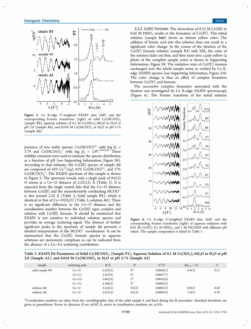
presence of two stable species: Ce(HCOO)2+ with log ß1 =1.79 and Ce(HCOO)2
+ with log ß2 = 2.97.10,19,20 Thesestability constants were used to estimate the species distributionas a function of pH (see Supporting Information, Figure S8).According to that estimate, the Ce(III) species of sample A2are composed of 42% Ce3+(aq), 41% Ce(HCOO)2+, and 17%Ce(HCOO)2
+. The EXAFS spectrum of this sample is shownin Figure 5. The spectrum reveals only a single peak of 9.6(3)O atoms at a Ce−O distance of 2.521(2) Å (Table 3). It isexpected from the single crystal data that the Ce−O distancebetween Ce(III) and the monodentately coodinating HCOO−
is also around 2.52 Å (Table 3, Solid sample P1), which isidentical to that of Ce−O(H2O) (Table 3, solution A1). Thereis no significant difference in the Ce−O distance and thecoordination number between the Ce(III) aquo complex andsolution with Ce(III) formate. It should be mentioned thatEXAFS is not sensitive to individual solution species andprovides an average scattering signal. The absence of furthersignificant peaks in the spectrum of sample A2 prevents adetailed interpretation of the HCOO− coordination. It can besummarized that the Ce(III) formate species in aqueoussolutions are monomeric complexes as can be indicated fromthe absence of a Ce−Ce scattering contribution.
3.2.2. Ce(IV) Formate. The electrolysis of 0.12 M Ce(III) in0.56 M HNO3 results in the formation of Ce(IV). This initialsolution (sample Init) shows an intense yellow color. Theaddition of formic acid into this solution does not result in asignificant color change. In the course of the titration of theCe(IV) formate solution (sample F1) with NH3 the color ofthe solution fades out first, and then turns into a pale yellow (aphoto of the complete sample series is shown in SupportingInformation, Figure S9. The oxidation state of Ce(IV) remainsunchanged over the whole sample series as verified by Ce K-edge XANES spectra (see Supporting Information, Figure S10.The color change is thus an effect of complex formationbetween Ce(IV) and formate.The successive complex formation associated with the
titration was investigated by Ce K-edge EXAFS spectroscopy(Figure 6). The Fourier transform of the initial solution
Figure 5. Ce K-edge k3-weighted EXAFS data (left) and thecorresponding Fourier transforms (right) of solid Ce(HCOO)3(sample P1), aqueous solution of 0.1 M Ce(NO3)3·6H2O in H2O atpH 2.0 (sample A1), and 0.018 M Ce(HCOO)3 in H2O at pH 5.74(sample A2).
Table 3. EXAFS Fit Parameters of Solid Ce(HCOO)3 (Sample P1), Aqueous Solution of 0.1 M Ce(NO3)3·6H2O in H2O at pH2.0 (Sample A1), and 0.018 M Ce(HCOO)3 in H2O at pH 5.74 (Sample A2)
sample scattering path R/Å N σ2/Å2 ΔEk=0 / eV F
solid sample P1 Ce−O 2.523(2) 9a 0.0084(1) 0.4(3) 0.11Ce−C1 3.431(8) 6a 0.0047(7)Ce−C2 3.601(6) 3a 0.0032(6)Ce−Ce 4.100(2) 2a 0.0062(3)
solution A1 Ce−O 2.522(2) 9.4(3) 0.0085(3) 0.8(4) 0.28solution A2 Ce−O 2.521(2) 9.6(3) 0.0089(4) 1.9(3) 0.78
aCoordination numbers are taken from the crystallographic data of the solid sample 1 and fixed during the fit procedure. Standard deviations aregiven in parentheses. Errors in distances R are ±0.02 Å, errors in coordination numbers are ±15%.
Figure 6. Ce K-edge k3-weighted EXAFS data (left) and thecorresponding Fourier transforms (right) of aqueous solutions with0.01 M Ce(IV), 0.5 M HNO3, and 1 M HCOOH with different pHvalues. The sample composition is listed in Table 1
Inorganic Chemistry Article
dx.doi.org/10.1021/ic400999j | Inorg. Chem. 2013, 52, 11734−1174311739
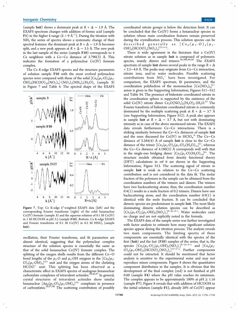
(sample Init) shows a dominant peak at R + Δ ∼ 1.9 Å. TheEXAFS spectrum changes with addition of formic acid (sampleF1) in the higher k-range (k > 9 Å−1). During the titration withNH3 the series of spectra shows a systematic change of theirspectral features: the dominant peak at R + Δ ∼ 1.9 Å becomessplit, and a new peak appears at R + Δ ∼ 3.5 Å. The new peakin the last sample of the series (sample F10) corresponds to 4Ce neighbors with a Ce−Ce distance of 3.790(3) Å. Thisindicates the formation of a polynuclear Ce(IV) formatecomplex.The Ce K-edge EXAFS spectra and the structure parameters
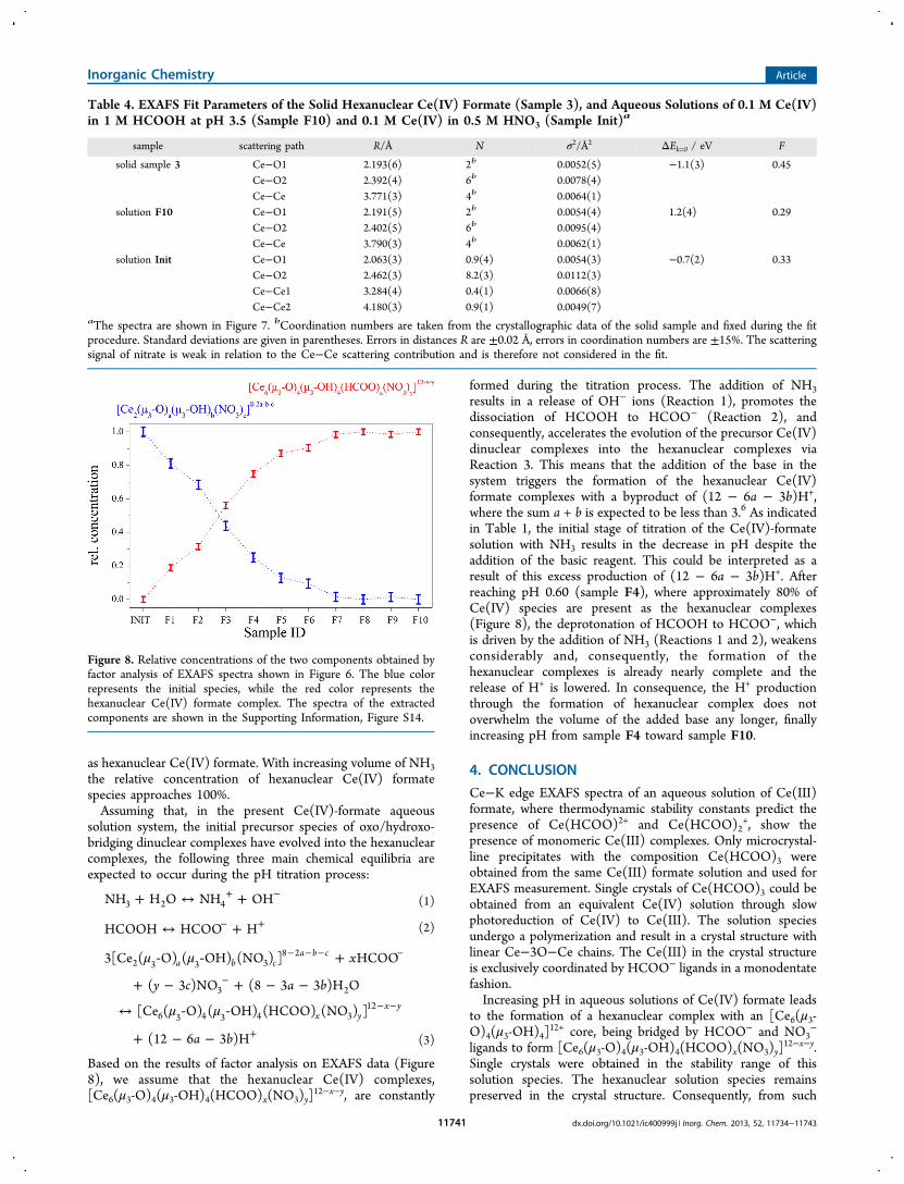
of solution sample F10 with the most evolved polynuclearspecies were compared with those of the solid [Ce6(μ3-O)4(μ3-OH)4(HCOO)10(NO3)4]·(NO3)3(NH4)5(H2O)5 (sample 3)in Figure 7 and Table 4. The spectral shape of the EXAFS
oscillation, their Fourier transforms, and fit parameters arealmost identical, suggesting that the polynuclear complexstructure of the solution species is essentially the same asthat of the solid hexanuclear Ce(IV) formate complex. Thesplitting of the oxygen shells results from the different Ce−Obond lengths of the μ3-O and μ3-OH oxygens in the [Ce6(μ3-O)4(μ3-OH)4]
12+ unit and the oxygen atoms of the chelatingHCOO− unit. This splitting has been observed as acharacteristic effect in EXAFS spectra of analogous hexanuclearcarboxylate complexes of tetravalent actinides.28,46,47 In general,crystal structures of tetravalent actinides show similarhexanuclear [An6(μ3-O)4(μ3-OH)4]
12+ complexes in presenceof carboxylates.28,47,48 The scattering contribution of possibly
coordinated nitrate groups is below the detection limit. It canbe concluded that the Ce(IV) forms a hexanuclear species insolution whose main coordination features remain preservedduring the crystallization process. This solution species can bed e s c r i b e d g e n e r a l l y a s [ C e 6 ( μ 3 - O ) 4 ( μ 3 -OH)4(HCOO)x(NO3)y]
12−x−y.There is wide agreement in the literature that a Ce(IV)
nitrate solution as in sample Init is composed of polymericspecies, mainly dimers and trimers.36,38b,49,50 The EXAFSspectrum of sample Init shows several peaks in the range R + Δ= 2.5−4.0 Å. The peaks may originate from Ce−Ce interaction,nitrate ions, and/or water molecules. Possible scatteringcontributions from NO3
− have been investigated. Forcomparison, the EXAFS spectrum, fit parameters, and thecoordination polyhedron of the mononuclear [Ce(NO3)6]
2−
anion is given in the Supporting Information, Figures S11−S12and Table S4. The presence of bidentate coordinated nitrate inthe coordination sphere is supported by the existence of thesolid Ce(IV) nitrate dimer Ce2O(NO3)6(H2O)6·2H2O.
51 TheFourier transform of bidentate coordinated nitrate is commonlydominated by the multiple scattering peak at R + Δ ∼ 3.7 Å(see Supporting Information, Figure S12). A peak also appearsin sample Init at R + Δ ∼ 3.7 Å, but not with dominatingintensity as in case of the above mentioned nitrate. The EXAFSdata reveals furthermore Ce−Ce interactions. There is astriking similarity between the Ce−Ce distances of sample Initand the ones discussed for Ce(IV) in HClO4.
6 The Ce−Cedistance of 3.284(4) Å of sample Init is close to the Ce−Cedistance of the trimer [Ce3(μ2-O)3(μ3-O)2(H2O)12]
2+, whereasthe Ce−Ce distance of 4.180(3) Å corresponds well with thatin the single-oxo bridging dimer [Ce2(μ2-O)(H2O)14]
6+. Thestructure models obtained from density functional theory(DFT) calculations in ref 6 are shown in the SupportingInformation, Figure S13. The scattering signal of nitrate insample Init is weak in relation to the Ce−Ce scatteringcontribution and is not considered in the data fit. The molarfraction of the polymers in the sample can be obtained from thecoordination numbers of the trimers and dimers. The trimershave two backscattering atoms; thus, the coordination number0.4(1) results in a mole fraction of 0.2 trimers. Dimers have onebackscattering atom, and the coordination number 0.9(1) isidentical with the mole fraction. It can be concluded thatdimeric species are predominant in sample Init. The most likelydominating dimeric solution species can be described as[Ce2(μ3-O)a(μ3-OH)b(NO3)c]
8−2a−b−c. Water molecules carryno charge and are not explicitly noted in the formula.The EXAFS data of the sample series was further investigated
with factor analysis to estimate how many significant solutionspecies appear during the titration process. The analysis revealstwo main components. The limiting spectra of thesecomponents are essentially identical with the spectra of thefirst (Init) and the last (F10) samples of the series, that is, thespecies [Ce2(μ3-O)a(μ3-OH)b(NO3)c]
8−2a−b−c and [Ce6(μ3-O)4(μ3-OH)4(HCOO)x(NO3)y]
12−x−y. Further componentscould not be extracted. It should be mentioned that factoranalysis is sensitive to the experimental noise and may notreproduce minor components. Figure 8 shows the quantitativecomponent distribution in the samples. It is obvious that thedevelopment of the final complex (red) is not finished at pH0.60 (sample F4) where the pH value reaches its minimum.The complex appears to be approximately 100% at pH ≥ 1.16(sample F7). Figure 8 reveals that with addition of HCOOH tothe initial solution (sample F1), already 20% of Ce(IV) appear
Figure 7. Top: Ce K-edge k3-weighted EXAFS data (left) and thecorresponding Fourier transforms (right) of the solid hexanuclearCe(IV) formate (sample 3) and the aqueous solution of 0.1 M Ce(IV)in 1 M HCOOH at pH 3.5 (sample F10). Bottom: Ce K-edge EXAFSand Fourier transforms of 0.1 M Ce(IV) in 0.5 M HNO3 (sampleInit).
Inorganic Chemistry Article
dx.doi.org/10.1021/ic400999j | Inorg. Chem. 2013, 52, 11734−1174311740
as hexanuclear Ce(IV) formate. With increasing volume of NH3the relative concentration of hexanuclear Ce(IV) formatespecies approaches 100%.Assuming that, in the present Ce(IV)-formate aqueous
solution system, the initial precursor species of oxo/hydroxo-bridging dinuclear complexes have evolved into the hexanuclearcomplexes, the following three main chemical equilibria areexpected to occur during the pH titration process:
+ ↔ ++ −NH H O NH OH3 2 4 (1)
↔ +− +HCOOH HCOO H (2)
μ μ
μ μ
‐ ‐ +
+ − + − −
↔ ‐ ‐
+ − −
− − − −
−
− −
+
x
y c a b
a b
3[Ce ( O) ( OH) (NO ) ] HCOO
( 3 )NO (8 3 3 )H O
[Ce ( O) ( OH) (HCOO) (NO ) ]
(12 6 3 )H
a b ca b c
x yx y
2 3 3 38 2
3 2
6 3 4 3 4 312
(3)
Based on the results of factor analysis on EXAFS data (Figure8), we assume that the hexanuclear Ce(IV) complexes,[Ce6(μ3-O)4(μ3-OH)4(HCOO)x(NO3)y]
12−x−y, are constantly
formed during the titration process. The addition of NH3results in a release of OH− ions (Reaction 1), promotes thedissociation of HCOOH to HCOO− (Reaction 2), andconsequently, accelerates the evolution of the precursor Ce(IV)dinuclear complexes into the hexanuclear complexes viaReaction 3. This means that the addition of the base in thesystem triggers the formation of the hexanuclear Ce(IV)formate complexes with a byproduct of (12 − 6a − 3b)H+,where the sum a + b is expected to be less than 3.6 As indicatedin Table 1, the initial stage of titration of the Ce(IV)-formatesolution with NH3 results in the decrease in pH despite theaddition of the basic reagent. This could be interpreted as aresult of this excess production of (12 − 6a − 3b)H+. Afterreaching pH 0.60 (sample F4), where approximately 80% ofCe(IV) species are present as the hexanuclear complexes(Figure 8), the deprotonation of HCOOH to HCOO−, whichis driven by the addition of NH3 (Reactions 1 and 2), weakensconsiderably and, consequently, the formation of thehexanuclear complexes is already nearly complete and therelease of H+ is lowered. In consequence, the H+ productionthrough the formation of hexanuclear complex does notoverwhelm the volume of the added base any longer, finallyincreasing pH from sample F4 toward sample F10.
4. CONCLUSION
Ce−K edge EXAFS spectra of an aqueous solution of Ce(III)formate, where thermodynamic stability constants predict thepresence of Ce(HCOO)2+ and Ce(HCOO)2
+, show thepresence of monomeric Ce(III) complexes. Only microcrystal-line precipitates with the composition Ce(HCOO)3 wereobtained from the same Ce(III) formate solution and used forEXAFS measurement. Single crystals of Ce(HCOO)3 could beobtained from an equivalent Ce(IV) solution through slowphotoreduction of Ce(IV) to Ce(III). The solution speciesundergo a polymerization and result in a crystal structure withlinear Ce−3O−Ce chains. The Ce(III) in the crystal structureis exclusively coordinated by HCOO− ligands in a monodentatefashion.Increasing pH in aqueous solutions of Ce(IV) formate leads
to the formation of a hexanuclear complex with an [Ce6(μ3-O)4(μ3-OH)4]
12+ core, being bridged by HCOO− and NO3−
ligands to form [Ce6(μ3-O)4(μ3-OH)4(HCOO)x(NO3)y]12−x−y.
Single crystals were obtained in the stability range of thissolution species. The hexanuclear solution species remainspreserved in the crystal structure. Consequently, from such
Table 4. EXAFS Fit Parameters of the Solid Hexanuclear Ce(IV) Formate (Sample 3), and Aqueous Solutions of 0.1 M Ce(IV)in 1 M HCOOH at pH 3.5 (Sample F10) and 0.1 M Ce(IV) in 0.5 M HNO3 (Sample Init)a
sample scattering path R/Å N σ2/Å2 ΔEk=0 / eV F
solid sample 3 Ce−O1 2.193(6) 2b 0.0052(5) −1.1(3) 0.45Ce−O2 2.392(4) 6b 0.0078(4)Ce−Ce 3.771(3) 4b 0.0064(1)
solution F10 Ce−O1 2.191(5) 2b 0.0054(4) 1.2(4) 0.29Ce−O2 2.402(5) 6b 0.0095(4)Ce−Ce 3.790(3) 4b 0.0062(1)
solution Init Ce−O1 2.063(3) 0.9(4) 0.0054(3) −0.7(2) 0.33Ce−O2 2.462(3) 8.2(3) 0.0112(3)Ce−Ce1 3.284(4) 0.4(1) 0.0066(8)Ce−Ce2 4.180(3) 0.9(1) 0.0049(7)
aThe spectra are shown in Figure 7. bCoordination numbers are taken from the crystallographic data of the solid sample and fixed during the fitprocedure. Standard deviations are given in parentheses. Errors in distances R are ±0.02 Å, errors in coordination numbers are ±15%. The scatteringsignal of nitrate is weak in relation to the Ce−Ce scattering contribution and is therefore not considered in the fit.
Figure 8. Relative concentrations of the two components obtained byfactor analysis of EXAFS spectra shown in Figure 6. The blue colorrepresents the initial species, while the red color represents thehexanuclear Ce(IV) formate complex. The spectra of the extractedcomponents are shown in the Supporting Information, Figure S14.
Inorganic Chemistry Article
dx.doi.org/10.1021/ic400999j | Inorg. Chem. 2013, 52, 11734−1174311741
solutions crystallizes a compound with the composition[ C e 6 ( μ 3 - O ) 4 ( μ 3 - O H ) 4 ( H C O O ) 1 0 ( N O 3 ) 4 ] ·(NO3)3(NH4)5(H2O)5 including four nitrate ligands in thehexanuclear complex. Using different titration agents (NH3 orNaOH) yields slightly different hexanuclear Ce(IV) formatecomplexes. A titration with NaOH resulted in the formation ofa hexanuclear complex with the composition [Ce6(μ3-O)4(μ3-OH)4(HCOO)10(NO3)2(H2O)3]·(H2O)9.5 with two nitrateligands in the hexanuclear complex.The different structures observed for Ce(III) and Ce(IV)
reflect the different influence of hydrolysis. In case of Ce(III)the hydrolysis occurs far above the onset of the formation offormate complexes at pH ∼ 2. In case of Ce(IV) hydrolyzedpolynuclear species already appear in an acidic solution withoutthe addition of basic reagents. The polynuclear Ce(IV) formatecomplex appears thus from a competing reaction betweenhydrolysis and ligation. The hydrolysis causes polymerizationthrough olation and oxolation, while the ligation by thecarboxylic groups of HCOO− prevents further hydrolyticpolymerization and, consequently, stabilizes the hexanuclearcomplex in an aqueous solution.
■ ASSOCIATED CONTENT*S Supporting InformationExperimental details, X-ray diffraction data (single crystals andpowder), species fraction in solution, XANES and EXAFSspectra and structural parameters, photos of the aqueoussolutions, cif files of the crystal structures. This material isavailable free of charge via the Internet at http://pubs.acs.org.
■ AUTHOR INFORMATIONCorresponding Author*E-mail: [email protected].
NotesThe authors declare no competing financial interest.
■ ACKNOWLEDGMENTSThe authors are thankful to Wouter Van Beek/SNBL forsupport during some EXAFS measurements and A. Scholz/HZDR and J. Wenzel/BAM for powder diffraction measure-ments.
■ REFERENCES(1) Binnemans, K. Application of tetravalent cerium compounds. InHandbook on the Physics and Chemistry of Rare Earths; Geschneider, K.A., Jr., Bunzli, J.-C. G., Pecharsky, V. K., Eds.; Elsevier: Amsterdam,The Netherlands, 2006; Vol. 36, pp 281−392.(2) Richens, D. T. The Chemistry of Aqua Ions; John Wiley & Sons:Chichester, U.K., 1997.(3) Habenschuss, A.; Spedding, F. H. J. Chem. Phys. 1980, 73, 442−450.(4) Buzko, V.; Sukhno, I.; Polushin, A. Int. J. Quantum Chem. 2011,111, 2705−2711.(5) Baes, Jr., C. F.; Mesmer, R. E. The Hydrolysis of Cations; JohnWiley & Sons: New York, 1976; Chapter 7, pp 138−146.(6) Ikeda-Ohno, A.; Tsushima, S.; Hennig, C.; Yaita, T.; Bernhard, G.Dalton Trans. 2012, 41, 7190−7192.(7) Ikeda-Ohno, A.; Hennig, C.; Weiss, S.; Yaita, T.; Bernhard, G.Chem.Eur. J. 2013, 19 (23), 7348−7360.(8) Wadsworth, E.; Duke, F. R.; Goetz, C. A. Anal. Chem. 1957, 29,1824−1825.(9) Maverick, A. W.; Yao, Q. Inorg. Chem. 1993, 32, 5626−5628.(10) Wood, S. A. Eng. Geol. 1993, 34, 229−259.
(11) Reutemann, W.; Kieczka, H. Formic acid. In Ullmann’sEncyclopedia of Industrial Chemistry; Wiley-VCH: Weinheim, Germany,2011.(12) Lu, W.; Wacker, T.; Gerbing,-Smentke, E.; Andrade, L. A.;Einsle, O. Science 2011, 332, 352−354.(13) Grasemann, M.; Laurenczy, G. Energy Environ. Sci. 2012, 5,8171−8181.(14) Stubenrauch, J.; Brosha, E.; Vohs, J. M. Catal. Today 1996, 28,431−441.(15) Senanayake, S. D.; Mullins, D. R. J. Phys. Chem. C 2008, 112,9744−9752.(16) Sridharan, V.; Menendez, J. C. Chem. Rev. 2010, 110, 3805−3849.(17) (a) Miyajima, Y.; Ishizuka, T.; Yamamoto, Y.; Sumaoka, J.;Komiyama, M. J. Am. Chem. Soc. 2009, 131, 2657−2662. (b) Aiba, Y.;Lonnberg, T.; Komiyama, M. Chem.Asian. J. 2011, 6, 2407−2411.(18) Tsubota, H. B. Chem. Soc. Jpn. 1962, 35, 640−644.(19) Kovar, L. E.; Powell, J. E. Stability constants of rare earths withsome weak carboxylic acids; Report TID-4500; Ames Laboratory, IowaState University: Ames, Iowa, 1966.(20) Martell, A. E.; Smith, R. M. NIST critically selected stabilityconstants of metal complexes; NIST standard reference database 46,Vers. 8.0; National Institute of Standards and Technology :Gaithersburg, MD, 2004.(21) Bolotovsky, R. L.; Bulkin, A. P.; Krutov, G. A.; Kudryashev, V.A.; Trunov, V. A.; Ulyanov, V. A.; Antson, O.; Hiismaki, P.; Poyry;Tiitta, A.; Loshmanov, A. A.; Furmanova, N. G. Solid State Commun.1990, 76, 1045−1049.(22) Go, Y. B.; Jacobson, A. J. Chem. Mater. 2007, 19, 4702−4709.(23) (a) Sheldrick, G. M. SHELXS-97, Program for the Solution ofCrystal Structures; Universitat Gottingen: Gottingen, Germany, 1997;(b) Sheldrick, G. M. SHELXL-97, Program for the Crystal StructureRefinement; Universitat Gottingen: Gottingen, Germany, 1997.(24) Ressler, T. J. Synchrotron Radiat. 1998, 5, 118.(25) George, G. N.; Pickering, I. J. EXAFSPAK, a suite of computerprograms for analysis of X-ray absorption spectra; Stanford SynchrotronRadiation Laboratory: Stanford, CA, 2000.(26) Ankudinov, A. L.; Ravel, B.; Rehr, J. J.; Conradson, S. D. Phys.Rev. B 1998, 58, 7565−7576.(27) (a) Rossberg, A.; Reich, T.; Bernhard, G. Anal. Bioanal. Chem.2003, 376, 631−638. (b) Malinowski, E. R. Anal. Chim. Acta 1980,122, 327−330.(28) Takao, S.; Takao, K.; Kraus, W.; Emmerling, F.; Scheinost, A.C.; Bernhard, G.; Hennig, C. Eur. J. Inorg. Chem. 2009, 4771−4775.(29) (a) Kotani, A.; Ogasawara, H.; Okada, K.; Thole, B. T.;Sawatzky, G. A. Phys. Rev. B 1989, 40, 65−73. (b) Kotani, A.;Kvashnina, K. O.; Butorin, S. M.; Glatzel, P. Eur. Phys. J. B 2012, 85,257−269.(30) Marabelli, F.; Wachter, P. Phys. Rev. B 1987, 36, 1238−1243.(31) Hanyu, T.; Ishii, H.; Yanagihara, M.; Kamada, T.; Miyahara, T.;Kato, H.; Naito, K.; Suzuki, S.; Ishii, T. Solid State Commun. 1985, 56,381−383.(32) Beck, D. D.; Capehart, T. W.; Hofman, R. W. Chem. Phys. Lett.1989, 159, 207−213.(33) Prieto, C.; Lagarde, P.; Dexpert, H.; Briois, V.; Villain, F.;Verdaguer, M. J. Phys. Chem. Solids 1992, 53, 233−237.(34) Antonio, M. R.; Soderholm, L. Inorg. Chem. 1994, 33, 5988−5993.(35) Briotis, V.; Lutzenkirchen-Hecht, D.; Villain, F.; Fonda, E.;Belin, S.; Griesebock, B.; Frahm, R. J. Phys. Chem. A 2005, 109, 320−329.(36) Blaustein, B. D.; Gryder, J. W. J. Am. Chem. Soc. 1957, 79, 540−547.(37) Miller, J. T.; Irish, D. E. Can. J. Chem. 1967, 45, 147−155.(38) (a) Jones, E. G.; Soper, F. G. J. Chem. Soc. 1935, 802−805.(b) Hardwick, T. J.; Robertson, E. Can. J. Chem. 1951, 29, 818−827.(c) Paulenova, A.; Creager, S. E.; Navratil, J. D.; Wei, Y. J. PowerSources 2002, 109, 431−438.(39) Lundgren, G. Ark. Kemi 1956, 5, 349−363.
Inorganic Chemistry Article
dx.doi.org/10.1021/ic400999j | Inorg. Chem. 2013, 52, 11734−1174311742

(40) Toledano, P.; Ribot, F.; Sanchez. C. R. Acad. Sci. Ser. II 1990,311, 1315−1320.(41) Mereacre, V.; Ako, M. A.; Akhtar, M. N.; Lindemann, A.; Anson,C. E.; Powell, A. K. Helv. Chim. Acta 2009, 92, 2507−2514.(42) Wang, R.; Carducci, M. C.; Zheng, Z. Inorg. Chem. 2000, 39,1836−1837.(43) Mudring, A.-V.; Timofte, T.; Babei, A. Inorg. Chem. 2006, 45,5162−5166.(44) Calvez, G.; Daiguebonne, C.; Guillou, O.; Le Dret, F. Eur. J.Inorg. Chem. 2009, 3172−3178.(45) Das, R.; Samara, R.; Baruah, J. B. Inorg. Chem. Commun. 2010,23, 793−795.(46) Takao, K.; Takao, S.; Scheinost, A. C.; Bernhard, G.; Hennig, C.Inorg. Chem. 2012, 51, 1336−1344.(47) Hennig, C.; Takao, S.; Takao, K.; Weiss, S.; Kraus, W.;Emmerling, F.; Scheinost, A. C. Dalton Trans. 2012, 41, 12818−12823.(48) (a) Lundgren, G. Ark. Kemi 1952, 5, 421−428. (b) Morkry, L.M.; Dearn, N. S.; Carrano, C. J. Angew. Chem., Int. Ed. Engl. 1996, 35,1497−1498. (c) Duval, P. B.; Burns, C. J.; Buschmann, W. E.; Clark,D. L.; Morris, D. E.; Scott, B. L. Inorg. Chem. 2001, 40, 5491−5496.(d) Berthet, J. C.; Thuery, P.; Ephritikhine, M. Chem. Commun. 2005,3415−3417. (e) Nocton, G.; Burdet, F.; Pecaut, J.; Mazzanti, M.Angew. Chem., Int. Ed. 2007, 46, 7574−7578. (f) Nocton, G.; Pecaut,J.; Filinchuk, Y.; Mazzanti, M. Chem. Commun. 2010, 46, 2757−2759.(g) Mougel, V.; Biswas, B.; Pecaut, J.; Mazzanti, M. Chem. Commun.2010, 46, 8648−8650. (h) Biswas, B.; Mougel, V.; Pecaut, J.; Mazzanti,M. Angew. Chem., Int. Ed. 2011, 123, 5863−5866. (i) Knope, K. E.;Wilson, R. E.; Vasiliu, M.; Dixon, D. A.; Soderholm, L. Inorg. Chem.2011, 50, 9696−9704. (j) Vasiliu, M.; Knope, K. E.; Soderholm, L.;Dixon, D. A. J. Phys. Chem. 2012, 116, 6917−6926. (k) Knope, K. E.;Soderholm, L. Chem. Rev. 2013, 113, 944−994. (l) Hennig, C.; Takao,S.; Takao, K.; Weiss, S.; Kraus, W.; Emmerling, F.; Meyer, M.;Scheinost, A. C. J. Phys. Conf. Series 2013, 430, 012116. (m) Falaise,C.; Volkringer, C.; Vigier, J.-F.; Henry, N.; Beaurain, A.; Loiseau, T.Chem.Eur. J. 2013, 19, 5324−5331.(49) Wiberg, K. B.; Ford, P. C. Inorg. Chem. 1968, 7, 369−373.(50) Duke, F. R.; Parchen, F. R. J. Am. Chem. Soc. 1956, 78, 1540−1543.(51) Guillou, N.; Auffredic, J. P.; Louer, J. Solid State Chem. 1994,112, 45−52.
Inorganic Chemistry Article
dx.doi.org/10.1021/ic400999j | Inorg. Chem. 2013, 52, 11734−1174311743


![Coordination-driven self-assembly of discrete Ru6 Pt6 ... · Coordination-driven self-assembly of discrete ... The [3 + 2] hexanuclear cages 3a and 3b were isolated in good yields](https://img.pdfslide.net/doc/110x75/5edc4710ad6a402d6666e17f/coordination-driven-self-assembly-of-discrete-ru6-pt6-coordination-driven-self-assembly.jpg)