Embed Size (px)
Citation preview
Cryst. Res. Technol. 39, No. 4, 353 – 358 (2004) / DOI 10.1002/crat.200310194
© 2004 WILEY-VCH Verlag GmbH & Co. KGaA, Weinheim
Crystal structure of 17α-acetoxyprogesterone, C23H32O4
Rajnikant*, Dinesh, Anshu, and Mousmi
Post-Graduate Department of Physics, University of Jammu, Jammu Tawi – 180 006, India
Received 9 January 2003, revised 26 March 2003, accepted 15 April 2003 Published online 1 April 2004
Key words crystal structure.
PACS 61.66.Hq
The crystal structure of the title compound (C23H32O4) has been determined by X-ray diffraction methods. It crystallizes in the orthorhombic space group P212121 with cell parameters: a = 10.86(1), b = 11.95(2), c = 15.65(5) Å and Z = 4. The structure has been refined to an R-value of 0.045 for 1610 observed reflections. Ring A exists in sofa conformation, Ring B adopts a distorted chair conformation while as Ring C assumes a chair conformation. The five membered ring D adopts a half-chair conformation. The crystal structure is stabilized by intra- and intermolecular C-H…O interactions.
© 2004 WILEY-VCH Verlag GmbH & Co. KGaA, Weinheim
1 Introduction
The steroid drugs occupy a conspicuous place in the modern therapeutic practice. The broad spectrum of biological activity within the group and the multiplicity of actions displayed by certain individual members make the steroids one of the most intriguing classes of biologically active compounds. The structure analysis of the title compound has been taken up as a part of our on-going crystallographic investigations on steroids [1-16]. The aim of the present work is to: i) obtain X-ray diffraction quality single crystals of a series of steroidal molecules using a variety of organic
solvent systems, ii) determine three-dimensional structure of these materials so as to find out a relationship between the solvent
system, crystal quality and crystal structure, iii) study the possible role of hydrogen bonding in intra- and intermolecular interactions.
2 Experimental
17α-Acetoxyprogesterone was prepared from 16-dehydropregnenolone acetate (I). Reaction of 16-dehydropregnenolone acetate with alkaline hydrogen peroxide furnished the epoxide (II), which was converted into the bromohydrin (III) with HBr in acetic acid. Catalytic hydrogenation (Pd/C) in methanol in the presence of ammonium acetate to neutralise HBr formed furnished IV which was formylated directly at C3 to give V. Acylation of the tert-hydroxyl group in the presence of p-toluenesulfonic acid gave the 17-acylate which was
converted directly into 17α-Acetoxyprogesterone (VI) by Oppenauer oxidation. The material was dissolved in chloroform and few drops of methanol were added to the solution to achieve quality crystallization. ____________________
* Corresponding author: e-mail: [email protected]

354 Rajnikant et al.: Crystal Structure of 17α-Acetoxyprogesterone, C23H32O4
© 2004 WILEY-VCH Verlag GmbH & Co. KGaA, Weinheim
The three-dimensional intensity data of the title compound were collected on Enraf-Nonius CAD-4
diffractometer [23]. The sample was exposed to MoKα radiation (λ = 0.71073 ). ω-2θ scan mode was
employed for data collection with θ range 2.14 to 24.98o. A total of 2045 reflections were found to be unique
(0 ≤ h ≤ 12, 0 ≤ k ≤ 14, 0 ≤ l ≤ 18) and 1610 reflections were treated as observed [Fo > 4σ (Fo)]. The data were collected for Lorentz and Polarization factors. No absorption and extinction correction were applied.
3 Structure analysis
The structure has been solved by direct methods using SHELXS86 [16]. Anisotropic refinement of the structure by least-squares methods has been carried out by using SHELXL93 [17]. All H-atom positions were fixed stereochemically. The final R-factor converged to R = 0.045 and wR (F2) = 0.130. Atomic scattering factors were taken from International Tables for Crystallography (1992, Vol. C). The crystallographic data are presented in Table 1.
Table 1 Crystal data and structure refinement details.
Crystal description Transparent rectangular plates Chemical formula C23H32O4 Molecular weight 372.49
Radiation, Wavelength (λ) MoKα, 0.71073Å Crystal system, space group Orthorhombic, P212121 Unit cell dimensions a=10.86(1), b=11.95(2), c=15.65(5)Å Volume 2030.9(8) Å3 Z, Calculated density 4, 1.218 Mg/m3
F(000) 808 Crystal size 0.4 x 0.3 x 0.25 mm
θ range for data collection 2.14 to 24.98 o
Indicex range 0 ≤ h ≤ 12, 0 ≤ k ≤14, 0≤ l ≤18 Reflections collected / unique 2520 / 2045
No. of observed reflections 1610 [ Fo > 4σ (Fo)] Data / restraints / parameters 2045 / 0 / 249 Goodness-of-fit on F2 1.037 Final R factor 0.045 wR 0.1302 Absolute structure parameter -1(2)
Final residual electron density -0.21 < ∆ρ < 0.27 e Å-3
Table 2 Atomic coordinates and equivalent isotropic thermal parameters (Å) for non-hydrogen atoms (e.s.d.’s are given in parentheses).
Atom X Y Z Ueq*
O(1) 0.6989(3) 0.6115(3) 0.3800(2) 0.084(1)
O(2) 0.6632(2) -0.1184(2) 0.6325(1) 0.043(1)
O(3) 0.6123(3) -0.2782(2) 0.6999(2) 0.063(1)
O(4) 0.8778(2) -0.2132(2) 0.7833(2) 0.061(1)
C(1) 0.8216(4) 0.3357(3) 0.4262(2) 0.053(1)
C(2) 0.8240(4) 0.4501(3) 0.3817(2) 0.065(1)
C(3) 0.7360(4) 0.5306(3) 0.4189(3) 0.056(1)
C(4) 0.6995(4) 0.5106(3) 0.5079(3) 0.054(1)
C(5) 0.7443(3) 0.4292(3) 0.5573(2) 0.045(1)
C(6) 0.7127(4) 0.4258(3) 0.6513(2) 0.056(1)
C(7) 0.6725(4) 0.3101(3) 0.6792(2) 0.052(1)
Cryst. Res. Technol. 39, No. 4 (2004) / www.crt-journal.org 355
© 2004 WILEY-VCH Verlag GmbH & Co. KGaA, Weinheim
C(8) 0.7706(3) 0.2230(2) 0.6586(2) 0.040(1)
C(9) 0.8003(3) 0.2244(2) 0.5616(2) 0.037(1)
C(10) 0.8332(3) 0.3421(3) 0.5241(2) 0.041(1)
C(11) 0.8947(3) 0.1338(3) 0.5393(2) 0.047(1)
C(12) 0.8558(3) 0.0161(3) 0.5673(2) 0.045(1)
C(13) 0.8242(3) 0.0134(2) 0.6636(2) 0.037(1)
C(14) 0.7279(3) 0.1047(3) 0.6816(2) 0.039(1)
C(15) 0.6858(4) 0.0804(3) 0.7729(2) 0.053(1)
C(16) 0.6901(4) -0.0482(3) 0.7797(2) 0.051(1)
C(17) 0.7547(3) -0.0917(3) 0.6980(2) 0.039(1)
C(18) 0.9425(3) 0.0295(3) 0.7173(2) 0.053(1)
C(19) 0.9637(3) 0.3786(3) 0.5496(3) 0.057(1)
C(20) 0.8401(3) -0.1922(3) 0.7125(2) 0.045(1)
C(21) 0.8834(4) -0.2574(3) 0.6365(2) 0.056(1)
C(22) 0.5980(3) -0.2128(3) 0.6430(2) 0.046(1)
C(23) 0.5052(4) -0.2248(3) 0.5726(3) 0.066(1)
* Ueq = (1/3) Σ Σ Uij ai* aj
* (ai.aj)
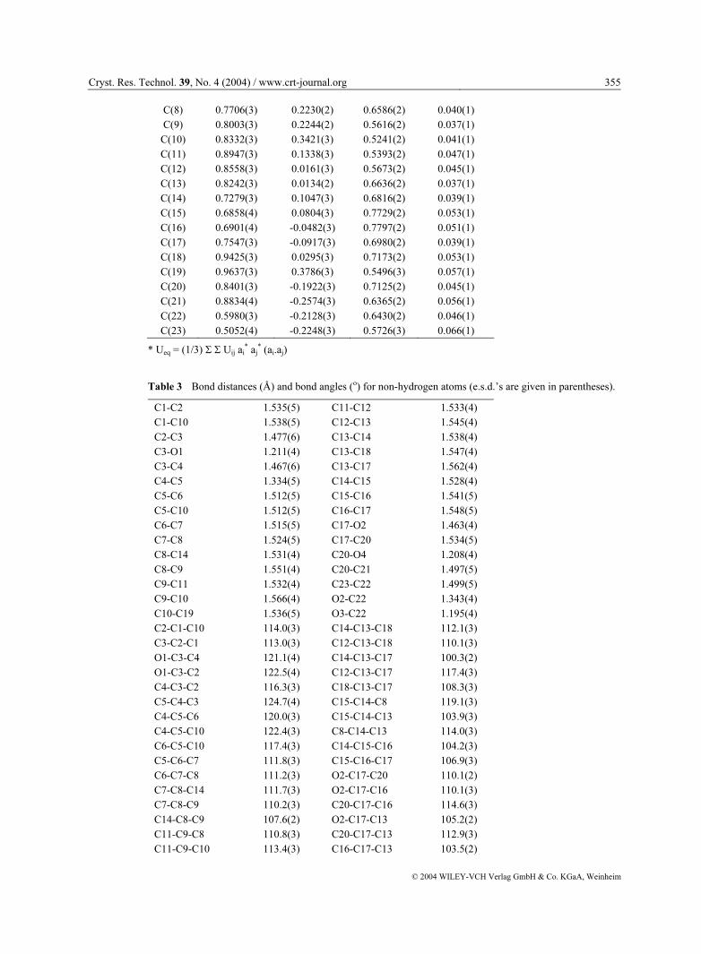
Table 3 Bond distances (Å) and bond angles (o) for non-hydrogen atoms (e.s.d.’s are given in parentheses).
C1-C2 1.535(5) C11-C12 1.533(4)
C1-C10 1.538(5) C12-C13 1.545(4)
C2-C3 1.477(6) C13-C14 1.538(4)
C3-O1 1.211(4) C13-C18 1.547(4)
C3-C4 1.467(6) C13-C17 1.562(4)
C4-C5 1.334(5) C14-C15 1.528(4)
C5-C6 1.512(5) C15-C16 1.541(5)
C5-C10 1.512(5) C16-C17 1.548(5)
C6-C7 1.515(5) C17-O2 1.463(4)
C7-C8 1.524(5) C17-C20 1.534(5)
C8-C14 1.531(4) C20-O4 1.208(4)
C8-C9 1.551(4) C20-C21 1.497(5)
C9-C11 1.532(4) C23-C22 1.499(5)
C9-C10 1.566(4) O2-C22 1.343(4)
C10-C19 1.536(5) O3-C22 1.195(4)
C2-C1-C10 114.0(3) C14-C13-C18 112.1(3)
C3-C2-C1 113.0(3) C12-C13-C18 110.1(3)
O1-C3-C4 121.1(4) C14-C13-C17 100.3(2)
O1-C3-C2 122.5(4) C12-C13-C17 117.4(3)
C4-C3-C2 116.3(3) C18-C13-C17 108.3(3)
C5-C4-C3 124.7(4) C15-C14-C8 119.1(3)
C4-C5-C6 120.0(3) C15-C14-C13 103.9(3)
C4-C5-C10 122.4(3) C8-C14-C13 114.0(3)
C6-C5-C10 117.4(3) C14-C15-C16 104.2(3)
C5-C6-C7 111.8(3) C15-C16-C17 106.9(3)
C6-C7-C8 111.2(3) O2-C17-C20 110.1(2)
C7-C8-C14 111.7(3) O2-C17-C16 110.1(3)
C7-C8-C9 110.2(3) C20-C17-C16 114.6(3)
C14-C8-C9 107.6(2) O2-C17-C13 105.2(2)
C11-C9-C8 110.8(3) C20-C17-C13 112.9(3)
C11-C9-C10 113.4(3) C16-C17-C13 103.5(2)
356 Rajnikant et al.: Crystal Structure of 17α-Acetoxyprogesterone, C23H32O4
© 2004 WILEY-VCH Verlag GmbH & Co. KGaA, Weinheim
C8-C9-C10 115.1(2) O4-C20-C21 121.0(3)
C5-C10-C19 107.7(3) O4-C20-C17 120.1(3)
C5-C10-C1 108.9(3) C21-C20-C17 118.7(3)
C19-C10-C1 110.4(3) C22-O2-C17 117.1(2)
C5-C10-C9 110.1(3) O3-C22-O2 124.9(3)
C19-C10-C9 111.6(3) O3-C22-C23 124.9(3)
C1-C10-C9 108.1(3) O2-C22-C23 110.2(3)
C9-C11-C12 113.5(3) C14-C13-C12 108.3(3)
C11-C12-C13 111.0(3)
Table 4 Some selected endocyclic torsion angles (°) for non-hydrogen atoms (e.s.d.’s are given in parentheses).
C1-C2-C3-C4 24.0(5) C4-C5-C10-C1 -21.4(5)
C2-C3-C4-C5 3.3(5) C6-C5-C10-C9 44.0(4)
C3-C4-C5-C6 171.9(3) C2-C1-C10-C5 48.0(5)
C3-C4-C5-C10 -4.3(6) C8-C9-C11-C12 55.3(4)
C10-C5-C6-C7 -51.2(5) C9-C11-C12-C13 -54.4(4)
C5-C6-C7-C8 56.3(4) C11-C12-C13-C14 53.7(4)
C6-C7-C8-C9 -57.0(4) C17-C13-C14-C15 46.0(3)
C14-C8-C9-C11 -55.5(3) C13-C14-C15-C16 -34.6(4)
C7-C8-C9-C10 52.2(4) C14-C15-C16-C17 9.3(4)
Table 5 Asymmetry parameters and Geometry of some intermolecular C-H…O hydrogen bonds (e.s.d.’s are given in parentheses).
Asymmetry Parameters
Ring A Ring C
∆Cs (C1) = 2.29 ∆Cs (C11) = 1.07
∆C2 (C9-C11) = 4.65
Ring B Ring D
∆C2 (C5-C10) = 5.61 ∆C2 (C13-C14) = 7.57
∆Cs (C10) = 0.73
Molecular interactions
C-H…O H…O(Å) C…O(Å) C-H…O(o)
C12-H12B...O2 2.42(1) 2.83(1) 104.0(1)
C14-H14...O2 2.48(1) 2.86(1) 102.9(1)
C16-H16A...O3 2.73(1) 3.14(1) 106.1(1)
C16-H16B...O4 2.41(1) 2.84(1) 105.9(1)
C21-H21C...O3 2.63(1) 3.12(1) 111.6(1)
C1-H1B...O4(i) 2.62(1) 3.44(1) 142.4(1)
C2-H2B...O3(i) 2.86(1) 3.58(1) 131.5(1)
C6-H6A...O3(ii) 2.99(1) 3.78(1) 139.7(1)
C7-H7B...O1(iii) 2.85(1) 3.56(1) 131.4(1)
C15-H15A...O3(iv) 2.72(1) 3.68(1) 169.6(1)
C19-H19B...O4(v) 2.62(1) 3.32(1) 129.5(1)
C18-H18C...O4(v) 2.73(1) 3.64(1) 160.3(1)
C23-H23B...O1(vi) 2.83(1) 3.67(1) 146.2(1)
C18-H18A...O1(vii)2.72(1) 3.59(1) 151.3(1)
Symmetry codes: i) -x + 1/2 + 1, -y, z – 1/2 ; ii) x, y + 1, z; iii) -x + ½ + 1, -y + 1, z + 1/2 iv) –x + 1, y + ½, -z + 1/2 + 1; v)
-x + 2, y + 1/2, -z +1/2 + 1 vi) x – 1/2, -y + 1/2, -z + 1; vii) x + 1/2, -y + 1/2, -z + 1
Cryst. Res. Technol. 39, No. 4 (2004) / www.crt-journal.org 357
© 2004 WILEY-VCH Verlag GmbH & Co. KGaA, Weinheim
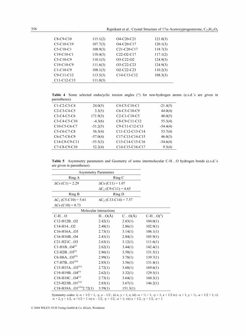
Fig. 1 General view of 17α-Acetoxyprogesterone indicating atomic numbering scheme.
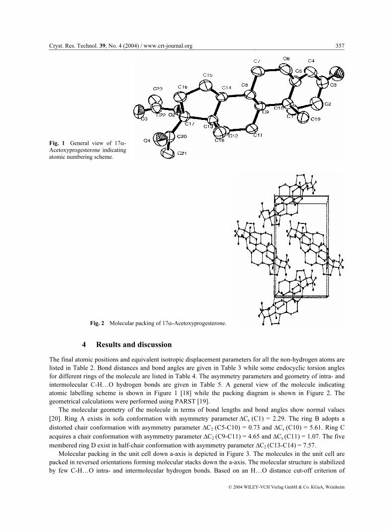
Fig. 2 Molecular packing of 17α-Acetoxyprogesterone.
4 Results and discussion
The final atomic positions and equivalent isotropic displacement parameters for all the non-hydrogen atoms are listed in Table 2. Bond distances and bond angles are given in Table 3 while some endocyclic torsion angles for different rings of the molecule are listed in Table 4. The asymmetry parameters and geometry of intra- and intermolecular C-H…O hydrogen bonds are given in Table 5. A general view of the molecule indicating atomic labelling scheme is shown in Figure 1 [18] while the packing diagram is shown in Figure 2. The geometrical calculations were performed using PARST [19].
The molecular geometry of the molecule in terms of bond lengths and bond angles show normal values
[20]. Ring A exists in sofa conformation with asymmetry parameter ∆Cs (C1) = 2.29. The ring B adopts a
distorted chair conformation with asymmetry parameter ∆C2 (C5-C10) = 0.73 and ∆Cs (C10) = 5.61. Ring C
acquires a chair conformation with asymmetry parameter ∆C2 (C9-C11) = 4.65 and ∆Cs (C11) = 1.07. The five
membered ring D exist in half-chair conformation with asymmetry parameter ∆C2 (C13-C14) = 7.57. Molecular packing in the unit cell down a-axis is depicted in Figure 3. The molecules in the unit cell are
packed in reversed orientations forming molecular stacks down the a-axis. The molecular structure is stabilized by few C-H…O intra- and intermolecular hydrogen bonds. Based on an H…O distance cut-off criterion of
358 Rajnikant et al.: Crystal Structure of 17α-Acetoxyprogesterone, C23H32O4
© 2004 WILEY-VCH Verlag GmbH & Co. KGaA, Weinheim
2.8Å [24], the oxygen atoms O1, O3 and O4 are a three-fold acceptors (C7-H7B…O1 2.85, C23-H23B…O1 2.83, C18-H18A…O1 2.72; C2-H2B…O3 2.86, C6-H6A…O3 2.99, C15-H15A…O3 2.72; C1-H1B…O4 2.62, C19-H19B…O4 2.62 and C18-H18C…O4 2.73 Å). With the increasing awareness of the role of weak hydrogen bonds in ligand-receptor complexes [21], and in view of the opinion that the approach geometry of hydrogen-bonding donor and acceptor groups in these complexes can be derived from the ligand crystal structures [22], the crystal structures of steroids stabilized by C-H…O hydrogen bonding are a source of valuable information.
Acknowledgements The corresponding author (Rajnikant) is thankful to Dr. B.D. Gupta, Regional Research Laboratory (CSIR), Jammu for providing the sample and Dr. Babu Varghese, RSIC, IIT Chennai for collecting the data. The funding as recieved from Council of Scientific and Industrial Research and University Grants Commission, Govt. of India under research project no. 03(0927)-01/EMR-II and F.10-82/ 2001 (SR-I), respectively is thankfully acknowledged.
References
[1] V. K. Gupta, Rajnikant, K. N. Goswami, and K. K. Bhutani, Cryst Res. Technol. 29, 77 (1994). [2] V. K. Gupta, Rajnikant, K. N. Goswami, S. K. Mazumdar, and K. K. Bhutani, Acta Cryst. C50, 789 (1994). [3] V. K. Gupta, K. N. Goswami, K. K. Bhutani, and R. M. Vaid, Molecular Materials 4, 303 (1994). [4] A. Singh, V. K. Gupta, Rajnikant, and K. N. Goswami, Cryst. Res. Technol. 29, 837 (1994). [5] A. Singh, V. K. Gupta, Rajnikant, K. N. Goswami, and K. K. Bhutani, Molecular Materials 4, 837 (1994). [6] A. Singh, V. K. Gupta, Rajnikant, K. N. Goswami, B. D. Gupta, and S. K. Banerjee, Molecular Materials 6, 53
(1996). [7] Rajnikant, V. K. Gupta, K. N. Goswami, D. K. Magotra, and K. K. Bhutani, Molecular Materials 12, 101 (2000). [8] Rajnikant, V. K. Gupta, J. Firoz, Shafiullah, and R. Gupta, Crystallography Reports 5, 857 (2000). [9] Rajnikant, V. K. Gupta, J. Firoz, Shafiullah, and R. Gupta, Cryst. Res. Technol. 36, 215 (2001).
[10] Rajnikant, V. K. Gupta, J. Firoz, Shafiullah, and R. Gupta, Cryst. Res. Technol. 36, 471 (2001). [11] Rajnikant, V. K. Gupta, E. H. Khan, S. Shafi, S. Hashmi, Shafiullah, B. Varghese, and Dinesh, Crystallography
Reports 46, 963 (2001). [12] Rajnikant, V. K. Gupta, E. H. Khan, S. Shafi, S. Hashmi, Shafiullah, B. Varghese, and Dinesh, Cryst. Res. Technol.
36, 1281 (2001). [13] Rajnikant, V. K. Gupta, J. Firoz, Shafiullah, and R. Gupta, Crystallography Reports 3 (2001) Mss. No. 2486 [In
Press]. [14] Rajnikant, V. K. Gupta, S. Hashmi, Shafiullah, B. Varghese, and Dinesh, Crystallography Reports, (2001) Mss. No.
2600 [In Press]. [15] Rajnikant, V. K. Gupta, E. H. Khan, S. Shafi, Shafiullah, and Dinesh, Bull. Pure and Appl. Sc., (2001) Mss. No.
2266/Phy [In Press]. [16] G. M. Sheldrick, SHELXS86: Program for the solution of Crystal Structures. University of Göttingen, Göttingen,
Germany (1986). [17] G. M. Sheldrick, SHELLS93: Program for the Refinement of Crystal Structures. University of Göttingen,
Göttingen, Germany (1993). [18] L. J. Farrugia, J. Appl. Cryst. 30, 565 (1997). [19] M. Nardelli, J. Appl. Cryst. 28, 659 (1995). [20] L. S. Bartell, J. Amer. Chem. Soc. 81, 3497 (1959). [21] M. C. Wahl and M. Sundaralingam, Trends Biochem. Sci. 22, 97 (1997). [22] G. Klebe, J. Mol. Biol. 237, 212 (1994). [23] ENRAF-NONIUS, CAD-4 Software, Version 5.0, Enraf-Nonius, Delft, The Netherlands, (1989). [24] G. R. Desiraju and T. Steiner, “The weak hydrogen bond in Structural Chemistry and Biology”, IUCr, Oxford
Press, (1999) 48.