Embed Size (px)
Citation preview
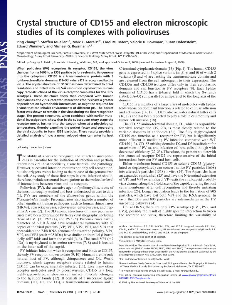
Crystal structure of CD155 and electron microscopicstudies of its complexes with poliovirusesPing Zhanga,1, Steffen Muellerb,1, Marc C. Moraisa,2, Carol M. Batora, Valorie D. Bowmana, Susan Hafensteina,Eckard Wimmerb, and Michael G. Rossmanna,3
aDepartment of Biological Sciences, Purdue University, 915 West State Street, West Lafayette, IN 47907-2054; and bDepartment of Molecular Genetics andMicrobiology, Stony Brook University, 280C Life Sciences Building, Stony Brook, NY 11794-5222
Edited by Gregory A. Petsko, Brandeis University, Waltham, MA, and approved October 8, 2008 (received for review August 8, 2008)
When poliovirus (PV) recognizes its receptor, CD155, the viruschanges from a 160S to a 135S particle before releasing its genomeinto the cytoplasm. CD155 is a transmembrane protein with 3Ig-like extracellular domains, D1–D3, where D1 is recognized by thevirus. The crystal structure of D1D2 has been determined to 3.5-Åresolution and fitted into �8.5-Å resolution cryoelectron micros-copy reconstructions of the virus–receptor complexes for the 3 PVserotypes. These structures show that, compared with humanrhinoviruses, the virus–receptor interactions for PVs have a greaterdependence on hydrophobic interactions, as might be required fora virus that can inhabit environments of different pH. The pocketfactor was shown to remain in the virus during the first recognitionstage. The present structures, when combined with earlier muta-tional investigations, show that in the subsequent entry stage thereceptor moves further into the canyon when at a physiologicaltemperature, thereby expelling the pocket factor and separatingthe viral subunits to form 135S particles. These results provide adetailed analysis of how a nonenveloped virus can enter its hostcell.
cell entry � receptor � virus
The ability of a virus to recognize and attach to susceptiblecells is essential for the initiation of infection and partially
determines viral host specificity, tissue tropism, and pathology.However, successful infection requires not only cell recognition,but also triggers events leading to the release of the genome intothe cell. Any study of these first steps in viral infection should,therefore, include structural investigations at the molecular levelof interactions between virus and its receptor.
Poliovirus (PV), the causative agent of poliomyelitis, is one ofthe most thoroughly studied and best-understood viruses to date(1). PVs are members of the Enterovirus genus within thePicornaviridae family. Picornaviruses also include a number ofother significant human pathogens, such as human rhinoviruses(HRVs), coxsackieviruses, echoviruses, enteroviruses, and hep-atitis A virus (2). The 3D atomic structures of many picornavi-ruses have been determined by X-ray crystallography, includingthose of PV1 (3), PV2 (4), and PV3 (5). Picornaviruses have adiameter of �310 Å and have icosahedral symmetry with 60copies of the viral proteins (VP) VP1, VP2, VP3, and VP4 thatencapsidate the 7-kb RNA genome of plus-strand polarity. VP1,VP2, and VP3 (each �35 kDa) have similar antiparallel �-barrel‘‘jelly-roll’’ folds and form the capsid (3, 6). The small VP4 (�7kDa) is myristylated at its amino terminus (7, 8) and is locatedon the inner wall of the capsid.
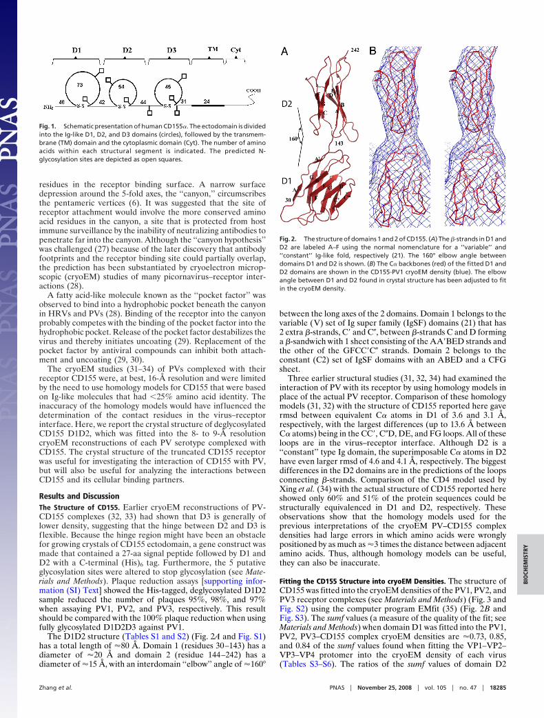
PV initiates infection when it recognizes and binds to CD155,the only PV receptor known to date (9, 10). Humans are the onlynatural host of PV, although chimpanzees and Old Worldmonkeys, which express receptors closely related to humanCD155, can be experimentally infected (11). Like many otherreceptor molecules used by picornaviruses, CD155 is a long,highly glycosylated, single-span cell surface molecule belongingto the Ig super family (12). It consists of 3 successive Ig-likedomains (D1, D2, and D3), a transmembrane domain and a
C-terminal cytoplasmic domain (13) (Fig. 1). The human CD155gene is expressed in 4 splice variants (�, �, �, and �) of which 2variants (� and �) are lacking the transmembrane domain andare released from the cell subsequent to their expression. TheCD155� and CD155� isotypes differ only in their cytoplasmicdomains and can function as PV receptors (9). Each Ig-likedomain of CD155 has a �-barrel fold in which the �-strands(labeled A–G) run parallel or antiparallel to the long axis of thedomain.
CD155 is a member of a large class of molecules with Ig-likefolds whose predominant function is related to cellular adhesionand activation (14, 15). CD155 also activates natural killer cells(16, 17) and has been reported to play a role in cell motility andtumor cell invasion (18).
The CD155 amino-terminal domain, D1, which is responsiblefor viral recognition (19, 20), is most closely related to thevariable domains in antibodies (21). The fully deglycosylatedCD155 can function as a receptor for PV, but is significantlymore efficient in mediating PV infection compared with WTCD155 (13). CD155 missing domains D2 and D3 is sufficient forattachment of PV to, and infection of, host cells although withdecreased efficiency (22, 23). Therefore, the present studies withthe truncated receptor D1D2 are representative of the initialinteractions between PV and host cells.
Either membrane-bound CD155 or soluble CD155 (glycosy-lated or deglycosylated) can convert native PV particles (160S)into altered A-particles (135S) in vitro (24). The A particles havean expanded capsid shell (25) and have the N-terminal extensionof VP1 and VP4 externalized. VP4 exits the viral capsid probablyvia a channel along the 5-fold axes of the capsid, entering the hostcell’s membrane after cell recognition and thereby initiatinginfection (26). Longer incubation leads to the formation of 80Sparticles, which have lost both VP4 and the RNA genome. Invivo, the 135S and 80S particles are intermediates in the PVuncoating pathway (24).
Unlike HRVs, there are only 3 PV serotypes (PV1, PV2, andPV3), possibly the result of highly specific interaction betweenthe receptor and virus, therefore limiting the variability of
Author contributions: P.Z., S.M., M.C.M., E.W., and M.G.R. designed research; P.Z., S.M.,C.M.B., and V.D.B. performed research; S.H. contributed new reagents/analytic tools; P.Z.and M.G.R. analyzed data; and P.Z. and M.G.R. wrote the paper.
The authors declare no conflict of interest.
This article is a PNAS Direct Submission.
Data deposition: The atomic coordinates have been deposited in the Protein Data Bank,www.pdb.org (PDB ID codes 3EOW, 3EPC, 3EPF, and 3EPD). The cryoreconstruction mapsreported in this paper have been deposited in the Array Express database, www.ebi.ac.uk/arrayexpress (accession nos. 6390, 6386, and 6387).
1P.Z. and S.M contributed equally to this work.
2Present address: Sealy Center for Structural Biology and Molecular Biophysics, Universityof Texas Medical Branch, 301 University Boulevard, Galveston, TX 77555-0647.
3To whom correspondence should be addressed. E-mail: [email protected].
This article contains supporting information online at www.pnas.org/cgi/content/full/0807848105/DCSupplemental.
© 2008 by The National Academy of Sciences of the USA
18284–18289 � PNAS � November 25, 2008 � vol. 105 � no. 47 www.pnas.org�cgi�doi�10.1073�pnas.0807848105
residues in the receptor binding surface. A narrow surfacedepression around the 5-fold axes, the ‘‘canyon,’’ circumscribesthe pentameric vertices (6). It was suggested that the site ofreceptor attachment would involve the more conserved aminoacid residues in the canyon, a site that is protected from hostimmune surveillance by the inability of neutralizing antibodies topenetrate far into the canyon. Although the ‘‘canyon hypothesis’’was challenged (27) because of the later discovery that antibodyfootprints and the receptor binding site could partially overlap,the prediction has been substantiated by cryoelectron microp-scopic (cryoEM) studies of many picornavirus–receptor inter-actions (28).
A fatty acid-like molecule known as the ‘‘pocket factor’’ wasobserved to bind into a hydrophobic pocket beneath the canyonin HRVs and PVs (28). Binding of the receptor into the canyonprobably competes with the binding of the pocket factor into thehydrophobic pocket. Release of the pocket factor destabilizes thevirus and thereby initiates uncoating (29). Replacement of thepocket factor by antiviral compounds can inhibit both attach-ment and uncoating (29, 30).
The cryoEM studies (31–34) of PVs complexed with theirreceptor CD155 were, at best, 16-Å resolution and were limitedby the need to use homology models for CD155 that were basedon Ig-like molecules that had �25% amino acid identity. Theinaccuracy of the homology models would have influenced thedetermination of the contact residues in the virus–receptorinterface. Here, we report the crystal structure of deglycosylatedCD155 D1D2, which was fitted into the 8- to 9-Å resolutioncryoEM reconstructions of each PV serotype complexed withCD155. The crystal structure of the truncated CD155 receptorwas useful for investigating the interaction of CD155 with PV,but will also be useful for analyzing the interactions betweenCD155 and its cellular binding partners.
Results and DiscussionThe Structure of CD155. Earlier cryoEM reconstructions of PV-CD155 complexes (32, 33) had shown that D3 is generally oflower density, suggesting that the hinge between D2 and D3 isflexible. Because the hinge region might have been an obstaclefor growing crystals of CD155 ectodomain, a gene construct wasmade that contained a 27-aa signal peptide followed by D1 andD2 with a C-terminal (His)6 tag. Furthermore, the 5 putativeglycosylation sites were altered to stop glycosylation (see Mate-rials and Methods). Plaque reduction assays [supporting infor-mation (SI) Text] showed the His-tagged, deglycosylated D1D2sample reduced the number of plaques 95%, 98%, and 97%when assaying PV1, PV2, and PV3, respectively. This resultshould be compared with the 100% plaque reduction when usingfully glycosylated D1D2D3 against PV1.
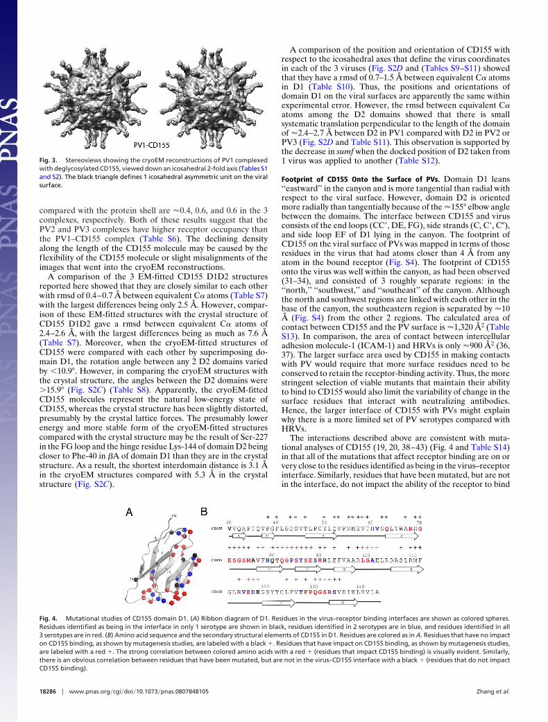
The D1D2 structure (Tables S1 and S2) (Fig. 2A and Fig. S1)has a total length of �80 Å. Domain 1 (residues 30–143) has adiameter of �20 Å and domain 2 (residue 144–242) has adiameter of �15 Å, with an interdomain ‘‘elbow’’ angle of �160°
between the long axes of the 2 domains. Domain 1 belongs to thevariable (V) set of Ig super family (IgSF) domains (21) that has2 extra �-strands, C� and C�, between �-strands C and D forminga �-sandwich with 1 sheet consisting of the AA�BED strands andthe other of the GFCC�C� strands. Domain 2 belongs to theconstant (C2) set of IgSF domains with an ABED and a CFGsheet.
Three earlier structural studies (31, 32, 34) had examined theinteraction of PV with its receptor by using homology models inplace of the actual PV receptor. Comparison of these homologymodels (31, 32) with the structure of CD155 reported here gavermsd between equivalent C� atoms in D1 of 3.6 and 3.1 Å,respectively, with the largest differences (up to 13.6 Å betweenC� atoms) being in the CC�, C�D, DE, and FG loops. All of theseloops are in the virus–receptor interface. Although D2 is a‘‘constant’’ type Ig domain, the superimposable C� atoms in D2have even larger rmsd of 4.6 and 4.1 Å, respectively. The biggestdifferences in the D2 domains are in the predictions of the loopsconnecting �-strands. Comparison of the CD4 model used byXing et al. (34) with the actual structure of CD155 reported hereshowed only 60% and 51% of the protein sequences could bestructurally equivalenced in D1 and D2, respectively. Theseobservations show that the homology models used for theprevious interpretations of the cryoEM PV–CD155 complexdensities had large errors in which amino acids were wronglypositioned by as much as �3 times the distance between adjacentamino acids. Thus, although homology models can be useful,they can also be inaccurate.
Fitting the CD155 Structure into cryoEM Densities. The structure ofCD155 was fitted into the cryoEM densities of the PV1, PV2, andPV3 receptor complexes (see Materials and Methods) (Fig. 3 andFig. S2) using the computer program EMfit (35) (Fig. 2B andFig. S3). The sumf values (a measure of the quality of the fit; seeMaterials and Methods) when domain D1 was fitted into the PV1,PV2, PV3–CD155 complex cryoEM densities are �0.73, 0.85,and 0.84 of the sumf values found when fitting the VP1–VP2–VP3–VP4 protomer into the cryoEM density of each virus(Tables S3–S6). The ratios of the sumf values of domain D2
Fig. 1. Schematic presentation of human CD155�. The ectodomain is dividedinto the Ig-like D1, D2, and D3 domains (circles), followed by the transmem-brane (TM) domain and the cytoplasmic domain (Cyt). The number of aminoacids within each structural segment is indicated. The predicted N-glycosylation sites are depicted as open squares.
Fig. 2. The structure of domains 1 and 2 of CD155. (A) The �-strands in D1 andD2 are labeled A–F using the normal nomenclature for a ‘‘variable’’ and‘‘constant’’ Ig-like fold, respectively (21). The 160° elbow angle betweendomains D1 and D2 is shown. (B) The C� backbones (red) of the fitted D1 andD2 domains are shown in the CD155-PV1 cryoEM density (blue). The elbowangle between D1 and D2 found in crystal structure has been adjusted to fitin the cryoEM density.
Zhang et al. PNAS � November 25, 2008 � vol. 105 � no. 47 � 18285
BIO
CHEM
ISTR
Y
compared with the protein shell are �0.4, 0.6, and 0.6 in the 3complexes, respectively. Both of these results suggest that thePV2 and PV3 complexes have higher receptor occupancy thanthe PV1–CD155 complex (Table S6). The declining densityalong the length of the CD155 molecule may be caused by theflexibility of the CD155 molecule or slight misalignments of theimages that went into the cryoEM reconstructions.
A comparison of the 3 EM-fitted CD155 D1D2 structuresreported here showed that they are closely similar to each otherwith rmsd of 0.4–0.7 Å between equivalent C� atoms (Table S7)with the largest differences being only 2.5 Å. However, compar-ison of these EM-fitted structures with the crystal structure ofCD155 D1D2 gave a rmsd between equivalent C� atoms of2.4–2.6 Å, with the largest differences being as much as 7.6 Å(Table S7). Moreover, when the cryoEM-fitted structures ofCD155 were compared with each other by superimposing do-main D1, the rotation angle between any 2 D2 domains variedby �10.9°. However, in comparing the cryoEM structures withthe crystal structure, the angles between the D2 domains were�15.9° (Fig. S2C) (Table S8). Apparently, the cryoEM-fittedCD155 molecules represent the natural low-energy state ofCD155, whereas the crystal structure has been slightly distorted,presumably by the crystal lattice forces. The presumably lowerenergy and more stable form of the cryoEM-fitted structurescompared with the crystal structure may be the result of Ser-227in the FG loop and the hinge residue Lys-144 of domain D2 beingcloser to Phe-40 in �A of domain D1 than they are in the crystalstructure. As a result, the shortest interdomain distance is 3.1 Åin the cryoEM structures compared with 5.3 Å in the crystalstructure (Fig. S2C).
A comparison of the position and orientation of CD155 withrespect to the icosahedral axes that define the virus coordinatesin each of the 3 viruses (Fig. S2D and (Tables S9–S11) showedthat they have a rmsd of 0.7–1.5 Å between equivalent C� atomsin D1 (Table S10). Thus, the positions and orientations ofdomain D1 on the viral surfaces are apparently the same withinexperimental error. However, the rmsd between equivalent C�atoms among the D2 domains showed that there is smallsystematic translation perpendicular to the length of the domainof �2.4–2.7 Å between D2 in PV1 compared with D2 in PV2 orPV3 (Fig. S2D and Table S11). This observation is supported bythe decrease in sumf when the docked position of D2 taken from1 virus was applied to another (Table S12).
Footprint of CD155 Onto the Surface of PVs. Domain D1 leans‘‘eastward’’ in the canyon and is more tangential than radial withrespect to the viral surface. However, domain D2 is orientedmore radially than tangentially because of the �155° elbow anglebetween the domains. The interface between CD155 and virusconsists of the end loops (CC�, DE, FG), side strands (C, C�, C�),and side loop EF of D1 lying in the canyon. The footprint ofCD155 on the viral surface of PVs was mapped in terms of thoseresidues in the virus that had atoms closer than 4 Å from anyatom in the bound receptor (Fig. S4). The footprint of CD155onto the virus was well within the canyon, as had been observed(31–34), and consisted of 3 roughly separate regions: in the‘‘north,’’ ‘‘southwest,’’ and ‘‘southeast’’ of the canyon. Althoughthe north and southwest regions are linked with each other in thebase of the canyon, the southeastern region is separated by �10Å (Fig. S4) from the other 2 regions. The calculated area ofcontact between CD155 and the PV surface is �1,320 Å2 (TableS13). In comparison, the area of contact between intercellularadhesion molecule-1 (ICAM-1) and HRVs is only �900 Å2 (36,37). The larger surface area used by CD155 in making contactswith PV would require that more surface residues need to beconserved to retain the receptor-binding activity. Thus, the morestringent selection of viable mutants that maintain their abilityto bind to CD155 would also limit the variability of change in thesurface residues that interact with neutralizing antibodies.Hence, the larger interface of CD155 with PVs might explainwhy there is a more limited set of PV serotypes compared withHRVs.
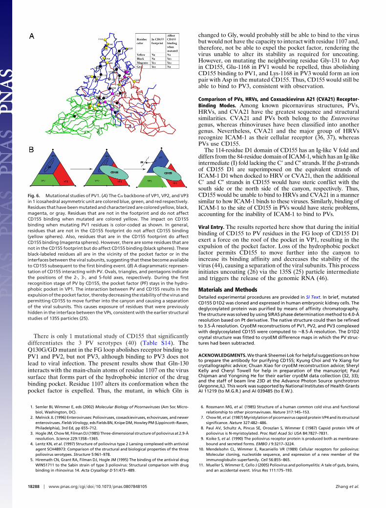
The interactions described above are consistent with muta-tional analyses of CD155 (19, 20, 38–43) (Fig. 4 and Table S14)in that all of the mutations that affect receptor binding are on orvery close to the residues identified as being in the virus–receptorinterface. Similarly, residues that have been mutated, but are notin the interface, do not impact the ability of the receptor to bind
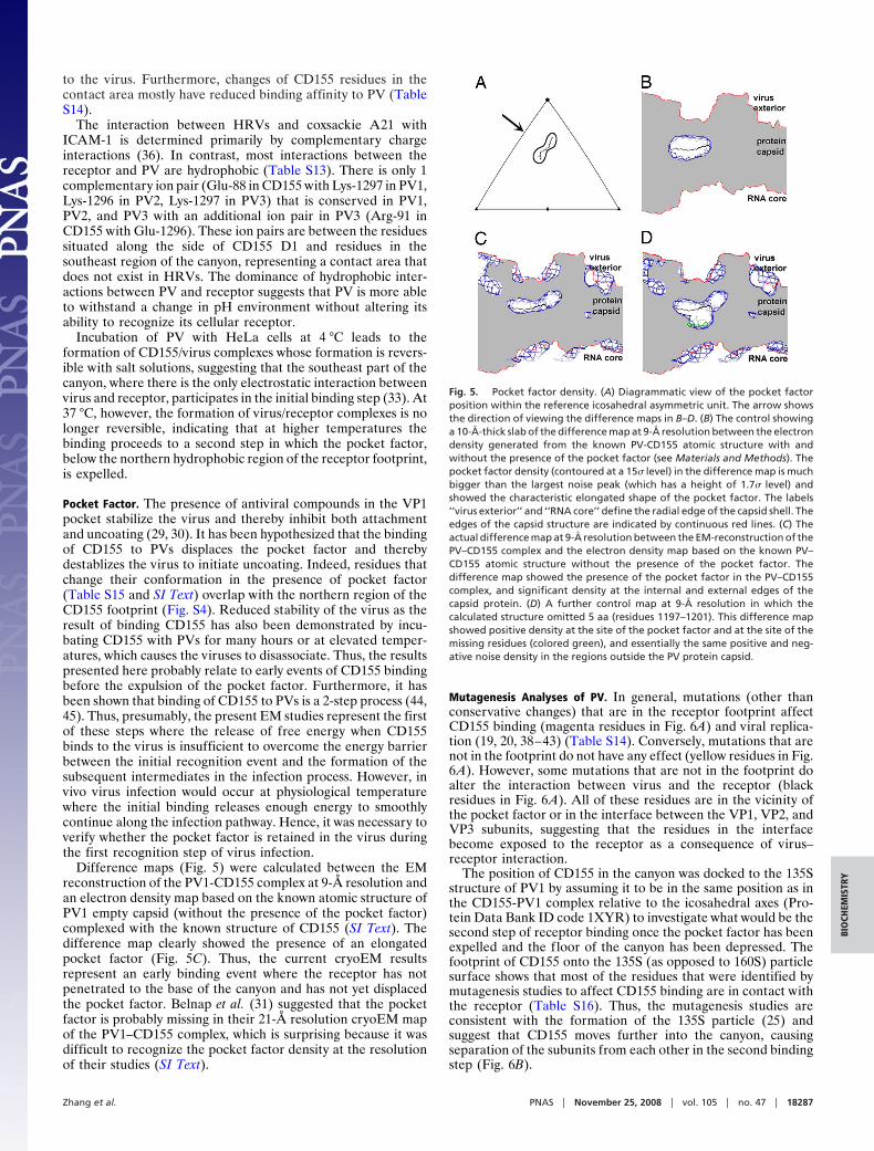
Fig. 3. Stereoviews showing the cryoEM reconstructions of PV1 complexedwith deglycosylated CD155, viewed down an icosahedral 2-fold axis (Tables S1and S2). The black triangle defines 1 icosahedral asymmetric unit on the viralsurface.
Fig. 4. Mutational studies of CD155 domain D1. (A) Ribbon diagram of D1. Residues in the virus–receptor binding interfaces are shown as colored spheres.Residues identified as being in the interface in only 1 serotype are shown in black, residues identified in 2 serotypes are in blue, and residues identified in all3 serotypes are in red. (B) Amino acid sequence and the secondary structural elements of CD155 in D1. Residues are colored as in A. Residues that have no impacton CD155 binding, as shown by mutagenesis studies, are labeled with a black �. Residues that have impact on CD155 binding, as shown by mutagenesis studies,are labeled with a red �. The strong correlation between colored amino acids with a red � (residues that impact CD155 binding) is visually evident. Similarly,there is an obvious correlation between residues that have been mutated, but are not in the virus–CD155 interface with a black � (residues that do not impactCD155 binding).
18286 � www.pnas.org�cgi�doi�10.1073�pnas.0807848105 Zhang et al.
to the virus. Furthermore, changes of CD155 residues in thecontact area mostly have reduced binding affinity to PV (TableS14).
The interaction between HRVs and coxsackie A21 withICAM-1 is determined primarily by complementary chargeinteractions (36). In contrast, most interactions between thereceptor and PV are hydrophobic (Table S13). There is only 1complementary ion pair (Glu-88 in CD155 with Lys-1297 in PV1,Lys-1296 in PV2, Lys-1297 in PV3) that is conserved in PV1,PV2, and PV3 with an additional ion pair in PV3 (Arg-91 inCD155 with Glu-1296). These ion pairs are between the residuessituated along the side of CD155 D1 and residues in thesoutheast region of the canyon, representing a contact area thatdoes not exist in HRVs. The dominance of hydrophobic inter-actions between PV and receptor suggests that PV is more ableto withstand a change in pH environment without altering itsability to recognize its cellular receptor.
Incubation of PV with HeLa cells at 4 °C leads to theformation of CD155/virus complexes whose formation is revers-ible with salt solutions, suggesting that the southeast part of thecanyon, where there is the only electrostatic interaction betweenvirus and receptor, participates in the initial binding step (33). At37 °C, however, the formation of virus/receptor complexes is nolonger reversible, indicating that at higher temperatures thebinding proceeds to a second step in which the pocket factor,below the northern hydrophobic region of the receptor footprint,is expelled.
Pocket Factor. The presence of antiviral compounds in the VP1pocket stabilize the virus and thereby inhibit both attachmentand uncoating (29, 30). It has been hypothesized that the bindingof CD155 to PVs displaces the pocket factor and therebydestablizes the virus to initiate uncoating. Indeed, residues thatchange their conformation in the presence of pocket factor(Table S15 and SI Text) overlap with the northern region of theCD155 footprint (Fig. S4). Reduced stability of the virus as theresult of binding CD155 has also been demonstrated by incu-bating CD155 with PVs for many hours or at elevated temper-atures, which causes the viruses to disassociate. Thus, the resultspresented here probably relate to early events of CD155 bindingbefore the expulsion of the pocket factor. Furthermore, it hasbeen shown that binding of CD155 to PVs is a 2-step process (44,45). Thus, presumably, the present EM studies represent the firstof these steps where the release of free energy when CD155binds to the virus is insufficient to overcome the energy barrierbetween the initial recognition event and the formation of thesubsequent intermediates in the infection process. However, invivo virus infection would occur at physiological temperaturewhere the initial binding releases enough energy to smoothlycontinue along the infection pathway. Hence, it was necessary toverify whether the pocket factor is retained in the virus duringthe first recognition step of virus infection.
Difference maps (Fig. 5) were calculated between the EMreconstruction of the PV1-CD155 complex at 9-Å resolution andan electron density map based on the known atomic structure ofPV1 empty capsid (without the presence of the pocket factor)complexed with the known structure of CD155 (SI Text). Thedifference map clearly showed the presence of an elongatedpocket factor (Fig. 5C). Thus, the current cryoEM resultsrepresent an early binding event where the receptor has notpenetrated to the base of the canyon and has not yet displacedthe pocket factor. Belnap et al. (31) suggested that the pocketfactor is probably missing in their 21-Å resolution cryoEM mapof the PV1–CD155 complex, which is surprising because it wasdifficult to recognize the pocket factor density at the resolutionof their studies (SI Text).
Mutagenesis Analyses of PV. In general, mutations (other thanconservative changes) that are in the receptor footprint affectCD155 binding (magenta residues in Fig. 6A) and viral replica-tion (19, 20, 38–43) (Table S14). Conversely, mutations that arenot in the footprint do not have any effect (yellow residues in Fig.6A). However, some mutations that are not in the footprint doalter the interaction between virus and the receptor (blackresidues in Fig. 6A). All of these residues are in the vicinity ofthe pocket factor or in the interface between the VP1, VP2, andVP3 subunits, suggesting that the residues in the interfacebecome exposed to the receptor as a consequence of virus–receptor interaction.
The position of CD155 in the canyon was docked to the 135Sstructure of PV1 by assuming it to be in the same position as inthe CD155-PV1 complex relative to the icosahedral axes (Pro-tein Data Bank ID code 1XYR) to investigate what would be thesecond step of receptor binding once the pocket factor has beenexpelled and the floor of the canyon has been depressed. Thefootprint of CD155 onto the 135S (as opposed to 160S) particlesurface shows that most of the residues that were identified bymutagenesis studies to affect CD155 binding are in contact withthe receptor (Table S16). Thus, the mutagenesis studies areconsistent with the formation of the 135S particle (25) andsuggest that CD155 moves further into the canyon, causingseparation of the subunits from each other in the second bindingstep (Fig. 6B).
Fig. 5. Pocket factor density. (A) Diagrammatic view of the pocket factorposition within the reference icosahedral asymmetric unit. The arrow showsthe direction of viewing the difference maps in B–D. (B) The control showinga 10-Å-thick slab of the difference map at 9-Å resolution between the electrondensity generated from the known PV-CD155 atomic structure with andwithout the presence of the pocket factor (see Materials and Methods). Thepocket factor density (contoured at a 15� level) in the difference map is muchbigger than the largest noise peak (which has a height of 1.7� level) andshowed the characteristic elongated shape of the pocket factor. The labels‘‘virus exterior’’ and ‘‘RNA core’’ define the radial edge of the capsid shell. Theedges of the capsid structure are indicated by continuous red lines. (C) Theactual difference map at 9-Å resolution between the EM-reconstruction of thePV–CD155 complex and the electron density map based on the known PV–CD155 atomic structure without the presence of the pocket factor. Thedifference map showed the presence of the pocket factor in the PV–CD155complex, and significant density at the internal and external edges of thecapsid protein. (D) A further control map at 9-Å resolution in which thecalculated structure omitted 5 aa (residues 1197–1201). This difference mapshowed positive density at the site of the pocket factor and at the site of themissing residues (colored green), and essentially the same positive and neg-ative noise density in the regions outside the PV protein capsid.
Zhang et al. PNAS � November 25, 2008 � vol. 105 � no. 47 � 18287
BIO
CHEM
ISTR
Y
There is only 1 mutational study of CD155 that significantlydifferentiates the 3 PV serotypes (40) (Table S14). TheQ130G/GD mutant in the FG loop abolishes receptor binding toPV1 and PV2, but not PV3, although binding to PV3 does notlead to viral infection. The present results show that Gln-130interacts with the main-chain atoms of residue 1107 on the virussurface that forms part of the hydrophobic interior of the drugbinding pocket. Residue 1107 alters its conformation when thepocket factor is expelled. Thus, the mutant, in which Gln is
changed to Gly, would probably still be able to bind to the virusbut would not have the capacity to interact with residue 1107 and,therefore, not be able to expel the pocket factor, rendering thevirus unable to alter its stability as required for uncoating.However, on mutating the neighboring residue Gly-131 to Aspin CD155, Glu-1168 in PV1 would be repelled, thus abolishingCD155 binding to PV1, and Lys-1168 in PV3 would form an ionpair with Asp in the mutated CD155. Thus, CD155 would still beable to bind to PV3, consistent with observation.
Comparison of PVs, HRVs, and Coxsackievirus A21 (CVA21) Receptor-Binding Modes. Among known picornavirus structures, PVs,HRVs, and CVA21 have the greatest sequence and structuralsimilarities. CVA21 and PVs both belong to the Enterovirusgenus, whereas rhinoviruses have been classified into anothergenus. Nevertheless, CVA21 and the major group of HRVsrecognize ICAM-1 as their cellular receptor (36, 37), whereasPVs use CD155.
The 114-residue D1 domain of CD155 has an Ig-like V fold anddiffers from the 84-residue domain of ICAM-1, which has an Ig-likeintermediate (I) fold lacking the C� and C� strands. If the �-strandsof CD155 D1 are superimposed on the equivalent strands ofICAM-1 D1 when docked to HRV or CVA21, then the additionalC� and C� strands in CD155 would have steric conflict with thesouth side or the north side of the canyon, respectively. Thus,CD155 would be unable to bind to HRVs and CVA21 in a mannersimilar to how ICAM-1 binds to these viruses. Similarly, binding ofICAM-1 to the site of CD155 in PVs would have steric problems,accounting for the inability of ICAM-1 to bind to PVs.
Viral Entry. The results reported here show that during the initialbinding of CD155 to PV residues in the FG loop of CD155 D1exert a force on the roof of the pocket in VP1, resulting in theexpulsion of the pocket factor. Loss of the hydrophobic pocketfactor permits CD155 to move further into the canyon toincrease its binding affinity and decreases the stability of thevirus (44), causing a separation of the viral subunits. This processinitiates uncoating (26) via the 135S (25) particle intermediateand triggers the release of the genomic RNA (46).
Materials and MethodsDetailed experimental procedures are provided in SI Text. In brief, mutatedCD155 D1D2 was cloned and expressed in human embryonic kidney cells. Thedeglycosylated protein was purified by means of affinity chromatography.The structure was solved by using SIRAS phase determination method to 4.0-Åresolution based on Pt derivative. The native structure could then be refinedto 3.5-Å resolution. CryoEM reconstructions of PV1, PV2, and PV3 complexedwith deglycosylated CD155 were computed to �8.5-Å resolution. The D1D2crystal structure was fitted to cryoEM difference maps in which the PV struc-tures had been subtracted.
ACKNOWLEDGMENTS. We thank Sheemei Lok for helpful suggestions on howto prepare the antibody for purifying CD155; Kyung Choi and Ye Xiang forcrystallographic advice; Chuan Xiao for cryoEM reconstruction advice; SherylKelly and Cheryl Towell for help in preparation of the manuscript; PaulChipman and Yongning He for their earlier cryoEM data collection (32, 33);and the staff of beam line 23D at the Advance Photon Source synchrotron(Argonne,IL). This work was supported by National Institutes of Health GrantsAI 11219 (to M.G.R.) and AI 039485 (to E.W.).
1. Semler BL Wimmer E, eds (2002) Molecular Biology of Picornaviruses (Am Soc Micro-biol, Washington, DC).
2. Melnick JL (1996) Enteroviruses: Polioviruses, coxsackieviruses, echoviruses, and newerenteroviruses. Fields Virology, eds Fields BN, Knipe DM, Howley PM (Lippincott–Raven,Philadelphia), 3rd Ed, pp 655–712.
3. Hogle JM, Chow M, Filman DJ (1985) Three-dimensional structure of poliovirus at 2.9-Åresolution. Science 229:1358–1365.
4. Lentz KN, et al. (1997) Structure of poliovirus type 2 Lansing complexed with antiviralagent SCH48973: Comparison of the structural and biological properties of the threepoliovirus serotypes. Structure 5:961–978.
5. Hiremath CN, Grant RA, Filman DJ, Hogle JM (1995) The binding of the antiviral drugWIN51711 to the Sabin strain of type 3 poliovirus: Structural comparison with drugbinding in rhinovirus 14. Acta Crysallogr D 51:473–489.
6. Rossmann MG, et al. (1985) Structure of a human common cold virus and functionalrelationship to other picornaviruses. Nature 317:145–153.
7. Chow M, et al. (1987) Myristylation of picornavirus capsid protein VP4 and its structuralsignificance. Nature 327:482–486.
8. Paul AV, Schultz A, Pincus SE, Oroszlan S, Wimmer E (1987) Capsid protein VP4 ofpoliovirus is N-myristoylated. Proc Natl Acad Sci USA 84:7827–7831.
9. Koike S, et al. (1990) The poliovirus receptor protein is produced both as membrane-bound and secreted forms. EMBO J 9:3217–3224.
10. Mendelsohn CL, Wimmer E, Racaniello VR (1989) Cellular receptors for poliovirus:Molecular cloning, nucleotide sequence, and expression of a new member of theimmunoglobulin superfamily. Cell 56:855–865.
11. Mueller S, Wimmer E, Cello J (2005) Poliovirus and poliomyelitis: A tale of guts, brains,and an accidental event. Virus Res 111:175–193.
Fig. 6. Mutational studies of PV1. (A) The C� backbone of VP1, VP2, and VP3in 1 icosahedral asymmetric unit are colored blue, green, and red respectively.Residues that have been mutated and characterized are colored yellow, black,magenta, or gray. Residues that are not in the footprint and do not affectCD155 binding when mutated are colored yellow. The impact on CD155binding when mutating PV1 residues is color-coded as shown. In general,residues that are not in the CD155 footprint do not affect CD155 binding(yellow spheres). Also, residues that are in the CD155 footprint do affectCD155 binding (magenta spheres). However, there are some residues that arenot in the CD155 footprint but do affect CD155 binding (black spheres). Theseblack-labeled residues all are in the vicinity of the pocket factor or in theinterfaces between the viral subunits, suggesting that these become availableto CD155 subsequent to the first binding event. (B) A diagrammatic represen-tation of CD155 interacting with PV. Ovals, triangles, and pentagons indicatethe positions of the 2-, 3-, and 5-fold axes, respectively. During the firstrecognition stage of PV by CD155, the pocket factor (PF) stays in the hydro-phobic pocket in VP1. The interaction between PV and CD155 results in theexpulsion of the pocket factor, thereby decreasing the stability of the virus andpermitting CD155 to move further into the canyon and causing a separationof the viral subunits. This causes exposure of residues that were previouslyhidden in the interface between the VPs, consistent with the earlier structuralstudies of 135S particles (25).
18288 � www.pnas.org�cgi�doi�10.1073�pnas.0807848105 Zhang et al.
12. Rieder E, Wimmer E (2002) Cellular receptors of picornaviruses: An overview. MolecularBiology of Picornaviruses, eds Semler BL Wimmer E (Am Soc Microbiol, Washington,DC), pp 61–70.
13. Bernhardt G, Bibb JA, Bradley J, Wimmer E (1994) Molecular characterization of thecellular receptor for poliovirus. Virology 199:105–113.
14. Mueller S, Wimmer E (2003) Recruitment of nectin-3 to cell–cell junctions throughtransheterophilic interaction with CD155, a vitronectin and poliovirus receptor thatlocalizes to �v�3 integrin-containing membrane microdomains. J Biol Chem278:31251–31260.
15. Ogita H, Takai Y (2006) Nectins and nectin-like molecules: Roles in cell adhesion,polarization, movement, and proliferation. IUBMB Life 58:334–343.
16. Bottino C, et al. (2003) Identification of PVR (CD155) and nectin-2 (CD112) as cellsurface ligands for the human DNAM-1 (CD226)-activating molecule. J Exp Med198:557–567.
17. Fuchs A, Cella M, Giurisato E, Shaw AS, Colonna M (2004) Cutting edge: CD96 (tactile)promotes NK cell-target cell adhesion by interacting with the poliovirus receptor(CD155). J Immunol 172:3994–3998.
18. Sloan K, et al. (2004) CD155/PVR plays a key role in cell motility during tumor cellinvasion and migration. BMC Cancer 4:73.
19. Bernhardt G, Harber J, Zibert A, deCrombrugghe M, Wimmer E (1994) The poliovirusreceptor: Identification of domains and amino acid residues critical for virus binding.Virology 203:344–356.
20. Morrison ME, He Y-J, Wien MW, Hogle JM, Racaniello VR (1994) Homolog-scanningmutagenesis reveals poliovirus receptor residues important for virus binding andreplication. J Virol 68:2578–2588.
21. Chothia C, Jones EY (1997) The molecular structure of cell adhesion molecules. AnnuRev Biochem 66:823–862.
22. Selinka HC, Zibert A, Wimmer E (1991) Poliovirus can enter and infect mammalian cellsby way of an intercellular adhesion molecule 1 pathway. Proc Natl Acad Sci USA88:3598–3602.
23. Selinka HC, Zibert A, Wimmer E (1992) A chimeric poliovirus/CD4 receptor conferssusceptibility to poliovirus on mouse cells. J Virol 66:2523–2526.
24. Hogle JM (2002) Poliovirus cell entry: Common structural themes in viral cell entrypathways. Annu Rev Microbiol 6:677–702.
25. Bubeck D, et al. (2005) The structure of the poliovirus 135S cell entry intermediate at10-Å resolution reveals the location of an externalized polypeptide that binds tomembranes. J Virol 79:7745–7755.
26. Bubeck D, Filman DJ, Hogle JM (2005) Cryoelectron microscopy reconstruction of apoliovirus–receptor–membrane complex. Nat Struct Mol Biol 12:615–618.
27. Smith TJ, Chase ES, Schmidt TJ, Olson NH, Baker TS (1996) Neutralizing antibody tohuman rhinovirus 14 penetrates the receptor-binding canyon. Nature 383:350–354.
28. Rossmann MG, He Y, Kuhn RJ (2002) Picornavirus–receptor interactions. Trends Mi-crobiol 10:324–331.
29. Rossmann MG (1994) Viral cell recognition and entry. Protein Sci 3:1712–1725.30. Smith TJ, et al. (1986) The site of attachment in human rhinovirus 14 for antiviral agents
that inhibit uncoating. Science 233:1286–1293.31. Belnap DM, et al. (2000) Three-dimensional structure of poliovirus receptor bound to
poliovirus. Proc Natl Acad Sci USA 97:73–78.32. He Y, et al. (2000) Interaction of the poliovirus receptor with poliovirus. Proc Natl Acad
Sci USA 97:79–84.33. He Y, et al. (2003) Complexes of poliovirus serotypes with their common cellular
receptor, CD155. J Virol 77:4827–4835.34. Xing L, et al. (2000) Distinct cellular receptor interactions in poliovirus and rhinoviruses.
EMBO J 19:1207–1216.35. Rossmann MG, Bernal R, Pletnev SV (2001) Combining electron microscopic with X-ray
crystallographic structures. J Struct Biol 136:190–200.36. Kolatkar PR, et al. (1999) Structural studies of two rhinovirus serotypes complexed with
fragments of their cellular receptor. EMBO J 18:6249–6259.37. Xiao C, et al. (2005) The crystal structure of coxsackievirus A21 and its interaction with
ICAM-1. Structure 13:1019–1033.38. Aoki J, Koike S, Ise I, Sato-Yoshida Y, Nomoto A (1994) Amino acid residues on human
poliovirus receptor involved in interaction with poliovirus. J Biol Chem 269:8431–8438.39. Colston E, Racaniello VR (1994) Soluble receptor-resistant poliovirus mutants identify
surface and internal capsid residues that control interaction with the cell receptor.EMBO J 13:5855–5862.
40. Harber J, Bernhardt G, Lu HH, Sgro JY, Wimmer E (1995) Canyon rim residues, includingantigenic determinants, modulate serotype-specific binding of polioviruses to mutantsof the poliovirus receptor. Virology 214:559–570.
41. Liao S, Racaniello VR (1997) Allele-specific adaptation of poliovirus VP1 B-C loopvariants to mutant cell receptors. J Virol 71:9770–9777.
42. Racaniello VR (1996) The poliovirus receptor: A hook, or an unzipper? Structure4:769–773.
43. Wien MW, Chow M, Hogle JM (1996) Poliovirus: New insights from an old paradigm.Structure 4:763–767.
44. McDermott BM, Jr, Rux AH, Eisenberg RJ, Cohen GH, Racaniello VR (2000) Two distinctbinding affinities of poliovirus for its cellular receptor. J Biol Chem 275:23089–23096.
45. Tsang SK, McDermott BM, Racaniello VR, Hogle JM (2001) Kinetic analysis of the effectof poliovirus receptor on viral uncoating: The receptor as a catalyst. J Virol 75:4984–4989.
46. Brandenburg B, et al. (2007) Imaging poliovirus entry in live cells. PLoS Biol 5:e183.
Zhang et al. PNAS � November 25, 2008 � vol. 105 � no. 47 � 18289
BIO
CHEM
ISTR
Y