Embed Size (px)
Citation preview
Crystal structure of Lon protease: moleculararchitecture of gated entry to a sequestereddegradation chamber
Sun-Shin Cha1,2,5,*, Young Jun An1,5,Chang Ro Lee1, Hyun Sook Lee1, Yeon-GilKim3, Sang Jin Kim1,2, Kae Kyoung Kwon1,2,Gian Marco De Donatis4, Jung-Hyun Lee1,2,Michael R Maurizi4,* andSung Gyun Kang1,2,*1Marine Biotechnology Research Center, Korea Ocean Research andDevelopment Institute, Ansan, Republic of Korea, 2Department ofMarine Biotechnology, University of Science and Technology, Daejeon,Republic of Korea, 3Beamline Division, Pohang Accelerator Laboratory,Pohang, Kyungbuk, Republic of Korea and 4Laboratory of Cell Biology,Center for Cancer Research, National Cancer Institute, Bethesda,MD, USA
Lon proteases are distributed in all kingdoms of life and
are required for survival of cells under stress. Lon is
a tandem fusion of an AAAþ molecular chaperone and
a protease with a serine-lysine catalytic dyad. We report
the 2.0-A resolution crystal structure of Thermococcus
onnurineus NA1 Lon (TonLon). The structure is a three-
tiered hexagonal cylinder with a large sequestered cham-
ber accessible through an axial channel. Conserved loops
extending from the AAAþ domain combine with an
insertion domain containing the membrane anchor to
form an apical domain that serves as a gate governing
substrate access to an internal unfolding and degradation
chamber. Alternating AAAþ domains are in tight- and
weak-binding nucleotide states with different domain or-
ientations and intersubunit contacts, reflecting intramole-
cular dynamics during ATP-driven protein unfolding and
translocation. The bowl-shaped proteolytic chamber is
contiguous with the chaperone chamber allowing inter-
nalized proteins direct access to the proteolytic sites with-
out further gating restrictions.
The EMBO Journal (2010) 29, 3520–3530. doi:10.1038/
emboj.2010.226; Published online 10 September 2010
Subject Categories: proteins; structural biology
Keywords: AAAþ protein; ATP-dependent protease;
compartmentalized protease; protein quality control; TonLon
Introduction
ATP-dependent proteases have important functions in cellular
and metabolic regulation as well as in protein quality control
(Goldberg and John, 1976; Hershko and Ciechanover, 1992;
Gottesman, 2003; Sauer et al, 2004; Maupin-Furlow et al,
2005). ATP-dependent proteases are bifunctional enzymes
consisting of protein unfolding chaperones coupled to
one of several families of broad specificity proteases.
Proteasomes, HslUV, and Clp proteases are assembled from
separate chaperone and protease components, and, to avoid
unregulated damage to cellular proteins, the proteases are
assembled into rings of six or seven subunits stacked face to
face with the active sites inside and almost totally inacces-
sible to proteins in the surrounding medium (Lowe et al,
1995; Bochtler et al, 1997; Groll et al, 1997; Wang et al, 1997;
Baumeister et al, 1998). The chaperone components recog-
nize specific features in protein substrates and catalyse ATP-
dependent unfolding and translocation of the substrate to the
proteolytic chamber through designed access channels
(Gottesman et al, 1998; Weber-Ban et al, 1999; Kim et al,
2000; Singh et al, 2000; Flynn et al, 2003; Martin et al, 2008).
Regulated gating of protein translocation occurs within the
chaperone rings and at a constricted entrance to the proteo-
lytic chamber.
The Lon and FtsH families of ATP-dependent proteases
function in the same way, but the chaperone and protease
components are encoded in tandem in a single subunit. As
with other ATP-dependent protease families, the functional
enzyme is formed by homotropic association of the ATPase
and protease domains into six- or seven-membered rings
(Stahlberg et al, 1999; Park et al, 2006). The crystal structure
of hexameric FtsH (Bieniossek et al, 2006; Suno et al, 2006)
reveals a sequestered degradation chamber encapsulated by
the chaperone and protease rings; however, crystal structures
of isolated domains and small fragments of Lon (Im et al,
2004; Botos et al, 2004a, b; Li et al, 2005) produced a
speculative model in which the proteolytic sites of the
hexamer were exposed and accessible from the medium,
suggesting that Lon might not have a sequestered degrada-
tion chamber or that, to form such a chamber, two hexamers
would need to interact to enclose the active sites. Our study
establishes that the active sites in Lon hexamers are oriented
away from the medium and are sequestered within an
aqueous chamber.
Orthologues of Lon are divided into two subgroups
(Rotanova et al, 2006): A type (A-Lons), which have a large
multi-lobed N-terminal domain together with the ATPase and
protease domains, and B type (B-Lons), which lack an
N domain, but have a membrane-anchoring region emerging
from the ATPase domain. B-Lons are found in Archaea, in
which they are the lone membrane-anchored ATP-dependent
protease. As Archaea lack FtsH and the Clp proteases, aReceived: 20 April 2010; accepted: 19 August 2010; published online:10 September 2010
*Corresponding authors. MR Maurizi, National Cancer Institute,37 Convent Dr, Bldg 37 Room 2128, Bethesda, MD 20892, USA.Tel.: þ 1 301 496 7961; Fax: þ 1 301 480 2284;E-mail: [email protected] or S-S Cha or SG Kang, MarineBiotechnology Research Center, Korea Ocean Research & DevelopmentInstitute, Ansan 426-744, Republic of Korea. Tel.: þ 82 31 400 6297,Fax: þ 82 31 406 2495; E-mail: [email protected] orTel.: þ 82 31 400 6248; Fax: þ 82 31 406 2495; E-mail: [email protected] authors contributed equally to this work
The EMBO Journal (2010) 29, 3520–3530 | & 2010 European Molecular Biology Organization | All Rights Reserved 0261-4189/10
www.embojournal.org
The EMBO Journal VOL 29 | NO 20 | 2010 &2010 European Molecular Biology Organization
EMBO
THE
EMBOJOURNAL
THE
EMBOJOURNAL
3520
B-Lon together with a cytosolic PAN/proteasome complex
carry out all the ATP-dependent proteolysis in those cells. The
soluble A-Lons are found in all bacteria and in eukaryotic
cell organelles, such as mitochondria and peroxisomes, and
are needed for recovery from various stress conditions
(Mizusawa and Gottesman, 1983; Suzuki et al, 1994; Ngo
and Davies, 2007). Lon proteases degrade multiple regulatory
and physiological targets including the cell division inhibitor,
SulA, in enteric bacteria (Mizusawa and Gottesman, 1983)
and the steroid acute regulatory protein in human mitochon-
dria (Granot et al, 2007), and they have been identified
as virulence factors in pathogenic bacteria (Ingmer and
Brondsted, 2009). Deficiencies in mammalian Lon, which
targets oxidatively damaged proteins, are correlated with
premature ageing (Ngo and Davies, 2007). Here, we report
the first structure of an intact, assembled Lon protease.
TonLon has an enclosed degradation chamber that is contig-
uous with the chamber formed by the chaperone domains.
Two conserved axial loops combine with a unique insertion
domain, which also contains the membrane anchor, to form a
gated portal through which substrates enter the protein-
processing chamber and are then exposed to the proteolytic
sites. We propose that this structure serves as a model for all
Lon proteases.
Results
Crystal structure of the TonLon hexamer
TonLon is a 635-residue protein belonging to the B-Lon
family. For crystallization and biochemical studies, we
generated a soluble form of TonLon by deleting the putative
membrane-anchoring region (residues 134–170). The
membrane anchor is found within a larger domain of
B110 residues inserted after the Walker-A helix in the AAAþdomain. We refer to this inserted domain as Ins1
(Supplementary Figure S1). The purified protein was enzy-
matically active for peptide cleavage and both ATP-dependent
and -independent protein degradation (Supplementary Figure
S2B; Supplementary Table I). To avoid self-degrading activity
during crystallization, we introduced two point mutations
(Ser523Ala and Lys566Ala) that abolished proteolytic activity,
but had little effect on ATPase activity (Supplementary
Table I). Hereafter, references to the TonLon structure apply
to the mutant with both the deletion and the point mutations.
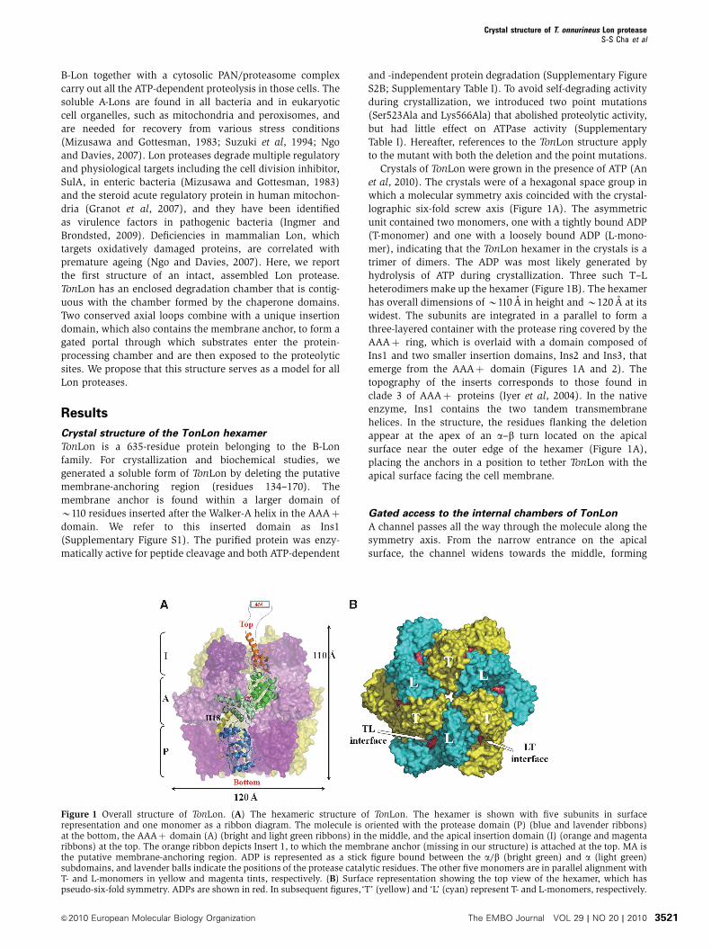
Crystals of TonLon were grown in the presence of ATP (An
et al, 2010). The crystals were of a hexagonal space group in
which a molecular symmetry axis coincided with the crystal-
lographic six-fold screw axis (Figure 1A). The asymmetric
unit contained two monomers, one with a tightly bound ADP
(T-monomer) and one with a loosely bound ADP (L-mono-
mer), indicating that the TonLon hexamer in the crystals is a
trimer of dimers. The ADP was most likely generated by
hydrolysis of ATP during crystallization. Three such T–L
heterodimers make up the hexamer (Figure 1B). The hexamer
has overall dimensions of B110 A in height and B120 A at its
widest. The subunits are integrated in a parallel to form a
three-layered container with the protease ring covered by the
AAAþ ring, which is overlaid with a domain composed of
Ins1 and two smaller insertion domains, Ins2 and Ins3, that
emerge from the AAAþ domain (Figures 1A and 2). The
topography of the inserts corresponds to those found in
clade 3 of AAAþ proteins (Iyer et al, 2004). In the native
enzyme, Ins1 contains the two tandem transmembrane
helices. In the structure, the residues flanking the deletion
appear at the apex of an a–b turn located on the apical
surface near the outer edge of the hexamer (Figure 1A),
placing the anchors in a position to tether TonLon with the
apical surface facing the cell membrane.
Gated access to the internal chambers of TonLon
A channel passes all the way through the molecule along the
symmetry axis. From the narrow entrance on the apical
surface, the channel widens towards the middle, forming
Figure 1 Overall structure of TonLon. (A) The hexameric structure of TonLon. The hexamer is shown with five subunits in surfacerepresentation and one monomer as a ribbon diagram. The molecule is oriented with the protease domain (P) (blue and lavender ribbons)at the bottom, the AAAþ domain (A) (bright and light green ribbons) in the middle, and the apical insertion domain (I) (orange and magentaribbons) at the top. The orange ribbon depicts Insert 1, to which the membrane anchor (missing in our structure) is attached at the top. MA isthe putative membrane-anchoring region. ADP is represented as a stick figure bound between the a/b (bright green) and a (light green)subdomains, and lavender balls indicate the positions of the protease catalytic residues. The other five monomers are in parallel alignment withT- and L-monomers in yellow and magenta tints, respectively. (B) Surface representation showing the top view of the hexamer, which haspseudo-six-fold symmetry. ADPs are shown in red. In subsequent figures,‘T’ (yellow) and ‘L’ (cyan) represent T- and L-monomers, respectively.
Crystal structure of T. onnurineus Lon proteaseS-S Cha et al
&2010 European Molecular Biology Organization The EMBO Journal VOL 29 | NO 20 | 2010 3521
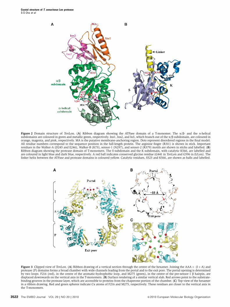
Figure 3 Clipped view of TonLon. (A) Ribbon drawing of a vertical section through the centre of the hexamer. Joining the AAAþ (IþA) andprotease (P) domains forms a broad chamber with wide channels leading from the portal and to the exit pore. The portal opening is determinedby two loops. F216 (red), in the centre of the aromatic-hydrophobic loop, and M275 (green), in the centre of the pre-sensor-1 b hairpin, aredisplaced downwards on the vertical axis in the T-monomers. (B) Surface rendering of a similar vertical slab. Red arrows point to the substrate-binding grooves in the protease layer, which are accessible to proteins from the chaperone portion of the chamber. (C) Top view of the hexamerin a ribbon drawing. Red and green spheres indicate Ca atoms of F216 and M275, respectively. These residues are closer to the vertical axis inthe T-monomers.
Figure 2 Domain structure of TonLon. (A) Ribbon diagram showing the ATPase domain of a T-monomer. The a/b- and the a-helicalsubdomains are coloured in green and metallic green, respectively. Ins1, Ins2, and In3, which branch out of the a/b subdomain, are coloured inorange, magenta, and pink, respectively. MA is the putative membrane-anchoring region. Dots represent disordered regions in the final model.All residue numbers correspond to the sequence position in the full-length protein. The arginine finger (R311) is shown in stick. Importantresidues in the Walker-A (D245 and E246), Walker-B (K73), sensor-1 (N297), and sensor-2 (R379) motifs are shown in sticks and labelled. (B)Ribbon diagram showing the protease domain of T-monomers. The S-subdomain and the K subdomain, with catalytic K566, are labelled andare coloured in light blue and dark blue, respectively. A red ball indicates conserved glycine residue (G441 in TonLon and G596 in EcLon). Thelinker helix between the ATPase and protease domains is coloured yellow. Catalytic residues, S523 and K566, are shown as balls and labelled.
Crystal structure of T. onnurineus Lon proteaseS-S Cha et al
The EMBO Journal VOL 29 | NO 20 | 2010 &2010 European Molecular Biology Organization3522
a cylindrical chamber, and becomes narrower again as it
passes through the last portion of the protease ring (Figure
3A and B). By analogy with other ATP-dependent proteases,
the channel opening in the apical domain likely serves as a
gateway for substrate entry and will be referred to as the
portal. The portal is formed by flexible loops contributed by
Ins2 and Ins3.
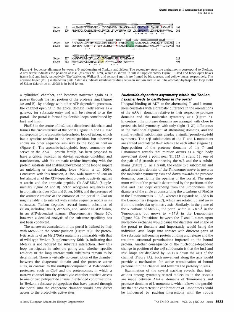
Phe216 in the centre of Ins2 has a disordered side chain and
frames the circumference of the portal (Figure 3A and C). Ins2
corresponds to the aromatic-hydrophobic loop of EcLon, which
has a tyrosine residue in the central position, but otherwise
shows no other sequence similarity to the loop in TonLon
(Figure 4). The aromatic-hydrophobic loop, commonly ob-
served in the AAAþ protein family, has been proposed to
have a critical function in driving substrate unfolding and
translocation, with the aromatic residue interacting with the
protein substrate and enabling movement of the loop to impart
an unfolding or translocating force (Martin et al, 2008).
Consistent with this function, a Phe216Ala mutant of TonLon
lost almost all of the ATP-dependent proteolytic activity against
a casein and the aromatic peptide, Glt-AAF-MNA (Supple-
mentary Figure 2A and B). EcLon recognizes sequences rich
in aromatic residues (Gur and Sauer, 2008), and the presence of
the aromatic residue at the entrance of the portal in TonLon
might enable it to interact with similar sequence motifs in its
substrates. TonLon degrades several known substrates of
EcLon, including UmuD, Arc-SulA, and Lambda N-GFP fusion,
in an ATP-dependent manner (Supplementary Figure 2C);
however, a detailed analysis of the substrate specificity has
not been conducted.
The narrowest constriction in the portal is defined by Ins3
with Met275 in the centre position (Figure 3C). The proteo-
lytic activity of an Met275Ala mutant is comparable with that
of wild-type TonLon (Supplementary Table I), indicating that
Met275 is not required for substrate interaction. How this
loop participates in substrate gating and whether specific
residues in the loop interact with substrates remain to be
determined. There is virtually no constriction of the chamber
between the chaperone domain and the protease active
sites, in contrast to the multiple-component ATP-dependent
proteases, such as ClpP and the proteasomes, in which a
narrow channel into the proteolytic chamber restricts access
to one or two polypeptide chains in extended conformations.
In TonLon, substrate polypeptides that have passed through
the portal into the chaperone chamber would have direct
access to the proteolytic active sites.
Nucleotide-dependent asymmetry within the TonLon
hexamer leads to oscillations in the portal
Unequal binding of ADP to the alternating T- and L-mono-
mers correlates with a dramatic difference in the orientations
of the AAAþ domains relative to their respective protease
domains and the molecular symmetry axis (Figure 5).
In contrast, the protease domains are arranged with close to
perfect six-fold symmetry, with only slight (1–21) differences
in the rotational alignment of alternating domains, and the
small a-helical subdomains display a similar pseudo-six-fold
symmetry. The a/b subdomains of the T- and L-monomers
are shifted and rotated 8–91 relative to each other (Figure 5).
Superposition of the protease domains of the T- and
L-monomers reveals that rotation occurs as a rigid body
movement about a point near Thr323 in strand 13, one of
the pair of b strands connecting the a/b and the a subdo-
mains (Figure 5). As a result, the entire a/b subdomain and
apical insertion domain of the T-monomer move in towards
the molecular symmetry axis and down towards the protease
domains, constricting the opening of the portal. The mini-
mum width of the portal is determined by the positions of the
Ins1 and Ins2 loops extending from the T-monomers. The
diameter of the circle circumscribing the a carbons of Phe216
in the T-monomers is B13 A, whereas it expands to B28 A in
the L-monomers (Figure 3C), which are rotated up and away
from the molecular symmetry axis. Similarly, in the plane of
the a carbons of Met275, the portal width is B8.5 A in the
T-monomers, but grows to B17 A in the L-monomers
(Figure 3C). Transitions between the T and L states upon
nucleotide exchange would cause the diameter and shape of
the portal to fluctuate and importantly would bring the
individual axial loops into contact with different parts of
the substrate, influencing protein binding and release and the
resultant structural perturbations imparted on the bound
protein. Another consequence of the nucleotide-dependent
change in position of the a/b subdomain is that the Ins2 and
Ins3 loops are displaced by 12–15 A down the axis of the
channel (Figure 3A). Such movement along the axis would
provide a mechanism for active translocation of bound
proteins into the channel and towards the proteolytic sites.
Examination of the crystal packing reveals that inter-
actions among symmetry-related molecules in the crystals
are made between AAAþ domains of T-monomers and
protease domains of L-monomers, which allows the possibi-
lity that the characteristic conformation of T-monomers could
be influenced by packing interactions with the robust
Figure 4 Sequence alignment between a/b subdomains of TonLon and EcLon. The secondary structure assignments correspond to TonLon.A red arrow indicates the position of Ins1 (residues 85–189), which is shown in full in Supplementary Figure S1. Red and black open boxesframe Ins2 and Ins3, respectively. The Walker-A, Walker-B, and sensor-1 motifs are framed by blue, green, and yellow boxes, respectively. Thearginine finger (R311) is shaded in pink. Asterisks indicate identical residues between TonLon and EcLon. The aromatic-hydrophobic loop motifof EcLon (Martin et al, 2008) is in bold letters.
Crystal structure of T. onnurineus Lon proteaseS-S Cha et al
&2010 European Molecular Biology Organization The EMBO Journal VOL 29 | NO 20 | 2010 3523
protease domains. Remarkably, the crystal structure of the
isolated AAAþ domain with a tightly bound ADP (unpub-
lished data), which was used for the structure determination
of TonLon (see ‘Materials and methods’), is virtually identical
to that of T-monomers with RMSD of 0.6 A for all atoms. The
identical conformation of the two AAAþ domains under
different crystal packing environments strongly suggests
that the observed conformation of T-monomers is not a
crystallographic artefact.
Structural basis of tight and weak binding of ADP
ADP is bound at the interface between the a/b and
a subdomains and is positioned to interact with highly
conserved residues that constitute functional motifs in the
majority of AAAþ proteins. The large a/b subdomain (re-
sidues 1–321) with its central five-stranded parallel b sheet
sandwiched by a-helices has three highly conserved motifs:
Walker A, which interacts with the b and g phosphates of
ATP; Walker B, which activates a water molecule that attacks
the b–g phosphodiester bond of ATP; and sensor-1, which
communicates with the g phosphate and is used to discrimi-
nate between ATP and ADP (Figures 2A and 4). The a/bsubdomain also has an arginine-finger residue (Arg311),
which is required for activation of ATPase activity upon
assembly. These residues are positioned to interact with the
tightly bound ADP in three of the subunits (Supplementary
Figure S4). Mutations in Walker-A or Walker-B residues or
in the arginine-finger residue caused a drastic decrease in
ATPase activity and ATP-dependent proteolytic activity
(Supplementary Table I). The smaller mostly a-helical sub-
domain (residues 322–412) contains the sensor-2 motif,
Arg379 (Figure 4), which is also positioned to interact with
bound nucleotide. The sensor-2 motif also serves to help
discriminate among nucleotide states.
The ADP situated at the subdomain interface in the
T-monomer (T-ATPase domain) (Figure 2A) is tightly bound
and shows strong electron density (Supplementary Figure
S3). In contrast, the electron density of ADP in the L-ATPase
domain is weak (Supplementary Figure S4), and the B factor
of ADP is much higher there (48.9 A2) than in the T-ATPase
domain (22.1 A2), implying lower occupancy and/or weaker
binding. Earlier studies with EcLon had suggested the
existence of high- and low-affinity states for the nucleotide-
binding sites in Lon oligomers (Menon and Goldberg, 1987a;
Vineyard et al, 2005; 2006). To confirm that ATP and ADP
bind unequally to different subunits of TonLon in solution,
we measured binding of ATPgS and ADP by isothermal
titration calorimetry. ATPgS bound tightly to TonLon
(KdB2mM) and the stoichiometry of binding was 3.2±0.04
per hexamer, and ADP bound even tighter (Kdo1 mM) with a
stoichiometry of 2.7±0.03 (Supplementary Figure S2D). To
evaluate the binding of ATP under assay conditions, we
measured the ATP concentration dependence of peptide
cleavage by TonLon. The ATP titration curve for activation
of peptidase activity was sigmoidal followed by a very steep
slope, which when fit to the Hill’s equation gave a Hill’s
coefficient of 3.4 (Supplementary Figure S2E). These data
suggest that unequal binding of ADP to adjacent subunits
observed in the crystal reflects an intrinsic property of
TonLon.
In the subunits with tightly bound ADP, the base is stacked
between Met75 in the a/b subdomain and Leu378 in the
a-helical subdomain, whereas N1, N3, and NH2 at C-6
make hydrogen-bonding interactions and the 20-OH of
ribose interacts with Glu342 at the entrance of the pocket
(Supplementary Figure S2). The diphosphate, nestled be-
tween the N-terminal end of helix-4 and the Walker-A
motif, makes multiple-polar interactions with the Walker-B
and sensor-1 motifs in the a/b subdomain and the sensor-2
motif in the a-helical subdomain. Notably, the only inter-
action of ADP with a neighbouring L-monomer (L-ATPase
domain) is the contact between the 30-OH of the ribose and
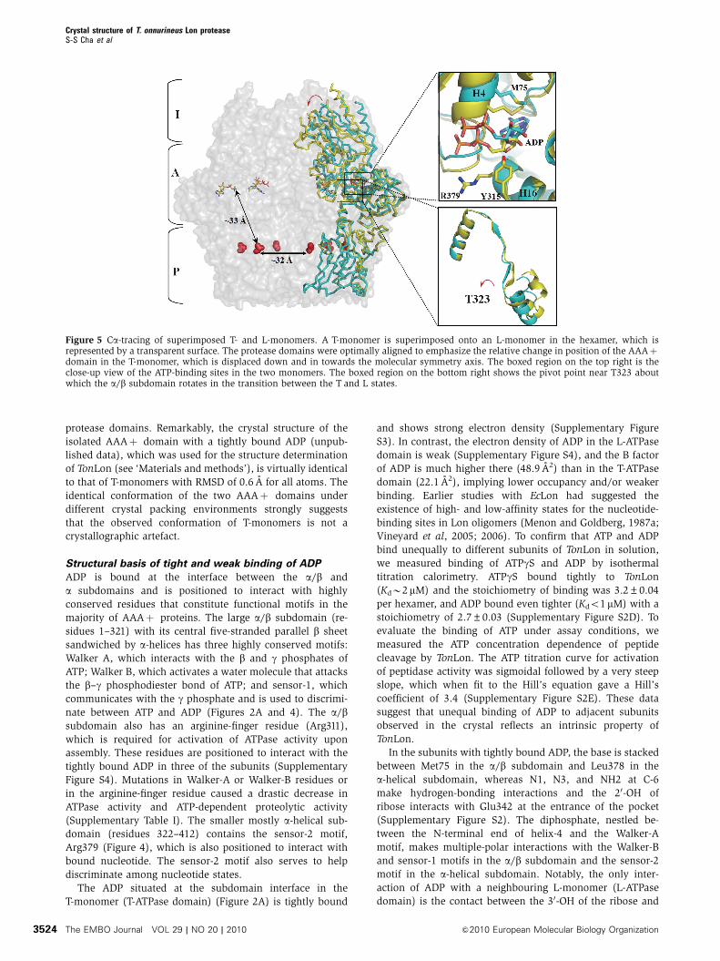
Figure 5 Ca-tracing of superimposed T- and L-monomers. A T-monomer is superimposed onto an L-monomer in the hexamer, which isrepresented by a transparent surface. The protease domains were optimally aligned to emphasize the relative change in position of the AAAþdomain in the T-monomer, which is displaced down and in towards the molecular symmetry axis. The boxed region on the top right is theclose-up view of the ATP-binding sites in the two monomers. The boxed region on the bottom right shows the pivot point near T323 aboutwhich the a/b subdomain rotates in the transition between the T and L states.
Crystal structure of T. onnurineus Lon proteaseS-S Cha et al
The EMBO Journal VOL 29 | NO 20 | 2010 &2010 European Molecular Biology Organization3524
Tyr315 (Figure 5; Supplementary Figure S4). When the T- and
L-monomers are superimposed, the rotation of the a/b sub-
domain is seen to produce a shift of B3 A in helix-4 including
the Walker-A motif (Figure 5). The change in position of
Met75 in helix-4 disturbs its interactions with the adenine
ring, and the accompanying change in position of the
ADP leads to the loss of the interactions between the
ribose and Tyr315 of the adjacent subunit and between
the a phosphate and the sensor-2 residue, Arg379.
Although ADP is more exposed to the medium in the L site,
it appears that a conformational change would be required
to allow dissociation of ADP from the T site, consistent
with experiments showing that protein substrate binding is
required to dislodge tightly bound ADP in EcLon protease
(Menon and Goldberg, 1987b).
The docking of alternating T-ATPase and L-ATPase do-
mains (Figure 3A) produces two different interfaces
(Figure 1B). We refer to the interface with a T site and tightly
bound ADP as the LT interface, and the one with an L site and
weakly bound ADP as the TL interface. At the LT interface,
3240 A2 of solvent-accessible area is buried between the
ATPase domains, and slightly less, 2730 A2, is buried at the
TL interface, suggesting that the TL interface is looser and
more mobile. The differences in the interfaces would be
expected to also affect the relative ATPase activity at the
respective sites as a consequence of the altered position of an
arginine-finger residue, Arg311 (Figure 2A), which is brought
closer to or further from the nucleotide in the adjacent active
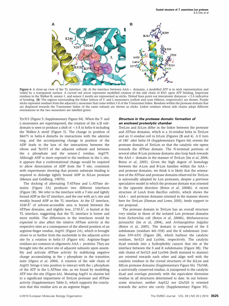
site by a rotation in helix-13 (Figure 6A). Arginine-finger
residues are common in oligomeric AAAþ proteins. They are
brought into the active site of adjacent subunits upon assem-
bly and activate ATPase activity by stabilizing negative
charge accumulating at the g phosphate in the transition
state (Ogura et al, 2004). A rotation of the side chain of
Arg311 brings it into position to interact with the g phosphate
of the ATP in the L-ATPase site, as we found by modelling
ATP into the site (Figure 6A). Mutating Arg311 to alanine led
to a significant impairment of TonLon protease and ATPase
activity (Supplementary Table I), which supports the conclu-
sion that this residue acts as an arginine finger.
Structure in the protease domain: formation of
an enclosed proteolytic chamber
TonLon and EcLon differ in the linker between the protease
and ATPase domains, which is a 16-residue helix in TonLon
and an 11-residue coil in EcLon (Figures 2B and 4). A U turn
of 1801 after helix-18 (Supplementary Figure S4) orients the
protease domain of TonLon so that the catalytic site opens
towards the ATPase domain. The N-terminal portions of
several other B-Lon protease domains also loop back towards
the AAAþ domain in the manner of TonLon (Im et al, 2004;
Botos et al, 2005). Given the high degree of homology
between the A-Lon and B-Lon families within the AAAþand protease domains, we think it is likely that the orienta-
tion of the ATPase and protease domains observed for TonLon
is universally adopted by Lon proteases, despite a previous
speculative model in which the protease domain was oriented
in the opposite direction (Botos et al, 2004b). A recent
structure of LonA from Bacillus subtilis, which shows the
AAAþ and protease domains oriented in the manner shown
here for TonLon (Duman and Lowe, 2010), lends support to
our proposal.
The protease domain in TonLon has an overall structure
very similar to those of the isolated Lon protease domains
from Escherichia coli (Botos et al, 2004b), Methanococcus
jannaschii (Im et al, 2004), and Archaeoglobus fulgidus
(Botos et al, 2005). The domain is composed of the S
subdomain (residues 441–538) and the K subdomain (resi-
dues 539–635) (Figure 2B), which harbour the catalytic
residues, Ser523 and Lys566, respectively. The catalytic
dyad extends into a hydrophobic canyon that sits at the
interface between the S and K subdomains (Figure 3B). The
side chains of Ser523 and Lys566 (both mutated to alanine)
are oriented towards each other and align well with the
catalytic residues in the crystal structures of the EcLon and
MjLon protease domains (Supplementary Figure S6). Thr548,
a universally conserved residue, is juxtaposed to the catalytic
dyad and overlaps precisely with the equivalent threonine
residue in all structures determined to date. In our holoen-
zyme structure, neither Asp522 nor Glu520 is oriented
towards the active site cavity (Supplementary Figure S5),
Figure 6 A close-up view of the TL interface. (A) At the interface between AAAþ domains, a modelled ATP is in stick representation andveiled by a transparent surface. A curved red arrow represents modelled rotation of the side chain of R311 upon ATP binding. Importantresidues in the Walker-B, sensor-1, and sensor-2 motifs are represented as sticks. Dotted lines point out interatomic distances o3 A indicativeof bonding. (B) The regions surrounding the linker helices of T- and L-monomers (yellow and cyan ribbons, respectively) are shown. Purplesticks represent residues from the adjacent L-monomer that come within 3 A of the T-monomer linker. Residues within the protease domain thatare displaced towards the T-monomer linker of the same subunit are shown as sticks. Linker residues whose side chains adopt differentorientations in the two monomers are labelled green.
Crystal structure of T. onnurineus Lon proteaseS-S Cha et al
&2010 European Molecular Biology Organization The EMBO Journal VOL 29 | NO 20 | 2010 3525
and neither would be in a position to make hydrogen bond
contact with the e amino of catalytic Lys566. Thus, it appears
unlikely that either of these residues is part of the catalytic
machinery in B-Lons as has been proposed (Im et al, 2004).
Viewed from the position of the linker, the protease
domain is shaped like a bowl with the exit pore at the
bottom. The axially positioned exit pore is formed by six
repeats of a long loop between strands 17 and 18 with the
disordered side chain of Glu470 protruding into the centre
(Figure 3A and B; Supplementary Figure S7). The width
of the exit pore is B13 A, sufficient to allow the passage of
unstructured cleavage products. A wide ledge runs around
the circumference of the bowl about half-way down. The six
active sites lie along the circumferential ring B28 A from the
centre, and adjacent sites are separated by B32 A (Figure 3B;
Supplementary Figure S6). The hydrophobic canyons with
the catalytic residues face towards the ATPase domain. The
ATPase ring forms a lid over the protease domain producing a
large hollow chamber, so that both unfolded or partially
folded substrate proteins that have entered the ATPase cham-
ber will easily encounter the active sites. Large protein
substrates and most peptides would only have access through
the axial channel in the AAAþ domain, but there are small
rectangular openings roughly 10 by 13 A near the linker in
each subunit through which small peptides or nucleotides
might pass. There is also some evidence of dynamic move-
ment in the protease domain, which could influence catalytic
activity as well as the accessibility of the active sites. The
backbone atoms of the individual protease domains overlay
with an RMSD of o1.0 A; however, as a result of the shift in
the linker in response to nucleotide binding and release, the
domains connected to the L ATPases are rotated slightly
relative to those of the T-ATPase subunits causing a displace-
ment of B1.5 A in the main chain along the periphery.
Mutations in Glu614, His665, and His667 of EcLon (Glu460,
His509, and Gln511 in TonLon, which are extensively
involved in interdomain contacts of the protease layer)
decrease ATPase and protease activity of EcLon (Oh et al,
1998; Starkova et al, 1998), pointing to the importance of the
conformational communication between the protease and
ATPase domains in coupling the two activities
Discussion
TonLon as a model for all Lon proteases
The structure of TonLon presented here confirms that the
proteolytic sites are located within a gated chamber that is
accessible to proteins only through restrictive axial channels.
To what extent does TonLon provide a model for A-Lons such
as EcLon? The protease domains (Supplementary Figures S5
and S6) as well as the a-helical subdomains of TonLon and
EcLon are homologous in their tertiary and quaternary struc-
tures. Sequence alignment confirms that all major functional
motifs are well matched (Figure 4; Supplementary Figure S1),
and sequence-based secondary structure analysis predicts
that EcLon has the insertions corresponding to Ins2 and
Ins3 of TonLon. Consequently, except for Ins1, a strategic
insertion needed to allow anchoring of TonLon to the cell
membrane, the a/b subdomains of TonLon and EcLon are
likely to exhibit close structural similarity.
A notable difference between EcLon and TonLon is the
linker between the ATPase and protease domains, which
is a helix in TonLon and an extended coil in EcLon
(Supplementary Figure S1). In a previous model of EcLon
(Botos et al, 2004a, b), the protease ring was proposed to take
the opposite orientation with the active sites directed away
from the ATPase and exposed to the medium. However, we
note that, in the crystal of the EcLon protease domain, the
N-terminal portion of the main chain adopts a hook-like
conformation reminiscent of the U turn that brings the
chain back towards the AAAþ domain in TonLon
(Supplementary Figure S5). Gly441 of TonLon, which is
part of the U turn (Figure 2B), is absolutely conserved in
Lon proteases, and the orthologous Gly596 contributes to
the hook in EcLon (Supplementary Figure S5). When the
a-subdomain of EcLon is superimposed onto that of TonLon,
the gap between the end of the terminal helix (Gln582) and
the glycine in the hook (Gly596) is B40 A. No portions of the
structure obstruct the region between Gln582 and Gly596,
and the 13 intervening residues are predicted by the PHDsec
programme (Rost et al, 2004) to adopt an extended coil.
Thus, the gap in EcLon could easily be bridged with
the domains in this orientation. We conclude that LonAs
and LonBs share a hexameric framework constructed from
homologous ATPase and protease domains, and that the
closed degradation chamber and the basic mechanism of
substrate-processing encrypted in this hexameric architecture
are common to both families.
A ‘bowl and lid’ design for sequestered proteolytic
chambers in single-polypeptide ATP-dependent
proteases
The architecture of TonLon is reminiscent of that of FtsH,
which is also a single-polypeptide ATP-dependent protease
(Bieniossek et al, 2006), and differs in several respects from
the multi-component proteases such as Clp and the protea-
somes. In TonLon and FtsH, there are only six active sites per
holoenzyme and they are located in a bowl-like chamber that
is covered with the hexameric assembly of ATPase domains
that serve as a concave lid, producing a large protein-proces-
sing chamber in which protein unfolding and degradation can
apparently occur simultaneously. The architectural design
explains several properties reported for Lon proteases. The
enclosed degradation chamber is consistent with biochemical
data showing that the majority of products of degradation by
Lon are small peptides (Maurizi, 1987; Nishii et al, 2005).
The contiguous chaperone and degradation chambers would
allow Lon to make initial cuts at multiple sites within protein
substrates and even to cleave native forms of some proteins
(Ondrovicova et al, 2005). The latter activity suggests that the
apical gate can open sufficiently to allow at least small
domains to enter without unfolding. Lon protease from
Thermococcus kodakaranesis KODI can degrade unfolded
proteins in the absence of ATP (Fukui et al, 2002). One
possibility is that, in the absence of tightly bound nucleotide,
all six a/b domains are rotated up and out in the manner of
the L subunits, which would generate an axial channel
sufficient to allow unfolded and perhaps even small folded
proteins to pass through.
In all ATP-dependent proteases, substrate access is con-
trolled at the apical surface of AAAþ domains through axial
loops whose positions are changed in response to rigid
domain movements as nucleotides bind and are hydrolysed
and released from the AAAþ domains. Multi-component
Crystal structure of T. onnurineus Lon proteaseS-S Cha et al
The EMBO Journal VOL 29 | NO 20 | 2010 &2010 European Molecular Biology Organization3526
proteases such as ClpXP, ClpAP, HslUV (ClpYQ), and 26 S
proteasomes, which function as dynamic complexes of cha-
perone and proteolytic components, also control substrate
access at the entrance to the protease. The sequestered
proteolytic chambers are constructed by joining two hepta-
mers (or hexamers in the case of ClpQ) face to face so that the
active sites are inside. Access to the chamber is through axial
channels, which are gated and regulated by interaction with
the chaperone. As the AAAþ components of multi-compo-
nent ATP-dependent proteases also function independently to
remodel protein structures and can release proteins that will
refold to the native state, gating at the point of entry to the
protease might provide a final checkpoint for deciding be-
tween degradative and refolding fates for specific proteins.
The use of a single gate may reflect that Lon selectively
targets unfolded proteins. In fact, several of the physiological
substrates of EcLon, l N-protein and the cell division inhi-
bitor, SulA, have structurally unstable regions (Gur and
Sauer, 2008). Such proteins might not be capable of refolding
if released, and a one-pass gating mechanism would assure
their efficient disposal.
Substrate-processing loops are manipulated by rigid
body movement of the a/b subdomain
The location of Ins2 and Ins3 on the apical surface forming a
cover over the axial channel places them in position to
engage substrates, control access to the interior, and actively
participate in unfolding and translocation of bound proteins
as has been proposed for similar structures in other AAAþproteins (Martin et al, 2008). The inserts jut out from the a/bsubdomain at an unusually sharp angle and thus extend from
the surface towards the membrane to which TonLon is
anchored in vivo, possibly to allow them to survey exposed
regions of membrane proteins and to facilitate the capture of
appropriate substrates. Phe216, located in an aromatic-hydro-
phobic loop of Ins2, and Met275, in the pre-sensor-1 b-hair-
pin loop, move in and down during the transition from the
L to the T state, whereas the corresponding residues move up
in the other three subunits. The narrowing of a region of the
portal would serve to grip substrates at specific locations and
the downwards motion could translocate them into the
interior. When the protein is too large to pass through the
narrow region of the portal, the vectorial movement of Ins2
during the ATP-powered L-T transition would tend to tug
proteins against the portal, generating counteractive forces
that would lead to unfolding (Sauer et al, 2004; Zhang et al,
2009). Therefore, the L-T transition is likely to be the
mechanism underlying both translocation and unfolding of
substrates in TonLon.
The buried ADP in the Tsite is consistent with studies with
EcLon showing that dissociation of ADP generated in situ is
slow and requires allosteric activation induced by protein
substrate binding (Menon and Goldberg, 1987a). Our direct
binding studies indicate that ADP binds to TonLon with a
Kdo1mM (Supplementary Figure 2D). The ATPase activity of
TonLon is enhanced approximately three-fold in the presence
of an unfolded protein substrate (Supplementary Table I),
which is also consistent with the studies of EcLon showing
that protein substrates accelerate the rate-limiting release of
ADP during the hydrolysis cycle. ADP/ATP exchange should
occur in the L domain, in which the ADP is more exposed and
exhibits a high B factor (Figure 3A).
If the L- and T-monomers are taken to represent conforma-
tional states before ATP binding and after ATP hydrolysis,
respectively, it is reasonable to propose that each monomer in
TonLon repeatedly interconverts between T- and L-conforma-
tions in concert with ATP hydrolysis cycles. A modelling
study in which an L-monomer is replaced with a T-monomer
shows a significant clash between a/b subdomains from
adjacent T-monomers (Supplementary Figure S8), indicating
that adjacent monomers cannot maintain the T-conformation
simultaneously. When an L-monomer adjacent to a T-mono-
mer undergoes an ATP-powered L-T transition, high-affinity
binding of ATP to the L state might impel a T-L transition of
the T-monomer to relieve the clash. We propose that the L-T
transition powered by ATP and substrate binding in an
L-monomer is coupled to T-L transition in an adjacent
T-monomer. Owing to the clash between adjacent T-mono-
mers, no more than three non-adjacent monomers may work
in concert in the hexamer. Studies with several AAAþproteins now point to a general mechanism in which ATP
hydrolysis occurs at different times in different subunits, in
either a random or alternating manner, and is facilitated
allosterically by nucleotide and substrate binding to other
subunits within the ring (Martin et al, 2005, 2007; Augustin
et al, 2009). The crystal structure presented here will serve as
a model to study similar details of how Lon proteases capture
and degrade their substrates and how those substrates are
recognized as appropriate targets by this important compo-
nent of cellular protein quality control.
The limited differences in conformations between the
T and L subunits suggest that Lon might undergo a smaller
range of motion than observed recently for ClpX (Glynn et al,
2009) and FtsH (Bieniossek et al, 2006, 2009), in which the
a-subdomain undergoes a displacement by as much as 801,
but is in line with motions proposed for HslU (Wang, 2004).
ATP binding to one or more subunits could induce a con-
formation different from that observed with ADP and thus add
additional asymmetry within the ring. A fuller understanding
of the conformational dynamics and range of motion will
require structural determination of multiple nucleotide states.
Materials and methods
Purification and crystallizationDetails of expression, purification, and crystallization of TonLonhave been described elsewhere (An et al, 2010). Protein concentra-tions were measured by absorbance using either experimentallydetermined absorption coefficients or absorption coefficientscalculated from the aromatic amino-acid content using the Pepstatsprogramme (http://helixweb.nih.gov/emboss/html/pepstats.html).Molar concentrations of TonLon refer to units of hexamers.
Structure determination and refinementIt was extremely difficult to obtain TonLon crystals of diffractionquality (An et al, 2010), which prohibited our using MIR or MADmethods to get phase information. As an alternative, we determinedto solve the structure of TonLon using molecular replacement. Asno high-resolution structural data was available for the AAAþdomain of Lon proteases, we first solved the structure of the AAAþdomain of TonLon, which we obtained by limited proteolysis. Thepurified full-length TonLon was treated with trypsin with 1:100(w/w) ratio for 30 min and the AAAþ domain was then separatedby gel filtration. The AAAþ domain was crystallized in aprecipitant solution containing 12% polyethylene glycol 4000,0.2 M potassium chloride, 0.01 M magnesium acetate, and 0.05 Mtri-sodium citrate dehydrate pH 4.5. The crystals belong to spacegroup P1 with unit cell parameters a¼ 40.83, b¼ 61.00, c¼ 76.32,
Crystal structure of T. onnurineus Lon proteaseS-S Cha et al
&2010 European Molecular Biology Organization The EMBO Journal VOL 29 | NO 20 | 2010 3527
a¼ 74.36, b¼ 86.48, and g¼ 83.89. To solve the structure of theisolated AAAþ domain by the MAD method, the seleno-methio-nine substituted AAAþ domain was prepared as above andcrystallized. MAD data sets at 2.0-A resolution were collected atthree wavelengths (peak, 0.97909; edge, 0.97934; remote, 0.96411)(Supplementary Table II). Selenium positions were located andphases were calculated using the programme SOLVE. The sub-sequent solvent flattening by RESOLVE gave rise to an interpretablemap, based on which the de novo model building was easilycompleted. The initial model containing ADP molecules wassubjected to several rounds of refinement and manual refitting,giving rise to a model with R and Rfree values to 25.99 and 30.18%,respectively. At this stage, we stopped the refinement withoutincorporating water molecules into the model, because watermolecules were not required to use the model for the subsequentmolecular replacement determination of TonLon.
The structure of TonLon was determined by molecular replace-ment with MolRep in the CCP4 programme suite. Structures of theisolated AAAþ domain of TonLon and the EcLon protease domain(Botos et al, 2004b) were separately used as search models. Theposition of the protease domain was first determined and then fixedin the subsequent search for the position of the isolated AAAþdomain. Refinement was performed with the maximum likelihoodalgorithm implemented in the CNS programme (Table I). Moleculargraphics manipulations and calculations of solvent-accessiblesurface area were performed with QUANTA (Molecular SimulationsInc., San Diego, CA). In the final model, a T-monomer containsresidues 18–130, 179–216, and 221–635 and an L-monomercontains residues 20–128, 178–216, and 222–634. The Ramachan-dran plot indicates 92.9% of non-glycine and non-proline residuesare in the most favoured regions, and no residues are in thedisallowed regions in the final model.
In vitro protein degradationTonLonDTM with wild-type catalytic residues in the proteasedomain was used for in vitro degradation assays. Proteolyticactivity against unfolded proteins was assayed with fluoresceinisothiocyanate casein (FITC casein) and with a casein (both fromSigma). The assay buffer consisted of 50 mM Tris–HCl, pH 8.0, and10 mM MgCl2, with or without 1 mM ATP. TonLonDTM, the F216Amutant, or the M275A mutant (50 nM hexamer) was incubated in200 ml of assay buffer at 701C with 2mM substrate. The fluorescenceincrease upon degradation of FITC casein was monitored using afluorescence spectrophotometer with excitation at 490 nm andemission at 525 nm. For a-casein degradation, aliquots wereremoved at timed intervals and mixed with SDS sample buffer at951C. After SDS–PAGE, the a casein remaining was detected bystaining with Coomassie blue.
Activity against folded substrates was assayed at 371C with0.2mM TonLon in the above buffer solution with and without1.5 mM ATP. The solution also contained 50 mM creatine phosphateand 0.3 U/ml of phosphocreatine kinase to regenerate ATP andminimize the accumulation of ADP. No degradation was observedwith the regenerating system alone. N-GFP is a fusion of the phage lN-protein with GFP-mut3.1 and was kindly provided by FatimaRasulova, LCB, NCI, NIH (unpublished data). E. coli UmuD wasprovided by Roger Woodgate (NGI, NICHD, NIH). Arc-SulA is afusion of the Arc repressor protein with the C-terminal 11 aminoacids of SulA and was provided by David McKay (StanfordUniversity School of Medicine). N-GFP, UmuD, and Arc-SulA werepresent in the assay solution at 4.0, 10, and B2 mM, respectively.Aliquots were quenched into SDS sample buffer and the proteinswere separated by SDS–PAGE. The remaining intact proteinwas detected by staining with Coomassie blue or, in the case ofArc-SulA, by silver staining.
Peptidase activityStock solutions of glutaryl-Ala-Ala-Phe-methoxynaphthyl amide(Glt-AAF-MNA) (Sigma) in DMSO were diluted to 0.3 mM in 50 mMTris/HCl buffer, pH 8.0, with 10 mM MgCl2, with or without 1 mMATP. After incubation at 701C with 12.5 nM TonLon hexamer forvarious times, the reaction was quenched by dilution into 2% SDSand the increase in fluorescence was measured in an AmincoBowman spectrofluorometer with excitation at 335 nm and emis-sion at 410. Continuous assays of peptidase were conducted using apeptide with the following sequence: aminobenzoic acid-Ala-Phe-His-Met-Ala-Leu-nitrotyrosine-Pro-Val (Fluo-FV) (Li et al, 2010).The fluorescence of the donor, nitrotyrosine, is reduced by thepresence of the acceptor, aminobenzoic acid. Increased fluores-cence emission at 415 nm upon cleavage of the peptide wasmonitored with excitation at 320 nm. Typical assay mixturescontained 50 mM creatine phosphate, 0.3 U/ml of phosphocreatinekinase as an ATP regenerating system, 0.25mM (100 mg/ml) ofTonLon hexamer, and 4 mM of Fluo-FV. Assays were conducted at371C with variable concentrations of ATP.
ATPase activityTo measure ATPase activity, TonLon (2mg) was incubated with1 mM ATP and 10 mM MgCl2 in 100ml of 50 mM Tris–HCl buffer(pH 8.0) at 371C for 15 min. Inorganic phosphate released wasdetermined from the increase in absorbance at 660 nm afteraddition of ammonium molybdate and malachite green. Stimulationof the ATPase activity of TonLon by a casein was investigated at acasein concentration of 0.3 mg/ml.
Nucleotide-binding assaysBinding of ATPgS and ADP to TonLon was measured by isothermaltitration calorimetry at 251C using a VP-ITC microcalorimeter(Microcal). A solution of 1.2 ml of 50 mM Tris/HCl, pH 8.0,containing 10 mM MgCl2 and 5mM hexamer TonLon was titratedwith ATPgS or ADP dissolved in the same buffer adjusted to10.2 mM MgCl2. ATPgS or ADP (0.2 mM)was added in nine aliquotsof 2.5ml each followed by 17 aliquots of 14ml each.
Addendum in revisionDuring the time this manuscript was under revision, a reportappeared online with a 3.4 A structure of B. subtilis LonA with ADPbound (Duman and Lowe, 2010). The subunit structure is consistentwith the orientation of the protease active sites with respect to theAAAþ domain that we observe in the TonLon hexamer andconfirms our proposal that this orientation is maintained in the LonA family as well.
Supplementary dataSupplementary data are available at The EMBO Journal Online(http://www.embojournal.org).
Acknowledgements
We thank the staff of beamline 17A at Photon Factory for help withdata collection. We are grateful to H-Y Kim and WC Lee at KoreaBasic Science Institute (KBSI) for the use of Rigaku MicroMax-007HF X-ray generator and to Susan Gottesman and MatthewHumbard (National Cancer Institute, Bethesda, MD) for comments
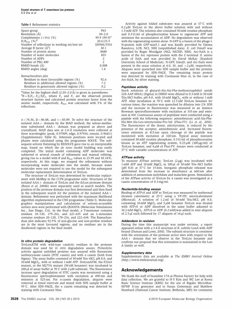
Table I Refinement statistics
Space group P63Resolution (A) 30–2.0Completeness (40 s) (%) 99.9 (99.9)a
R/Rfree (%)b 20.6/23.5Number of reflections in working set/test set 103966/5516Average B factor (A2) 30.1Number of protein atoms 8680Number of water molecules 835Number of ADPs 2Number of PEG 400 2RMSD bonds (A) 0.008RMSD angles (deg) 1.4
Ramachandran plotResidues in most favourable regions (%) 92.6Residues in additional allowed regions (%) 7.2Residues in generously allowed regions (%) 0.2
aValue for the highest shell (2.03–2.0 A) is given in parentheses.bR¼S|Fo�Fc|/SFo, where Fo and Fc are the observed proteinstructure factors and calculated protein structure factor from theatomic model, respectively. Rfree was calculated with 5% of thereflections.
Crystal structure of T. onnurineus Lon proteaseS-S Cha et al
The EMBO Journal VOL 29 | NO 20 | 2010 &2010 European Molecular Biology Organization3528
on the manuscript. This work was supported by KORDI in-houseprogramme (PE98513), the Marine and Extreme Genome ResearchCenter programme, and the Development of BiohydrogenProduction Technology Using Hyperthermophilic Archaea pro-gramme of the Ministry of Land, Transport, and Maritime Affairs,Republic of Korea. MRM and GMDD are supported by the intramuralresearch programme of the Center for Cancer Research, NCI, NIH,
Bethesda, MD. The atomic coordinates have been deposited in theProtein Data Bank (accession code 3K1J).
Conflict of interest
The authors declare that they have no conflict of interest.
References
An YJ, Lee CR, Supangat S, Lee HS, Lee JH, Kang SG, Cha SS (2010)Crystallization and preliminary X-ray crystallographic analysis ofLon from Thermococcus onnurineus NA1. Acta Crystallogr Sect FStruct Biol Cryst Commun 66: 54–56
Augustin S, Gerdes F, Lee S, Tsai FT, Langer T, Tatsuta T (2009) Anintersubunit signaling network coordinates ATP hydrolysis by m-AAA proteases. Mol Cell 35: 574–585
Baumeister W, Walz J, Zuhl F, Seemuller E (1998) The proteasome:paradigm of a self-compartmentalizing protease. Cell 92:367–380
Bieniossek C, Niederhauser B, Baumann UM (2009) The crystalstructure of apo-FtsH reveals domain movements necessary forsubstrate unfolding and translocation. Proc Natl Acad Sci USA106: 21579–21584
Bieniossek C, Schalch T, Bumann M, Meister M, Meier R, BaumannU (2006) The molecular architecture of the metalloprotease FtsH.Proc Natl Acad Sci USA 103: 3066–3071
Bochtler M, Ditzel L, Groll M, Huber R (1997) Crystal structure ofheat shock locus V (HslV) from Escherichia coli. Proc Natl AcadSci USA 94: 6070–6074
Botos I, Melnikov EE, Cherry S, Khalatova AG, Rasulova FS, TropeaJE, Maurizi MR, Rotanova TV, Gustchina A, Wlodawer A (2004a)Crystal structure of the AAA+ a domain of E. coli Lon protease at1.9A resolution. J Struct Biol 146: 113–122
Botos I, Melnikov EE, Cherry S, Kozlov S, Makhovskaya OV,Tropea JE, Gustchina A, Rotanova TV, Wlodawer A(2005) Atomic-resolution crystal structure of the proteolyticdomain of Archaeoglobus fulgidus Lon reveals the conformationalvariability in the active sites of lon proteases. J Mol Biol 351:144–157
Botos I, Melnikov EE, Cherry S, Tropea JE, Khalatova AG, RasulovaF, Dauter Z, Maurizi MR, Rotanova TV, Wlodawer A, Gustchina A(2004b) The catalytic domain of Escherichia coli Lon protease hasa unique fold and a Ser-Lys dyad in the active site. J Biol Chem279: 8140–8148
Duman RE, Lowe J (2010) Crystal structures of Bacillus subtilis LonProtease. J Mol Biol 401: 653–670
Flynn JM, Neher SB, Kim YI, Sauer RT, Baker TA (2003)Proteomic discovery of cellular substrates of the ClpXP proteasereveals five classes of ClpX-recognition signals. Mol Cell 11:671–683
Fukui T, Eguchi T, Atomi H, Imanaka T (2002) A membrane-boundarchaeal Lon protease displays ATP-independent proteolytic ac-tivity towards unfolded proteins and ATP-dependent activity forfolded proteins. J Bacteriol 184: 3689–3698
Glynn SE, Martin A, Nager AR, Baker TA, Sauer RT (2009)Structures of asymmetric ClpX hexamers reveal nucleotide-de-pendent motions in a AAA+ protein-unfolding machine. Cell139: 744–756
Goldberg AL, John ACS (1976) Intracellular protein degradation inmammalian and bacterial cells. Annu Rev Biochem 45: 747–803
Gottesman S (2003) Proteolysis in bacterial regulatory circuits.Annu Rev Cell Dev Biol 19: 565–587
Gottesman S, Roche E, Zhou Y, Sauer RT (1998) The ClpXP andClpAP proteases degrade proteins with carboxy-terminalpeptide tails added by the SsrA-tagging system. Genes Dev 12:1338–1347
Granot Z, Melamed-Book N, Bahat A, Orly J (2007) Turnover ofStAR protein: roles for the proteasome and mitochondrialproteases. Mol Cell Endocrinol 265–266: 51–58
Groll M, Ditzel L, Lowe J, Stock D, Bochtler M, Bartunik HD, HuberR (1997) Structure of 20S proteasome from yeast at 2.4 Aresolution. Nature 386: 463–471
Gur E, Sauer RT (2008) Recognition of misfolded proteins by Lon, aAAA(+) protease. Genes Dev 22: 2267–2277
Hershko A, Ciechanover A (1992) The ubiquitin system for proteindegradation. Annu Rev Biochem 61: 761–807
Im YJ, Na Y, Kang GB, Rho SH, Kim MK, Lee JH, Chung CH, Eom SH(2004) The active site of a Lon protease from Methanococcusjannaschii distinctly differs from the canonical catalytic Dyad ofLon proteases. J Biol Chem 279: 53451–53457
Ingmer H, Brondsted L (2009) Proteases in bacterial pathogenesis.Res Microbiol 160: 704–710
Iyer LM, Leipe DD, Koonin EV, Aravind L (2004) Evolutionaryhistory and higher order classification of AAA+ ATPases.J Struct Biol 146: 11–31
Kim YI, Burton RE, Burton BM, Sauer RT, Baker TA (2000)Dynamics of substrate denaturation and translocation by theClpXP degradation machine. Mol Cell 5: 639–648
Li DHS, Chung YS, Gloyd M, Joseph E, Ghirlando R, Wright GD,Cheng YQ, Maurizi MR, Guarne A, Ortega J (2010)Acyldepsipeptide antibiotics induce the formation of a structuredaxial channel in ClpP: a model for the ClpX/ClpA bound state ofClpP. Chem Biol (in press)
Li M, Rasulova F, Melnikov EE, Rotanova TV, Gustchina A,Maurizi MR, Wlodawer A (2005) Crystal structure of the N-terminal domain of E. coli Lon protease. Protein Sci 14:2895–2900
Lowe J, Stock D, Jap B, Zwickl P, Baumeister W, Huber R (1995)Crystal structure of the 20S proteasome from the archaeon T.acidophilum at 3.4 A resolution. Science 268: 533–539
Martin A, Baker TA, Sauer RT (2005) Rebuilt AAA+ motorsreveal operating principles for ATP-fuelled machines. Nature437: 1115–1120
Martin A, Baker TA, Sauer RT (2007) Distinct static and dynamicinteractions control ATPase-peptidase communication in aAAA+ protease. Mol Cell 27: 41–52
Martin A, Baker TA, Sauer RT (2008) Pore loops of the AAA+ ClpXmachine grip substrates to drive translocation and unfolding. NatStruct Mol Biol 15: 1147–1151
Maupin-Furlow JA, Gil MA, Humbard MA, Kirkland PA,Li W, Reuter CJ, Wright AJ (2005) Archaeal proteasomesand other regulatory proteases. Curr Opin Microbiol 8:720–728
Maurizi MR (1987) Degradation in vitro of bacteriophage lambda Nprotein by Lon protease from Escherichia coli. J Biol Chem 262:2696–2703
Menon AS, Goldberg AL (1987a) Binding of nucleotides to the ATP-dependent protease La from Escherichia coli. J Biol Chem 262:14921–14928
Menon AS, Goldberg AL (1987b) Protein substrates activate theATP-dependent protease La by promoting nucleotide binding andrelease of bound ADP. J Biol Chem 262: 14929–14934
Mizusawa S, Gottesman S (1983) Protein degradation in Escherichiacoli: the lon gene controls the stability of SulA protein. Proc NatlAcad Sci USA 80: 358–362
Ngo JK, Davies KJ (2007) Importance of the Lon protease inmitochondrial maintenance and the significance of decliningLon in aging. Ann N Y Acad Sci 1119: 78–87
Nishii W, Suzuki T, Nakada M, Kim YT, Muramatsu T, Takahashi K(2005) Cleavage mechanism of ATP-dependent Lon proteasetoward ribosomal S2 protein. FEBS Lett 579: 6846–6850
Ogura T, Whiteheart SW, Wilkinson AJ (2004) Conserved arginineresidues implicated in ATP hydrolysis, nucleotide-sensing, andinter-subunit interactions in AAA and AAA+ ATPases. J StructBiol 146: 106–112
Oh JY, Eun YM, Yoo SJ, Seol JH, Seong IS, Lee CS, Chung CH (1998)LonR9 carrying a single Glu614 to Lys mutation inhibits the ATP-dependent protease La (Lon) by forming mixed oligomeric com-plexes. Biochem Biophys Res Commun 250: 32–35
Crystal structure of T. onnurineus Lon proteaseS-S Cha et al
&2010 European Molecular Biology Organization The EMBO Journal VOL 29 | NO 20 | 2010 3529
Ondrovicova G, Liu T, Singh K, Tian B, Li H, Gakh O, Perecko D,Janata J, Granot Z, Orly J, Kutejova E, Suzuki CK (2005) Cleavagesite selection within a folded substrate by the ATP-dependent lonprotease. J Biol Chem 280: 25103–25110
Park SC, Jia B, Yang JK, Van DL, Shao YG, Han SW, Jeon YJ, ChungCH, Cheong GW (2006) Oligomeric structure of the ATP-dependentprotease La (Lon) of Escherichia coli. Mol Cells 21: 129–134
Rost B, Yachdav G, Liu J (2004) The PredictProtein server. NucleicAcids Res 32: W321–W326
Rotanova TV, Botos I, Melnikov EE, Rasulova F, Gustchina A,Maurizi MR, Wlodawer A (2006) Slicing a protease: structuralfeatures of the ATP-dependent Lon proteases gleaned from in-vestigations of isolated domains. Protein Sci 15: 1815–1828
Sauer RT, Bolon DN, Burton BM, Burton RE, Flynn JM, Grant RA,Hersch GL, Joshi SA, Kenniston JA, Levchenko I, Neher SB,Oakes ES, Siddiqui SM, Wah DA, Baker TA (2004) Sculpting theproteome with AAA(+) proteases and disassembly machines.Cell 119: 9–18
Singh SK, Grimaud R, Hoskins JR, Wickner S, Maurizi MR(2000) Unfolding and internalization of proteins by the ATP-dependent proteases ClpXP and ClpAP. Proc Natl Acad Sci USA97: 8898–8903
Stahlberg H, Kutejova E, Suda K, Wolpensinger B, Lustig A, SchatzG, Engel A, Suzuki CK (1999) Mitochondrial Lon of Saccharo-myces cerevisiae is a ring-shaped protease with seven flexiblesubunits. Proc Natl Acad Sci USA 96: 6787–6790
Starkova NN, Koroleva EP, Rumsh LD, Ginodman LM, Rotanova TV(1998) Mutations in the proteolytic domain of Escherichia coli
protease Lon impair the ATPase activity of the enzyme. FEBS Lett422: 218–220
Suno R, Niwa H, Tsuchiya D, Zhang X, Yoshida M, Morikawa K(2006) Structure of the whole cytosolic region of ATP-dependentprotease FtsH. Mol Cell 22: 575–585
Suzuki CK, Suda K, Wang N, Schatz G (1994) Requirement for theyeast gene LON in intramitochondrial proteolysis and mainte-nance of respiration. Science 264: 273–276
Vineyard D, Patterson-Ward J, Berdis AJ, Lee I (2005) Monitoringthe timing of ATP hydrolysis with activation of peptide cleavagein Escherichia coli Lon by transient kinetics. Biochemistry 44:1671–1682
Vineyard D, Patterson-Ward J, Lee I (2006) Single-turnover kineticexperiments confirm the existence of high- and low-affinityATPase sites in Escherichia coli Lon protease. Biochemistry 45:4602–4610
Wang J (2004) Nucleotide-dependent domain motions within ringsof the RecA/AAA(+) superfamily. J Struct Biol 148: 259–267
Wang J, Hartling JA, Flanagan JM (1997) The structure of ClpP at2.3 A resolution suggests a model for ATP-dependent proteolysis.Cell 91: 447–456
Weber-Ban EU, Reid BG, Miranker AD, Horwich AL (1999) Globalunfolding of a substrate protein by the Hsp100 chaperone ClpA.Nature 401: 90–93
Zhang F, Wu Z, Zhang P, Tian G, Finley D, Shi Y (2009) Mechanismof substrate unfolding and translocation by the regulatory particleof the proteasome from Methanococcus jannaschii. Mol Cell 34:485–496
Crystal structure of T. onnurineus Lon proteaseS-S Cha et al
The EMBO Journal VOL 29 | NO 20 | 2010 &2010 European Molecular Biology Organization3530