Embed Size (px)
Citation preview

O O
Crystal structures of synthetic 7 A and 10 A manganates substituted by mono- and divalent
cations
KENSHI KUMA
Department of Chemistry, Faculty of Fisheries, Hokkaido University, 3-1-1 Minato-cho, Hakodate, Hokkaido 041, Japan
AKIRA Ustn
Marine Geology Department, Geological Survey of Japan, 1-1-3 Higashi, Tsukuba, Ibaraki 305, Japan
AND
WILLIAM PAPLAWSKY, BENJAMIN GEDULIN AND GUSTAF ARRHEN1US
Scripps Institution of Oceanography, University of California at San Diego, La Jolla, CA 92093, USA
Abstract
The crystal structures of synthetic 7 .~ and 10 Jk manganates, synthetic birnessite and buserite, substituted by mono- and divalent cations were investigated by X-ray and electron diffractions. The monoclinic unit cell parameters of the subcell of lithium 7 A manganate, which is one of the best ordered manganates, were obtained by computing the X-ray powder diffraction data: a = 5.152 A, b = 2.845 A, c = 7.196 A, 13 = 103.08 ~ On the basis of the indices obtained by computing the X-ray diffraction data of Li 7 A manganate, monovalent Na, K and Cs and divalent Be, Sr and Ba 7 A manganates were interpreted as the same monoclinic structure with 13 = 100-103 ~ as that of Li 7 A manganate, from their X-ray diffraction data. In addition, divalent Mg, Ca and Ni 10 A manganates were also interpreted as the same monoclinic crystal system with 13 = 90-94 ~ The unit cell parameters, especially a, c and 13, change possibly with the type of substituent cation probably because of the different ionic radius, hydration energy and molar ratio of substituent cation to manganese. However, these diffraction data, except for those of Sr and Ba 7 A and Ca and Ni 10 A manganates, reveal only some parts of the host manganese structure with the edge-shared [MnO6] octahedral layer. On the other hand, one of the superlattice reflections observed in the electron diffractions was found in the X-ray diffraction lines for heavier divalent cations Sr and Ba 7 A and Ca and Ni 10 A manganates. The reflection presumably results from the substituent cation position in the interlayer which is associated with the vacancies in the edge-shared [MnO6] layer and indicates that the essential vacancies are linearly arranged parallel to the b-axis. Furthermore, the characteristic superlattice reflection patterns for several cations, Li, Mg, Ca, Sr, Ba and Ni, manganates were interpreted that the substituent cations are regularly distributed in the interlayer according to the exchange percentage of substituent cation to Na + . In contrast, the streaking in the a-direction observed strongly in the electron diffractions for heavier monovalent cations, K and Cs, manganates probably results from the disordering of their cations in the a-direction in the interlayer.
KEYWORDS: 7 ,~ manganate, 10 A manganate, X-ray diffraction, electron diffraction, crystal structure.
Introduction
IN several studies the marine 7 ,~ and 10 ,/k manganates in manganese nodules and crusts are
concluded as being structurally/identical or similar to synthetic 7 A and 10 A manganates and terrestrial minerals such as birnessite, buserite
Mineralogical Magazine, September 1994, Vol. 58, pp. 425-447 �9 Copyright the Mineralogical Society
426 K. KUMA E T AL.
and todoroki te (Buser and Griitter, 1956; Arrhenius et al., 1979; Giovanoli and Briitsch, 1979; Giovanoli, 1980; Hariya, 1980; Arrhenius and Tsai, 1981; Crane~ 1981; Mellin, 1981). The synthetic 7 A and 10 A manganates offer simple analogues of the phyllomanganate minerals birnessite and buserite, which appear to typify the hydrothermal manganese oxides deposited along submarine oceanic spreading centers (Moore and Vogt, 1976; Corliss et al., 1978) and island arcs (Cronan et al., 1982; Usui et al., 1989; Usui and Nishimura, 1992). This mineralogy differs from the todorokite-vernadite of marine manganese nodules of low-temperature, hydro- genous origin (Bums and Burns, 1977, 1978; Turner et al., 1982). However, the crystal structures of synthetic 7 and 10 A manganates as well as marine manganates are still incompletely known because of their small crystallite sizes, precluding single crystal analysis by X-ray diffrac- tion, structural transformation and analysis. The most common crystalline manganese phases identified in manganese nodules and crusts by X- ray powder diffraction patterns are expandable and fixed 10 A manganates, buserite and todorokite, which were characterized by X-ray diffraction lines at 9.6-9.7, 4.80-4.85, 2.40-2.45 and 1.40-1.42,4,, and 7 A manganate, birnessite, at 7.0-7.3 and 3.5-3.6 A as well as others around 2.40-2.45 and 1.40-1.42 .~ (Glasby, 1972; Giovanoli and Biirki, 1975; Burns and Burns, 1977, 1979, 1981; Usui et al., 1978, 1989). Such lines occur in X-ray. diffraction patterns of synthetic 7 .~ and 10 A manganates reported in derivatives of a synthetic.sodium manganese oxide hydrate, Na 7 A and l0 A manganates (Feitknecht and Marti, 1945; Wadsley, 1950a, b; McKenzie, 1971; Giovanoli, 1980).
Na 10 A manganate with cation exchange properties is synthesized by oxidation of freshly precipitated Mn(OH)2 suspension in cold aqueous NaOH with flowing 02 gas (Wadsley, 1950a, b; Giovanoli et al., 1970a, 1975; Crane, 1981). In air at room temperature, the synthetic Na 10 A manganate dehydrates readily to Na 7 A manganate, resulting in contraction from 10 A to 7 A basal spacing. The cation exchange in Na 10 A manganate by divalent cations with high hydra- tion energy, such as Cu, Ni, Co, Ca and Mg, stabilize the 10 A spacing against contraction to 7 ,~ with decreasing strength Cu >> Ni > Zn >> Ca > Mg (Giovanoli et al., 1975; Giovanoli and Briitsch, 1979; Usui, 1979; Crane 1981). The synthetic Na manganate consists of elongated platelets which give an electron diffraction pattern with pseudohexagonal symmetry and weak superlattice reflections (Giovanoli et al.,
1970a). In the structure of synthetic Na 7 manganate, Na4Mn~4027-9H20, proposed by Giovanoli et al. (1970a), one out of every six [MnO6] octahedra is vacant, and Mn 2+ and/or
3 + Mn are considered to lie above and below these vacancies in the edge-shared [MnO6] octahedral layer. The positions of the Na + ions in the intermediate layer are uncertain. The X-ray and electron diffraction measurements (Giovanoli et al., 1970a, b) proposed that synthetic Na 7 A manganate is a double layer manganate (IV) structure resembling other manganates such as chalcophanite (Zn2Mn6Ot4.6H20) and that the orthorhombic unit cell parameters are a = 8.54 A, b = 15.39 ,~ and c = 14.26 A. However, Post and Veblen (1990) determined the crystal structures o f the subcells of synthetic Na, Mg and K 7 A manganates by the Rietveld method and electron diffraction. The subcells have the monoclinic unit cell parameters: Na: a = 5.175 ,~, b = 2.850 ,~, c = 7.337 A, 13 = 103.18~ Mg: a = 5.049 A, b = 2.845 A, c ~ 7.051 A, 13 ~ 96.65~ K: a = 5.149 ,~, b = 2.843 A, c = 7.176 A, 13 = 100.76 ~ Only the b parameter is relatively constant for all three phases at 2.84-2.85 A, which is the intralayer M n - M n distance. The wide range of unit cell parameters are evidence of the flexibility of the structural framework. The sheets of water molecules and hydroxyl groups are located between layers of edge-shared [MnO6] octahedra. Synthetic 10 ,~ manganate is considered to have a similar layered structure, but additional OH- ions and H20 molecules are present so that the vacancy- ordered layers of [MnO6] octahedra are 9.6-10.1 �9 ~ apart (Giovanoli, 1980).
The fact that the manganate crystal size does not exceed the order of I ~tm excludes the possibility of complete solution of the crystal structure by X-ray diffraction analysis, which requires single crystals at least a hundred times larger. Excellent single exposures are obtained by selected area electron diffraction analysis, but dynamic diffraction effects preclude quantitative interpretation of the interactions of the diffracted beams. However, it is possible to decide the unit cell parameters of the various manganate structures and the precise location of the exchangeable interlayer cations by combination of the symmetry information from single crystal electron diffraction and cell dimension data obtainable from X-ray powder diffraction. In this study, the unit cell parameters of synthetic 7 .~ and 10 A manganates substituted by various cations were determined from their diffraction data. In addition, their characteristic electron diffraction patterns were interpreted from a point of view for the substituent cations.
SYNTHETIC MANGANATES 427
Materials and methods
Synthetic manganate samples. Na 10 A, manganate as a host material was synthesized by oxidation of freshly precipitated Mn(OH)2 in alkaline suspension, supersaturated with oxygen, at low temperature for 1 day (Wadsley, 1950a, b; Giovanoli et al., 1970a, 1975; Crane, 1981). 1000 ml of 0.5 M MnC12 solution cooled to 0~ were added to 1250 ml of 5 M NaOH solution cooled to 0~ on an iced bath and stirred with a Teflon- coated magnetic stirrer, followed by bubbling molecular oxygen at a flow rate of 25 ml/min for 1 day. The degree of oxidation of the divalent Mn ions determines the product and crystal size formed, resulting in the formation of thin two dimensional 8-MnO2 (Buser and.Graf, 1955a, b) and/or small crystalline Na 10 A manganate at higher oxygen flow rate. The precipitate was filtered through a 0.45 ~tm Millipore filter and washed with distilled water until the pH of the filtrate had decreased to the range 9.5-10.0. To ensure full hydration, keeping the 10 A structure stable, the product was stored as aqueous alkaline suspension, from which aliquots were removed as needed for the various investigations.
The cation substitution was carried out by shaking 1 g of the synthetic Na 10 ,~ manganate into 50 ml of each 0.5 M solution of monovalent (Li, K and Cs chlorides) and divalent (Be, Mg, Ca, St, Ba and Ni chlorides) cations. The cation exchange reaction time was 3 days. Each synthetic manganate substituted with cations was filtered through a 0.45 ~tm filter, rinsed repeatedly with d is t i l led wate r and dr ied in air at room temperature for at least 3 days.
Analyses of manganese and substituent cations. The contents of manganese and substituent cations, except for Cs and Be, in the synthetic manganates were analyzed by an atomic absorp- tion spectrophotometer (AA) after digestion of each sample (25 mg) suspended into 100 ml of distilled water with 20 ml of concentrated HC1 and then suitable di lut ion. The molar rat io of substituent cation to manganese in each manga- nate was consequently determined.
X-ray powder diffraction analysis. The crystal structures of manganates substituted by various cations, in addition of Na 7 A manganate as a host material, were studied by X-ray powder diffrac- t ion, which was car r ied out on a i r -dr ied manganates to establish the rank of contraction of the basal c-spacing, d (001), to obtain three dimensional information and to determine the unit cell parameters of the different manganate structures by combination with two dimensional information (a-b plane) from electron diffraction.
All air-dried manganates were allowed to equili- brate at ambient laboratory conditions for at least 3 days. General Electric XRD-5 diffractometers modified for automated input and data processing were used with a step size of 0.02 ~ and count time of 0.5 see at 20 range from 6 ~ to 70~ radiation employed was filtered Cu-K~t (40 kV, 20 mA) and measured with a Philips scintillation counter.
Electron diffraction and transmission electron microscopy image. A small amount of each synthetic manganate from well dispersed aqueous suspensions was placed on carbon coated formvar film microscope grids. The image and electron diffraction of the single crystal was observed by a transmission electron microscope, Hitachi H500 instrument, at accelerating voltage of 100 kV. The directions in the diffraction pattern were deter- mined by superimposing the image of the long edge of a molybdenum trioxide (MOO3) crystal in order to obtain the correlation of directions in the image and the diffraction pattern (Beeston et al., 1972). In addition, the accurate value of camera length was de termined from the unit cell parameter of gold in the electron diffraction pattern.
Results and discussion
The manganates substituted by alkali, Li, Na, K and Cs, and alkali earth, Be, Sr and Ba, cations collapsed to 7 A spacing at ambient atmospheric water vapour pressure (Table 1). In contrast, Mg, Ca and Ni manganates retained the 10 A basal spacing with the weakly coordinated water molecules in the interlayer under air dried condition probably because of the higher hydra- tion energy of Mg 2+, Ca 2+ and Ni 2+ (Table 1). In fact, X-ray diffraction of Mg and Ca manganates dehydrated by heating at 60 and 50~ respec- tively, for 3 hr confirmed the transition to 7 A spacing (Table 1) resulting from the loss of coordinated water (8 -9 mass %) in the interlayer measured by a thermogravimetr ic analysis. Tejedor-Tejedor and Paterson (1978) emphasized the effect of relative humidity on the stability and lattice dimensions of the synthetic 10 A manga- nates substituted by alkali and alkali earth cations. They reported that all the monovalent cation derivatives, including unsubstituted Na manga- nate, collapsed immediately to 7 A spacing, while Mg and Ca manganates imparted the most stability of 10 A spacing. It has been reported that transition metal divalent cations such as Co, Ni, Cu and Zn, which are more tightly built into the structure of 10 A manganate, stabilize 10 A spacing (Giovanoli et al., 1975; Usui, 1979; Giovanoli, 1980; Crane, 1981). Although Be 2+
428 K. KUMA ET AL.
TABLE 1. Ionic radii and hydration energy of substituent cations, composition of the synthetic manganates analysed by A A and basal c-spacing of the manganates observed in X-ray powder diffraction patterns
Substituent Ionic Hydration Me/Mn Mn Me Exchange c-spacing cation radius energy percentage d(001)
(charge) ,~ -kcal/mol molar ratio wt.% wt.% % A 1) 2) 3) 4)
(+ 1) Li 0.68 122 0.345 45.7 1.99 89 7.006 Na 0.97 98 0.388 44.4 7.21 100 7.128 Na* 5) 10.140 K 1.33 81 0.296 45.0 9.47 76 7.032 Cs 1.67 68 - 43.2 - - 7.268
(+2) Be 0.35 591 - 49.5 - - 7.200 Mg 0.66 456 0.161 49.3 3.52 83 9.606 Mg* 6) 6.834 Ca 0.99 381 0.111 45.3 3.65 57 9.984 Ca* 6) 7.012 Sr 1.12 346 0.098 48.1 7.51 51 7.008 Ba 1.34 315 0.071 49.3 8.77 37 7.042
Ni 0.69 594 0.168 44.0 7.92 87 9.654
1): Ahrens ionic radii (CN=VI) (Ahrens, 1952; Shannon and Prewitt, 1969). 2): Hydration energy (Rosseinsky, 1965) 3): Exchange percentage of substituent cation to Na § on the basis of the molar ratio of Na/Mn for Na
manganate. 4): Basal c-spacings calculated from 2 x d(002) and 3 x d(003) in the X-ray diffraction data for 7 A and 10
manganates, respectively (Table 2, 3 and 4). 5): Basal c-spacing for Na 10 A manganate (moist sample without drying). 6): Basal c-spacings for Mg and Ca 7 A manganates (samples dried at 60 and 50~ respectively, for 1 day). -: not determined
has the highest hydration energy in this study, Be manganate collapsed to 7 A spacing under air dried condition probably because of the smallest ionic radius (Table 1). These results suggest that the strength of the water complex rather than the size of cations primarily determines the interplanar spacing and the stability of I0 A manganate structure. The infrared spectra of 10 A and 7 A manganates (Potter and Rossman, 1979) indicated that synthetic 10 A and 7 A manganates have analogous structures, the contraction from 10 ,~ to 7 A basal plane spacing in X-ray diffraction patterns being the result of water loss alone ra ther than a s t ruc tura l rear rangement of [MnO6] octahedral framework.
Unit cell parameters of synthetic 7,4 and 10 ,,1 manganates. It is possible.to derive the unit cell parameters of synthetic 7 A and 10 A manganates
structure by combination of information from the cell dimension data from X-ray powder diffraction and single crystal electron diffraction.
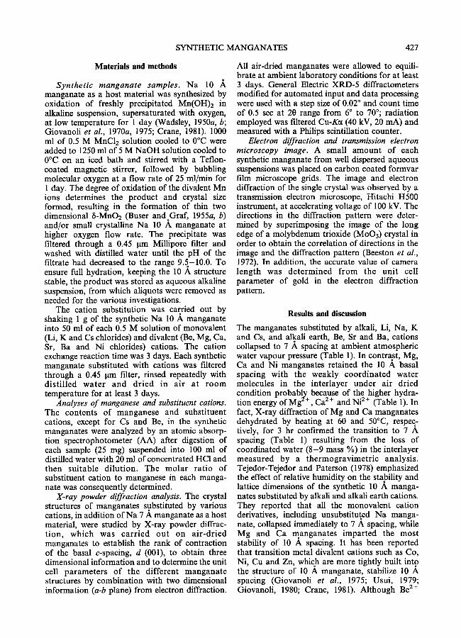
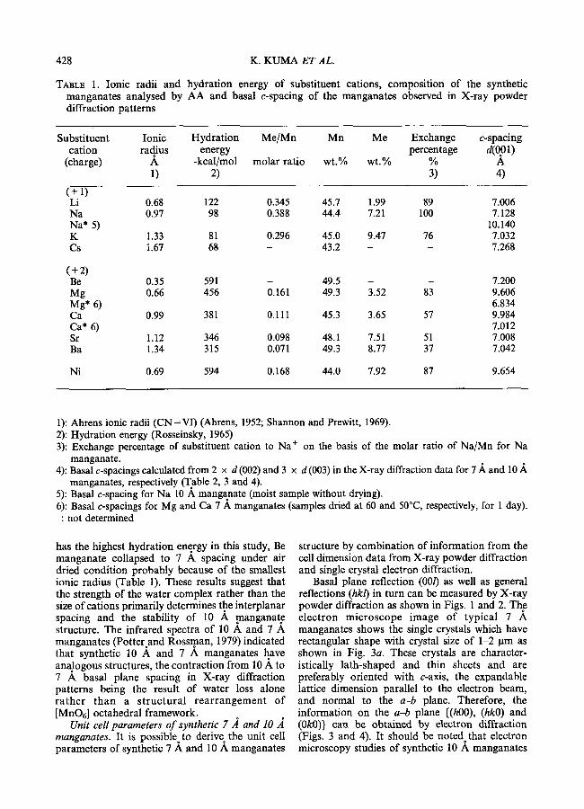
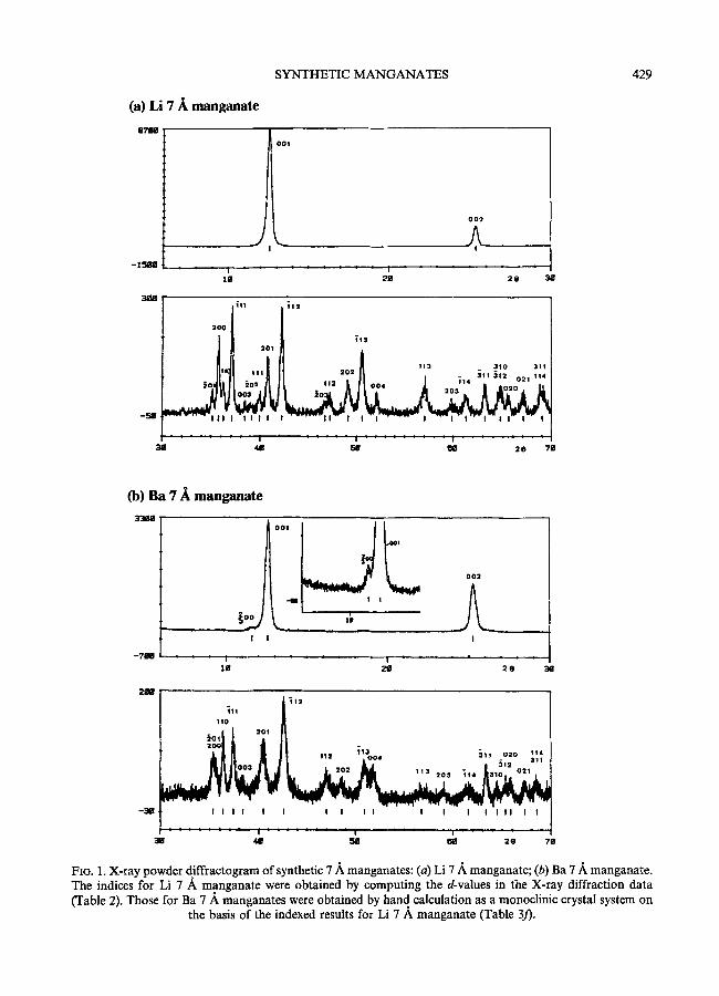
Basal plane reflection (00/) as well as general reflections (hkl) in turn can be measured by X-ray powder diffraction as shown in Figs. 1 and 2. The electron microscope image of typical 7 A manganates shows the single crystals which have rectangular shape with crystal size of 1-2 lam as shown in Fig. 3a. These crystals are character- istically lath-shaped and thin sheets and are preferably oriented with c-axis, the expandable lattice dimension parallel to the electron beam, and normal to the a-b plane. Therefore, the information on the a-b plane {(h00), (hkO) and (0k0)} cart be obtained by electron diffraction (Figs. 3 and 4). It should be noted that electron microscopy studies of synthetic 10 ,~ manganates
SYNTHETIC MANGANATES
(a) Li 7 ~, manganate
oTim
J
0 0 2
l a
i l l
200
201
1 I I I
i 311 411
113
113 3 1 0 311 2 0 2 ~111 - - 3 1 2 21 114
5 8 O 8 2 e 7 8
429
(b) Ba 7 ~ manganate
- 7 R I
!
�9 4 1 i , i I , I | , I ,
O O 2
!
2 e ~
2~W ~11 i l :
110 2 0 1
~ 11: "=oo, ~ . o:o 114
0 3 - 3 1 2 311
I I I I ! i I I I I I I I ! I I ! i !
Flo. I. X-ray powder diffractogram of synthetic 7 ~. manganates: (a) Li 7 .~. manganate; (b) Ba 7/k manganate. The indices for Li 7 A manganate were obtained by computing the d-values in the X-ray diffraction data (Table 2). Those for Ba 7 .A manganates were obtained by hand calculation as a monoclinic crystal system on
the basis of the indexed results for Li 7 A manganate (Table 3/).
430 K. KUMA E T AL.
(a) Ca lO/~ manganate
o o , y..a l 002 -lss ,, a ~. t
~00
! I
0 0 3
,A. !
l m 2 ~ 2 0 3 8
-,IB
3~I
110 2 0 1
2 0 1 111
~176 J~o2 I
/ I L i t ~'".~, oo~';, ~ ~o, ~o: oo. ~" ,e,' I
I I I I II I I I I | I I I I I I II II I
I i . . . . . ! - i 411 ~i~ 81 2e 711
(b) Ni 10 A manganate
O01
2 1 e
- 3 8
O O 2
! ! !
: 1 ' ' ' ' ' " " i ' ' ' ' ' ' ' ' l g ~ 2 G 3 8
2 0 0 0 4 1 2
i l 2 0 0 1 1 I d, ~ o o s " ' - 3 1 o 3 1 2
313 2 0 4 0 2 0 ,1111 I l l .o.~ : i I.,, o..
I I ! I I I I I I I I I I I I f
38 ~ ~ ~ 20
FIG. 2. X-ray powder diffractograms of synthetic 10 lk manganates: (a) Ca 10 A, manganate; (b) Ni 10 A manganate. The indices were obtained by hand calculation as a monoclinic crystal system on the basis of the
indexed results for Li 7 A manganate (Table 4b and c).

a
-~b
a
431
(c) Na manganate (b) Li manganate
SYNTHETIC MANGANATES
(a) Cs manganate
(d) K manganate
~31o i �84
/ 2~o
t 3~o
i
(e) Cs manganate
�9 9
~020
PO '
t220
?020
- - b
I ~ /r
FIG. 3. Electron micrograph and electron diffraction patterns in the a-b plane of synthetic monovalent cation manganates: (a) electron micrograph of typical manganate (Cs manganate); (b) Li manganate; (c) Na manganate; (d) K manganate; (e) Cs manganate. The reflection marked with arrow: (% 0 0). The indices were obtained as an orthorhombic crystal system in the a-b plane on the basis of fundamental pseudohexagonal
reflections.

432 K. KUMA ET AL.
(a) Be manganate
0
~310
~'110
r
(b) Mg manganate
(c) Ca manganate
, . ~ : ~ : . : : J , : . o Q310
�9 . ~ , . �9 . ~
�9 ; ~200 " : : ' 2 2 0
(d) Sr manganate
e 0 2 0
.220
~o2o
(e) Ba manganate (f) Ni manganate
110
"220
~, 020
~ 2 2 0
": o2o
=-b
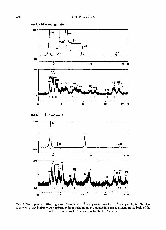
FIG, 4. Electron diffraction patterns in the a-b plane of synthetic divalent cation manganates: (a) Be manganate; (b) Mg manganate, (c) Ca manganate; (d) Sr manganate; (e) Ba manganate; (]) Ni manganate. The reflection marked with arrow: (% 0 0). The indices were obtained as an orthorhombic crystal system in the a-b
plane on the basis of fundamental pseudohexagonal reflections.

SYNTHETIC MANGANATES 433
TABLE 2. Indexed interplanar spacings of Li 7 A manganate obtained by computing the X-ray diffraction data. (a=5.152 A, b=2.845 A, c=7.196 A, 13=103.08 ~
Line No. Intensity d(m~a,.) d(e~tle.) hkl d(meas.)~d(eale.) I/Io A A
( x 100) ( x lO00)
1 100 7.016 7.009 001 7 2 17 3.503 3.505 002 -2 3 1 2.553 2.553 301 0 4 2 2.509 2.509 200 0 5 1 2.475 2.475 110 0 6 3 2.420 2.420 i l l 0 7 < 1 2.336 2.336 003 0 8 < 1 2.299 2.302 302 -3 9 < 1 2.256 2.256 111 0
10 2 2.209 2.209 201 0 11 3 2.136 2.137 i12 -1 12 < 1 1.941 1.943 303 -2 13 < 1 1.923 1.923 112 0 14 1 1.850 1.851 202 -1 15 2 1.802 1.802 i13 0 16 1 1.751 1.752 004 -1 17 1 1.612 1.611 113 1 18 < 1 1.543 1.544 203 -1 19 < 1 1.511 1.512 i14 -1 20 1 1.470 1.470 311 0 21 1 1.438 1.442 310 -4
1.436 312 2 22 < 1 1.422 1.422 020 0 23 < 1 1.394 1.394 021 0 24 1 1.361 1.361 311 0
1.360 114 1
d(me~s.) and d(cale.) were d-spacings measured from X-ray diffraction pattern (Fig. 1) and calculated by computing the X-ray diffraction data, respectively.
result in a transformation from an initial 10 ,~ basal spacing to 7 A due to the exposure of the samples to high vacuum in the electron micro- scope.
These electron diffraction patterns contain the fundamental and imperfect hexagonal reflections. Only first order reflections appear for Be 7 A manganate (Fig. 4a), while those for the other cation manganates show superlattice reflections in the a-b plane (Li, K, Cs, Mg, Ca, Sr, Ba and Ni manganates; Figs. 3 and 4) and/or streaking in the a-direction (Na, K and Cs manganates; Fig. 3). Only observation of fundamental hexagonal reflections for Be manganate may be because of lower scattering power of light element Be and/or lower exchange percentage of Be to Na +. The fundamental hexagonal reflections for all the cation manganates were temporarily interpreted as an isotropic manganate layer structure based on edge-shared [MnOt] octahedra. The morphology
and electron diffraction data indicate that the observed hexagonal symmetry is a pseudosym- metry; the true unit cell appears to be actually or thorhombic or monoclinic. All the cat ion manganates displayed strong reflections, (200), (020), (110), (310), etc. in Figs. 3 and 4. These fundamental lattice reflections presumably appear as a result of the ordered edge-shared [MnOt] octahedra extending in the a-b plane.
In this study, the Li 7 A manganate structure is one of the best ordered manganates in X-ray diffraction (Fig. la). The diffraction data of Li 7 ,~ manganate was therefore selected to find the unit cell from the X-ray powder diffraction pattern by computing the diffraction data in the programme of which principles were described by Visser (1969). The indexed result (Table 2 and Fig. la) indicates that the Miller indices can be expressed as a monoclinc crystal system by only combination between basal spacing, (00/), observed in X-ray
434 K. KUMA ET AL.
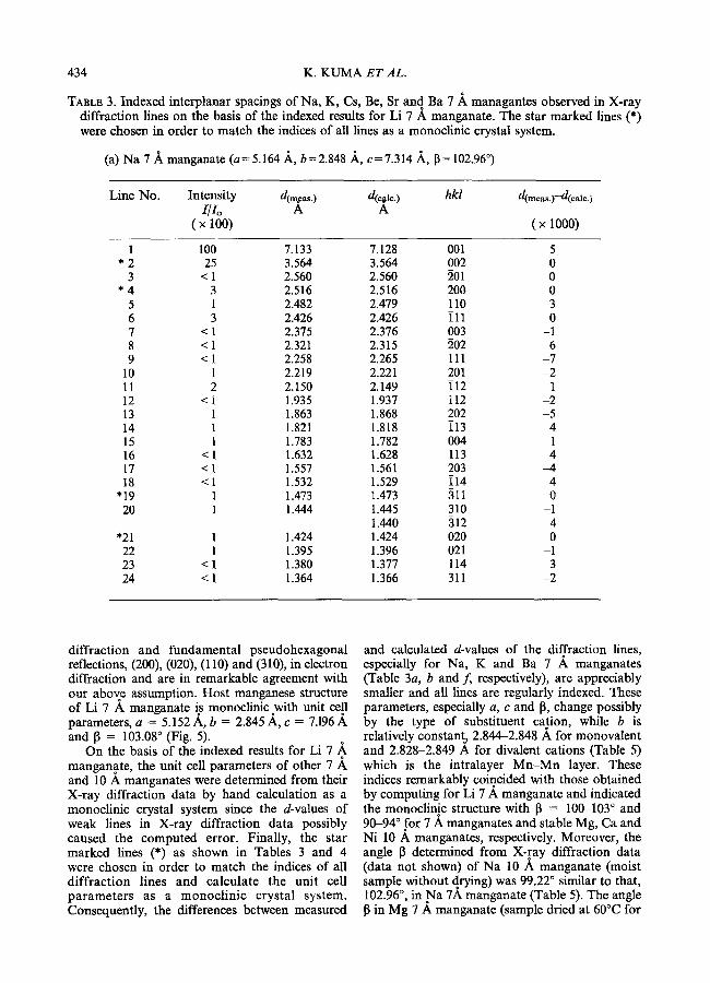
TABLE 3. Indexed interplanar spacings of Na, K, Cs, Be, Sr and Ba 7 ,~ managantes observed in X-ray diffraction lines on the basis of the indexed results for Li 7 A manganate. The star marked lines (*) were chosen in order to match the indices of all lines as a mort,clinic crystal system.
(a) Na 7 A, manganate (a=5.164 ,~, b=2.848 A, c=7.314 A_, 13= 102.96 ~
Line No. Intensity d(n~,s.) d(c~lc.) hkl d( . . . . . ) -d(ca le . ) XlXo A A
( x loo) (x moo)
1 100 7.133 7.128 001 5 * 2 25 3.564 3.564 002 0
3 < 1 2.560 2.560 201 0 * 4 3 2.516 2.516 200 0
5 1 2.482 2.479 110 3 6 3 2.426 2.426 i l l 0 7 < 1 2.375 2.376 003 -1 8 < 1 2.321 2.315 202 6 9 < 1 2.258 2.265 111 -7
10 1 2.219 2.221 201 -2 11 2 2.150 2.149 i12 1 12 < 1 1.935 1.937 112 -2 13 1 1.863 1.868 202 -5 14 1 1.821 1.818 i13 4 15 1 1.783 1.782 004 1 16 < 1 1.632 1.628 113 4 17 < 1 1.557 1.561 203 -4 18 < 1 1.532 1.529 i14 4
"19 1 1.473 1.473 311 0 20 1 1.444 1.445 310 -1
1.440 312 4 "21 1 1.424 1.424 020 0 22 1 1.395 1.396 021 -1 23 < 1 1.380 1.377 114 3 24 < 1 1.364 1.366 311 -2
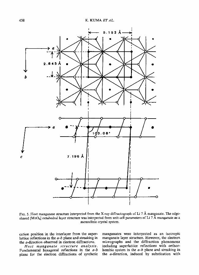
diffraction and fundamental pseudohexagonal reflections, (200), (020), (110) and (310), in electron diffraction and are in remarkable agreement with our above assumption. Host manganese structure of Li 7 ,~ manganate is monoclinic.with unit ce.ll parameters, a = 5.152 A, b = 2.845 A, c = 7.196 A and 13 = 103.08 ~ (Fig. 5).
On the basis of the indexed results for Li 7 A, manganate, the unit cell parameters of other 7 ,~ and 10 A manganates were determined from their X-ray diffraction data by hand calculation as a monoclinic crystal system since the d-values of weak lines in X-ray diffraction data possibly caused the computed error. Finally, the star marked lines (*) as shown in Tables 3 and 4 were chosen in order to match the indices of all diffraction lines and calculate the unit cell parameters as a monoclinic crystal system. Consequently, the differences between measured
and calculated d-values of the diffraction lines, especially for Na, K and Ba 7 A. manganates (Table 3a, b and f, respectively), are appreciably smaller and all lines are regularly indexed. These parameters, especially a, c and 13, change possibly by the type of substituent cation, while b is relatively constanta 2.844-2.848 A for monovalent and 2.828-2.849 A for divalent cations (Table 5) which is the intralayer M n - M n layer. These indices remarkably coincided with those obtained by computing for Li 7 A manganate and indicated the monoclinic structure with 13 = 100-103 ~ and 90-94 ~ for 7 A manganates and stable Mg, Ca and Ni 10 A manganates, respectively. Moreover, the angle ~ determined from X-ray diffraction data (data not shown) of Na 10 A manganate (moist sample without drying) was 99.22 ~ similar to that, 102.96 ~ in Na 7A manganate (Table 5). The angle 13 in Mg 7 A manganate (sample dried at 60~ for
SYNTHETIC MANGANATES
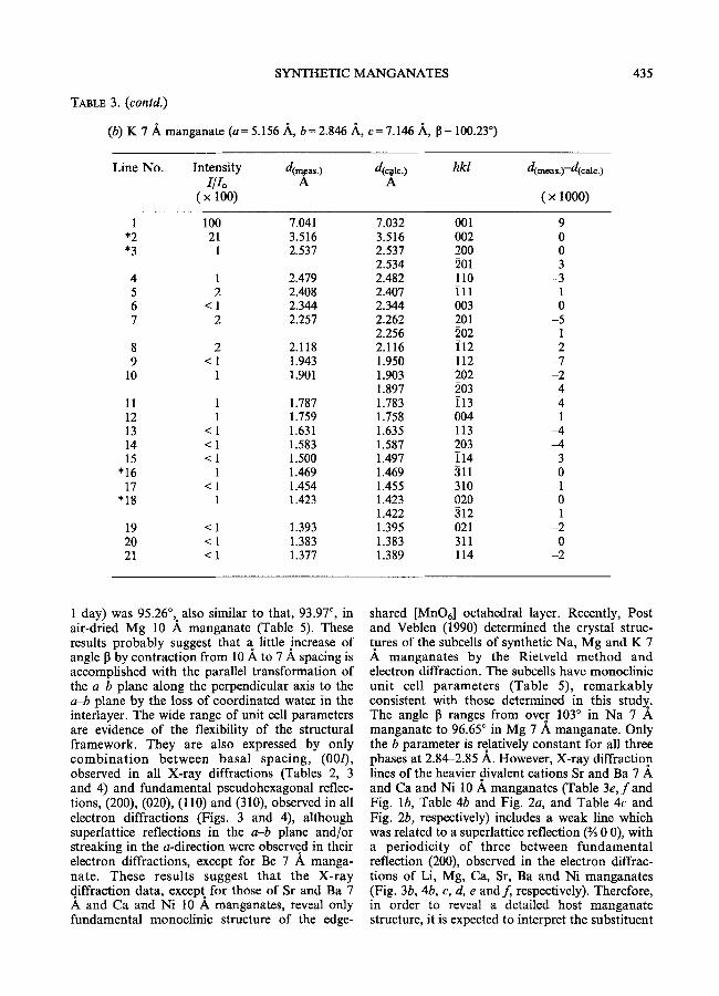
TABLE 3. (contd.)
(b) K 7 .~, manganate (a=5.156 ,~, b=2.846 ,~, c=7.146 A, 13= 100.23 ~
435
Line No. Intensity d(~as.) d(~l~.) hkl IIio A A
( x lOO)
d(meas.)"-d(calc.)
(x 1000)
1 100 7.041 7.032 001 *2 21 3.516 3.516 002 *3 1 2.537 2.537 200
2.534 201 4 1 2.479 2.482 110 5 2 2.408 2.407 i l l 6 < 1 2.344 2.344 003 7 2 2.257 2.262 201
2.256 202 8 2 2.118 2.116 i12 9 < 1 1.943 1.950 112
10 1 1.901 1.903 202 1.897 203
11 1 1.787 1.783 i13 12 1 1.759 1.758 004 13 < 1 1.631 1.635 113 14 < 1 1.583 1.587 203 15 < 1 1.500 1.497 114
"16 1 1.469 1.469 311 17 < 1 1.454 1.455 310
"18 1 1.423 1.423 020 1.422 312
19 < 1 1.393 1.395 021 20 < 1 1.383 1.383 311 21 < 1 1.377 1.389 114
9 0 0 3
-3 1 0
-5 1 2
-7 -2
4 4 1
-4 -4
3 0
-1 0 1
-2 0
-2
1 day) was 95.26~ also similar to that, 93.97 ~ in air-dried Mg 10 A manganate (Table 5). These results probably suggest that a little increase of angle 13 by contraction from 10 A to 7 ,~ spacing is accomplished with the parallel transformation of the a-b plane along the perpendicular axis to the a-b plane by the loss of coordinated water in the interlayer. The wide range of unit cell parameters are evidence of the flexibility of the structural framework. They are also expressed by only combina t i on between basa l spacing, (00/), observed in all X-ray diffractions (Tables 2, 3 and 4) and fundamental pseudohexagonal reflec- tions, (200), (020), (110) and (310), observed in all electron diffractions (Figs. 3 and 4), although superlattice reflections in the a-b plane and/or streaking in the a-direction were observed in their electron diffractions, except for Be 7 A manga- hate. These results suggest that the X-ray diffraction data, except for those of Sr and Ba 7 .& and Ca and Ni 10 A manganates, reveal only fundamental monoclinic structure of the edge-
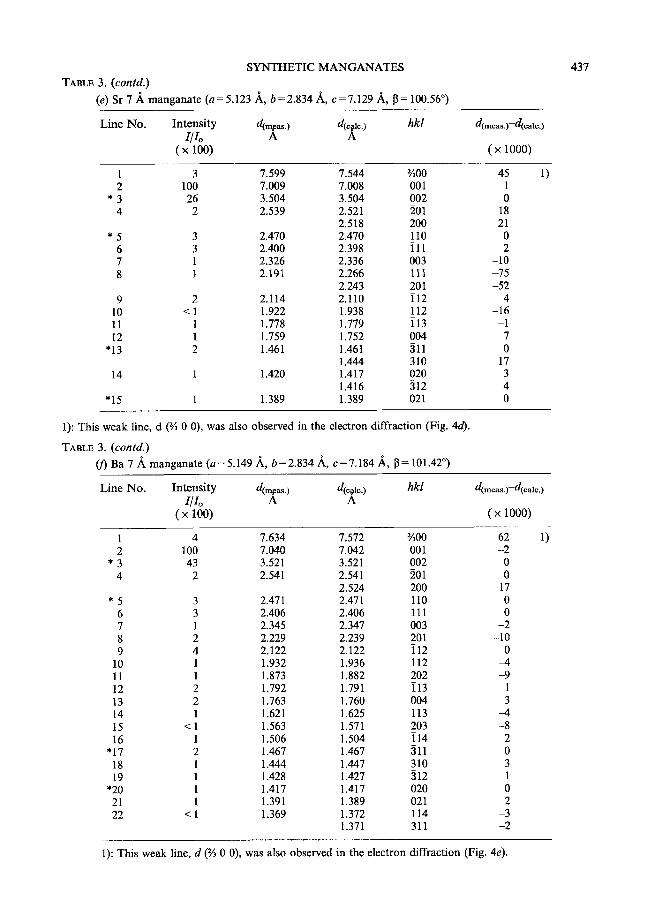
shared [MnO6] octahedrat layer. Recently, Post and Veblen (1990) determined the crystal struc- tures of the subcells of synthetic Na, Mg and K 7 .& manganates by the Rietveld method and electron diffraction. The subcells have monoclinic uni t cell parameters (Table 5), remarkably consistent with those determined in this study. The angle 13 ranges from over 103 ~ in Na 7 A manganate to 96.65 ~ in Mg 7 A manganate. Only the b parameter is relatively constant for all three phases at 2.84-2.85 A. However, X-ray diffraction lines of the heavier divalent cations Sr and Ba 7 and Ca and Ni 10 A manganates (Table 3e, f a n d Fig. lb, Table 4b and Fig. 2a, and Table 4c and Fig. 2b, respectively) includes a weak line which was related to a superlattice reflection (% 0 0), with a periodicity of three between fundamental reflection (200), observed in t he electron diffrac- tions of Li, Mg, Ca, Sr, Ba and Ni manganates (Fig. 3b, 4b, c, d, e and f, respectively). Therefore, in order to reveal a detailed host manganate structure, it is expected to interpret the substituent
436 K. K U M A ET AL.
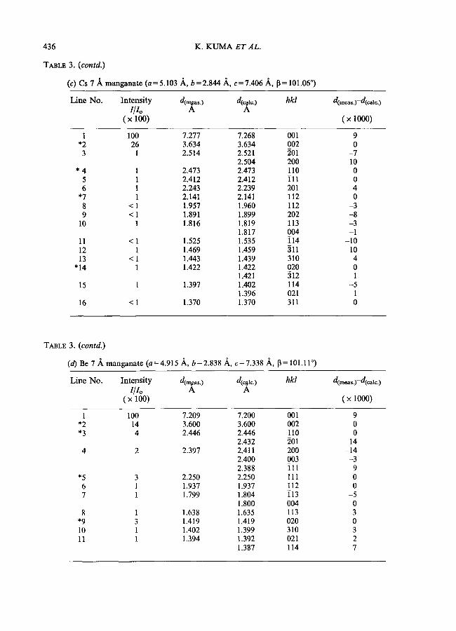
TABLE 3. (contd.)
(c) Cs 7 A manganate (a=5.103 ~,, b=2.844 .~, c=7.406 ~,, 13= 101.06 ~
Line No. Intensity d(r~as.) d(r162162 hkl I/Io A A
( x 100)
d(meas.)-d(cale.)
(• lOOO)
1 100 7.277 7.268 001 *2 26 3.634 3.634 002 3 1 2.514 2.521 201
2.504 200 * 4 1 2.473 2.473 110
5 1 2.412 2.412 i l l 6 1 2.243 2.239 201
*7 1 2.141 2.141 i12 8 < 1 1.957 1.960 112 9 < 1 1.891 1.899 202
10 1 1.816 1.819 i13 1.817 004
11 < I 1.525 1.535 i14 12 1 1.469 1.459 311 13 < 1 1.443 1.439 310
"14 1 1.422 1.422 020 1.421 312
15 1 1.397 1.402 114 1.396 021
16 < 1 1.370 1.370 311
9 0
-7 10 0 0 4 0
-3 -8 -3 -1
-10 10 4 0 1
-5 1 0
TABLE 3. (contd.)
(d) Be 7 A manganate (a=4.915 A, b=2.838 A, c=7.338 A,, 13=101.11 ~
Line No. Intensity d(m~as.) d(r hkl Z/Zo h A
( x 100)
d(meas.)-d(calc.)
( x 1000)
1 100 7.209 7.200 001 *2 14 3.600 3.600 002 *3 4 2.446 2.446 110
2.432 201 4 2 2.397 2.411 200
2.400 003 2.388 i l l
*5 3 2.250 2.250 111 6 1 1.937 1.937 112 7 1 1.799 1.804 i13
1.800 004 8 1 1.638 1.635 113
*9 3 1.419 1.419 020 10 l 1.402 1.399 310 11 1 1.394 1.392 021
1.387 114
9 0 0
14 -14
-3 9 0 0
-5 0 3 0 3 2 7
SYNTHETIC M A N G A N A T E S
TABLE 3. (contd.) (e) Sr 7 ,~ manganate (a=5.123 .A, b=2.834 ,~, c=7.129 A, 13= 100.56 ~
437
Line No. Intensi ty d(~as.) d(c#.) hkl 1/lo A A
( x 100)
d(meas.)-d(cale.)
( • 1000)
1 3 7.599 7.544 �90 45 2 100 7.009 7.008 001 1
* 3 26 3.504 3.504 002 0 4 2 2.539 2.521 201 18
2.518 200 21 * 5 3 2.470 2.470 110 0
6 3 2.400 2.398 i l l 2 7 1 2.326 2.336 003 -10 8 1 2.191 2.266 111 -75
2.243 201 -52 9 2 2.114 2.110 i12 4
10 < 1 1.922 1.938 112 -16 11 1 1.778 1.779 i13 -1 12 1 1.759 1.752 004 7
"13 2 1.461 1.461 311 0 1.444 310 17
14 1 1.420 1.417 020 3 1.416 212 4
"15 1 1.389 1.389 021 0
1): This weak line, d (�90 0 0), was also observed in the electron diffraction (Fig. 4d).
TABLE 3. (contd.) (10 Ba 7 ,~ manganate ( a = 5.149 A, b=2.834 ,~, c=7.184 ,~, 13 = 101.42 ~
Line No. Intensi ty d(m,eas.) d(c~lc. ) hkl d( . . . . . )-d(calc.) I/Io A A
( x loo ) ( x looo)
1 4 7.634 7.572 %00 62 2 100 7.040 7.042 001 -2
* 3 43 3.521 3.521 002 0 4 2 2.541 2.541 201 0
2.524 200 17 * 5 3 2.471 2.471 110 0
6 3 2.406 2,406 i l l 0 7 1 2.345 2.347 003 -2 8 2 2.229 2.239 201 -10 9 4 2.122 2.122 i12 0
10 1 1.932 1.936 112 -4 11 1 1.873 1.882 202 -9 12 2 1.792 1.791 i13 1 13 2 1.763 1.760 004 3 14 1 1.621 1.625 113 -4 15 < 1 1.563 1.571 203 -8 16 1 1.506 1.504 i14 2
"17 2 1.467 1.467 311 0 18 1 1.444 1.447 310 -3 19 1 1.428 1.427 312 1
*20 1 1.417 1.417 020 0 21 1 1.391 1.389 021 2 22 < 1 1.369 1.372 114 -3
1.371 311 -2
1)
1): This weak line, d (% 0 0), was also observed in the electron diffraction (Fig. 4e).
438 K. KUMA ET AL.
b
/ C
5 . 1 5 2 ~ ! I
I
o 1
/- Z_2 ?-- > a 0 - - - ~ �9
~ 3 . 0 8 *
7. 1 9 6 .,l i
I I I I
e - - ' t - - ~ I / �9
i d
FIG. 5. Host manganese structure interpreted from the X-ray diffractograph of Li 7 A man/~anate. The edge- shared [MnO6] octahedral layer structure was interpreted from unit cell parameters of Li 7 A manganate as a
monoclinic crystal system.
cation position in the interlayer from the super- lattice reflections in the a-b plane and streaking in the a-direction observed in electron diffractions.
H o s t m a n g a n a t e s t r u c t u r e ana ly s i s . Fundamental hexagonal reflections in the a-b plane for the electron diffractions of synthetic
manganates were interpreted as an isotropic manganate layer structure. However, the electron micrographs and the diffraction phenomena including superlattice reflections with orthor- hombic system in the a-b plane and streaking in the a-direction, induced by substitution with
SYNTHETIC MANGANATES 439
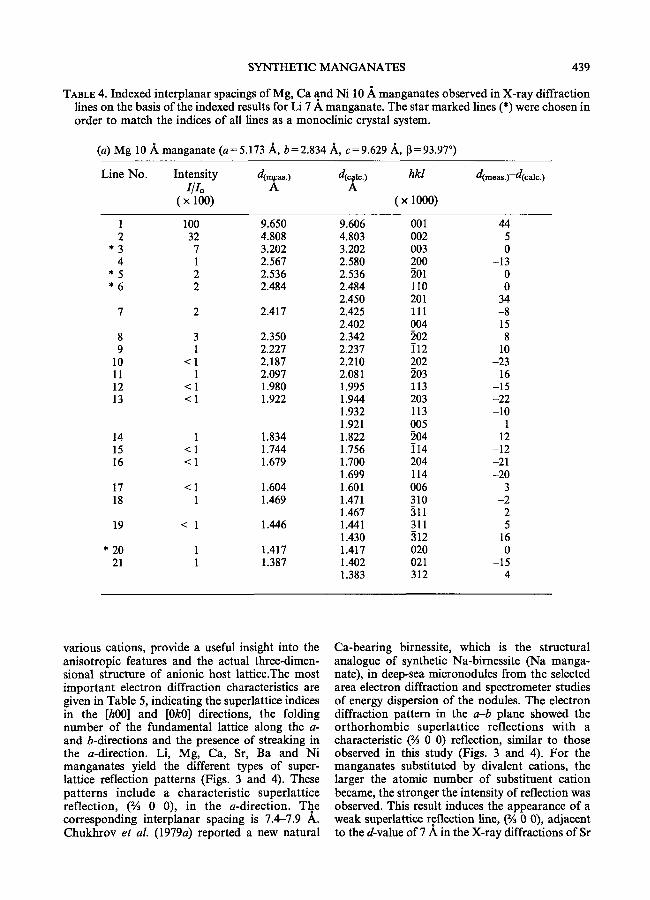
TABLE 4. Indexed interplanar spacings of Mg, Ca and Ni 10 A manganates observed in X-ray diffraction lines on the basis of the indexed results for Li 7 A manganate. The star marked lines (*) were chosen in order to match the indices of all lines as a monoclinic crystal system.
(a) Mg 10 ,~ manganate (a= 5.173 ,~,, b=2.834 ~,, c=9.629 A, 13=93.97 ~
Line No. Intensity a~e,s.) d(c#c.) hkl d( . . . . . )-d(ealc.) 1/lo A A
( x 100) ( x 1000)
1 100 9.650 9.606 001 44 2 32 4.808 4.803 002 5
* 3 7 3.202 3.202 003 0 4 1 2.567 2.580 200 -13
* 5 2 2.536 2.536 201 0 * 6 2 2.484 2.484 110 0
2.450 201 34 7 2 2.417 2.425 i l l -8
2.402 004 15 8 3 2.350 2.342 202 8 9 1 2.227 2.237 i12 -10
10 < 1 2.187 2.210 202 -23 11 1 2.097 2.081 203 16 12 < 1 1.980 1.995 i13 -15 13 < 1 1.922 1.944 203 -22
1.932 113 -10 1.921 005 1
14 1 1.834 1.822 204 12 15 < 1 1.744 1.756 i14 -12 16 < 1 1.679 1.700 204 -21
1.699 114 -20 17 < 1 1.604 1.601 006 3 18 1 1.469 1.471 310 -2
1.467 311 2 19 < 1 1.446 1.441 311 5
1.430 312 16 * 20 1 1.417 1.417 020 0
21 1 1.387 1.402 021 -15 1.383 312 4
various cations, provide a useful insight into the anisotropic features and the actual three-dimen- sional structure of anionic host lattice.The most important electron diffraction characteristics are given in Table 5, indicating the superlattice indices in the [h00] and [0k0] directions, the folding number of the fundamental lattice along the a- and b-directions and the presence of streaking in the a-direction. Li, Mg, Ca, Sr, Ba and Ni manganates yield the different types of super- lattice reflection patterns (Figs. 3 and 4). These patterns include a characteristic superlattice reflection, (% 0 0), in the a-direction. The corresponding interplanar spacing is 7.4-7.9 A. Chukhrov et al. (1979a) reported a new natural
Ca-bearing birnessite, which is the structural analogue of synthetic Na-birnessite (Na manga- nate), in deep-sea micronodules from the selected area electron diffraction and spectrometer studies of energy dispersion of the nodules. The electron diffraction pattern in the a-b plane showed the orthorhombic superlattice reflections with a characteristic (% 0 0) reflection, similar to those observed in this study (Figs. 3 and 4). For the manganates substituted by divalent cations, the larger the atomic number of substituent cation became, the stronger the intensity of reflection was observed. This result induces the appearance of a weak superlattice reflection line, (% 0 0), adjacent to the d-value of 7 A in the X-ray diffractions of Sr
440 K. KUMA ET AL.
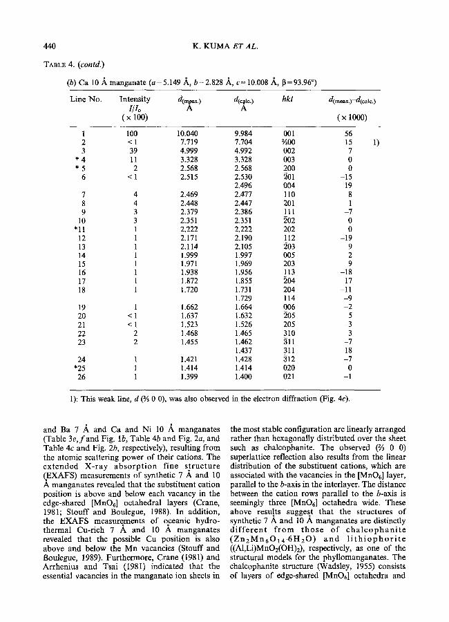
TABLE 4. (contd.)
(b) Ca 10 A manganate (a=5.149 A, b=2.828 ]k, c= 10.008 A,, 13=93.96 ~
Line No. Intensity d(m~as.) d(cr hkl d( . . . . . )-d(ca,r I/Io A A
( x 100) ( x 1000)
1 100 10.040 9.984 001 56 2 < 1 7.719 7.704 �90 15 3 39 4.999 4.992 002 7
* 4 11 3.328 3.328 003 0 * 5 2 2.568 2.568 200 0
6 < 1 2.515 2.530 201 -15 2.496 004 19
7 4 2.469 2.477 110 8 8 4 2.448 2.447 201 1 9 3 2.379 2.386 111 -7
10 3 2.351 2.351 202 0 *11 1 2.222 2.222 202 0
12 1 2.171 2.190 112 -19 13 1 2.114 2.105 203 9 14 1 1.999 1.997 005 2 15 1 1.971 1.969 203 9 16 1 1.938 1.956 113 -18 17 1 1.872 1.855 204 17 18 1 1.720 1.731 204 -11
1.729 114 -9 19 1 1.662 1.664 006 -2 20 < 1 1.637 1.632 205 5 21 < 1 1.523 1.526 205 -3 22 2 1.468 1.465 310 3 23 2 1.455 1.462 511 -7
1.437 311 18 24 1 1.421 1.428 312 -7
*25 1 1.414 1.414 020 0 26 1 1.399 1.400 021 -1
1)
1): This weak line, d (�90 0 0), was also observed in the electron diffraction (Fig. 4c).
and Ba 7 .~ and Ca and Ni 10 .~. manganates (Table 3e, f a n d Fig. lb, Table 4b and Fig. 2a, and Table 4c and Fig. 2b, respectively), resulting from the atomic scattering power of their cations. The extended X-ray absorp t ion fine s t ructure (,EXAFS) measurements of synthetic 7 ,~ and 10 A manganates revealed that the substituent cation position is above and below each vacancy in the edge-shared [MnO6] octahedral layers (Crane, 1981; Stouff and Boulegue, 1988). In addition, the EXAFS measurements of oceanic hydro- thermal Cu-rich 7 A and 10 A manganates revealed that the possible Cu position is also above and below the Mn vacancies (Stouff and Boulegue, 1989). Furthermore, Crane (1981) and Arrhenius and Tsai (1981) indicated that the essential vacancies in the manganate ion sheets in
the most stable configuration are linearly arranged rather than hexagonally distributed over the sheet such as chalcophanite. The observed (% 0 0) superlattice reflection also results from the linear distribution of the substituent cations, which are associated with the vacancies in the [MnO6] layer, parallel to the b-axis in the interlayer. The distance between the cation rows parallel to the b-axis is seemingly three [MnO6] octahedra wide. These above results suggest that the structures of synthetic 7 A and 10 A manganates are distinctly d i f f e r e n t f rom those of c h a l c o p h a n i t e ( Z n E M n 6 0 1 4 . 6 H 2 0 ) a n d l i t h i o p h o r i t e ((A1,Li)MnOE(OH)2), respectively, as one of the structural models for the phyllomanganates. The chalcophanite structure (Wadsley, 1955) consists of layers of edge-shared [MnO6] octahedra and
SYNTHETIC MANGANATES
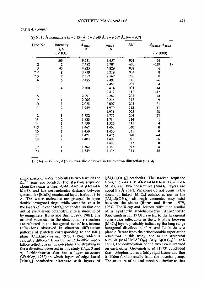
TAaL8 4. (contd.)
(c) Ni 10 A manganate (a= 5.134 ,~, b=2.849 .~, c=9.657 ,~, 13 = ~90 ~
441
Line No. Intensity d ( ~ . ) d(~l~.) hkl d( . . . . . ) - d ( e a l e . )
I/Io A A ( x 100) ( x 1000)
1 100 9.631 9.657 001 -26 2 2 7.482 7.701 �90 -219 3 45 4.823 4.829 002 -6
* 4 8 3.219 3.219 003 0 * 5 2 2.567 2.567 200 0
6 5 2.485 2.491 110 -6 2.481 201 4
7 4 2.400 2.414 004 -14 2.412 111 -12
8 3 2.291 2.267 202 24 9 4 2.205 2.214 112 -9
10 1 2.028 2.007 203 21 11 2 1.959 1.970 113 -11
1.931 005 28 12 1 1.782 1.759 204 23 13 2 1.733 1.734 114 - 1 14 1 1.530 1.526 115 4
"15 2 1.467 1.467 310 0 16 1 1.450 1.450 311 0 17 2 1.421 1.425 020 -4 18 2 1.403 1.409 021 -6
1.403 312 0 19 1 1.362 1.366 022 -4 20 1 1.349 1.335 313 14
1)
1): This weak line, d (�90 was also observed in the electron diffraction (Fig. 4./).
single sheets of water molecules between which the Zn 2+ ions are located. The stacking sequence along the c-axis is thus: -O-Mn-O-Zn-H20-Zn-O- Mn-O, and the perpendicular distance between consecutive [MnO6] octahedral layers is about 7.16 A. The water molecules are grouped in open double hexagonal rings, while vacancies exist in the layers of linked [MnOd octahedra, so that one out of every seven octahedral sites is unoccupied by manganese (Burns and Burns, 1979, 1981). The ordered vacancies in the chalcophanite structure are reflected in the hexagonal net of less intense reflections observed in electron diffract ion patterns of platelets corresponding to the (001) plane (Chukhrov et al., 1978, 1979b), which is evidently different from the orthorhombic super- lattice reflections in the a-b plane and streaking in the a-direction observed in this study (Figs. 3 and 4). Li thiophori te also has a layer structure (Wadsley, 1952) in which layers of edge-shared [MnO6] oc tahedra a l ternate with layers of
[(AI,Li)(OH)6 ] octahedra. The stacked sequence along the c-axis is: -O-Mn-O-OH-(A1,Li)-OH-O- Mn-O, and two consecutive [MnO6] layers are about 9.5 A apart. Vacancies do not occur in the sheets of linked [MnOr] octahedra, nor in the [(AI,Li)(OH)6], although vacancies may exist between the sheets (Bums and Burns, 1979, 1981). The X-ray and electron diffraction studies of a synthet ic s to ich iomet r ic l i t h iophor i t e (Giovanoli et al., 1973) have led to the hexagonal superlattice reflections in the a-b plane between [MnO6] layers, probably indicating the long-range hexagonal distribution of A1 and Li in the a-b plane different from the orthorhombic superlattice reflections in this study, and to the structural
4 + 2 + + formula [Mn5 Mn Oi2]-.[Al4Li2(OH)12] indi- cating the composition of the two layers stacked on each other. Giovanoli et al. (1973) concluded that litbiophorite has a fairly rigid lattice and that it differs fundamentally from the buserite group. The structure of natural asbolane, similar to that
442
d(i/3 1 §
d(5 /3 1
K. KUMA E T AL.
0)/ /
o o~m
+ o r - -
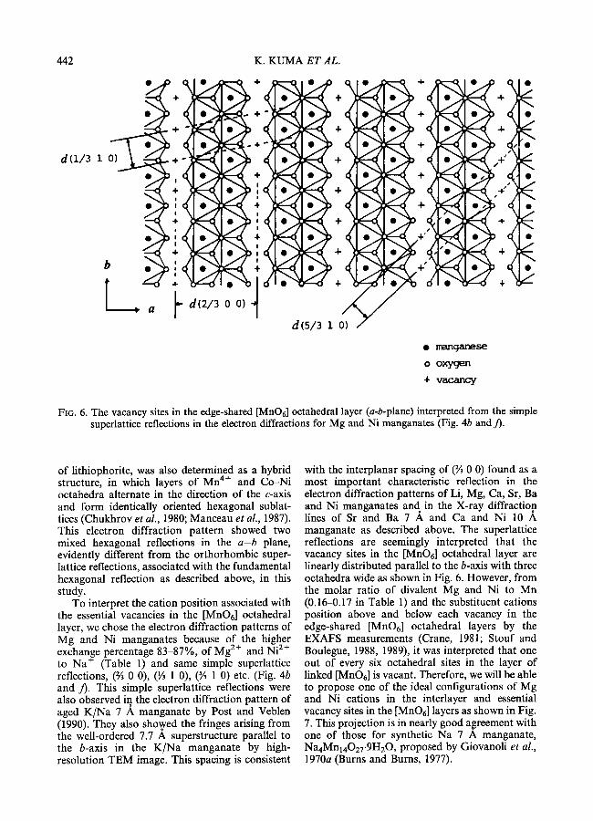
FIG. 6. The vacancy sites in the edge-shared [MnO6] octahedral layer (a-b-plane) interpreted from the simple superlattice reflections in the electron diffractions for Mg and Ni manganates (Fig. 4b and/).
of lithiophorite, was also determined as a hybrid structure, in which layers of Mn 4+ and Co-Ni octahedra alternate in the direction of the c-axis and form identically oriented hexagonal sublat- tices (Chukhrov et al., 1980; Manceau et al., 1987). This electron diffraction pattern showed two mixed hexagonal reflections in the a - b plane, evidently different from the orthorhombic super- lattice reflections, associated with the fundamental hexagonal reflection as described above, in this study.
To interpret the cation position associated with the essential vacancies in the [MnO6] octahedral layer, we chose the electron diffraction patterns of Mg and Ni manganates because of the higher exchange percentage 83-87%, of Mg 2+ and Ni 2+ to Na (Table 1) and same simple superlattice reflections, (36 0 0), (�89 1 0), (% 1 0) etc. (Fig. 4b and / ) . This simple superlattice reflections were also observed in the electron diffraction pattern of aged K/Na 7 ~, manganate by Post and Veblen (1990). They also showed the fringes arising from the well-ordered 7.7 A superstructure parallel to the b-axis in the K/Na manganate by high- resolution TEM image. This spacing is consistent
with the interplanar spacing of (2/3 0 0) found as a most important characteristic reflection in the electron diffraction patterns of Li, Mg, Ca, Sr, Ba and Ni manganates and in the X-ray diffraction lines of Sr and Ba 7 A and Ca and Ni 10 A manganate as described above. The superlattice reflections are seemingly interpreted that the vacancy sites in the [MnO6] octahedral layer are linearly distributed parallel to the b-axis with three octahedra wide as shown in Fig. 6. However, from the molar ratio of divalent Mg and Ni to Mn (0.16-0.17 in Table 1) and the substituent cations position above and below each vacancy in the edge-shared [MnO6] octahedral layers by the EXAFS measurements (Crane, 1981; Stouf and Boulegue, 1988, 1989), it was interpreted that one out of every six octahedral sites in the layer of linked [MnO6] is vacant. Therefore, we will be able to propose one of the ideal configurations of Mg and Ni cations in the interlayer and essential vacancy sites in the [MnO6] layers as shown in Fig. 7. This projection is in nearly good agreement with one of those for synthetic Na 7 A manganate, Na4Mn14027.gH20, proposed by Giovanoli et al., 1970a (Burns and Bums, 1977).
SYNTHETIC MANGANATES 443
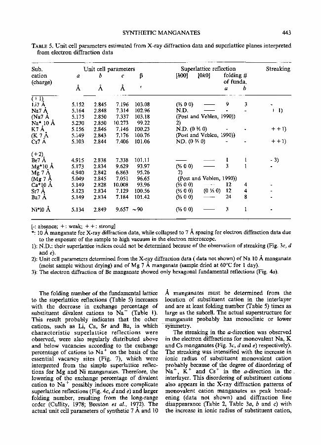
TABLE 5. Unit cell parameters estimated from X-ray diffraction data and superlattice planes interpreted from electron diffraction data
Sub. Unit cell parameters Superlattice reflection cation a b c I 3 [hO0] [OkO] folding # (charge) of funda.
A A A ~ a b
Streaking
(+ 1). Li7 A 5.152 2.845 7.196 103.08 (�90 0 0) 9 Na7 .~ 5.164 2.848 7.314 102.96 N.D. (Na7 .~. , 5.175 2.850 7.337 103.18 (Post and Veblen, 1990)) Na* 10 A 5.230 2.850 10.273 99.22 2) K7 A 5.156 2.846 7.146 100.23 N.D. (0 �90 0) (K 7 .~. 5.149 2.843 7.176 100.76 (Post and Veblen, 1990)) Cs7 A. 5.103 2.844 7.406 101.06 ND. (0 �90 0)
(+2). Be7 A 4.915 2.838 7.338 101.11 1 1 Mg*IO_A 5.173 2.834 9.629 93.97 (�90 0 0) 3 1 Mg 7 A 4.940 2.842 6.863 95.26 2) (Mg 7 A, 5.049 2.845 7 . 0 5 1 96.65 (Post and Veblen, 1990)) Ca*10 A 5.149 2.828 10.008 93.96 (% 0 0) 12 4 Sr7 A, 5.123 2.834 7.129 100.56 (% 0 0) (0 �89 0) 12 4 Ba7 A 5.149 2.834 7.184 101.42 (% 0 0) 24 8
Ni*10 ,~, 5.134 2.849 9.657 ~90 (% 0 0) 3 1
3 + 1)
- + + 1 )
- + + 1 )
- 3 )
[-: absence; +: weak; + +: strong] *: 10 A manganate for X-ray diffraction data, while collapsed to 7 .~ spacing for electron diffraction data due
to the exposure of the sample to high vacuum in the electron microscope. 1): N.D.: their superlattice indices could not be determined because of the observation of streaking (Fig. 3c, d
and e). 2): Unit cell parameters determined from the X-ray diffraction data ( data not shown) of Na 10 .A manganate
(moist sample without drying) and of Mg 7 A manganate (sample dried at 60~ for 1 day). 3): The electron diffraction of Be manganate showed only hexagonal fundamental reflections (Fig. 4a).
The folding number of the fundamental lattice to the supedattice reflections (Table 5) increases with the decrease in exchange percentage of substituent divalent cations to Na + (Table 1). This result probably indicates that the other cations, such as Li, Ca, Sr and Ba, in which cha rac te r i s t i c super la t t i ce ref lect ions were observed, were also regularly distributed above and below vacancies according to the exchange percentage of cations to Na + on the basis of the essential vacancy sites (Fig. 7), which were interpreted from the simple superlattice reflec- tions for Mg and Ni manganates. Therefore, the lowering of the exchange percentage of divalent cation to Na + possibly induces more complicate superlattice reflections (Fig. 4c, d and e) and larger folding number, resulting from the long-range order (Cullity, 1978; Beeston et al., 1972). The actual unit cell parameters of synthetic 7 A and 10
manganates must be determined from the location of substituent cation in the interlayer and are at least folding number (Table 5) times as large as the subcell. The actual superstructure for manganate probably has monoclinic or lower S ~ e t r y .
The streaking in the a-direction was observed in the electron diffractions for monovalent Na, K and Cs manganates (Fig. 3c, d and e) respectively). The streaking was intensified with the increase in ionic radius of substituent monovalent cation probably because of the degree of disordering of Na +, K § and Cs + in the a-direction in t h e interlayer. This disordering of substituent cations also appears in the X-ray diffraction patterns of monovalent cation manganates as peak broad- ening (data not shown) and diffraction line disappearance (Table 2, Table 3a, b and c) with the increase in ionic radius of substituent cation,

444 K. KUMA E T AL.
b
i I
i I I I
I I
~ . . . . . . . . r . r . - ' '
T r 6
d(o o i) | 0 0 0 (9
,,, ~ ~ . !. I.I.I ,i.I. I~
C
1
0 0 0 0 0 I I
I
. . . .
! !
6 6 o o
d(2/3 0 0) o ox%,gen
(9 Mg2+, Ni 2+
|
+ vacancy
FIG. 7. The proposed synthetic manganate structure indicating the essential vacancy sites in the edge-shared [MnO6] octahedral layer and possible cation position in the interlayer.
resulting from the structural disorder. Post and Veblen (1990) reported the observation of streaking in the a-direction in the electron
diffraction patterns of Na and K 7 .~ manga- nate, suggesting structural disorder. The (% 0 0) diffraction line, which was observed in the X-ray

SYNTHETIC MANGANATES 445
diffractions for the heavier divalent Sr and Ba 7 A and Ca and Ni 10 ]~ manganates was not found in those for the monovalent cation manganates. This result also indicates the disordering of Na +, K + and Cs + in the a-direction in the interlayer although their cations associate with essential vacancy sites regularly distributed in the layer of linked [MnO6] octahedra as shown in Fig. 7. In addition to the streaking observed in the a- direction, the electron diffraction patterns of K and Cs manganates (Fig. 3d and e) include the same simple superlattice reflections, (0 �90 0) and (1 �89 0), probably due to several ordered arrange- ments of K + and Cs + in different interlayer regions in the same crystal as suggested by Post and Veblen (1990).
From the above results, the X-ray powder diffraction patterns for synthetic 7 A and 10 A manganates predominantly reveal only funda- mental monoclinic structure of the edge-shared [MnOd octahedral layer. The subcells of all synthetic 7 A and 10 A manganates in this study have monoclinc unit cell parameters of which the angle 13, a and c parameters change possibly with the type of substituent cation and the b parameter is relatively constant, in excellent agreement with those of Na, K and Mg 7 A manganates reported by Post and Veblen (1990). However, the characteristic superlattice reflections in the elec- tron diffraction patterns supply the information that several substituent cations are regularly distributed in the interlayer and occupy different positions depending upon the type of cation. The substituent cation position in the interlayer is attributed to essential vacancy sites regularly distributed in the layer of edge-shared [MnO6] octahedra (Fig. 7), the exchange percentage of substituent cation to Na + and the ionic radius. In general, the electron and powder X-ray diffraction patterns of the synthetic birnessites and buserites are very similar to those of most natural samples, while the powder diffraction peaks for natural phases are more broadened, indicating a greater degree of structural disorder. The natural birnessites and buserites typically have more than one type of interlayer cation, which probably contributes to the structural disorder, and may have different numbers of interlayer cations than our synthetic phases.
Acknowledgements
We thank Prof. K. Matsunaga, Hokkaido University, for technical arrangements of the sample analysis by atomic absorption spectro- photometer.
References
Ahrens, L. H. (1952) The use of ionization potentials. Part 1. ionic radii of the elements. Geochim. Cosmochim. Acta, 2, 155-69.
Arrhenius, G. and Tsai, A. G. (1981) Structure, phase transformation and prebiotic catalysis in marine manganate minerals. Scripps Inst. Oceanogr. Ref. Set., 81, 1-19.
Arrhenius, G., Cheung, K. Crane, S., Fisk, M., Frazer, J., Korkisch, J., Mellin, T., Nakao, S., Tsai, A., and Wolf, G. (1979) Counterions in marine manganates. In La Gendse des Nodules de Mangandse, (C. Lalou, ed.), Colloq. Int. C. N. R. S., 289, 333-56.
Beeston, B. E. P., Home, R. W. and Markham, R. (1972) Electron diffraction and optical diffraction techniques. In Practical Methods in Electron Microscopy, (A. M. Glauert, ed.), Vol. 1, Part II, North-Holland/Elsevier, New York, 444 pp.
Burns, R. G. and Burns, V. M. (1977) The mineralogy and crystal chemistry of deep-sea manganese nodules, a polymetallic resource of the twenty-first century. Phil Trans. R. Soc. Lond. A., 286, 283-301.
Bums, R. G. and Burns, V. M. (1979) Manganese oxides. In Marine Minerals, (R. G. Burns, ed.), Short Course Notes, Vol. 6, Miner. Soc. Amer., Washington, D. C., pp. 1-46.
Burns, R. G. and Burns, V. M. (1981) Authigenic oxides. In The Sea, VoL 7: The Oceanic Litho- sphere, ((2. Emiliani, ed.), John Wiley, New York, pp. 875-914.
Burns, V. M. and Burns, R. G. (1978) Post- depositional metal enrichment processes inside manganese nodules from the north equatorial Pacific. Earth Planet. Sci. Lett., 39, 341-8.
Buser, W. and Graf, P. (1955a) Radiochemische Untersuchungen an Festk6rpern, III. Ionen- und Isotopenaustauschreaktionen an Mangandioxy- den und Manganiten. Helv. Chim. Acta, 38, 810-29.
Buser, W. and Graf, P. (1955b) Differenzierung von Mangan (II) manganit und 8-MnO2 durch Oberfl/ichenmessung nach Brunauer-Emmett- Teller. Helv. Chim. Acta, 38, 830-4.
Buser, W. and Griitter, A. (1956) l]ber die Nature der Manganknollen. Schweiz Mineral Petrogr. Mitt., 36, 49-62.
Chukhrov, F. V., Gorshkov, A. I., Rudnitskaya, E. S. and Sivtsov, A. V. (1978) The characteristics of birnessite. Izvest. Akad. Nauk SSSR, ser. geol., no. 9, 67-76.
Chukhrov, F. V., Gorshkov, A. I., Sivtsov, A. V. and Berezovskaya, V. V. (1979a) A new 14 ]k mineral of the birnessite group in deep-sea micronodules. Nature, 280, 136-7.
446 K. KUMA ET AL.
Chukhrov, F. V., Gorshkov, A. I., Sivtsov, A. V. and Berezovskaya, V. V. (1979b) New mineral phases of oceanic manganese microconcretions, lzvest. Akad. Nauk SSSR,, ser. geol., no. 1, 83-90.
Chukhrov, F. V., Gorshkov, A. I., Vitovskaya, I. V., Drits, V. A., Sivtsov, A. I. and Rudnitskaya, Ye. S. (1980) Crystallochemical nature of Co-Ni asbolan. AN SSSR lzvestiya, ser. geoL, no. 6, 73-81. (Trans. Internat. Geol. Rev., 24, 598-604 (1982)).
Corliss, J. B., Lyle, M., Dymond, J. and Crane, K. (1978) The chemistry of hydrothermal mounds near the Galapagos rift. Earth Planet. Sci. Lett., 40, 12-24.
Crane, S. E. (1981) Structural chemistry of the marine manganate minerals. Ph.D. thesis (unpubl.), Uni- versity of California, San Diego, 296 pp.
Cronan, D. S., Glasby, G. P., Moorby, S. A., Thomson, J., Knedler, K. E. and McDougall, J. C. (1982) A submarine hydrothermal manganese deposit from the south-west Pacific island arc. Nature, 298, 456-8.
Cullity, B. D. (1978) Elements of X-ray diffraction, 2nd ed., Addison-Wesley, 555 pp.
Feitknecht, W. and Marti, W. (1945) ~Iber die Oxydation von Mangan(II) hydroxid mit moleku- larem Sauerstoff. Helv. Chim. Acta, 28, 129-56.
Giovanoli, R. (1980) On natural and synthetic manganese nodules. In Geology and Geochemistry of Manganese, (I. M. Varentsov and Gy. Grass- elly, eds.), Akad. Kiado, Budapest, Vol. 1, pp. 159 - 202.
Giovanoli, R. and Brfitsch, R. (1979) Uber die Oxidhydroxide des Mn(IV) mit Schichtengitter. 5 Mitteilung: Stfchiometrie, Austauschverhalten und die Rolle bei der Bindung von Tiesee- Mangankonkretionen. Chimia, 33, 372-6.
Giovanoli, R. and Biirki, P. (1975) Comparison of x- ray evidence of marine manganese nodules and non-marine manganese ore deposits. Chimia, 29, 266-9.
Giovanoli, R., Stahli, E. and Feitknecht, W. (1970a) l:Iber Oxidhydroxide des vierwertigen Mangans mit Schichtengitter. 1. Natrium-mangan (II,III) manganat (IV). Hetv. Chim. Acta, 53, 209-20.
Giovanoli, R., Stahli, E. and Feitknecht, W. (1970b) Uber Oxihydroxide des vierwertigen Mangans mit Schichtengitter. 2. Mangan (II)-Manganat (IV). Helv. Chim. Acta, 53, 453-64.
Giovanoli, R., Bfihler, H. and Sokolowska, K. (1973) Synthetic lithiophorite: electron microscopy and x-ray diffraction. J. Microsc., 18, 271-84.
Giovanoli, R., Biirki, P., Giuffredi, S. and Stumm, W. (1975) Layer structured manganese oxide hydroxides. IV: The buserite group; structure stabilization of transition elements. Chimia, 29, 517-20.
Glasby, G. P. (1972) The mineralogy of manganese nodules from a range of marine environments. Mar. GeoL, 13, 57-72.
Hariya, Y. (1980) On the geochemistry and formation of manganese dioxide deposits. In Geology and Geochemistry of Manganese (I. M. Varentsov and Gy. Grasselly, eds.), Akad. Kiado, Budapest, Vol. 1, pp. 353-66.
Manceau, A., Llorca, S. and Calas, G. (1987) Crystal chemistry of cobalt and nickel in lithiophorite and asbolane from New Caledonia. Geochim. Cosmo- chim. Acta, 51, 105-13.
McKenzie, R. M. (1971) The synthesis of birnessite, cryptomelane, and some other oxides and hydro- xides of manganese. Mineral. Mag., 28, 493-503.
Mellin, T. (1981) Structural chemistry of synthetic manganate and iron compounds: implication for geochemistry of marine ferromanganese deposits. Ph.D. thesis (unpubl.), University of G6teborg, G6teborg, 237 pp.
Moore, W. S. and Vogt, P. R. (1976) Hydrothermal manganese crusts from two sites near the Galapagos spreading axis. Earth Planet. Sci. Lett., 29, 349-56.
Post, J. E. and Veblen, D. R. (1990) Crystal structure determinations of synthetic sodium, magnesium, and potassium birnessite using TEM and the Rietveld method. Amer. Mineral., 75, 477-89.
Potter, R. M. and Rossman, G. R. (1979) The tetravalent manganese oxides: identification, hy- dration, and structural relationships by infrared spectroscopy. Amer. Mineral., 64, 1199-218.
Rosseinsky, D. R. (1965) Electrode potentials and hydration theories and correlations. Chem. Rev., 65, 467-90.
Shannon, R. D. and Prewitt, C. T. (1969) Effective ionic radii in oxides and fluorides. Acta. Crystal- logr., B25, 925-46.
Stouff, P. and Boulegue, J. (1988) Synthetic 10-A and 7-A phyllomanganates: their structures as deter- mined by EXAFS. Amer. Mineral., 73, 1162-9.
Stouff, P. and Boulegue, J. (1989) Geochemistry and crystallochemistry of oceanic hydrothermal man- ganese oxyhydroxides showing Mn-Cu associa- tion. Geochim. Cosmochim. Acta, 53, 833-43.
Tejedor-Tejedor, M. I. and Paterson, E. (1978) Reversibility of lattice collapse in synthetic buserite. Int. Clay Conf., 27, 501-8.
Turner, S., Siegel, M. D. and Buseck, P. R. (1982) Structural features of todorokite intergrowths in manganese nodules. Nature, 296, 841-3.
Usui, A. (1979) Mineralogical study of marine manganese nodules, synthesis of hydrous manga- nese oxides, and their implication to the genesis and geochemistry. Ph.D. thesis (unpubl.), University of Tokyo, Tokyo, 175 pp.
Usui, A. and Nishimura, A. (1992) Submersible

SYNTHETIC MANGANATES 447
observations of hydrothermal manganese deposits on the Kaikata seamount, Izu-Ogasawara (Bonin) arc. Mar. Geol., 106, 203-16.
Usui, A., Takenouchi, S. and Shoji, T. (1978) Mineralogy of deep sea manganese nodules and synthesis of manganese oxides: implication to genesis and geochemistry. Mining Geol., 28, 405-20. (In Japanese with English abstr.).
Usui, A., Melline, T. A., Nohara, M. and Yuasa, M. (1989) Structural stability of marine 10 ,~ manganates from the Ogasawara (Bonin) Arc: Implication for low-temperature hydrothermal activity. Mar. Geol., 86, 41-56.
Visser, J. W. (1969) A fully automatic program for finding the unit cell from powder data. Appl.
Cryst., 2, 89-95. Wadsley, A. D. (1950a) A hydrous manganese oxide
with exchange properties. J. Amer. Chem. Soc., 72, 1782-4.
Wadsley, A. D. (1950b) Synthesis of some hydrated manganese minerals. Amer. Mineral., 35, 485-99.
Wadsley, A. D. (1952) The structure of lithiophorite, (A1,Li)MnO2(OH)2. Acta Crystallogr., 5, 676-80.
Wadsley, A. D. (1955) The crystal structure of chalcophanite, ZnMn3OT.3H20. Acta Crystal- logr., 8, 165-72.
[Manuscript received 13 September 1993: revised I November 1993]


![[ ]K [ ]RX [ ]OH K' [ ] - Journal of Chemical and ... · Chaurasia [1] studied the kinetics of hydrolysis of 4-bromoaniline-phosphoro mono-amidate, which is a substituted ester of](https://img.pdfslide.net/doc/110x75/5e316ab0705c5a092e15ccfe/-k-rx-oh-k-journal-of-chemical-and-chaurasia-1-studied-the.jpg)