Embed Size (px)
Citation preview
?
Crystallography NewsBritish Crystallographic Association
Issue No. 115 December 2010ISSN 1467-2790
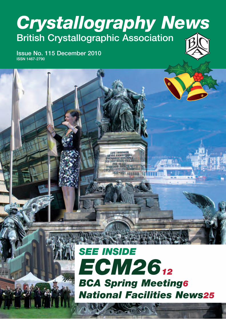
SEE inSidE ECM2612BCA Spring Meeting6national Facilities news25

The Rigaku Desktop Crystallization Workbench completely automates protein crystallization – using the
same technologies that support some of the largest structural biology eff orts all over the world – and fits
comfortably on two lab benches.
Isn’t it time to stop wasting time in the lab doing tedious manual labor?
Rigaku Americas Corporation phone: 281-362-2300 e-mail: [email protected] Europe phone: +[44] 1732 763367
www.riga
ku.com
/autom
ation
SC-XRDthink forward
X8 PROTEUMYour Personal Beamline � High-throughput screening and SAD-phasing quality data in an
uncompromised package
� Compact goniometer-mounted source ensures highest beam stability and most reliable alignment
� PLATINUM135CCD detector with large dynamic range and high sensitivity
� Kappa goniometer for easy sample handling
� Powerful, easy-to-use PROTEUM2 softwarewww.bruker.com
X8 PROTEUM 279 x 210 mm Farbe.indd 1 03.09.2010 13:45:27
Crystallography News December 2010 1111
This month’s cover:
Crystallography News December 2010
Contents
BCA Administrative Office, David Massey
Northern Networking Events Ltd.
Glenfinnan Suite, Braeview House
9/11 Braeview Place
East Kilbride G74 3XH
Tel: +44 (0)1355 244 966
Fax: +44 (0)1355 249 959
e-mail: [email protected]
CRYSTALLOGRAPHY NEWS is published quarterly (March, June, September and December) by the British Crystallographic Association, and printed by William Anderson and Sons Ltd, Glasgow. Text should preferably be sent electronically as MSword documents (any version - .doc, .rtf or .txt files) or else on a PC disk. Diagrams and figures are most welcome, but please send them separately from text as .jpg, .gif, .tif, or .bmp files. Items may include technical articles, news about people (e.g. awards, honours, retirements etc.), reports on past meetings of interest to crystallographers, notices of future meetings, historical reminiscences, letters to the editor, book, hardware or software reviews. Please ensure that items for inclusion in the March 2011 issue are sent to the Editor to arrive before 25 January 2011.
Carl Schwalbe15 St. Augustine Dr., Droitwich, Worcs WR9 8QRTel: 01905 775257e-mail: [email protected]
The British Crystallographic Association is a Registered Charity (#284718)As required by the DATA PROTECTION ACT, the BCA is notifying members that we store your contact information on a computer database to simplify our administration. These details are not divulged to any others without your permission. You may inspect your entry during the Annual Meeting, or otherwise by application to the BCA Administrative Office. We will be happy to amend entries at any time.
Designed & printed by Wm. Anderson & Sons Ltd.34 Loanbank Quadrant, Glasgow.Tel: 0141 440 2881e-mail: [email protected]
From the Editor . . . . . . . . . . . . . . . . . . . . . . . . . . . . 2
Council Members . . . . . . . . . . . . . . . . . . . . . . . . . . . . 3
From the President . . . . . . . . . . . . . . . . . . . . . . . . . . . 4
Puzzle Corner . . . . . . . . . . . . . . . . . . . . . . . . . . . . . . 5
BCA Spring Meeting 2011 . . . . . . . . . . . . . . . . . . . . . 6
ECM Darmstadt . . . . . . . . . . . . . . . . . . . . . . . . . . . . 12
BCA IG XRF Meeting Report . . . . . . . . . . . . . . . . . . 15
BCA IG XRD Meeting Report . . . . . . . . . . . . . . . . . . 18
The Bragg Lecture . . . . . . . . . . . . . . . . . . . . . . . . . . 21
News . . . . . . . . . . . . . . . . . . . . . . . . . . . . . . . . . . . . 21
Books . . . . . . . . . . . . . . . . . . . . . . . . . . . . . . . . . . . . 23
Hermann and Mauguin . . . . . . . . . . . . . . . . . . . . . . . 24
Central Facility News . . . . . . . . . . . . . . . . . . . . . . . . 25
News from the Groups . . . . . . . . . . . . . . . . . . . . . . . 29
Meetings of Interest . . . . . . . . . . . . . . . . . . . . . . . . . 31
ECM26 venue, Star speaker, excursion and dinner.
Crystallography News December 201022
By the time this issue hits the newsstands our three November group meetings will have already taken place, but there should still be an opportunity to attend the Biological Structures Group meeting on Wednesday, December 15, at the University of Reading. The subject is
‘Metal-protein interactions and their role in molecular transport and cell signalling.’ More information is available at http://www.reading.ac.uk/biologicalsciences/businessdevelopment/biosci-BCAwintermeet.aspx
For macromolecular crystallographers there is even more good news: CCP4 will once again hold a Study Weekend in January 2011. The topic is “Model Building & Refinement & Validation”. The venue is familiar to us and well-liked, namely the University of Warwick, and the dates are 6-7 January for the main meeting, preceded by a Diamond MX user meeting on the afternoon of the 5th and a satellite session on ‘What’s New in CCP4’ first thing in the morning of the 6th. The complete programme can be accessed at http://www.cse.scitech.ac.uk/events/CCP4_2011/programme.html.
We also feature the first draft of the programme for the 2011 BCA Spring Meeting at the University of Keele. Thanks to its central location and its proximity to Manchester Airport, Keele is easily reachable from anywhere in the U.K. or indeed the world. Already the deadline for oral presentation has passed, but time remains for the submission of poster abstracts. Young Crystallographers still have an opportunity (until January 14) to submit abstracts for oral presentation at the YC’s meeting, which precedes the full BCA meeting. No doubt the BCA’s exquisite judgment in its selection of Plenary Lecturers will once again be affirmed. In 2009 Venki Ramakrishnan was one of our Plenary Lecturers; later that year he was awarded the Nobel Prize. At our most recent meeting in 2010 Simon Billinge delivered the Teaching Plenary lecture, making pair distribution functions intelligible to non-physicists. Along with Takeshi Egami, who was his thesis advisor, he has just been awarded the 2010 Hanawalt Prize for excellence in X-ray powder diffraction by the International Centre for Diffraction Data.
This issue features reports on some very interesting recent meetings, starting with the two BCA Industrial Group joint XRD and XRF meetings in May. At a time when even some of our fellow scientists regard crystallography as “just another technique” it is important to emphasise the unique
perspective that crystallography can contribute to so many areas of science. Our Industrial Group has continued its distinguished record of forging and maintaining links with related disciplines, this time from bio-implantation to brewing. Part of David Beveridge‘s presentation was particularly close to home, identifying deposits formed on the wall of his X-ray lab. If only, in the present financial climate, they had been Au, or even FeS
2!
I am indebted to Georgina Rosair for contributing her perspective on the 26th European Crystallographic Meeting in Darmstadt to complement my own. In the usual ECM procedure several microsymposia ran concurrently both in the morning and in the afternoon, topped and tailed by plenary lectures. Therefore it is impossible for one person, or even two people, to summarise the wealth of information on offer. This year seemed to require even more heartbreaking decisions than usual, to miss one interesting microsymposium in order to attend an even more interesting one. I must also confess that I succumbed to the delights of two very enjoyable conference excursions, to the Heavy Ion Research facility and to Heidelberg. The atmosphere in Darmstadt of happy pan-European collaboration prompted me to write about a much earlier example when this was the exception rather than the rule: the collaboration of Carl Hermann and Charles Mauguin to produce the space group notation that we use today.
I am very pleased to include in this issue the second of Dave Allan’s articles on the National Facilities, this time describing Diamond’s beamlines for macromolecular crystallography. Surely no other field of crystallography has been so energised by the advent and further development of synchrotron radiation. It is reassuring to know that crystallographers are not the only people who appreciate the usefulness and potential of the Diamond synchrotron: George Osborne told the House of Commons that this is one of the projects that will escape the budget cuts.
Another forward-looking scientific project that is still proceeding is the Grand Challenge on Directed Assembly of Extended Structures with Targeted Properties (DAESTP). Paul Raithby gives us an update on its progress, particularly activities in which we can participate.
We salute the recent formation of the Irish Crystallographic Association and look forward to harmonious collaboration between the ICA and the BCA. Our Irish readers are reminded how close Keele is to Ireland. Your presence at the next BCA meeting will be particularly welcome: if Elspeth gets her wish and we have a ceilidh, you can show us how it’s done!
Carl Schwalbe
From the Editor
Crystallography News December 2010 33Crystallography News December 2010 33
BCA Council 2010COUNCIL MEMBERS
President (2012)Prof. Elspeth F. Garman Department of BiochemistrySouth Parks RoadOXFORD OX1 [email protected]
Vice President (2013) Dr David R. AllanDiamond Light SourceDiamond House, CHiLtOnOxfordshire, OX11 0DEtel. 01235 [email protected]
Secretary (2010) Dr Georgina Rosair School of EPS - Chemistry Perkin Building Heriot-Watt University EDinBURGH EH14 4AS tel: 0131 451 8036/4241 [email protected]
Treasurer (2011) Dr Harry R. PowellMRC Laboratory of Molecular Biology MRC Centre, Hills Road CAMBRiDGE CB2 2QH tel: (01223) 402423 [email protected]
ORDINARY MEMBERSDr David Beveridge (2012) Harman technology - iLFORD Phototown Lane, Mobberley, KnUtSFORD WA16 7JL tel: 01565 650000David.Beveridge@ harmantechnology.com
Dr Arwen Pearson (2013)Astbury Centre for Structural Molecular Biology, institute for Molecular and Cellular Biology,Astbury Building, LEEDS, LS2 9Jttel: 0113 343 [email protected]
Dr Alexandra Griffin (2012)Oxford Diffraction Ltd. 10 Mead Road,Oxford industrial Park,Yarnton,OxfordshireOX5 1QU [email protected]
GROUP REPRESENTATIVES Biological Structures Dr Darren Thompson Department of Biochemistry University of SussexBRiGHtOn Bn1 9QGtel: 01273 [email protected]
Chemical Crystallography Dr Peter Wood Cambridge Crystallographic Data Centre, 12 Union Road,CAMBRiDGE, CB2 1EZ.tel: 01223 [email protected]
Industrial Dr Anne Kavanagh AstraZenecaMACCLESFiELD, SK10 2nAtel. 01625 [email protected]
Physical Crystallography Dr Matt TuckerStFC Rutherford Appleton Laboratory DiDCOt OX11 0QX tel: 01235 [email protected]
Young Crystallographers Susanne Coles (née Huth)School of ChemistryUniversity of SouthamptonSOUtHAMPtOn SO17 1BJtel: 023 8059 [email protected]
CO-OPTED MEMBERS Dr. Andrés E. Goeta (2012)Department of ChemistryDurham UniversityScience Site, South RoadDURHAM DH1 3LEtel.: 0191 334 [email protected]
Prof. Paul FewsterPAnalytical Research Centre Sussex innovation Centre BRiGHtOn Bn1 9SB tel: 01273 704422 paul.fewster @ panalytical.com
Full committee details on the BCA website www.crystallography.org.ukSpring Meeting Registration and Subscriptions:
www.crystallography-meetings.org.uk
EX-OFFICIO MEMBERS
Education CoordinatorDr Michael R. ProbertDepartment of ChemistryDurham UniversityScience Site, South RoadDURHAM DH1 3LEtel: 0191 334 [email protected]
Editor “Crystallography News”Prof Carl H. Schwalbe15 St. Augustine Drive, Droitwich, Worcs WR9 8QRtel: 01905 [email protected]
WebmasterDr Richard Cooperinhibox Ltd.Pembroke House36-37 Pembroke St.OXFORD OX1 1BPtel: 01865 [email protected]
GROUP CHAIRMEN
Biological Structures GroupProf Vilmos FulopSchool of Life SciencesUniversity of WarwickCOVEntRY CV4 7ALtel: 024 7657 [email protected]
Chemical Crystallography GroupDr Andrew D. BondDepartment of Physics and ChemistryUniversity of Southern Denmark, 5230 ODEnSE M, DEnMARKtel: +45 6550 [email protected]
Industrial GroupDr Anne Kavanagh AstraZenecaMACCLESFiELD, SK10 2nAtel. 01625 [email protected]
Physical Crystallography GroupProf. David KeeniSiS Facility, Rutherford Appleton LaboratoryHarwell Science and innovation CampusDiDCOt Oxfordshire OX11 0QXtel: 01235 [email protected]
Young Crystallographers Susanne Coles (née Huth)School of ChemistryUniversity of SouthamptonSOUtHAMPtOn SO17 1BJtel: 023 8059 [email protected]
(The dates in parentheses indicate the end of the term of office).
Crystallography News December 201044
From the PresidentDear MeMBerAs the leaves fall outside and the evenings darken, we are grappling to understand the effects of the Government’s Comprehensive Spending Review and the Browne Report, both on the costs for future undergraduates at our Universities and on UK research efforts. Regarding the
latter, there seems to be some relief that the overall science budget will be frozen for the next four years (effectively approximately a 9% cut in real terms, taking inflation into account) rather than suffering the reduction that had been forecast in some quarters. Obtaining a commitment to flat funding can be partly attributed to widespread lobbying by the science community over the last few months. However, dealing with the ramifications of a frozen budget will be challenging, and it is not yet clear how the cake will be divided between the research councils.
For the undergraduates of the future, there are real concerns that students from less well off families will not be able to seriously contemplate going to University and taking on the ensuing level of debt that much higher tuition fees will bring. On one hand I am truly saddened by this development, having myself benefitted hugely from the system of the 1960s and 70s whereby I was in receipt of a full maintenance Grant from Northumberland Education Authority (£330 per annum which was certainly enough for survival!) who also (invisibly) paid all the tuition fees. This was despite the fact that I failed the 11+ examination and thus had not been offered a place in a Northumbrian Grammar School. My brother and sister were both students at the same time as I, and also received full grants. In today’s climate we would not have had the hugely positive experience of Higher Education and the opportunities it brings. However, on the other hand, I do see that our Universities cannot be funded to the extent they were while other areas bear the brunt of the effects of Deficit UK. It is to be hoped that when the detail of the fees increases emerge, some provision will be made to give support to students who need it in order to be able to study.
Moving over the Channel, the ECA in Darmstadt at the end of August attracted nearly 1000 participants and 45 trade stands: very impressive totals. There were some really interesting sessions, and I was happy to see many acquaintances from Europe and beyond there. I had last visited Darmstadt in 1979 when, as a card carrying Nuclear Physicist, I went on a conference outing to GSI
while in Frankfurt. It was raining then and sadly it rained on me again in August too: just unlucky, not a permanent condition, the locals assured me! I was struck by the friendliness of the locals when I got lost, and asked the way of a young mother with her baby son who had a map of Darmstadt propped up on the pushchair hood. She kindly showed me her map and then to my amazement called me by name: it transpired that she had been on a crystallography course on which I had taught over 15 years ago. Small world indeed.
Changing tack, as President I have the very enjoyable task annually of inviting nominations for new Honorary Members of the BCA. Honorary Membership is the highest membership accolade of the BCA, and is awarded to a small number of colleagues who have contributed significantly to crystallography and to the work of the BCA. Last year Bill Clegg, Venki Ramakrishnan and David Taylor became our most recent Honorary Members, taking the total number to 22. In the coming year we anticipate electing one or two new Honorary Members. Please send your nominations, together with a short supporting case to me at [email protected] by 31st January, 2011. For information, a list of our 22 current Honorary Members is now available at http://crystallography.org.uk/honorary-members (thanks to Richard Cooper our WWW Master).
The 2011 Keele Spring Meeting planning is now well advanced, and details can be found later in this issue. I encourage all members to bring as many of their group as is feasible to the meeting, since it promises to be an exciting event, with several very interesting ‘cross over’ sessions between the groups. The Young Crystallographers will again hold their pre-meeting event on Monday 11th April and the morning of Tuesday 12th April.
Unfortunately this year, due to various undergraduate and postgraduate teaching commitments, I am only able to attend one of the one-day autumn/winter Group meetings. By chance this is the BSG in Reading since it is after the end of our undergraduate term, but I assure you there is no favouritism involved! I very much enjoyed ‘doing the rounds’ last year and chatting to a cross section of Members. I hope that these have been productive: I heard today that the 3rd and 4th November Industrial Meeting at Diamond was very successful, and Dave Allan, our indefatigable Vice-President, was able to be there in my stead and to say a word about the BCA and upcoming (2013) ECA meeting in Warwick.
Wishing you all a Merry Christmas and Happy 2011.
Crystallography News December 2010 55
BCA Corporate MembershipThe BCA values its close ties with commercial companies involved with crystallography. To enhance these contacts, the BCA offers Corporate Membership. Corporate Membership is available on an annual basis starting from 1 January to 31 March and includes the following benefits:
• Upto10freeBCAmembershipsforyouremployees.
• A10%discountonexhibitionstandsontheannual BCA Spring Meeting, OR - A promotional poster at the annual BCA Spring Meeting.
• FreeinsertintheannualSpringMeeting delegate bag.
• Twofreefullregistrationstotheannual Spring Meeting.
• Tencomplimentarycopiesofthequarterly BCA newsletter.
• CorporateMemberswillbelistedineveryBCAnewsletter and on the BCA Web Site with links to your corporate site.
the cost of this membership is £750.00 per annumto apply for Corporate Membership, or if you have anyenquiries,pleasecontact:
David Massey | BCA Administrative Officenorthern networking Events LtdGlenfinnan SuiteBraeview House, 9/11 Braeview PlaceEast Kilbride G74 3XHtel: +44 (0)1355 244 966 Fax: +44 (0)1355 249 959e-mail [email protected]
Corporate Members
Rigaku
Oxford Cyrosystems
Oxford Diffraction
CCDC
iCDD
incoatec
CCG
Molecular Dimensions
Bruker
thermo Fisher
PAnalytical
Crystallography News December 2010 55
Puzzle CornerWhile in Darmstadt for ECM26 we could see the laboratory where many of the heaviest known elements were made. Once the discovery of a new element has been confirmed, the discoverer gets the opportunity to name it. However, the historical record is littered with element names no longer used because they are ancient, provisional or based on mistaken claims. For the following list of non-standard element names give the accepted names and write down the chemical symbols. From the initial letter of each symbol extract a seasonal message. (For an enjoyable ramble through “elementymology” visit http://elements.vanderkrogt.net/index.php .)
Hydrargyrum
Anglohelvetium
Florentium
Illinium
Aldebaranium
Norium
Nebulium
Cassiopium
Stibium
•
Unnilhexium
Alabamine
Columbium
Masurium
Emanium
Crystallography News December 20106
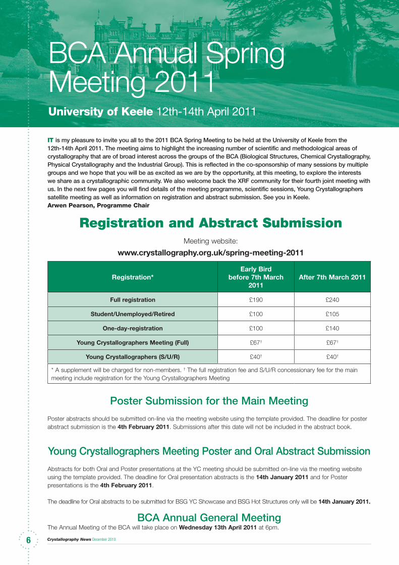
BCA Annual Spring Meeting 2011University of Keele 12th-14th April 2011
6
it is my pleasure to invite you all to the 2011 BCA Spring Meeting to be held at the University of Keele from the 12th-14th April 2011. The meeting aims to highlight the increasing number of scientific and methodological areas of crystallography that are of broad interest across the groups of the BCA (Biological Structures, Chemical Crystallography, Physical Crystallography and the Industrial Group). This is reflected in the co-sponsorship of many sessions by multiple groups and we hope that you will be as excited as we are by the opportunity, at this meeting, to explore the interests we share as a crystallographic community. We also welcome back the XRF community for their fourth joint meeting with us. In the next few pages you will find details of the meeting programme, scientific sessions, Young Crystallographers satellite meeting as well as information on registration and abstract submission. See you in Keele.Arwen Pearson, Programme Chair
Poster Submission for the Main Meeting
Poster abstracts should be submitted on-line via the meeting website using the template provided. The deadline for poster abstract submission is the 4th February 2011. Submissions after this date will not be included in the abstract book.
Young Crystallographers Meeting Poster and Oral Abstract Submission
Abstracts for both Oral and Poster presentations at the YC meeting should be submitted on-line via the meeting website using the template provided. The deadline for Oral presentation abstracts is the 14th January 2011 and for Poster presentations is the 4th February 2011.
The deadline for Oral abstracts to be submitted for BSG YC Showcase and BSG Hot Structures only will be 14th January 2011.
BCA Annual General MeetingThe Annual Meeting of the BCA will take place on Wednesday 13th April 2011 at 6pm.
registration and abstract SubmissionMeeting website:
www.crystallography.org.uk/spring-meeting-2011
Programme Details
Registration*Early Bird
before 7th March 2011
After 7th March 2011
Full registration £190 £240
Student/Unemployed/Retired £100 £105
One-day-registration £100 £140
Young Crystallographers Meeting (Full) £67† £67†
Young Crystallographers (S/U/R) £40† £40†
* A supplement will be charged for non-members. † The full registration fee and S/U/R concessionary fee for the main meeting include registration for the Young Crystallographers Meeting
Crystallography News December 2010 7777
youNg CryStallograPherS Satellite MeetiNg
Monday 11th April 2011
1-3 pm YC Session 1YC Industrial PlenaryMatthew Johnson (GlaxoSmithKline) “Industrial Action: Striking at the heart of Materials Science”
3:30-5pm YC Session 2YC Biological PlenaryArwen Pearson (University of Leeds)“Getting more than just diffraction from a crystal: How complementary methods can add to your experiment”
5:30-7pm YC Session 3YC AGMPoster flash presentations
7-9pm Poster Session with dinner and wine
Tuesday 11th April
9-10:30am YC Session 4Parkin Lecture Nominations should be sent to Anna Stevenson (YCG Secretary/Treasurer)
10:30-11:15 YC Session 5Professional Development
Abstract Deadlines for the YC SatelliteOral presentations: 14th January 2011 Posters: 4th February 2011
Abstracts can be submitted at:http://crystallography.org.uk/spring-meeting-2011
XrF WorkShoP & MeetiNg
Tuesday 12th April 2011
11-12:30 XRF Workshop 1Sample preparationChair: Margaret West
1:30-3pm XRF Workshop 2Sample preparationChair: Margaret West
3:30-5pm XRF Workshop 3CalibrationChair: Ros Schwarz
Wednesday 13th April
9-9:45 XRF/IG Plenary10:15-11:45 XRF Session 4New DevelopmentsChair: Mark Ingham
1:30-3pm XRF Session 5ApplicationsChair: David Beveridge
3:30-5pm XRF Session 6ApplicationsChair: David Beveridge
5:15-6pm XRF Session 7Applications in XRD/XRF (Joint with IG)Chair: David Beveridge
Thursday 14th April
9-9:45am XRF Keynote Novel Techniques“It began with a helping hand”Margaret West, West X-ray Solutions Ltd.
10:15-11:45am XRF Session 8Novel TechniquesChair: David Taylor
12-1:30pm XRF Session 9 (joint with PCG)Cultural HeritageChairs: David Taylor and W. Kockelmann
Programme Details
Crystallography News December 20108
BiologiCal StruCtureS grouP
Tuesday 12th April 201111:30-12:30 BSG Plenary Lecture in honour of Kathleen LonsdaleJohn Helliwell (University of Manchester)
1:30-3pm BSG Session 1Twinning and PseudosymmetryChair: Eleanor Dodson
3:30-5pm BSG Session 2Protein Crystallization: dealing with low solubility proteins and protein-ligand complexes Chair: Ray Owens
3:30-5pm (Joint with CCG) Macro and small molecule crystallography Chair: Paul Raithby
Wednesday 13th April 201110:15-11:45 BSG Session 3Membrane proteins Chair: Chris Tate11:45-12:30 BSG AGM
1:30-3pm BSG Session 4(Joint with CCG & PCG)Radiation Damage Chair: Colin Nave
3:30-5pm BSG Session 5Cell walls - synthesis, virulence and inhibitors Chair: Klaus Futterer
3:30-5pm (joint with CCG)Time resolved structural scienceChair: Dave Allan
Thursday 14th April 201110:15-11:45 BSG Session 6BSG Young Crystallographer Showcase
12-1:30pm BSG Session 7Hot structures Chair: Ravi Acharya
CheMiCal CryStallograPhy grouP
Tuesday 12th April 20111:30-3pm CCG Session 1From Molecular to SupramolecularChair: Peter Byrne
1:30-3pm (joint with PCG)New Developments at Diamond Chair: Helen Maynard-Casely & David Keen
3:30-5pm CCG Session 2Macro and small molecule crystallographyChair: Paul Raithby
Wednesday 13th April 201110:15-11:45 CCG Session 3 (joint with IG)Crystallization Chair: Louise Male
12:30-1:15 CCG AGM
1:30-3pm CCG Session 4Structure/Property Correlations in Luminescent Materials Chair: Andrew Bond (Includes the CCDC/CCG Prize Lecture)
1:30-3pm (joint with BSG & PCG)Radiation Damage Chair: Colin Nave
3:30-5pm CCG Session 5 (joint with BSG)Time resolved structural science Chair: Dave Allan
5:15-6pm CCG Plenary LectureJudith Howard, University of Durham
Thursday 14th April 201110:15-11:45 CCG Session 6(joint with PCG)Dynamic Data, dealing with limited dataChair: Peter Wood
12-1:30pm CCG Session 7Getting more from diffraction dataChair: Hazel Sparkes
Programme Details Programme Details
Crystallography News December 2010 9999
PhySiCal CryStallograPhy grouP
Tuesday 12th April 20111:30-3pm PCG Session 1New Developments at DiamondChair: Helen Maynard-Casely & David Keen
3:30-5pm PCG Session 2Local StructureChair: Andrew Goodwin & Matt Tucker
Wednesday 13th April 201110:15-11:45am PCG Session 3High Pressure and Energetic Materials Chair: Christoph Salzmann & Matt Tucker
11:45-12:30pm PCG AGM1:30-3pm PCG Session 4(joint with CCG & BSG)Radiation Damage Chair: Colin Nave
3:30-5pm PCG Session 5(joint with BSG & CCG)Time Resolved Structural Science Chair: Dave Allan
3:30-5pm (joint with IG)Stress-strain Microstructure
Thursday 14th April 20119-9:45am PCG Plenary LectureGilberto Artioli (Padova)
10:15-11:45 PCG Session 6(joint with IG)Materials Science: white beam methods Chair: Bob Cernik
10:15-11:45 (joint with CCG)Dynamic data, dealing with limited data Chair: Peter Wood
12-1:30pm PCG Session 7(joint with XRF)Cultural Heritage Chair: W. Kockelmann & David Taylor
iNDuStrial grouP
Tuesday 12th April 20111:30-3pm Sample Preparation (joint with XRFChair: Margaret West
Wednesday 13th April 20119-9:45am IG/XRF Plenary Lecture10:15-11:45am IG Session 1(joint with CCG) Crystallization Chair: Louise Male
11:45-12:30pm IG AGM
1:30-3pm IG Session 2 TBA
3:30-5pm IG Session 3(joint with PCG) Stress-strain Microstructure
5:15-6pm (joint with XRF)Applications in XRD/XRFChair: David Beveridge
Thursday 14th April 201110:15-11:45 IG Session 4(joint with PCG)Materials Science - White beam methodsChair: Bob Cernik
12-1:30pm IG Session 5Materials Science - Powder methodsTo include the IG Young Crystallographer Prize Lecture
Programme Details Programme Details
Crystallography News December 201010
Meeting organisation
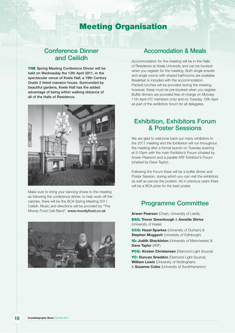
Conference Dinner and Ceilidh
the Spring Meeting Conference Dinner will be held on Wednesday the 13th April 2011, in the spectacular venue of Keele Hall, a 19th Century Grade 2 listed mansion house. Surrounded by beautiful gardens, Keele Hall has the added advantage of being within walking distance of all of the Halls of Residence.
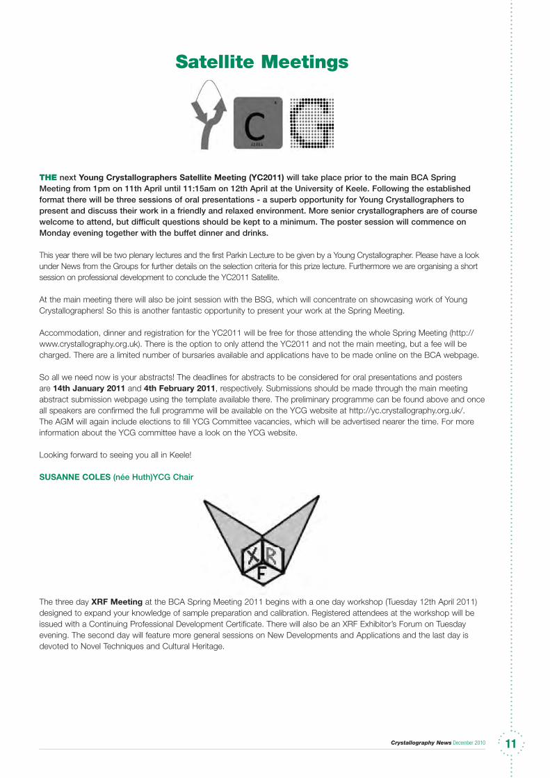
Make sure to bring your dancing shoes to the meeting as following the conference dinner, to help work off the calories, there will be the BCA Spring Meeting 2011 Ceilidh. Music and directions will be provided by “The Moody Food Ceili Band”. www.moodyfood.co.uk
Accomodation & Meals
Accommodation for the meeting will be in the Halls of Residence at Keele University and can be booked when you register for the meeting. Both single ensuite and single rooms with shared bathrooms are available. Breakfast is included with the accommodation. Packed lunches will be provided during the meeting, however, these must be pre-booked when you register. Buffet dinners are provided free-of-charge on Monday 11th April (YC members only) and on Tuesday 12th April as part of the exhibitors forum for all delegates.
Exhibition, Exhibitors Forum & Poster Sessions
We are glad to welcome back our many exhibitors to the 2011 meeting and the Exhibition will run throughout the meeting after a formal launch on Tuesday evening at 5:15pm with the main Exhibitor’s Forum (chaired by Arwen Pearson) and a parallel XRF Exhibitor’s Forum (chaired by Dave Taylor).
Following the Forum there will be a buffet dinner and Poster Session, during which you can visit the exhibitors as well as peruse the posters. As in previous years there will be a BCA prize for the best poster.
Programme Committee
Arwen Pearson (Chair), University of Leeds,
BSg: Trevor Greenhough & Annette Shrive (University of Keele)
CCg: Hazel Sparkes (University of Durham) & Stephen Moggach (University of Edinburgh)
ig: Judith Shackleton (University of Manchester) & Dave Taylor (XRF)
PCg: Kirsten Christensen (Diamond Light Source)
yC: Duncan Sneddon (Diamond Light Source), William Lewis (University of Nottingham) & Susanne Coles (University of Southhampton)
Crystallography News December 2010 1111
Satellite Meetings
the next Young Crystallographers Satellite Meeting (YC2011) will take place prior to the main BCA Spring Meeting from 1pm on 11th April until 11:15am on 12th April at the University of Keele. Following the established format there will be three sessions of oral presentations - a superb opportunity for Young Crystallographers to present and discuss their work in a friendly and relaxed environment. More senior crystallographers are of course welcome to attend, but difficult questions should be kept to a minimum. The poster session will commence on Monday evening together with the buffet dinner and drinks.
This year there will be two plenary lectures and the first Parkin Lecture to be given by a Young Crystallographer. Please have a look under News from the Groups for further details on the selection criteria for this prize lecture. Furthermore we are organising a short session on professional development to conclude the YC2011 Satellite.
At the main meeting there will also be joint session with the BSG, which will concentrate on showcasing work of Young Crystallographers! So this is another fantastic opportunity to present your work at the Spring Meeting.
Accommodation, dinner and registration for the YC2011 will be free for those attending the whole Spring Meeting (http://www.crystallography.org.uk). There is the option to only attend the YC2011 and not the main meeting, but a fee will be charged. There are a limited number of bursaries available and applications have to be made online on the BCA webpage.
So all we need now is your abstracts! The deadlines for abstracts to be considered for oral presentations and posters are 14th January 2011 and 4th February 2011, respectively. Submissions should be made through the main meeting abstract submission webpage using the template available there. The preliminary programme can be found above and once all speakers are confirmed the full programme will be available on the YCG website at http://yc.crystallography.org.uk/. The AGM will again include elections to fill YCG Committee vacancies, which will be advertised nearer the time. For more information about the YCG committee have a look on the YCG website.
Looking forward to seeing you all in Keele!
SUSANNE COLES (née Huth)YCG Chair
The three day XRF Meeting at the BCA Spring Meeting 2011 begins with a one day workshop (Tuesday 12th April 2011) designed to expand your knowledge of sample preparation and calibration. Registered attendees at the workshop will be issued with a Continuing Professional Development Certificate. There will also be an XRF Exhibitor’s Forum on Tuesday evening. The second day will feature more general sessions on New Developments and Applications and the last day is devoted to Novel Techniques and Cultural Heritage.
Crystallography News December 201012
ECM26 DarmstadtiF I had to sum up in a single word the 26th ECM in Darmstadt, it would be …generosity. Starting with warm words of welcome from our genial host Prof. Hartmut Fuess, the relaxed feeling continued at the reception after the opening ceremony. This was not one of those receptions where you had to elbow your research collaborators aside in order to get to a rapidly diminishing supply of food and drink, nor the ACA system of drinks tickets which ensured fairness but recalled wartime rationing. In Darmstadt a plentiful supply of delicious German food and beer encouraged old friends to meet and chat. Lunches were similarly conducive to making and renewing friendships. Instead of overpriced sandwiches sold by the conference venue, simple but filling hot lunches were provided free of charge. Thus most participants stayed in the building, and it was easy to find speakers from the morning sessions and discuss their presentations.
Our conference venue, the Darmstadtium, was interesting in itself. It had no immediately obvious symmetry, and many surfaces were neither horizontal nor vertical. On the first evening it rained for an hour or two. Where several sloping glass roofs converged, the central channel became a sparkling water feature illuminated by the lights from below. The seminar rooms were named after elements, mostly with international standard names ending in –ium. However, “Wasserstoff” and “Kohlenstoff” became “hydrogenium” and “carbonium”. Not having attended any sessions in “carbonium”, I did not find out whether participants received a jolt of positive charge there.
Already on the opening night (Sunday) we enjoyed a very stimulating lecture by Prof. Claude Lecomte, winner of the Perutz Prize. Beginning with a brief but clear exposition of the physics behind charge density determination and a generous acknowledgment of the pioneers of the technique, he went on to outline state-of-the-art results and challenges for the future. His own group has carried out a number of charge density studies on oligopeptides with a view to generalising the results to proteins. Studies based on very high-resolution data from protein crystals are starting to bear fruit, notably in the visualisation of protein-ligand interactions.
Placement of the microsymposium about crystallographic teaching on Monday morning rather than the last day of the conference signified the importance assigned to this topic by the organizers. Katherine Kantardjieff started the session with a thorough survey of the situation in the United States. Although the results of crystallography feature prominently in textbooks serving university science curricula, she communicated the bad news that the theory and practice of crystallography are being squeezed out of the syllabus. Even if an institution has the will to teach crystallography, it may lack the means in terms of staff and facilities. This gloom was offset by shining examples of the creative use of technology
to enable academic staff and students even at small colleges to access tutorial material and run experiments remotely on shared equipment. An example of the ultimate in outreach activity has been provided by our entomologist colleagues. BugScope enables children and teachers in elementary schools anywhere in the world to send specimens to the University of Illinois-Chicago for preparation and microscopy, viewing the resulting images over the Internet.
Next, Bob Sweet described the RapiData macromolecular crystallography course at the National Synchrotron Light Source, Brookhaven. Intended for postgraduate students but also established biological scientists, the course begins on a Sunday with optional lectures about fundamentals. Monday is devoted to instruction about data collection, and Tuesday to structure solution. Dividing into groups, each with a tutor, participants put what they have learned into practice on a beamline from Tuesday night to Friday. The bond formed within groups and with tutors is a strong point.
Annette Faust described a comprehensive set of MX tutorials that is available over the Internet at http://www.helmholtz-berlin.de/forschung/funkma/soft-matter/forschung/bessy-mx/tutorial/index_en.html . Covering all aspects from motivation and biological interest to data collection and processing through to structure determination, it is intended to help students starting out, students with a data set, lecturers and software developers. Great flexibility is allowed in the use of data: real data sets where the methods are known to have worked are available, advice is given on commercially available proteins known to give good data on home lab equipment, and users can submit their own original data sets.
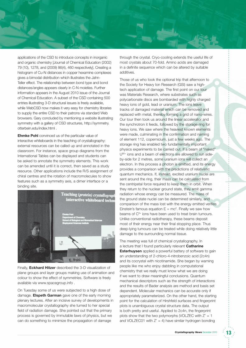
Gary Battle surveyed the many ways in which the Cambridge Structural Database can be used to enrich the teaching of chemistry. Starting with the common-sense propositions that 3-D visualization helps students to understand stereochemistry, and consideration of experimental errors lets students appreciate the limitations of real data, he described
Crystallography News December 2010 13131313
applications of the CSD to introduce concepts in inorganic and organic chemistry [Journal of Chemical Education (2002) 79 (10), 1278, and (2009) 86(4), 460 respectively]. Creating a histogram of Cu-N distances in copper hexamine complexes gives a bimodal distribution which illustrates the Jahn-Teller effect. The relationship between bond type and bond distances/angles appears clearly in C-N moieties. Further information appears in the August 2010 issue of the Journal of Chemical Education. A subset of the CSD containing 500 entries illustrating 3-D structural issues is freely available, while WebCSD now makes it very easy for chemistry libraries to supply the entire CSD to their patrons via standard Web browsers. Gary concluded by mentioning a website illustrating symmetry with a gallery of CSD structures, http://symmetry.otterbein.edu/index.html .
Ehmke Pohl convinced us of the particular value of interactive whiteboards in the teaching of crystallography: external resources can be called up and annotated in the classroom. For instance, space group diagrams from the International Tables can be displayed and students can be asked to annotate the symmetry elements. This work can be amended until it is correct, then saved as a study resource. Other applications include the R/S assignment of chiral centres and the rotation of macromolecules to show features such as a symmetry axis, a dimer interface or a binding site.
Finally, Eckhard Hitzer described the 3-D visualization of plane groups and layer groups making use of animation and colour to show the effect of symmetries. Software is freely available via www.spacegroup.info .
On Tuesday some of us were subjected to a high dose of damage. Elspeth Garman gave one of the early morning plenary lectures. After an incisive survey of developments in macromolecular crystallography she turned to her special field of radiation damage. She pointed out that the primary process is governed by immutable laws of physics, but we can do something to minimize the propagation of damage
through the crystal. Cryo-cooling extends the useful life of most crystals about 70-fold. Amino acids are damaged in a definite sequence which can be altered by suitable additives.
Those of us who took the optional trip that afternoon to the Society for Heavy Ion Research (GSI) saw a high-tech application of damage. The first point on our tour was Materials Research, where substrates such as polycarbonate discs are bombarded with highly charged heavy ions of gold, lead or uranium. The ions leave tracks of damaged material which can be removed and replaced with metal, thereby forming a grid of nano-wires. Our tour then took us around the linear accelerator and the synchrotron it feeds, followed by the storage ring for heavy ions. We saw where the heaviest known elements were made, culminating in the confirmation and naming of element 112, copernicium, just a few weeks ago. The storage ring has enabled two fundamentally important physics experiments to be carried out. If a beam of “naked” U92+ ions and a beam of electrons are allowed to run side-by-side for 2 metres, some uranium ions will collect an electron. In this process a photon is emitted, and its energy provides a comparison with the predictions of relativistic quantum mechanics. If, instead, excited uranium nuclei are sent around the ring, their mass can be calculated from the centripetal force required to keep them in orbit. When they return to the nuclear ground state, they emit gamma radiation whose energy can be measured. The mass of the ground state nuclei can be determined similarly, and comparison of the mass lost with the energy emitted verifies Einstein’s famous equation E = mc2. Finally we saw how beams of C6+ ions have been used to treat brain tumours. Unlike conventional radiotherapy, these beams deposit most of their energy near their final stopping place. Thus deep-lying tumours can be treated while doing relatively little damage to the surrounding normal tissue.
The meeting was full of chemical crystallography. In a lecture that I found particularly relevant Catharine Esterhuysen applied a powerful battery of software to gain an understanding of 2-chloro-4-nitrobenzoic acid (2c4n) and its cocrystal with nicotinamide. She began by warning people like me who enjoy dabbling in computational chemistry that we really must know what we are doing if we want to draw meaningful conclusions. Quantum mechanical descriptors such as the strength of interactions and the results of Bader analysis are method and basis set dependent. Molecular mechanics can be accurate only if appropriately parameterized. On the other hand, the starting point for the calculation of Hirshfeld surfaces and fingerprint plots is unambiguous crystal structure data. The output is both pretty and useful. Applied to 2c4n, the fingerprint plots show that the two polymorphs (VOLZEC with Z’ = 1 and VOLZEC01 with Z’ = 4) have similar hydrogen bonding

Crystallography News December 201014
but the latter has more pi stacking. The cocrystal of 2c4n and nicotinamide exhibits disorder affecting the Cl atom and one H atom of 2c4n. Molecular orbital calculations with counterpoise correction for basis set superposition error correlate reasonably well with the Hirshfeld surfaces as to the nature and strength of interactions in a series of nicotinamide co-crystals.
The session “Structure-properties relationship in molecular crystals” was started by Chick Wilson (below) with a typically effervescent presentation and finished by me. In between I was impressed by a scholarly contribution from Krzystof Wozniak from Warsaw which is described more fully in Georgina’s report.
We added to our education on Thursday evening, although this time the subject was gastronomy. The conference
dinner was held in a Grand Duke’s palatial hunting lodge outside Darmstadt. Instead of the usual dinner with three set courses served by waiters there was a large central buffet table with a wide variety of local dishes selected by Hartmut Fuess, carefully annotated and complemented by fragrant local wines. Therefore we could fill our plates with small samples of everything and come back for more of the dishes we liked best.
Bright and early the next morning, at 7:45, the survivors among us assembled for the conference excursion. We were whisked off by coach to the Rhineland, where our first stop was the monument at Niederwald, on a steep hill near Rüdesheim. In a typically elaborate 19th century style it commemorates the German victory in the Franco-Prussian war. At the top is a huge figure of Germania, (right) staring triumphantly across the Rhine in the direction of France—which prompted some ironic comments from our French colleagues. Next we proceeded to the riverfront at Rüdesheim, where we boarded a cruise boat for a trip downstream along the most romantic part of the Rhine to the Lorelei. Numerous hills were surmounted by castles, many of which had been destroyed by the French in the 1690’s and restored by wealthy German industrialists in the 1890’s. Passing the Lorelei, we admired the geology of the steep cliff jutting into the river. Unfortunately the beautiful maiden who is supposed to sit atop the cliff combing her golden hair and distracting mariners and crystallographers was off duty at the time. Our destination was St. Goar, where our boat docked long enough for us to enjoy a stroll around the picturesque town. Once back on board, we satisfied the appetite created by our walk with a “typisch deutsch” lunch as we cruised back to Rüdesheim. We departed in a good mood, looking forward to the delights of the Avon in 2013.
Carl Schwalbe
Georgina Rosair’s Report
DarMStaDt is half an hour by bus from Frankfurt airport and my hotel was walking distance from the conference centre. A gentle walk in the morning helps to foster a refreshed mind for lectures. The opening ceremony and reception were in the Darmstadium conference centre where we were treated to local wines, beers and nibbles whilst I met up with colleagues from Spain and Italy as well as the UK.
Elspeth Garman’s keynote on radiation damage to proteins was an eye opener. As a small molecule crystallographer, my experience of radiation damage was limited, the crystal just decayed; but Elspeth showed us that in proteins you could be determining a different structure because radiation induced radicals react with the protein itself. The teaching microsymposium was well attended and Katherine Kantardjieff gave us a very useful link to an ACS document which describes what material should be included in teaching crystallography (http://sites.nationalacademies.org/PGA/biso/IUCr/ and scroll down to the ‘Crystallography in the University Curricula’ section to download the .pdf file). On the chemical crystallography front, charge density is becoming a more widespread and significant feature of structure determination as evidenced by many lectures on this topic. Krzysztof Wozniak recommended us to use low angle data to determine H atom positions more accurately when neutron data was not available. He also exhorted us to use more detailed descriptions of electron density when it came to describing intermolecular interactions. There’s a new interest group for Art and Crystallography and Jan Fabry from Prague told
Crystallography News December 2010 1515
BCA IG XRF
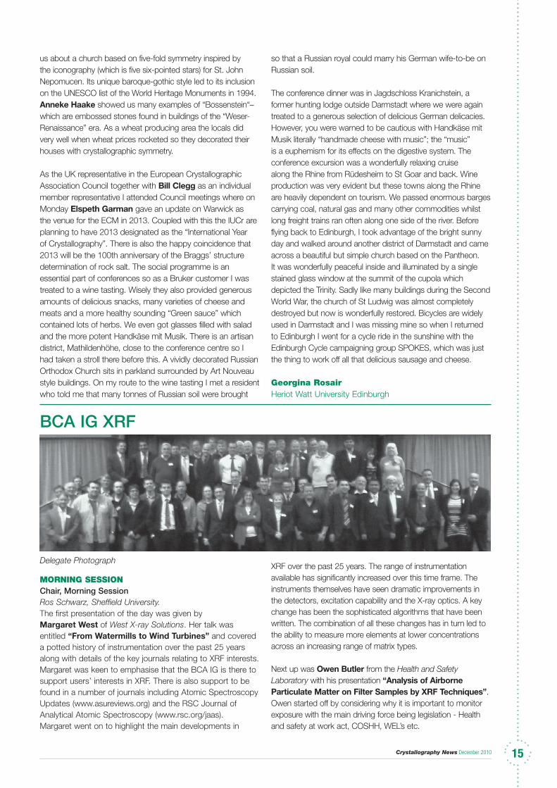
Delegate Photograph
MorNiNg SeSSioNChair, Morning SessionRos Schwarz, Sheffield University.The first presentation of the day was given by Margaret West of West X-ray Solutions. Her talk was entitled “From Watermills to Wind Turbines” and covered a potted history of instrumentation over the past 25 years along with details of the key journals relating to XRF interests. Margaret was keen to emphasise that the BCA IG is there to support users’ interests in XRF. There is also support to be found in a number of journals including Atomic Spectroscopy Updates (www.asureviews.org) and the RSC Journal of Analytical Atomic Spectroscopy (www.rsc.org/jaas). Margaret went on to highlight the main developments in
XRF over the past 25 years. The range of instrumentation available has significantly increased over this time frame. The instruments themselves have seen dramatic improvements in the detectors, excitation capability and the X-ray optics. A key change has been the sophisticated algorithms that have been written. The combination of all these changes has in turn led to the ability to measure more elements at lower concentrations across an increasing range of matrix types.
Next up was Owen Butler from the Health and Safety Laboratory with his presentation “Analysis of Airborne Particulate Matter on Filter Samples by XRF Techniques”. Owen started off by considering why it is important to monitor exposure with the main driving force being legislation - Health and safety at work act, COSHH, WEL’s etc.
us about a church based on five-fold symmetry inspired by the iconography (which is five six-pointed stars) for St. John Nepomucen. Its unique baroque-gothic style led to its inclusion on the UNESCO list of the World Heritage Monuments in 1994. Anneke Haake showed us many examples of “Bossenstein“–which are embossed stones found in buildings of the “Weser-Renaissance” era. As a wheat producing area the locals did very well when wheat prices rocketed so they decorated their houses with crystallographic symmetry.
As the UK representative in the European Crystallographic Association Council together with Bill Clegg as an individual member representative I attended Council meetings where on Monday Elspeth Garman gave an update on Warwick as the venue for the ECM in 2013. Coupled with this the IUCr are planning to have 2013 designated as the “International Year of Crystallography”. There is also the happy coincidence that 2013 will be the 100th anniversary of the Braggs’ structure determination of rock salt. The social programme is an essential part of conferences so as a Bruker customer I was treated to a wine tasting. Wisely they also provided generous amounts of delicious snacks, many varieties of cheese and meats and a more healthy sounding “Green sauce” which contained lots of herbs. We even got glasses filled with salad and the more potent Handkäse mit Musik. There is an artisan district, Mathildenhöhe, close to the conference centre so I had taken a stroll there before this. A vividly decorated Russian Orthodox Church sits in parkland surrounded by Art Nouveau style buildings. On my route to the wine tasting I met a resident who told me that many tonnes of Russian soil were brought
so that a Russian royal could marry his German wife-to-be on Russian soil.
The conference dinner was in Jagdschloss Kranichstein, a former hunting lodge outside Darmstadt where we were again treated to a generous selection of delicious German delicacies. However, you were warned to be cautious with Handkäse mit Musik literally “handmade cheese with music”; the “music” is a euphemism for its effects on the digestive system. The conference excursion was a wonderfully relaxing cruise along the Rhine from Rüdesheim to St Goar and back. Wine production was very evident but these towns along the Rhine are heavily dependent on tourism. We passed enormous barges carrying coal, natural gas and many other commodities whilst long freight trains ran often along one side of the river. Before flying back to Edinburgh, I took advantage of the bright sunny day and walked around another district of Darmstadt and came across a beautiful but simple church based on the Pantheon. It was wonderfully peaceful inside and illuminated by a single stained glass window at the summit of the cupola which depicted the Trinity. Sadly like many buildings during the Second World War, the church of St Ludwig was almost completely destroyed but now is wonderfully restored. Bicycles are widely used in Darmstadt and I was missing mine so when I returned to Edinburgh I went for a cycle ride in the sunshine with the Edinburgh Cycle campaigning group SPOKES, which was just the thing to work off all that delicious sausage and cheese.
georgina rosairHeriot Watt University Edinburgh
Crystallography News December 20101616
Owen has been working on ISO 10882-1:2001. Health and safety in welding and allied processes - Sampling of airborne particles and gases in the operator’s breathing zone - Part 1. He summarised the elements that can be determined by XRF. From his experience the use of XRF “wins” over ICP-MS owing to dissolution difficulties that can bias results, even those from accredited labs.
Sampling and sample handling is very critical along with the analytical capability of the technique. For this type of analysis there is a wide range of acceptable “inhalable” dust sampler designs but not all have usable attributes for the XRF. It is important to choose one where the sample will be collected evenly across the filter. The material of construction of the filter itself is also critical. It needs to be structurally strong for the XRF. Quartz filters are not a good idea as the particulates can sink in to the filter causing depth effects. Teflon can also be a problem because not enough sample may be collected for XRF analysis.
Another factor to consider is calibration. Dried aerosol from a nebulised solution is the current preferred technique. However there is a new development in the area of nano-printing. In this a scan is taken of the deposition pattern from the sample filter, this pattern is then replicated with standard solutions onto a filter using a nano-printer.
Following on from Owen was Colin Slater, from the University of Birmingham, who was very enthusiastic about his presentation “X-ray Speciation of Amorphous Bioceramics”. The Materials Chemistry Group that Colin is working with is interested in creating resorbable hard tissue replacement for bone and teeth. This is where the bone graft is slowly dissolved away and replaced with natural bone. The body’s own biological synthetic pathways are used which results in enhanced re-growth response leading to shortened healing times. Recent work from the group demonstrated that the presence of amorphous condensed phosphates (formed in situ) in Brushite cements has been shown to improve biological and mechanical properties.
The aim of Colin’s work is to synthesise and characterise amorphous M2P2O7 phases (where M = Mg, Ca, Sr) and to investigate their chemical and biological properties. As part of this he is using a range of analytical techniques including PXRD, MAS-NMR, TGA and XRF. Using the Bruker S8 Tiger with a 3kW Rh X-ray source Colin is running either pressed pellets, loose powders or fused beads for 1 or 20 minutes, depending on sample preparation. This work is being used for the determination of stoichiometry and salt contamination of the amorphous pyrophosphates. Results to date based on lithium metaborate fused pellets and semi-quantitative analyses on the XRF have shown good correlation between theoretical and measured values.
Richard Morris of Morris X-ray Analytical Ltd. presented a summary of his Environmental Sciences MSc research project entitled “Urban Mining - An Investigation of the Analysis of PGMs in Road Dust Using Bench top EDS XRF”. His hypothesis was: Is it a feasible proposition to analyse road dust for platinum group metals (PGMs) quickly using EDS XRF with a 50W source?
Platinum group metals are used in catalytic converters. They degrade with use and time resulting in Pt, Rh and Os being deposited at the roadside. How to measure these? Richard prepared the samples by using micronized silver sand as the blank and matrix for standard addition for calibration purposes. Both the samples and standards were gy-ro milled, mixed with Licowax C and then pressed at 10tons in a 32mm pellet die. Dr Dave Tunrow kindly ran the samples using the Spectro XEPOS at Wolverhampton University. To date no statistical analysis has been carried out on the results.
Having presented his results so far Richard opened up a discussion to ask for assistance, as he has been experiencing problems with inhomogeneous pellets that are not structurally robust. A suggestion from the audience was that it might be associated with decompression shearing. This is apparently quite prevalent for Si based samples and it is critical that the pressure is released slowly and evenly during pressing. Another suggestion was to create a fused pellet first, grind this and then repress into a pellet. Obviously care would need to be taken with the choice of metal of construction of the crucible.
Lunch was then served combined with the opportunity to talk with the exhibitors and sponsors of the day.
Meeting Speakers
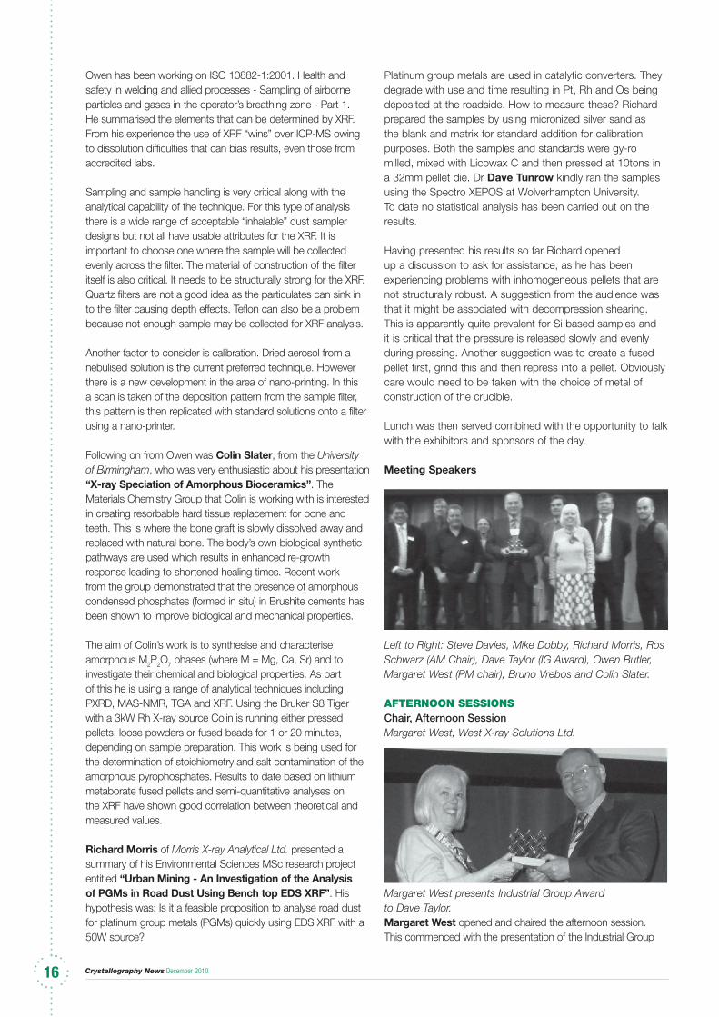
Left to Right: Steve Davies, Mike Dobby, Richard Morris, Ros Schwarz (AM Chair), Dave Taylor (IG Award), Owen Butler, Margaret West (PM chair), Bruno Vrebos and Colin Slater.
aFterNooN SeSSioNSChair, Afternoon SessionMargaret West, West X-ray Solutions Ltd.
Margaret West presents Industrial Group Award to Dave Taylor.Margaret West opened and chaired the afternoon session. This commenced with the presentation of the Industrial Group
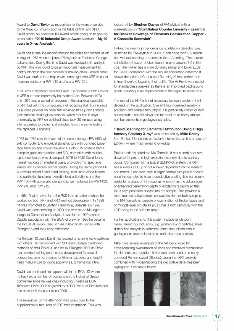
Crystallography News December 2010 17171717
Award to David Taylor as recognition for his years of service to the X-ray community both in the fields of XRF and XRD. David graciously accepted his award before going on to give his presentation “2010 Industrial Group Award Lecture - My 40 years in X-ray Analysis”.
David had a time line running through his slides and started us off in August 1963 when he joined Pilkington’s at Eccleston Grange Laboratories. During this time David was involved in tin analysis by XRF. This was found to be an important measurement to control bloom in the float process of making glass. Several times David was drafted in to help cover some night shift XRF tin count measurements on a PW1210 and later a PW1212.
1970 was a significant year for David. He became a Shift Leader in XRF but most importantly he married Ann. Between 1970 and 1972 was a period of progress in the analytical capability of XRF but with the consequence of replacing staff. Iron in sand as a loose powder on Mylar film replaced three junior analysts (colorimetric), whilst glass analysis, which required 5 days chemically, by XRF on polished discs took 30 minutes using intensity ratios to a chemical standard from the same factory - this replaced 8 analysts.
1973 to 1975 saw the dawn of the computer age, PW1450 with 8kb computer and empirical alpha factors with punched paper tape back up and colour televisions. Colour TV screens had a complex glass composition and SiO2 correction with manual alpha coefficients was developed. 1976 to 1980 David found himself working on medieval glass, photochromic spectacle lenses and Cosecure animal pellets. During the same period Pt/Au revolutionised fused bead making, calculated alpha factors and synthetic standards revolutionised calibrations and the PW1400 with automatic sample changer replaced the PW1450, PW1210 and PW1212.
In 1981 David moved in to the R&D labs at Lathom where he worked on both XRF and XRD method development. In 1988 he was promoted to Section Head X-ray analysis. By 1990 David was concentrating on XRD and was made Manager of Inorganic Composition Analysis. It was in the 1990’s where David’s association with the BCA-IG grew. In 1996 he became the Industrial Group Chair. In 1998 David finally parted with Pilkington’s and took early retirement.
For the past 12 years David has focused on sharing his knowledge with others. He has worked with St Helens College developing methods on their PW2400 and his ex Pilkington XRD kit. David has provided training and method development for several companies, summer courses for German students and taught glass manufacture to young apprentices, to name but a few.
David has continued his support within the BCA -IG where he has held a number of positions on the Industrial Group committee since he was chair including 5 years as BCA Treasurer. From 2002 he joined the ICDD Board of Directors and has been their treasurer since 2006.
The remainder of the afternoon was given over to the suppliers/manufacturers of XRF instrumentation. This was
kicked-off by Stephen Davies of PANalytical with a presentation on “Scintillation Counter Linearity - Essential for Blanket Coverage of Elements Heavier than Copper - A Crocodile Sandwich”.
Hi-Per, the new high performance scintillation detector, was launched by PANalytical in 2009. It can cope with 3.5 million cps without needing to decrease the mA setting. The current scintillation detector chokes (dead time) at around 1.5 million cps. The Hi-Per has a wide dynamic range and lower LLDs for Cd-Ru compared with the regular scintillation detector. It allows detection of Ce, La and Ba using K-lines rather than L-lines therefore lowering their LLDs. The Hi-Per is very useful for standardless analysis as there is an improved background profile resulting in an improvement in the signal to noise ratio.
The use of the Hi-Per is not necessary for every system. It will depend on the application. Overall it has increased sensitivity, precision and sample throughput. It is particularly useful for high concentration special alloys and for medium to heavy atomic number elements in geological samples.
“Rapid Scanning for Elemental Distribution Using a High Intensity Capillary X-ray” was presented by Mike Dobby from Bruker. I found this particularly informative, as it is an area of ED-XRF where I had limited knowledge.
Bruker’s offer is called the M4 Tornado. It has a small spot size, down to 25 µm, and high excitation intensity due to capillary optics. Compared with a typical SEM/XMA system the -XRF has a lower LOD, up to 500x lower dependant on the element and matrix, it can work with a larger sample size plus it doesn’t need the samples to have a conductive coating. It is particularly useful for analysis of thin coatings where it has the advantages of enhanced penetration depth of excitation radiation so that the X-rays penetrate deeper into the sample. This provides a more representative sample characterisation for bulk samples. The M4 Tornado is capable of examination of thicker layers and of multiple-layer structures plus it has a high sensitivity with the LOD being in the sub-nm-range.
Further applications for this system include single point measurement for inclusions, e.g. pigments and particles, linear distribution analysis in sediment cores, area distribution in geological or electronic samples and ultra trace analysis.
Mike gave several examples of the M4 being used for HyperMapping examination of icons and medieval manuscripts for elemental composition. It has also been used on a badly corroded Roman sword (Gladius). Using the -XRF analysis combined with HyperMapping the decorative detail has been highlighted. See image below.
Crystallography News December 201018
The technique can also be used to detect document forgeries including bank notes. It has been applied to the analysis of PCB of cell phones for toxic elements and elemental distribution in biological samples such as Daphnia and leaves. As well as 2D analysis it is possible to present data in 3D format. This has been put to use in the profiling of geological samples.
This is obviously a very powerful technique with a very wide range of applications and suited to an equally wide range of sample types.
The penultimate presentation was given by Bruno Vebros of PANalytical “Is the Euro Really as Good as They Claim? A semi-quantitative analysis of 2-Euro coins from different countries”. This was an irreverent but entertaining overview of the rules and regulations of the euro along with a listing of the “16” member states! He reviewed the number of Mints that are producing Euros and the countries they have supplied.
Analytically Bruno’s focus was XRF analysis of the inner portion of 2 Euro coins (borrowed from his nieces). His particular interest was the Chi2 relationship between the Zn Kα and the Zn Kβ lines.
The overall conclusion was that the scrap value of a 2 Euro coin is greater than the face value owing to the marked increase of Zn on the world commodities markets!
The day was concluded with a presentation from Ros Schwarz. Her presentation was an “Overview of the use of Fundamental Parameters in XRF”. This was her personal view of the strengths and weaknesses of fundamental parameter methods in XRF. Ros
started off by asking the following question “All platforms and manufacturers offer fundamental parameters but what does this mean to the average analyst?”
Ros considered the generalities of: acquire the spectrum, extract the net intensities, calculate concentrations, iterate and then normalise. She followed with the increasingly empirical drift compensation, dark matrix correction, instrument sensitivity correction, element sensitivity, what type of standards, overlap coefficients and restricted or extended element sets. And finally the biggie of the sample itself - size, shape, homogeneous, wet or dry etc.
Then on the instrumentation and algorithm side Ros posed the following questions: What exactly is the software doing to get net intensities? What is the quality of the spectrum fit? What effect might uncertainties in the fundamental parameters have? What effect might the real geometry and configuration of the spectrometer have? Is normalisation always a good thing? How do I use the dark matrix?
Ros concluded that checks can be made against CRMs however fundamental parameters remains a tricky technique. It is full of assumptions where the matrix is not standard and often has a complicated chemistry. Does the average lab customer expect too much?
Thanks to all speakers, sponsors and participants for a thoroughly enjoyable and informative day.
aliSoN Burke Huntsman Pigments
XRD Meeting “Between the Sheets”13th May 2010 BritiSh geologiCal Survey (BgS), Keyworth, Nottingham
MorNiNg SeSSioN
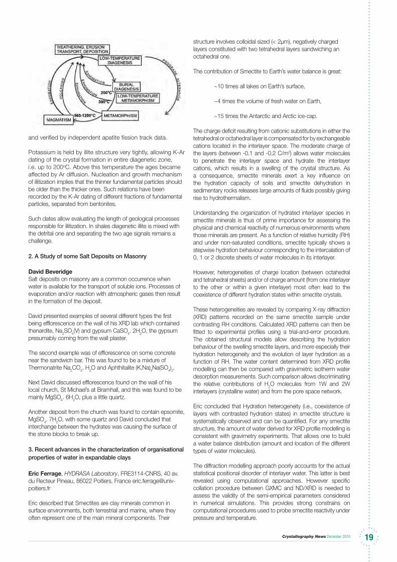
1. Illite-Smectite in Basin Analysis (K-Ar dating and estimation of the maximum paleotemperatures) Jan Srodo n Institute of Geological Sciences PAN, Senacka 1, 31002 Kraków, Poland, [email protected]
Jan explained that the clay minerals smectite and kaolinite are the most abundant products of chemical interaction between lithosphere, atmosphere, and hydrosphere. At high temperatures smectite and kaolinite react to form illite and chlorite. This Smectite-to-illite reaction is unique because of the abundance of these components (30% of the mass of sedimentary rocks), and the reaction mechanism,
which produces mixed-layer illite-smectite as intermediate products which indicate the reaction temperatures. The potassium content of illite allows K-Ar dating of the process.
Successive steps of the illitization of smectite can be followed by XRD measurements of the illite:smectite ratio or of the thickness of illite “zones” of the mixed-layer crystals (so called “fundamental particles”). Calibration of these measurements with respect to the maximum temperatures experienced by the rock is provided by the studies of young sedimentary basins (e.g. the Tertiary Podhale-Orava basin)
Crystallography News December 2010 1919
and verified by independent apatite fission track data. Potassium is held by illite structure very tightly, allowing K-Ar dating of the crystal formation in entire diagenetic zone, i.e. up to 200oC. Above this temperature the ages became affected by Ar diffusion. Nucleation and growth mechanism of illitization implies that the thinner fundamental particles should be older than the thicker ones. Such relations have been recorded by the K-Ar dating of different fractions of fundamental particles, separated from bentonites. Such dates allow evaluating the length of geological processes responsible for illitization. In shales diagenetic illite is mixed with the detrital one and separating the two age signals remains a challenge.
2. A Study of some Salt Deposits on Masonry
David Beveridge Salt deposits on masonry are a common occurrence when water is available for the transport of soluble ions. Processes of evaporation and/or reaction with atmospheric gases then result in the formation of the deposit. David presented examples of several different types the first being efflorescence on the wall of his XRD lab which contained thenardite, Na2SO4(V) and gypsum CaSO4. 2H2O, the gypsum presumably coming from the wall plaster. The second example was of efflorescence on some concrete near the sandwich bar. This was found to be a mixture of Thermonatrite Na2CO3. H2O and Aphthitalite (K,Na)3Na(SO4)2. Next David discussed efflorescence found on the wall of his local church, St Michael’s at Bramhall, and this was found to be mainly MgSO4. 6H2O, plus a little quartz. Another deposit from the church was found to contain epsomite, MgSO4. 7H2O, with some quartz and David concluded that interchange between the hydrates was causing the surface of the stone blocks to break up. 3. Recent advances in the characterization of organisational properties of water in expandable clays
Eric Ferrage, HYDRASA Laboratory, FRE3114-CNRS, 40 av. du Recteur Pineau, 86022 Poitiers, France [email protected]
Eric described that Smectites are clay minerals common in surface environments, both terrestrial and marine, where they often represent one of the main mineral components. Their
structure involves colloidal sized (< 2µm), negatively charged layers constituted with two tetrahedral layers sandwiching an octahedral one.
The contribution of Smectite to Earth’s water balance is great:
~10 times all lakes on Earth’s surface,
~4 times the volume of fresh water on Earth,
~15 times the Antarctic and Arctic ice-cap.
The charge deficit resulting from cationic substitutions in either the tetrahedral or octahedral layer is compensated for by exchangeable cations located in the interlayer space. The moderate charge of the layers (between -0.1 and -0.2 C/m2) allows water molecules to penetrate the interlayer space and hydrate the interlayer cations, which results in a swelling of the crystal structure. As a consequence, smectite minerals exert a key influence on the hydration capacity of soils and smectite dehydration in sedimentary rocks releases large amounts of fluids possibly giving rise to hydrothermalism.
Understanding the organization of hydrated interlayer species in smectite minerals is thus of prime importance for assessing the physical and chemical reactivity of numerous environments where those minerals are present. As a function of relative humidity (RH) and under non-saturated conditions, smectite typically shows a stepwise hydration behaviour corresponding to the intercalation of 0, 1 or 2 discrete sheets of water molecules in its interlayer.
However, heterogeneities of charge location (between octahedral and tetrahedral sheets) and/or of charge amount (from one interlayer to the other or within a given interlayer) most often lead to the coexistence of different hydration states within smectite crystals.
These heterogeneities are revealed by comparing X-ray diffraction (XRD) patterns recorded on the same smectite sample under contrasting RH conditions. Calculated XRD patterns can then be fitted to experimental profiles using a trial-and-error procedure. The obtained structural models allow describing the hydration behaviour of the swelling smectite layers, and more especially their hydration heterogeneity and the evolution of layer hydration as a function of RH. The water content determined from XRD profile modelling can then be compared with gravimetric isotherm water desorption measurements. Such comparison allows discriminating the relative contributions of H2O molecules from 1W and 2W interlayers (crystalline water) and from the pore space network.
Eric concluded that Hydration heterogeneity (i.e., coexistence of layers with contrasted hydration states) in smectite structure is systematically observed and can be quantified. For any smectite structure, the amount of water derived for XRD profile modelling is consistent with gravimetry experiments. That allows one to build a water balance distribution (amount and location of the different types of water molecules).
The diffraction modelling approach poorly accounts for the actual statistical positional disorder of interlayer water. This latter is best revealed using computational approaches. However specific collation procedure between GXMC and ND/XRD is needed to assess the validity of the semi-empirical parameters considered in numerical simulations. This provides strong constrains on computational procedures used to probe smectite reactivity under pressure and temperature.
Crystallography News December 201020
4. Anion Exchange Materials for use in Radiochemical ApplicationsAndy Butterworth; Dept. of Chemistry, Loughborough University Andy described how the compounds of the form Cu2(OH)3 X, X = Ac, NO3 have been prepared and that their properties as ion exchangers for various ions including iodide, oxyiodide species, perrhenate and antimonate have been investigated. These materials are stable in strong base and show good selectivity for iodine in small scale reactions with solutions containing a variety of anions. Structural work has been carried out using diffraction data collected at DIAMOND and a paper is currently being written on the family of materials and their exchange properties. A number of new layered hydroxides have also been prepared containing other cations such as Zn and Co. As a side project, synthetic routes for the 3 polymorphs of copper hydrochloride are being investigated with a focus on botallackite formation using Cu2(OH)3(Ac) as a starting material.
5. Geology of BeerJenny Huggett, Petroclays Jenny started by telling the audience that beer is mostly water and that most brewers have traditionally obtained their water from the local aquifer. The pH and key ions in the water affect the brewing process and as these chemical factors are strongly influenced by the nature of the aquifer rock, it follows that geology has a direct influence on brewing. The brewing process is water intensive; first there is the mashing – malted grain in hot water, this resulting wort is then boiled with hops and the wort is then fermented with yeast. Jenny then described the effect of geologically sourced ions on beer.
Ca2+ stabilises the enzyme amylase which helps breakdown of starch from malt in the mash tun and in later processes too. It precipitates any phosphate and so increases the acidity of the wort. Acidity influences strength and character of the fermentation and the microbiological stability of the enzyme processes. Ca2+ promotes flocculation of the yeast during fermentation.
Mg2+ ions produce a sour to bitter taste in beer but they also retard phosphate precipitation and stop the necessary increase in acidity. Both Na+ and K+ give beer a salty to sour taste and at >10 ppm K+ is noticeably laxative!
Sulphate is also very important in brewing because It helps break down protein, allows full extraction of bitter oils from hops and reacts with Mg2+ to form MgSO4 which is itself bitter.
The geographical locations of aquifers were then described which explains why beers from different regions have distinctively different tastes and colours but as they say, the proof of the beer is in the drinking and Jenny brought a selection with her for us all to try. These included:
riCharD MorriS
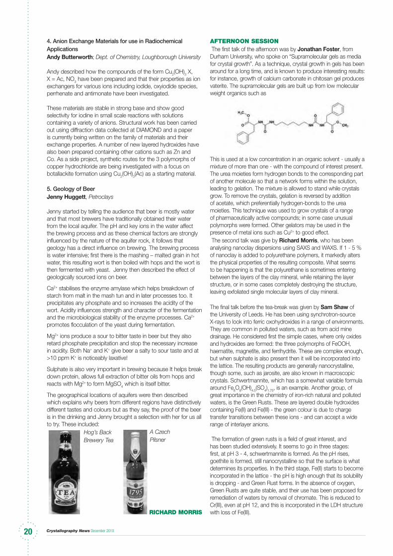
aFterNooN SeSSioN The first talk of the afternoon was by Jonathan Foster, from Durham University, who spoke on “Supramolecular gels as media for crystal growth”. As a technique, crystal growth in gels has been around for a long time, and is known to produce interesting results: for instance, growth of calcium carbonate in chitosan gel produces vaterite. The supramolecular gels are built up from low molecular weight organics such as
This is used at a low concentration in an organic solvent - usually a mixture of more than one - with the compound of interest present. The urea moieties form hydrogen bonds to the corresponding part of another molecule so that a network forms within the solution, leading to gelation. The mixture is allowed to stand while crystals grow. To remove the crystals, gelation is reversed by addition of acetate, which preferentially hydrogen-bonds to the urea moieties. This technique was used to grow crystals of a range of pharmaceutically active compounds; in some case unusual polymorphs were formed. Other gelators may be used in the presence of metal ions such as Cu2+ to good effect. The second talk was give by Richard Morris, who has been analysing nanoclay dispersions using SAXS and WAXS. If 1 - 5 % of nanoclay is added to polyurethane polymers, it markedly alters the physical properties of the resulting composite. What seems to be happening is that the polyurethane is sometimes entering between the layers of the clay mineral, while retaining the layer structure, or in some cases completely destroying the structure, leaving exfoliated single molecular layers of clay mineral. The final talk before the tea-break was given by Sam Shaw of the University of Leeds. He has been using synchrotron-source X-rays to look into ferric oxohydroxides in a range of environments. They are common in polluted waters, such as from acid mine drainage. He considered first the simple cases, where only oxides and hydroxides are formed: the three polymorphs of FeOOH, haematite, magnetite, and ferrihydrite. These are complex enough, but when sulphate is also present then it will be incorporated into the lattice. The resulting products are generally nanocrystalline, though some, such as jarosite, are also known in macroscopic crystals. Schwertmannite, which has a somewhat variable formula around Fe8O8(OH)4.5(SO4)1.75, is an example. Another group, of great importance in the chemistry of iron-rich natural and polluted waters, is the Green Rusts. These are layered double hydroxides containing Fe(II) and Fe(III) - the green colour is due to charge transfer transitions between these ions - and can accept a wide range of interlayer anions.
The formation of green rusts is a field of great interest, and has been studied extensively. It seems to go in three stages: first, at pH 3 - 4, schwertmannite is formed. As the pH rises, goethite is formed, still nanocrystalline so that the surface is what determines its properties. In the third stage, Fe(II) starts to become incorporated in the lattice - the pH is high enough that its solubility is dropping - and Green Rust forms. In the absence of oxygen, Green Rusts are quite stable, and their use has been proposed for remediation of waters by removal of chromate. This is reduced to Cr(III), even at pH 12, and this is incorporated in the LDH structure with loss of Fe(III).
Hog’s Back Brewery Tea
A Czech Pilsner

Crystallography News December 2010 2121
After a tea-break in which to rest my wrist, Nicholas Funnell of the University of Edinburgh spoke about the behaviour of Krogmann’s salt at high pressures. This is another mixed-valence species, K2Pt(CN)4Br0.3.3.2H2O, which crystallises in the unusual space group P4mm. The structure contains channels in which are to be found the bromide (or other halide ion) and water. The platinocyanide groups lie along the edges of the unit cell and behave as one-dimensional metals, conducting along the chains but not in the other directions. At high pressures - up to 50 kbar - phase changes have been reported, but this could
not be reproduced. What seems to be happening is that Peierls distortions cause the formation of a band gap - the substance becomes a semiconductor. The phenomena are clearly complex and might even include a chemical change. The final talk was by Ian Hutchinson, of the University of Leicester. He described the challenges of designing a radioisotope source XRD/XRF instrument to be conveyed to Mars on an ESA mission. The detectors have to be cooled to ensure stability.DaviD BeveriDge
The Bragg LecturerePort of the Bragg Lecture Fund (BLF) Committee Chairman Prof S E V Phillips for the period 2007 to 2010 inclusive (4 years)
I offer here my report here in the BCA Newsletter as outgoing Chairman of the Bragg Lecture Fund Committee.
The membership of the Committee in my period (basically a member drawn from each institution where Sir William Henry and Sir William Lawrence Bragg had worked) was:-
S E V Phillips (Chair) ( Research Complex at Harwell/University of Leeds)L N Johnson (Oxford)
J Bella (Manchester)
J E Davies (Cambridge)
C R A Catlow (UCL)
F A J L James (RI)
E Garman (BCA)
D M Paul (IoP)
The Chair of the Committee immediately preceding me was Prof J R Helliwell (Manchester). My successor as Leeds representative is Prof Rik Brydson, and the replacement Chair will be selected by the Committee.
The expenses of the Bragg Lecturer are handled by The Royal Institution of Great Britain (RIGB) who administers the Bragg Lecture Fund as per Rule 5 of the Bragg Lecture Fund Constitution and Rules (currently under review).
We consulted closely by email and selected Prof Sir John Meurig Thomas as the 2010 Bragg Lecturer (Warwick BCA, The RI and University of Leeds).
The Lecturer delivers two or more presentations, one always at The RIGB. In 2010 the others were at the BCA Spring Meeting and the University of Leeds.
The Bragg Lectures have thereby been successfully continued, and the full list of lecturers appears below:
1962 Paul P Ewald (Leeds & RI)1965 Dame Kathleen Lonsdale (Melbourne, Adelaide &
Perth)1968 Dorothy Hodgkin (Manchester)1970 B E Warren (RI)1973 R W G Wyckoff (Cambridge)1981 Henry Lipson (RI & Leeds)1982 Michael M Woolfson (Manchester & Cambridge)1985 Sir David Phillips (Leeds & RI)1987 Brian W Matthews (Perth & Adelaide)1993 Sir Gordon Cox and Max Perutz
(Manchester BCA and The RI)1994 A M Glazer (Newcastle BCA and The RI)1996 K C Holmes (Cambridge BCA and The RI)1997 Durward W J Cruickshank (The RI and Leeds BCA)1999 Jack Dunitz (chemistry) (The RI and IUCr
Glasgow Congress)2002 Dave Stuart (biology) (The RI and Reading BCA)2005 John Finney (physics) (The RI and UMIST BCA)2007 Roger Penrose (maths) (The RI and Canterbury BCA)2010 Sir John Meurig Thomas (chemistry) (Warwick BCA,
The RI and University ofLeeds)
I wish to thank all my Bragg Lecture Fund Committee colleagues, The RIGB and of course the Bragg Lecturer himself, Prof Sir John Meurig Thomas, during my term of office.
S e v PhilliPS, Director, Research Complex at Harwell
NEWSGrand Challenge Network Launchedthe EPSRC sponsored Grand Challenge Network “Directed Assembly of Extended Structures with Targeted Properties (DAESTP)” was formally launched together with another Network entitled “Dial-a-Molecule” at an inaugural meeting at the ICC in Birmingham on Wednesday, 20th October. The meeting was attended by almost 250 delegates from the areas of chemistry, chemical engineering, materials science,
physics, and biology together with a range of industrialists. After a brief welcome from Richard Whitby (Southampton) the principal investigator for the Dial-a-Molecule Network, the meeting opened with a short talk from Andrew Bourn, Head of Physical Sciences at the EPSRC. This was followed by introductions to the two Networks from the two network principal investigators Richard Whitby and Paul Raithby (for DAESTP), and the importance of the networks to UK industry was highlighted by David Hollinshead (AstraZeneca) and Neil Feeder (Pfizer).
The first plenary of the day was given by Professor Steve Ley FRS (Cambridge) who highlighted the benefits of “flow chemistry” and its importance in synthetic applications leading the achievement of the Dial-a-Molecule Grand Challenge.
Crystallography News December 201022
Control and Prediction of the Organic Solid State - 30 March 2011the 2011 meeting of the CPOSS project will be held in the Old Refectory at University College London on Wednesday 30th March 2011. The theme of the meeting will be “Towards Understanding the Pharmaceutical Solid State”, and will include speakers from the Solid State Pharmaceutical Cluster in Ireland and the Cambridge Crystallographic Data Centre. To complement the recurring theme of polymorphism causing
catastrophic problems in pharmaceutical production drugs, the successes of the recent International Blind Test of Crystal Structure Prediction will be highlighted. The meeting is free of charge to all academic groups, thanks to the sponsorship of the CPOSS Industrial Alliance. Lunch and coffee is included, and all participants are welcome to bring posters for the extended networking sessions. Full details of the programme are available at http://www.cposs.org.uk/ To register, please complete the online form, email Louise Price ([email protected]) or write to Dr Louise Price, Department of Chemistry, University College London, 20 Gordon Street London WC1H 0AJ.
2010 Hanawalt Award the International Centre for Diffraction Data takes pleasure in announcing that Dr. Takeshi Egami of the Joint Institute for Neutron Sciences, University of Tennessee, Knoxville, TN, and Oak Ridge National Laboratory, Oak Ridge, TN, and Dr. Simon J.L. Billinge of Applied Physics & Applied Mathematics, Columbia University, New York, NY were selected to receive the 2010 J.D. Hanawalt Award for excellence in the field of X-ray powder diffraction.
The presentation of the award took place at the 59th Annual Denver X-ray Conference, Denver, Colorado, U.S.A., on 4 August 2010. The Award was established to recognize outstanding achievement in the XRD field within the last five years. Takeshi and Simon were honored for their brilliant work in developing and extending the technique of Pair Distribution Function analysis of non-Bragg structures as exemplified in their book, “Underneath the Bragg Peaks”.
A special session entitled “Nanostructure Studies using the Atomic Pair Distribution Function” was held at the Denver X-ray Conference, where Drs. Egami and Billinge presented their Hanawalt Award lectures. Dr. Egami’s presentation focused on Recent Advances in the Pair Distribution Function Technique, while Dr. Billinge spoke on Structure at the Nanoscale: Atomic Pair Distribution Function Analysis of Nanostructured Materials.
Simon Billinge received his B.A. in Materials Science at the University of Oxford in 1986, and Ph.D. in Materials Science and Engineering at the University of Pennsylvania in 1992. Dr. Billinge first worked at Michigan State University as an Assistant Professor of physics and astronomy, and by 2003 he worked his way up to the title of Professor. In 2008, Dr. Billinge arrived at Columbia University as a Professor, as well as a Senior
Scientist at Brookhaven National Lab, Upton, NY. His research includes: nanoscale structure-property relationships in functional nanomaterials studied using novel X-ray and neutron scattering techniques coupled with advanced computing, as well as, solving the nanostructure problem.
Dr. Egami received his Bachelor’s degree in Applied Physics in 1968 at the University of Tokyo. In 1971, he received his Ph.D. in Materials Science from the University of Pennsylvania. Dr. Egami began teaching at the University of Pennsylvania in 1973 as an Assistant Professor. He became a Professor in 1980, and then Chair of the Materials Science and Engineering Department from 1997 to 2002. While teaching at Penn, Dr. Egami was Dr. Billinge’s thesis advisor in 1992. Dr. Egami is currently a Distinguished Professor at University of Tennessee, Knoxville and has written over 360 technical papers.
After lunch there were question and answers sessions followed by break-out sessions that were targeted at gaining community input into the next developments of the two networks. A lot of useful ideas were generated and some willing volunteers were recruited for the workshops and “sandpits” that are being planned.
The day finished with the second plenary from Professor Matthew Rosseinsky FRS (Liverpool) who developed the self-assembly theme and showed the importance of understanding the correlation between structure, property and function at the supramolecular level and how this would lead to assembling
materials with targeted properties.
Further information on the Directed Assembly Grand Challenge can be found on the website www.daestp.org.uk while enquiries about joining the network and about network events can be obtained by e-mailing [email protected]. Network developments and news about events can be followed through the twitter feed @DA_Challenge.
Paul raithBy
Congratulations to Dr. Takeshi Egami and Dr. Simon J.L. Billinge on receiving this prestigious award!
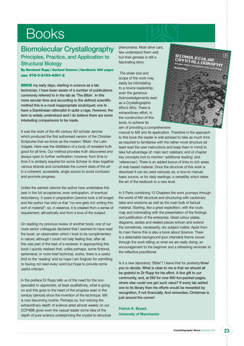
Crystallography News December 2010 2323
Biomolecular CrystallographyPrinciples, Practice, and Application to Structural BiologyBy Bernhard rupp | Garland Science | hardback: 850 pages
isbn: 978-0-8153-4081-2
SiNCe my early days, starting in science as a lab technician, I have been aware of a number of publications commonly referred to in the lab as ‘The Bible’. In this more secular time and according to the defined scientific method this is a most inappropriate soubriquet; one to have a Dawkinsian rationalist in quite a rage. However, the term is widely understood and I do believe there are some interesting comparisons to be made.
It was the work of the 4th century AD scholar Jerome which produced the first authorised version of the Christian Scriptures that we know as the modern ‘Bible’: the Latin Vulgate. Here was the distillation of a body of revealed truth good for all time. Our method provides truth discovered and always open to further verification; however, from time to time it is similarly required for some Scholar to draw together various strands and consolidate the current state of the art in a coherent, accessible, single source to avoid confusion and promote progress.
Unlike the sainted Jerome the author here undertakes this task in the full acceptance, even anticipation, of eventual redundancy. 5 years in preparation (Jerome took a bit longer) and the author has told us that “no-one gets rich writing this sort of material”; so, in essence, it is created from a sense of requirement, altruistically and from a love of the subject.
On reading my previous review of another book, one of our more senior colleagues declared that I seemed to have read the book; an observation which I took to be complimentary in nature; although I could not help feeling that, after all, this was part of the task of a reviewer. In approaching this book I quickly realised that, unlike perhaps, some fictional, ephemeral, or more brief technical, works, there is a useful limit to the ‘reading’ and so hope I am forgiven for admitting to having not read every word but hope to provide some useful criticism.
In the preface Dr Rupp tells us of the need for the non-specialist to appreciate, at least qualitatively, what is going on and this goes to the heart of the progress seen in the century (almost) since the invention of the technique. MX is now becoming routine. Perhaps so, but noticing the extraordinary depth of science aired almost weekly on our CCP4BB gives even the casual reader some idea of the depth of pure science underpinning the crystal-to-structure
phenomena. Most drive cars, few understand them well, but their genesis is still a fascinating story.
The sheer size and scope of the work may easily be intimidating to a novice readership; even the generous Acknowledgements read as a Crystallographic Who’s Who. There is extraordinary effort, in the construction of this book, to achieve its aim of providing a comprehensive manual to MX and its application. Therefore in the approach to this book the reader is well advised to take as much time as required to familiarise with the rather novel structure (at least read the user instructions and keep them in mind) to take full advantage of: main text; sidebars; end of chapter key concepts (not to mention ‘additional reading’ and ‘references’). There is an added bonus of links to rich areas of web based material. Once the structure of this work is absorbed it can be used variously as, a how-to manual, basic source, or for daily readings; a versatility which takes the art of the textbook to a new level.
In 5 Parts containing 13 Chapters the work journeys through the world of MX structure and structuring with cautionary tales and wisdoms as well as the main bulk of factual material. Starting, like a great expedition, with the route map and culminating with the presentation of the findings and justification of the enterprise. Great colour plates, diagrams, asides and related pieces enliven and extend the sometimes, necessarily, dry subject matter. Apart from its main theme this is also a book about Science. There is a detectable background (pun intended) theme woven through the work telling us what we are really doing; an encouragement to the beginner and a refreshing reminder to the reflective practitioner.
Is it a new laboratory ‘Bible’? I leave that for posterity/time/you to decide. What is clear to me is that we should all be grateful to Dr Rupp for his effort. A fine gift to our community, and, at £60 for over 800 fun-packed pages, where else could one get such value? If every lab added one to its library then his efforts would be rewarded by recognition, if not financially. And remember, Christmas is just around the corner!
Patrick K. BryantUniversity of Manchester
Books
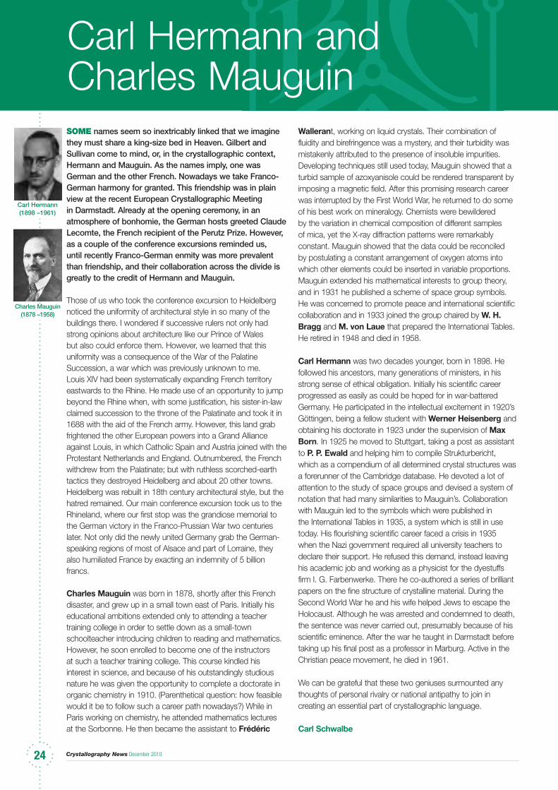
Crystallography News December 201024
Carl Hermann and Charles MauguinSoMe names seem so inextricably linked that we imagine they must share a king-size bed in Heaven. Gilbert and Sullivan come to mind, or, in the crystallographic context, Hermann and Mauguin. As the names imply, one was German and the other French. Nowadays we take Franco-German harmony for granted. This friendship was in plain view at the recent European Crystallographic Meeting in Darmstadt. Already at the opening ceremony, in an atmosphere of bonhomie, the German hosts greeted Claude Lecomte, the French recipient of the Perutz Prize. However, as a couple of the conference excursions reminded us, until recently Franco-German enmity was more prevalent than friendship, and their collaboration across the divide is greatly to the credit of Hermann and Mauguin.
Those of us who took the conference excursion to Heidelberg noticed the uniformity of architectural style in so many of the buildings there. I wondered if successive rulers not only had strong opinions about architecture like our Prince of Wales but also could enforce them. However, we learned that this uniformity was a consequence of the War of the Palatine Succession, a war which was previously unknown to me. Louis XIV had been systematically expanding French territory eastwards to the Rhine. He made use of an opportunity to jump beyond the Rhine when, with some justification, his sister-in-law claimed succession to the throne of the Palatinate and took it in 1688 with the aid of the French army. However, this land grab frightened the other European powers into a Grand Alliance against Louis, in which Catholic Spain and Austria joined with the Protestant Netherlands and England. Outnumbered, the French withdrew from the Palatinate; but with ruthless scorched-earth tactics they destroyed Heidelberg and about 20 other towns. Heidelberg was rebuilt in 18th century architectural style, but the hatred remained. Our main conference excursion took us to the Rhineland, where our first stop was the grandiose memorial to the German victory in the Franco-Prussian War two centuries later. Not only did the newly united Germany grab the German-speaking regions of most of Alsace and part of Lorraine, they also humiliated France by exacting an indemnity of 5 billion francs.
Charles Mauguin was born in 1878, shortly after this French disaster, and grew up in a small town east of Paris. Initially his educational ambitions extended only to attending a teacher training college in order to settle down as a small-town schoolteacher introducing children to reading and mathematics. However, he soon enrolled to become one of the instructors at such a teacher training college. This course kindled his interest in science, and because of his outstandingly studious nature he was given the opportunity to complete a doctorate in organic chemistry in 1910. (Parenthetical question: how feasible would it be to follow such a career path nowadays?) While in Paris working on chemistry, he attended mathematics lectures at the Sorbonne. He then became the assistant to Frédéric
Wallerant, working on liquid crystals. Their combination of fluidity and birefringence was a mystery, and their turbidity was mistakenly attributed to the presence of insoluble impurities. Developing techniques still used today, Mauguin showed that a turbid sample of azoxyanisole could be rendered transparent by imposing a magnetic field. After this promising research career was interrupted by the First World War, he returned to do some of his best work on mineralogy. Chemists were bewildered by the variation in chemical composition of different samples of mica, yet the X-ray diffraction patterns were remarkably constant. Mauguin showed that the data could be reconciled by postulating a constant arrangement of oxygen atoms into which other elements could be inserted in variable proportions. Mauguin extended his mathematical interests to group theory, and in 1931 he published a scheme of space group symbols. He was concerned to promote peace and international scientific collaboration and in 1933 joined the group chaired by W. H. Bragg and M. von Laue that prepared the International Tables. He retired in 1948 and died in 1958.
Carl Hermann was two decades younger, born in 1898. He followed his ancestors, many generations of ministers, in his strong sense of ethical obligation. Initially his scientific career progressed as easily as could be hoped for in war-battered Germany. He participated in the intellectual excitement in 1920’s Göttingen, being a fellow student with Werner Heisenberg and obtaining his doctorate in 1923 under the supervision of Max Born. In 1925 he moved to Stuttgart, taking a post as assistant to P. P. Ewald and helping him to compile Strukturbericht, which as a compendium of all determined crystal structures was a forerunner of the Cambridge database. He devoted a lot of attention to the study of space groups and devised a system of notation that had many similarities to Mauguin’s. Collaboration with Mauguin led to the symbols which were published in the International Tables in 1935, a system which is still in use today. His flourishing scientific career faced a crisis in 1935 when the Nazi government required all university teachers to declare their support. He refused this demand, instead leaving his academic job and working as a physicist for the dyestuffs firm I. G. Farbenwerke. There he co-authored a series of brilliant papers on the fine structure of crystalline material. During the Second World War he and his wife helped Jews to escape the Holocaust. Although he was arrested and condemned to death, the sentence was never carried out, presumably because of his scientific eminence. After the war he taught in Darmstadt before taking up his final post as a professor in Marburg. Active in the Christian peace movement, he died in 1961.
We can be grateful that these two geniuses surmounted any thoughts of personal rivalry or national antipathy to join in creating an essential part of crystallographic language.
Carl Schwalbe
Carl Hermann(1898 –1961)
Charles Mauguin(1878 –1958)
Crystallography News December 2010 2525
National Facilities NewsFolloWiNg on from our quick tour of the small-molecule single-crystal diffraction beamline, I19, and the powder-diffraction beamline, I11, at Diamond in the last issue of Crystallography News, we have now made our way to the other side of the synchrotron building where the macromolecular crystallography beamlines are located. Conveniently, the beamlines are grouped together in zones 4, 5 and 6 to form the Macromolecular Crystallography (MX) village. Currently there are five beamlines in the village with I02, I03 and I04 among the first tranche of beamlines to be built in the Phase-I construction of Diamond with the later addition of I24 and I04-1 during Phase II. An additional MX beamline, I23, will be one of the first to come online as part of the Phase-III construction, which will extend the village into zone 3. It almost goes without saying that macromolecular crystallography is well catered for at Diamond but the investment in beamlines reflects the demand for beamtime from this very large and high-profile user community.
Much of the pioneering work on the utilisation of synchrotron radiation from the first generation sources for diffraction was driven by the protein crystallography community and an excellent account of the international effort of this early work is given in a review by Liz Duke and Louise Johnson (Macromolecular crystallography at synchrotron radiation sources: current status and future developments, Proceedings of the Royal Society A (2010): Online ISSN: 1471-2946). These first generation sources were principally designed for the particle physics community and, although they had sufficiently good stability for those experiments, the drifts in the position of the electron beam, with the accompanying variations in X ray intensity, made it difficult to record accurate diffracted intensity data. This drove the demand for dedicated, second-generation, synchrotron sources and the SRS at Daresbury Laboratory, which was commissioned in 1981, was the first of these facilities to be built. The macromolecular crystallography community has benefited from the availability of synchrotron radiation from the earliest days of user operations at the SRS and many of the current synchrotron-based MX can be traced back to ideas and innovations developed there. A number were ultimately realised at the European Synchrotron Radiation Facility, and other third-generation sources.
The first MX beamline built at Daresbury was station 7.2 which was located on a bending magnet source. This became operational in 1982 soon after the synchrotron became available to users. With the later addition of a superconducting wiggler, a second dedicated MX station was built, 9.6, which was also operated at a fixed wavelength. It was with the subsequent development of station 9.5 that energy tuning became available which provided the opportunity to contribute to the development of the anomalous dispersion phasing technique (MAD) which was being implemented at facilities elsewhere at that time. The final complement of MX stations at Daresbury, 14.1 and
14.2, became operational in the early “noughties” and they provided an opportunity to develop techniques for narrow-gap insertion device technology in advance of Diamond. As these beamlines were positioned in a very congested area in the SRS experimental hall, the hutches were tiny and the geometrical constraints only allowed fixed wavelengths. A degree of tunability across the Se edge was possible on 14.2, however, to allow the MAD technique – considered by then to be a fundamental technique for MX studies. Station 14.2 was also used in the early development of automation hardware and software for the now ubiquitous data pipeline. In parallel to these later developments at SRS, the first MX beamline at the ESRF, BM14, became operational in the mid nineties, at about the same time as the ESRF itself was made available to users, and this transformed the capabilities of MX in the determination of extremely complex proteins. With the success of this initial beamline, ID14 was constructed in the late nineties, with 4 stations on the beamline sharing 3 inline undulators. Radiation damage started to become a major issue on these beamlines, especially on ID14-4 the straight-through station, and this was emphasised, big-time, by the later addition of beamline ID29.
The MX beamlines at Diamond have benefited directly from the experience gained at the SRS, the ESRF and other facilities world-wide. The phase-I beamlines (I02, I03 and I04) were built simultaneously and they are pretty much identical in design. In fact, to the casual observer it would be difficult to tell which beamline you were on from the interior of the experiments hutches – though the beamline scientists would take great issue with this. The first impression you get when you walk into the experiments hutch of I04, for example, is the presence of the large blue A-frame structure which provides support for the motorised detector platform. The A-frame almost fills the hutch and its size helps to provide the necessary stiffness to support the detector, an ADSC Quantum 315r which weighs the best part of 141 kg, with the required ~ 10 µm positional stability. The detector has an impressive 315 mm × 315 mm active area (6140 × 6140 pixels) and is composed of 9 tiled CCD chips each with its own demagnifying taper. As protein crystals diffract to only very low Bragg angles a large area detector at 2θ = 0 is all that is required for the collection of data to the full resolution of the samples and the mechanical complexity of a variable detector angle, especially considering the weight and cooling requirements, can be avoided. There is some provision, however, to provide data collections at reasonably high 2θ angles if required. The goniometer itself is also relatively simple as only one rotation axis is necessary to provide reasonable reciprocal space coverage, given the limited resolution of the diffraction data. As proteins often crystallise in fairly high symmetry space groups, with orthorhombic P2
12121 being a common example, a random orientation of the crystal on its mounting loop generally means that one crystal will give complete data sets (typically greater than about 98 - 99 %) with a redundancy in the final data set of 3 or greater. Occasionally crystals may be oriented on the
Crystallography News December 201026
loop with a reciprocal lattice vector aligned almost parallel with the ω rotation axis making the collection of a complete data set impossible from one sample. In cases where the samples have a needle, or rod, habit then custom designed sample mounts are available to fix the orientation of the principal axis of the crystal at 30° or 45° to the ω axis, or users occasionally bend the flexible nylon loops to reorientate the crystal. Mini-kappa systems are available also to permit best orientation of the crystal and work is underway on I04-1 and I04 at Diamond to implement these. The intense synchrotron X-ray beam will disrupt bonds in the protein, often at key binding sites, well in advance of any noticeable degradation in the quality of the diffraction data recorded on the detector images, although cryocooling to 100 K alleviates these issues substantially. The dose limit for each sample is calculated using programs such as RADDOSE, developed by Elspeth Garman’s group (Paithankar et. al. (2009) J. Synch. Rad. 16, 152-163), where parameters such as sample composition and sample volume are used to establish the maximum exposure time in the beam, which also needs to have a carefully calibrated flux for the energy in use. To minimise the exposure time and obtain the most efficient use of each crystal, data collection strategies can be implemented after an initial indexing has been carried out. Several strategy options (refinement set, SAD, low dose, user specific) for data collection are now automatically calculated for collected test images using EDNA and the results presented clearly for the user to then select and use in the GDA interface. For a limited number of systems, where the samples can diffract to relatively high resolution, say 0.9 Å to 1.0 Å, then an additional sample rotation may be required in the data collection strategy to enhance reciprocal space coverage. The additional sample orientation is achieved through the use of a mini-Kappa axis placed on the main omega circle.
The goniometry allows for an extremely open environment around the sample position and a relatively unhindered route for the robotic sample changer. Even so the cryojet, x-ray scatter guard, beam-stop and sample viewing back-light are all motorised and are driven in a carefully choreographed set of manoeuvres as the samples are mounted and dismounted. The whole sample change process takes less than 30 seconds with the samples being transferred from the dewar to the goniometer, and back again, with no build up of ice. This allows a batch of crystals to be screened prior to the best being chosen for subsequent data collection without any concern for sample degradation – save for slight beam damage during screening. It is now standard practice for the sample crystals to be mounted on loops, pre-frozen and transported to Diamond in a dewar ready to be loaded into the robotic sample changer. Apart from loading the robot, which can be done by the beamline scientists if the samples are couriered in advance of the experiment, then the users need not enter the experimental hutch for the remainder of their beamtime with the sample changes, crystal centring, screening and data collections carried out entirely from
the control room. Indeed, some users carry out experiments via remote access from their home institution without ever visiting Diamond. Apart from the sample changes, there is additional automation in data processing and analysis so that in ideal cases, and where a starting model is known, a preliminary electron density difference map can be produced within a few minutes of the data collection finishing. This functionality has been introduced through the implementation of the EDNA data analysis pipeline at Diamond (developed in collaboration with the ESRF) which has transformed the efficiency and ease of use of the MX beamlines. It allows users to plan their experiments and prepare their samples well in advance of their visit and, crucially, with extremely rapid data analysis available the experiments can be adapted during beamtime. The automation of beamline hardware control and data reduction and structure solution is not possible without the excellent support and development provided by the controls, data acquisition and scientific software teams at Diamond, some of whom also act as local contacts for users.
On I02, I03, I04 and I24 the wavelength can be tuned between 5 keV and 25 keV which covers the range for the more favoured absorption edges of Se, Zn, and Fe, and for heavy atom soaks Hg, Pt and Au. Fine tuning the energy accurately to the required absorption edge is achieved through the use of a fluorescence detector which measures the fluorescence of the metal atoms present in or bound to the protein. This allows subtle changes in absorption edge position from that of the elemental atom arising from any local bonds to be taken into account. The phase-III beamline, I23, is being designed to specifically cover the 2.1 – 12 keV energy range in which the K edges of the elements Cl, P, S and Ca occur, which are present in natural, or native, proteins, and will allow MAD techniques to be performed without the need for the samples to be doped with heavy elements. Air absorption will be significant and the experiments will need to be carried out with both the goniometer and detector located within a vacuum vessel. Side-station I04-1 has a fixed energy (13.53 keV which is equivalent to ~0.916 Å – an energy chosen where a strong anomalous signal can be recorded for Se and most commonly used heavy atom derivatives therefore making many SAD experiments possible) and although its experiments hutch is located on the floor space between I04 and I03 (in the position that would otherwise have been occupied by B03) it has its own undulator source. This is positioned just upstream of the I04 undulator, in the same straight section of the synchrotron, and a vertically mounted monochromator crystal housed within the I04 optics hutch deflects the beam horizontally into the I04-1 experiments hutch. The I04-1 experimental instrumentation is aligned at the same offset angle within the experiments hutch, almost corner to corner, while the hutch itself is neatly positioned within its fan between the other beamlines.
Beamline I24, which became operational in mid phase-II,
National Facilities News...Cont.
Crystallography News December 2010 2727
has been designed with microfocusing optics to provide beam sizes ranging from 5 µm to 30 µm so that full structure determinations can be obtained from very small crystals and to allow multiple data collections from different positions on a single, larger, crystal. For a reasonable chance of a successful outcome on the other MX beamlines, crystals with dimensions on the order of 50 µm to 100 µm are considered ideal but often crystals of this size are difficult, if not impossible, to obtain. Membrane proteins, for example, are notoriously difficult to crystallise and easily fall into this “awkward customer” category. Although they are essential for a very large variety of biological functions, including molecular transport, the relay of signals and respiration, and they comprise up to 30% of the proteins encoded by eukaryotic (nucleus containing) cells, there are fewer than 20 independent structures of integral membrane proteins currently reported. To put this into perspective the number of structure determinations of all proteins now runs into the many thousands. In part, I24 was built to meet the very demanding challenges of determining structures from very small membrane protein crystals. As membrane protein crystals are extremely fragile, and are less amenable to being transported cryogenically, it is no coincidence that the Membrane Protein Laboratory (set up as collaboration between Imperial College, London, and Diamond) is situated in the same sector of the synchrotron building as the beamline. The optics of I24 are broadly similar to the other MX beamlines with the exception that it has a set of microfocussing mirrors. To provide optimum performance, they need to be located as close to the sample position as possible and this has been achieved by mounting the single-axis goniometer tightly up against the mirrors’ vacuum vessel. The experimental hutch is otherwise broadly reminiscent of the other MX beamlines with the very obvious exception that there is the option for the use of two detectors: one large Rayonix CCD detector and an even larger Dectris Pilatus 6M detector. The latter, which has an active area of 431 mm × 448 mm (2463 × 2527 pixels) is an example of a new generation of photon-counting area detectors which are revolutionising not only MX but other areas of x-ray diffraction where an exceptionally large dynamic range and an extremely rapid frame rate (of up to 12 Hz for the 6M and 200 Hz for the 100K) are required but very high spatial resolution is less of a consideration. As the Pilatus allows some degree of energy discrimination significant improvements can be made in the reduction of background and this, coupled with the impressive dynamic range, allows the very accurate intensity determination of extremely weak reflections. The microfocus beam on I24 allows a range of new techniques to be developed as the beam is sufficiently small to allow different parts of a “standard” sized crystal to be probed: for relatively mosaic crystals this allows the part of a crystal exhibiting the best diffraction qualities to be used; and the same crystal can be used for a much longer period, before it has to be replaced due to radiation damage, as a fresh volume of the sample can be illuminated (with this process repeated often several times). The highly focused nature of the beam can also be used to screen samples prior to selection for subsequent data collection and the goniometer can be converted to hold and accurately translate crystallisation plates which are brought in directly from the Membrane Protein Laboratory. Although the data obtained from this screening setup aren’t yet of sufficient
quality for structure determinations, there is increasing interest in developing these techniques further with specially modified plates to allow greater angular access for both the incident and diffracted beams and to reduce the background with different, and thinner, plate materials.At the time of writing this piece, the phase-I MX beamlines are being prepared for a series of upgrades. This will involve a rolling programme of work to improve the end-station equipment, to upgrade the detectors, and to perform improvements and upgrades to the optic systems. As the work will be extremely invasive individual beamlines will be withdrawn from the user programme with the remaining operational beamlines supporting the scheduled MX beamtime. Following these improvements, beamlines I02, I03 and I04 will have much greater stability, faster data collection speeds, more rapid sample throughput and improved beam focussing options. This will ensure that the beamlines remain internationally competitive and offer World-class facilities for the UK protein crystallography community for the foreseeable future.
The scientists working within the Macromolecular Crystallography village reflect the international effort in this area. With the names grouped according to main beamline affiliation they are, deep breath: I02, Thomas Sorensen, James Sandy, Juan Sanchez-Weatherby; I03, Katherine McAuley, James Nicholson and Mark Williams; I04, Dave Hall, Ralf Flaig, Pierpaolo Romano; I04-1, Jose Brandao-Neto, Alice Douangamath; I23, Armin Wagner and Vitaly Mykhaylyk; and I24, Gwyndaf Evans, Robin Owen, Danny Axford and David Waterman. Michael Engel is the MX Industrial Liaison Scientist. Alun Ashton leads the efforts in downstream data processing at Diamond and he, and his team, have been responsible for the implementation of the data pipeline, and the development of EDNA, across all MX beamlines. Given the overlap between the Phase-I beamlines there is a considerable degree of shared effort when it comes to supporting users and these beamlines can now schedule short ~ 1shift (8 hour) beamtime periods with morning and early evening start times. The beamline scientists are happy to discuss any queries you may have about the beamlines and their peripheral laboratories including the range of sample preparation facilities that they offer. The Diamond web site (www.diamond.ac.uk) has links to the beamline pages and details of the Membrane Protein Laboratory, including details of how to apply for access, can be found at http://www.diamond.ac.uk/Home/MPL.html. In the next issue we’ll complete our orbit of the synchrotron building by looking at facilities for surface diffraction and magnetic structure determination.
I’d like to thank Liz Duke for her patient help in the preparation of this short article. She is one of the original members of the Life Sciences Division at Diamond and she was responsible for beamlines I02, I03 and I04 through their design, construction and commissioning periods. She is now Principal Beamline Scientist on the Phase-III beamline Full Field Cryo X-ray Tomography of Biological Cells, B24. I’d also like to thank Dave Hall, Armin Wagner and Katherine McAuley for some useful comments on the manuscript.

Crystallography News December 201028
The experiments hutch of I04 showing: the detector support frame (SF); the detector (DET); and the goniometer (GON) in the foreground
A closer view of the I04 goniometer showing: the omega axis air-bearing (OM); the microglide motorised goniometer head (XYZ); the on-axis sample viewing microscope (MIC); the cryojet (CRY); and the collimation for the fluorescence detector (FD)
The robotic sample changer in the I04 experiments hutch. The end effector is reaching into the sample dewar. The goniometer and detector are shown in the background.
A dry dewar and its packing case used to transport pre-mounted sample crystals to Diamond.
The x-ray transport pipe linking the optics hutch of I04, the yellow structure on the right, to the experiments hutch of I04-1, the yellow structure on the left. The image shows the walkway between the two beamlines, facing away from the synchrotron ratchet wall.
The I24 detector frame with the Rayonix CCD detector, above, in its parked position and the Pilatus 6M detector, below.

Crystallography News December 2010 2929
BCA BSG Winter Meeting The Biological Structures Group Winter Meeting will be held on Wednesday December 15th 2010 at the University of Reading. Registration opens from 10:00 am in Black Horse House with lectures starting at 11:00 in the Palmer Building on the Whiteknights Campus.
The general focus of the meeting is ‘Metal-protein interactions and their role in molecular transport and cell signalling.’
The aim of the conference is to highlight recent structure-function studies of proteins involved in molecular transport and signalling processes where metal ions play a crucial role.Nominations for speakers and other enquiries should be made to the organiser, Dr Kim Watson, by e-mail ( [email protected] ).
Posters We particularly welcome student posters. If you would like to present a poster, please submit an abstract (max 300 words) to the organiser by e-mail.
Deadline for abstract submission is Wednesday 1st December 2010.
RegistrationAs usual, the registration fee will include one year’s membership to the BCA, which can be refunded upon request after the meeting should you not wish to remain a member of this prestigious society, or if you have already paid!
Closing date for registration is Monday 6th December 2010.
AccommodationIf you require accommodation, please contact the meeting organiser, Kim Watson, by email ([email protected]).
WebsiteThe link for this meeting is http://www.reading.ac.uk/biologicalsciences/businessdevelopment/biosci-BCAwintermeet.aspx
Call For Nominations!
The Committee of the YCG would like to invite all crystallographers to submit nominations for candidates for the first Parkin Lecture to be held at the YC2011 Satellite Meeting on 12th April 2011 at the University of Keele.The Parkin Lecture has been established as the prize lecture of the YCG in recognition of the outstanding contributions of the late Dr Andrew Parkin to the YCG of the BCA and the candidate should be a Young Crystallographer (undergraduate or graduate student or a crystallographer within five years of graduation), who has been recognised for outstanding contributions to any of the following:
Promoting science,
Raising public awareness of science,
Teaching crystallography/science or
Originality in outreach and teaching activities.
If you think you know of a Young Crystallographer who has excelled in any of these areas then why not nominate him or her for the Parkin Lecture! Nominations should consist of a supporting statement (max 1 page A4) and may include copies of any relevant publications though not essential. The nomination documents should be sent to Anna Stevenson (YCG Secretary) at [email protected] no later than Wednesday, 19th January 2011. The rules for the Parkin Lecture and details regarding the nomination process can be found on the YCG website http://yc.crystallography.org.uk/.
Susanne Coles (née Huth)YCG Chair
News from the Groups
Crystallography News December 201030
CCP4 Study Weekend - January 2011
We cordially invite you to participate in this year’s Study Weekend at the Warwick Conferences, University of Warwick. Once again, we have put together an exciting scientific programme for Thursday and Friday, either side of the traditional conference dinner. Please also check out the satellite meetings which may be of interest - we have scheduled in a session of What’s New in CCP4. The Study Weekend is a chance to catch up with old friends, but is also a chance to meet the CCP4 staff who will be there in force to demonstrate the latest software and to answer questions.
This year, the topic for the Study Weekend is “Model Building & Refinement & Validation”. In keeping with previous CCP4 meetings, the lectures will focus on the presentation and discussion of advanced methods and techniques developed and used by the leaders in the field.
Scientific OrganisersRoberto Steiner - King’s College London (UK)Bernhard Rupp - k. k. Hofkristallamt (USA)
Administrative OrganisersShirley Miller ([email protected]) Damian Jones ([email protected])Laura Johnston ([email protected])Wendy Cotterill ([email protected])
RegistrationWe will operate a two-tiered registration fee, which benefits those who register early! The registration fee includes all meals during the conference, including the conference dinner on Thursday evening at the Chancellors Suite, Warwick Conferences.
Register between 13 September and 21 November - the fee is £210 Register between 22 November and 10 December - the fee is £260
regiStratioN CloSeS oN 10 DeCeMBer!
For those applying for a standard bursary (registration fee and one night’s standard accommodation) the registration fee of £210 is the only fee that will apply. If you are intending to apply for a standard bursary please ensure that you read this section.
NOTE: STRICTLY NO CASH WILL BE ACCEPTED FOR PAYMENT AT THE CONFERENCE.STFC now use Shared Services to administer payments and no cash can be taken at the event to pay for attendance at the Study Weekend. You will need to pay beforehand by using the methods included in these web pages. If you require invoicing, please email Shirley Miller in good time to ensure payments have been received before the conference.
Crystallography News December 2010 3131
Meetings of interestFurther information may be obtained from the websites given. If you have news of any meetings to add to list please send them to the Editor, [email protected]. Assistance from the IUCr website is gratefully acknowledged.
8-10 December 2010
8th International Conference on X-ray Investigations of
Polymer Structure, XIPS 2010, Wroclaw, Poland.
www.xips2010.ath.bielsko.pl
13-14 December 2010
SPEM2010. 1st International Workshop on Scanning
Photo Electron Microscopy and Ambient Pressure XPS,
Trieste, Italy.
www.elettra.eu/spem2010
14-16 December 2010
Condensed Matter and Materials Physics (CMMP10),
Coventry.
www.cmmp.org.uk
15 December 2010
BCA BSG Winter Meeting,
University of Reading.
www.reading.ac.uk/biologicalsciences/
businessdevelopment/
biosci-BCAwintermeet.aspx
13-14 December 2010
SPEM2010. 1st International Workshop on Scanning
Photo Electron Microscopy and Ambient Pressure XPS,
Trieste, Italy.
www.elettra.eu/spem2010
12-14 January 2011
BILL2011, Grenoble, France.
www.ill.eu/news-events/events/bill2011
17-18 January 2011
IPANEMA 2011. Synchrotron Radiation for Ancient
Materials. Satellite Workshop, L’Orme des Merisiers,
France.
www.synchrotron-soleil.fr/
Workshops/2011/SatelliteIPANEMA11/Welcome
17-18 January 2011RIXS2011. New Prospects for Resonant Inelastic Soft X-ray Scattering. Satellite Workshop, St Aubin, France.www.synchrotron-soleil.fr/Workshops/2011/SatelliteSoft-RIXS
19-20 January 20116th SOLEIL Users’ Meeting, Palaiseau and St Aubin, France.www.synchrotron-soleil.fr/Workshops/2011/SUM11/24 January - 3 February 2011 FullProf School-2011, 4th ILL annual School on Neutron Diffraction Data, Grenoble and Allevard, France.www.ill.eu/instruments-support/instruments-groups/groups/dif/FPSchool/
21-26 February 2011AAFS 2011. American Association of Forensic Sciences Annual Meeting, Chicago, IL, USA.www.aafs.org/aafs-2011-annual-meeting
26 March - 3 April 2011 13th Intensive Teaching School in X-ray Structure Analysis Durham.www.dur.ac.uk/durham.x-ray-school/
27-31 March 2011ACS National Meeting & Exposition. Spring 2011, Anaheim, CA, USA.http://portal.acs.org/portal/PublicWebSite/meetings/spring2011/index.htm
30 March 2011 Control and Prediction of the Organic Solid State - Towards Understanding the Pharmaceutical Solid State www.cposs.org.uk/
11-14 April 2011BCA Spring Meeting, Keele University.http://crystallography.org.uk/spring-meeting-20116-15 April 2011Synchrotron Radiation & Free Electron Lasers. Joint US-Cern-Japan-Russia School, Erice, Italy.www.cern.ch/schools/CAS
Crystallography News December 2010 3131
9-13 May 2011E-MRS Spring Meeting and IUMRS ICAM 2011, Nice, France.www.emrs-strasbourg.com/index.php?option=com_content&task=view&id=359&ltemid=134
9-13 May 2011X-ray Techniques for Materials Research--From Laboratory Sources to Free Electron Lasers. Symposium of E-MRS Spring Meeting, Nice, France.www.emrs-strasbourg.com/index.php?option=com_content&task=view&Itemid=134&id=394
10-14 May 2011 ICSG 2011 International Conference on Structural Genomics, Toronto ON, Canada. www.sgc.utoronto.ca/ICSG2011/
28 May - 2 June 2011American Crystallographic Association Meeting, New Orleans, LA, USA.www.amercrystalassn.org/content/pages/2011-homepage
2-12 June 2011 The Power of Powder Diffraction, Erice, Italy.www.crystalerice.org/Erice2011/2011pd.htm
2-12 June 2011 Electron Crystallography: New Methods to Explore Structure and Properties of the Nano World, Erice, Italy.www.crystalerice.org/Erice2011/2011ec.htm13-16 June 2011 Nanotech Conference & Expo 2011, Boston MA, USA.www.techconnectworld.com/Nanotech2011/
13-26 June 2011Zürich, Switzerland.The Zürich School of Crystallography 2011. Bring Your Own Crystals.www.oci.uzh.ch/group.pages/linden/zsc/
26-30 June 20115th International Workshop on Crystal Growth Technology, Berlin, Germany.http://iwcgt5.ikz-berlin.de/
17-21 July 2011 ECNS 2011. 5th European Conference on Neutron Scattering, Prague Czech Republic.www.ecns2011.org/joomla_15
17-22 July 2011Thin Film and Crystal Growth Mechanisms. Gordon Research Conference, Biddeford, ME, USA.www.grc.org/programs.aspx?year=2011&program=thinfilm
1-5 August 2011DXC 2011. 60th Annual Denver X-ray Conference, Denver, CO, USA.www.dxcicdd.com/
7-12 August 2011X-ray Science. Gordon Research Conference, Waterville, ME, USA.www.grc.org/programs.aspx?year=2011&program=xray
22-29 August 2011 IUCr2011. XXII Congress and General Assembly, Madrid, Spain.www.iucr2011madrid.es/
13-16 September 2011ISIC18. 18th International Symposium on Industrial Crystallization, Zürich, Switzerland.http://www.isic18.ethz.ch/
25-29 August 201328th European Crystallographic Meeting, University of Warwick.http://www.crystallography.org.uk/
Crystallography News December 201032 Crystallography News December 2010
PANalytical Ltd. 7310 IQ Cambridge,Waterbeach,Cambridge,CB25 9AY t +44 (0)1223 203480f +44 (0)1223 [email protected]
EMPYREAN
The world of X-ray diffractionis no longer fl at
-6000 -4000 -2000 0 2000 4000 600010
100
1K
10K
100K
1M
10M
100M
Omega/2Theta [s]
Inte
nsi
ty [
cou
nts
]
0D
2D
1D
3D
Position 2Theta [deg.] (Copper (Cu))
Inte
nsi
ty [
cou
nts
]
4000
3000
2000
1000
020 30 40 50 60 70 80 90 100 110 120 130 140
Inte
nsi
ty [
cou
nts
]
Position 2Theta [deg.] (Copper Cu))
FWHM = 0.026 °2θ
is no longer fl at
The new Empyrean from PANalytical is truly innovative, with cutting-edge technology in every aspect. Empyrean brings the idea of the perfect XRD platform to life:
• The widest range of samples• The highest data quality on every
sample, no compromises• Exceptional tube performance• The highest performance goniometer • 2nd generation PreFIX for optics and
sample platforms• PIXcel3D: the only detector for 0D, 1D,
2D and even 3D applications• Unmatched area detector dynamic
range, linearity and resolution• See inside your samples with the
world’s fi rst 3D detector
The only XRD platform that does it all
• Powders• Thin fi lms• Nanomaterials• Solid objects
Cutting-edge technology. Ultimate commitment.
The Analytical X-ray Company
PN7305.indd 1 19-02-2010 07:35:08
BCA 2011 Keele UniversityDate for your Diary
PDF-4+ 2010More Data • More Data Mining
The most comprehensive collection of inorganic powder patterns
Phase identification using physical, chemicaland crystallographic data
❖ Comprehensive materials database featuring 301,282 data sets
Standardized, edited data from four crystallographic databases
❖ Utilize quantitative analysis methods
147,393 Data sets with atomic parameters for Rietveld analysis
207,075 Data sets with I/Ic for Reference Intensity Ratio (RIR)
301,282 Data sets with full digital patternsfor total pattern analysis
CONFIDENCE COMES WITH GREAT RESULTS
Before PDF-4+ After PDF-4+
International Centre for Diffraction Data12 Campus Boulevard
Newtown Square, Pennsylvania 19073-3273 U.S.A.Phone: 610.325.9814 • Toll-free: 866.378.9331 (U.S. & Canada)
Fax: 610.325.9823www.icdd.com • www.dxcicdd.com
ICDD, the ICDD logo and PDF are registered in the U.S. Patent & Trademark Office.

CoolersOnly
For further information visit www.oxcryo.com
That’s all we do. Why?
Well, we believe that being entirely focused on one area of expertise, allows us to develop and manufacture the most advanced, reliable and easy-to-use range of coolers available for X-ray crystallography.
For over 20 years we have remained firmly at the heart of the global crystallographic community, listening to our customers and continually developing our products into what they are today - products that can deliver exactly what you really want.
From the wide temperature range liquid nitrogen Cryostream series, to the new compact non-liquid Desktop Cooler, there is a device suited to your exact need.
So, although we produce only coolers, we produce the only coolers that are the preferred choice of the crystallographic world - Oxford Cryosystems coolers.
Cryostream 700 series & Cryostream Plus Open flow LN2 cooler: 80-500 KCobra & Cobra Plus Non-liquid nitrogen cooler: 80-500 KN-HeliX Open flow helium cooler: 28-300 KDesktop Cooler Compact non-liquid cooler: 170-290 KPheniX Closed cycle powder cryostat: 12-290 K
0816 - OxCryo-BCA-09-10-v1.indd 1 14/7/10 00:26:15