Embed Size (px)
Citation preview
1
CT: Diagnosis of Pulmonary Embolism
Jud W Gurney MD FACR
University of NebraskaMedical Center
The movie Hunt for Red October is about about a silentdeadly submarine, a useful metaphor for the diagnosis ofpulmonary embolus.
2
The Hunt for Red October
Find the clot!
CT finds the clot
PE is all about finding the clot. Examine the literature on PEand you’ll find that the seminal papers all deal with finding theclot. In this lecture, we will review CT for diagnosing PE. CTvisualizes the clot, and has rapidly emerged as an importantclinical tool which will ultimately provide new insights into thenatural history of this process.
3
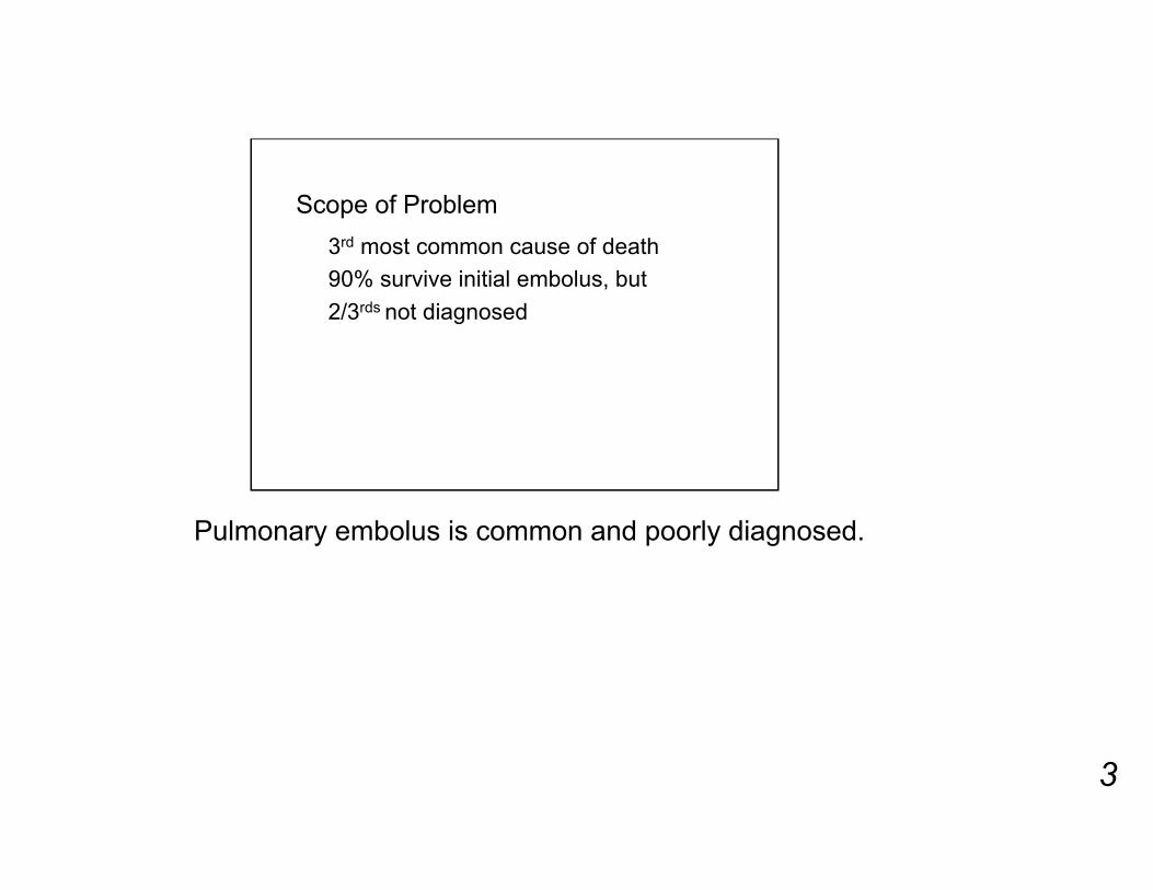
Scope of Problem
3rd most common cause of death
90% survive initial embolus, but
2/3rds not diagnosed
Pulmonary embolus is common and poorly diagnosed.
4
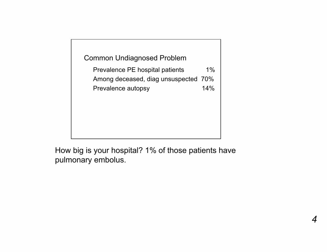
Common Undiagnosed Problem
Prevalence PE hospital patients 1%
Among deceased, diag unsuspected 70%
Prevalence autopsy 14%
How big is your hospital? 1% of those patients havepulmonary embolus.

5
Intensively studied
Medline search : pulmonary embolus
> 16,000 articles since 1966
> 1 article/day
Even though the world’s literature is voluminous, I wouldassert that we know very little about the natural history of thisdisorder.
6
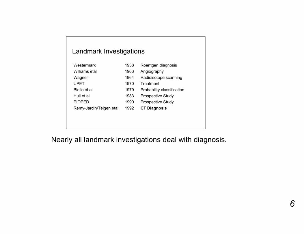
Landmark Investigations
CT Diagnosis1992Remy-Jardin/Teigen etal
Prospective Study1990PIOPED
Prospective Study1983Hull et al
Probability classification1979Biello et al
Treatment1970UPET
Radioisotope scanning1964Wagner
Angiography1963Williams etal
Roentgen diagnosis1938Westermark
Nearly all landmark investigations deal with diagnosis.
7
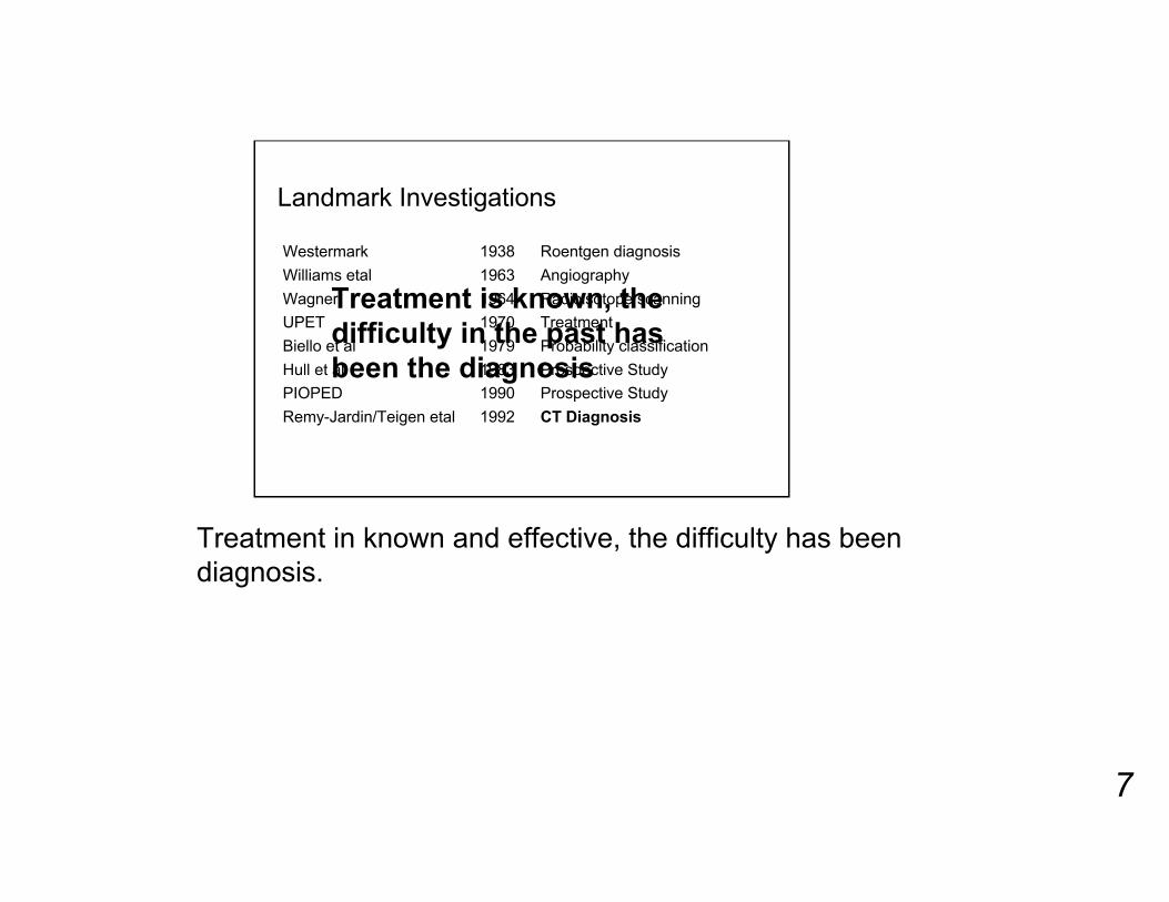
Landmark Investigations
CT Diagnosis1992Remy-Jardin/Teigen etal
Prospective Study1990PIOPED
Prospective Study1983Hull et al
Probability classification1979Biello et al
Treatment1970UPET
Radioisotope scanning1964Wagner
Angiography1963Williams etal
Roentgen diagnosis1938Westermark
Treatment is known, thedifficulty in the past has been the diagnosis
Treatment in known and effective, the difficulty has beendiagnosis.
8
Who’s at risk
CHFMIMalignancyShockObesityPregnancyRecent SurgeryTraumaBedridden
This is a common list of predisposing conditions. Note thatthis encompasses a very large patient population.
9
Who’s at riskCHFMIMalignancyShockObesityPregnancyRecent SurgeryTraumaBedridden
In short, Any hospitalized patient
Remember that prevalence is estimated at 1%.
10
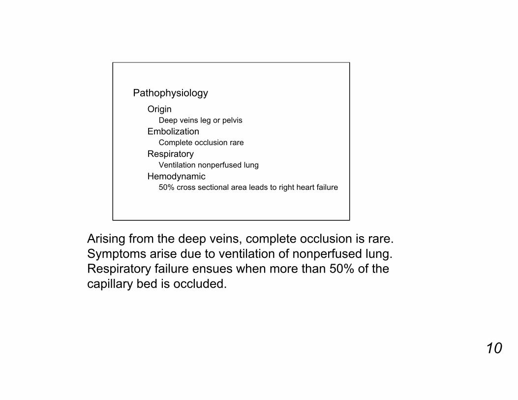
Pathophysiology
OriginDeep veins leg or pelvis
EmbolizationComplete occlusion rare
RespiratoryVentilation nonperfused lung
Hemodynamic50% cross sectional area leads to right heart failure
Arising from the deep veins, complete occlusion is rare.Symptoms arise due to ventilation of nonperfused lung.Respiratory failure ensues when more than 50% of thecapillary bed is occluded.
11
Clinical suspicion
History & physical exam nonspecificSilent embolism estimated in 40%
Moser Jama 1994
Problem, Who to Study?
Clinicians have great difficulty in deciding who to test forpulmonary embolus. In fact, many patients don’t have signs orsymptoms.
12
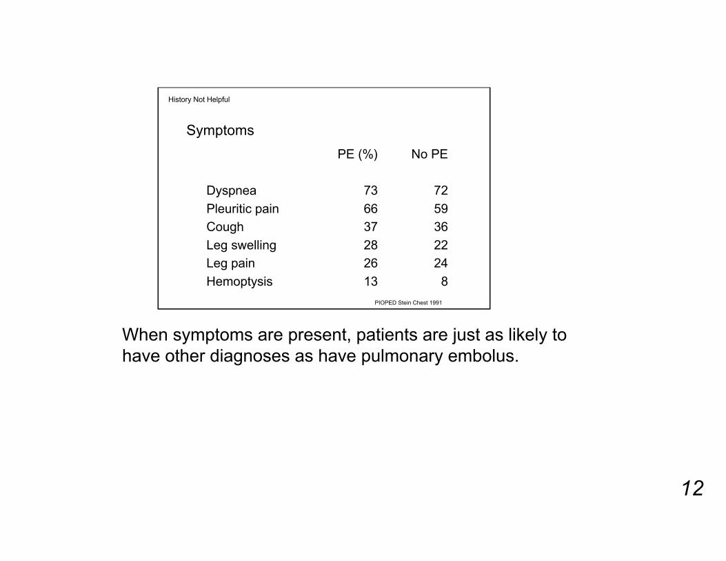
Symptoms
History Not Helpful
813Hemoptysis
2426Leg pain
2228Leg swelling
3637Cough
5966Pleuritic pain
7273Dyspnea
No PEPE (%)
PIOPED Stein Chest 1991
When symptoms are present, patients are just as likely tohave other diagnoses as have pulmonary embolus.
13
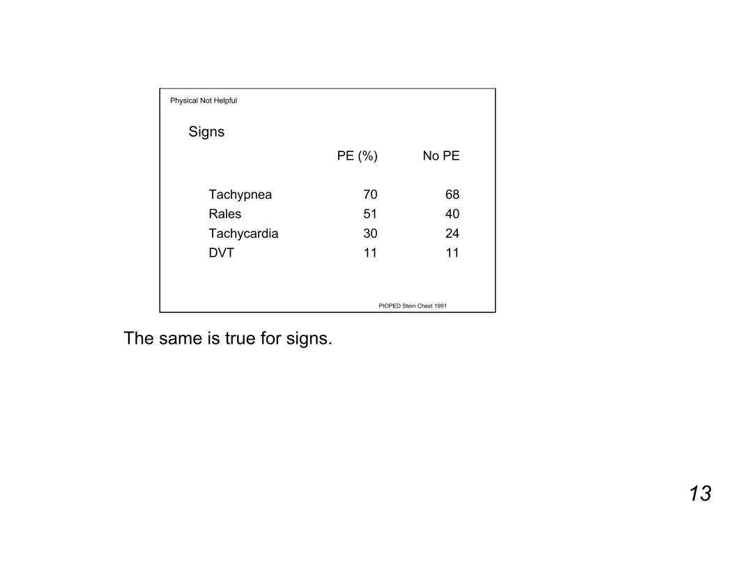
Signs
Physical Not Helpful
1111DVT
2430Tachycardia
4051Rales
6870Tachypnea
No PEPE (%)
PIOPED Stein Chest 1991
The same is true for signs.
14
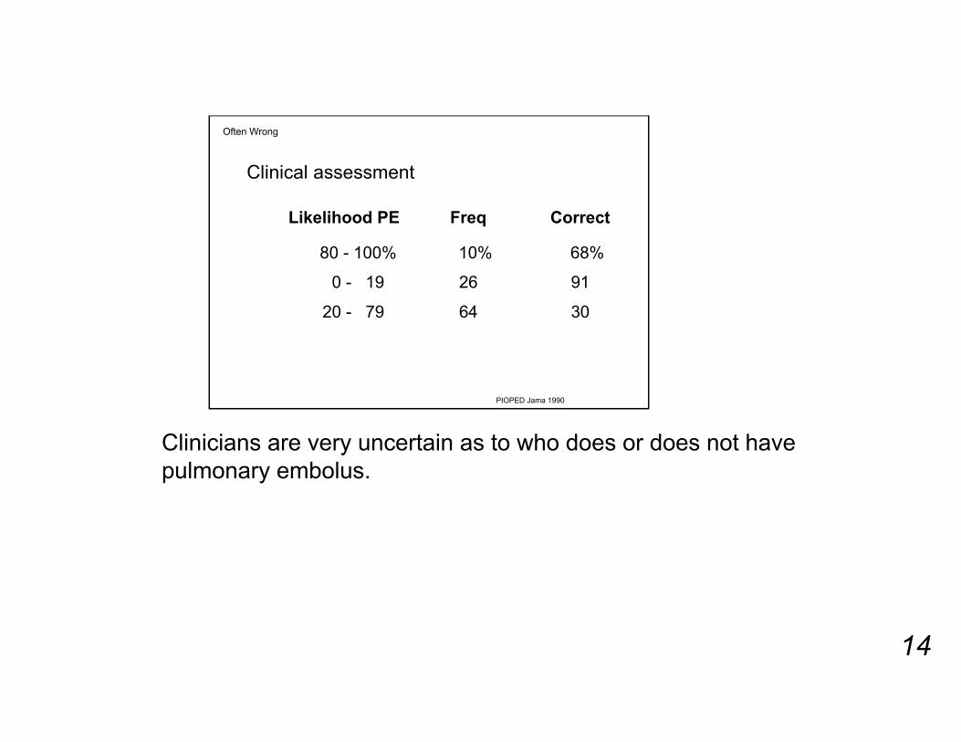
Clinical assessment
Often Wrong
3064 20 - 79
9126 0 - 19
68% 10% 80 - 100%
CorrectFreqLikelihood PE
PIOPED Jama 1990
Clinicians are very uncertain as to who does or does not havepulmonary embolus.
15
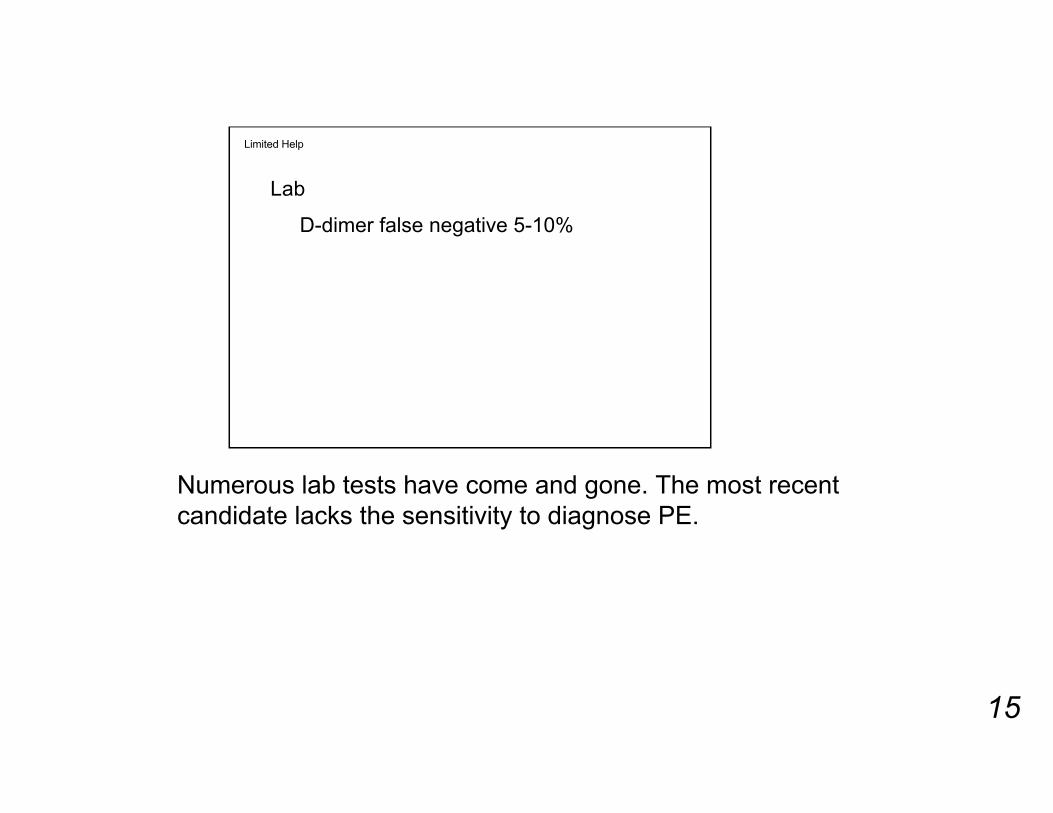
Lab
Limited Help
D-dimer false negative 5-10%
Numerous lab tests have come and gone. The most recentcandidate lacks the sensitivity to diagnose PE.
16
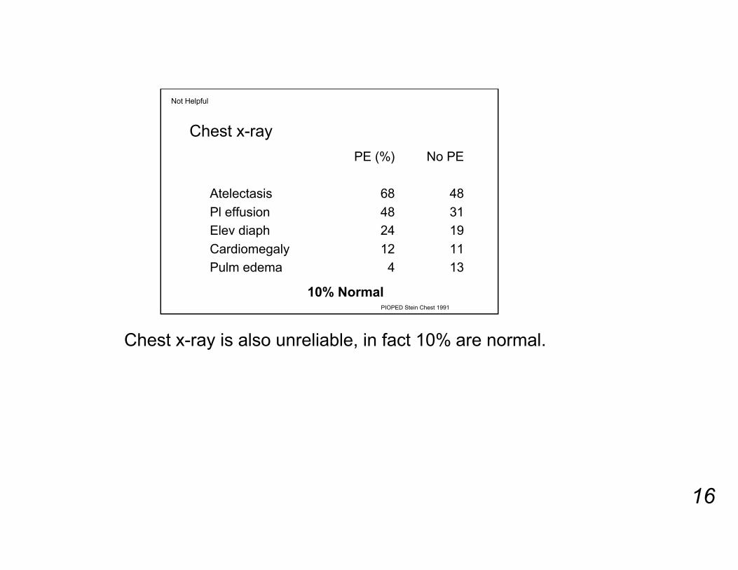
Not Helpful
Chest x-ray
134Pulm edema
1112Cardiomegaly
1924Elev diaph
3148Pl effusion
4868Atelectasis
No PEPE (%)
10% NormalPIOPED Stein Chest 1991
Chest x-ray is also unreliable, in fact 10% are normal.
17
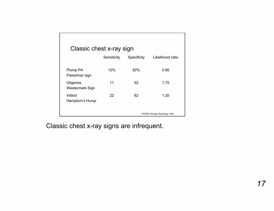
Classic chest x-ray sign
1.208222Infarct
Hamptom’s Hump
1.759211Oligemia
Westermark Sign
0.6682%12%Plump PA
Fleischner sign
Likelihood ratioSpecificitySensitivity
PIOPED Worsley Radiology 1993
Classic chest x-ray signs are infrequent.
18
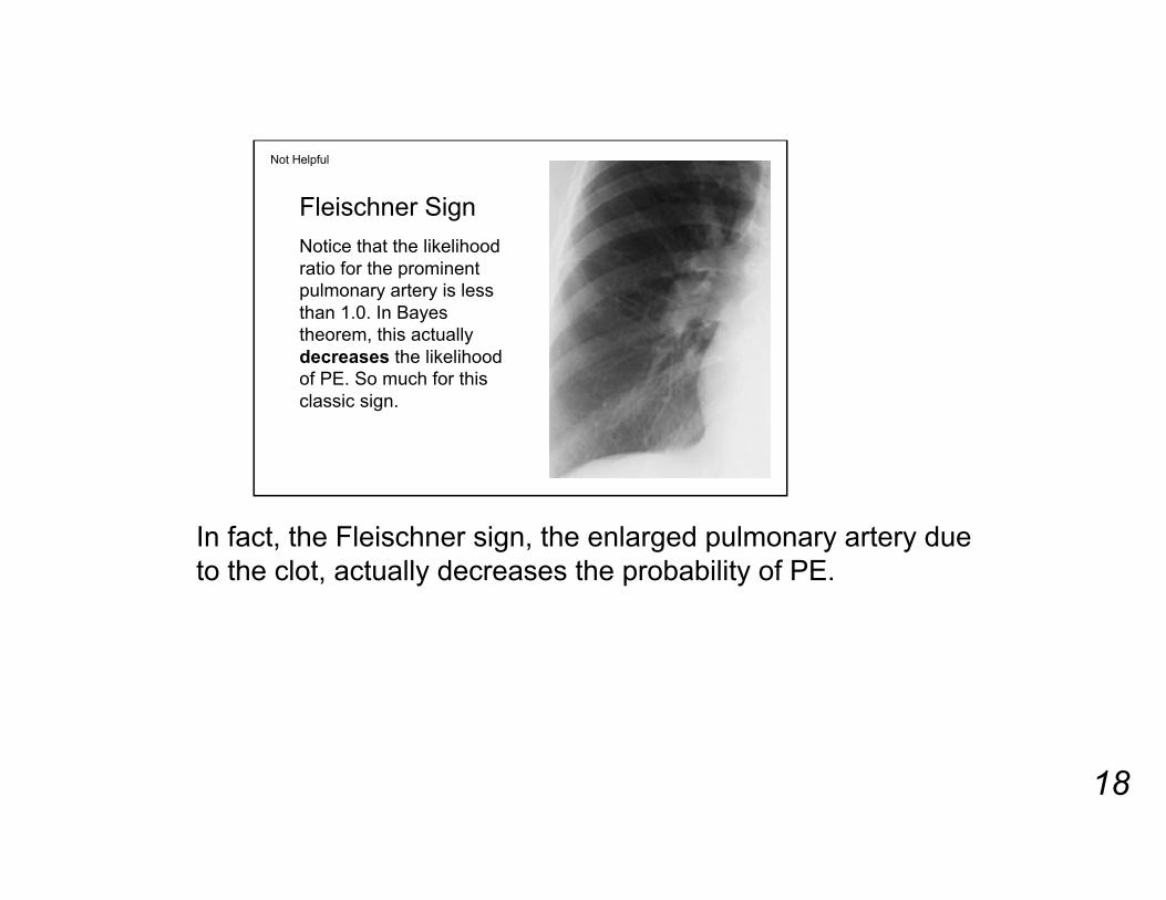
Fleischner Sign
Notice that the likelihoodratio for the prominentpulmonary artery is lessthan 1.0. In Bayestheorem, this actuallydecreases the likelihoodof PE. So much for thisclassic sign.
Not Helpful
In fact, the Fleischner sign, the enlarged pulmonary artery dueto the clot, actually decreases the probability of PE.
19
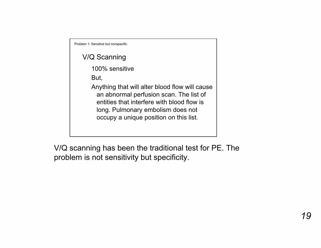
V/Q Scanning
100% sensitive
But,
Anything that will alter blood flow will causean abnormal perfusion scan. The list ofentities that interfere with blood flow islong. Pulmonary embolism does notoccupy a unique position on this list.
Problem 1: Sensitive but nonspecific
V/Q scanning has been the traditional test for PE. Theproblem is not sensitivity but specificity.

20
PIOPED
Problem 2: Frequently Not Helpful
71/8829/1273Indeterminate/Low
128813High Probability
96414Normal
No PEPEFreq (%)V/Q Classification
Based on V/Q patterns, patients are classified into fourcategories. Unfortunately, most patients fall into the low -indeterminate category in which too much uncertainty exists.

21
Reader Agreement: V/Q
Problem 3: Poor Agreement
70%Indeterminate/Low
95%High Probability
94%Normal
AgreementScan Classification
PIOPED JAMA 1990
In addition, the largest category has poor interobserveragreement.
22
The main problem with V/Q scanning:Don’t see clot, only the secondaryeffects of the clot
However,
The main problem with V/Q scanning is that the test does notprimarily visualize the clot, only the secondary effects of theclot. Historically, all tests that rely on secondary signs, suchas hypotonic duodenography for pancreatic masses, havebeen abandoned as soon as a test modality became availablethat could depict the primary pathology.

23
For years, Nuc Med has promisedinfarct avid agents
Alderson PO. Radiology State of the Art 164: 1987
Wellman HN. Sem Nucl Med 16: 1986
Time’s up!
Broken promises
Nuclear medicine physicians have been unsuccessful indeveloping in infarct avid agent.

24
It’s the clot stupid
To paraphrase James Carville.
25
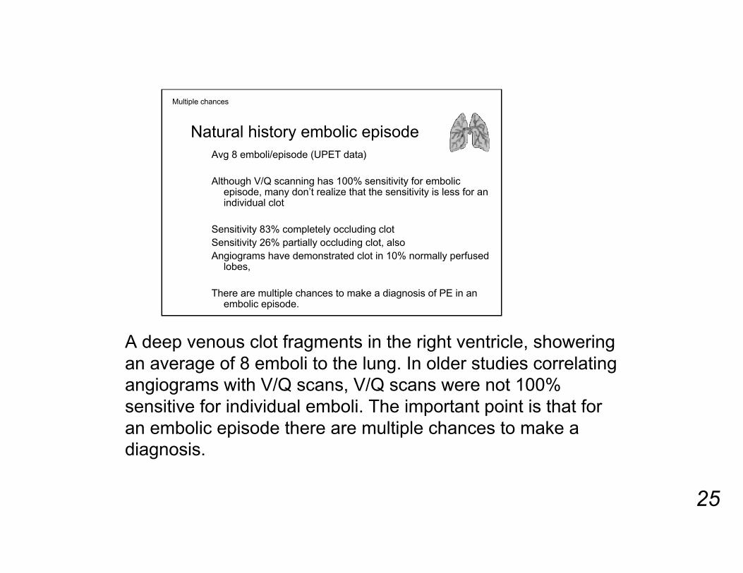
Natural history embolic episodeAvg 8 emboli/episode (UPET data)
Although V/Q scanning has 100% sensitivity for embolicepisode, many don’t realize that the sensitivity is less for anindividual clot
Sensitivity 83% completely occluding clotSensitivity 26% partially occluding clot, alsoAngiograms have demonstrated clot in 10% normally perfused
lobes,
There are multiple chances to make a diagnosis of PE in anembolic episode.
Multiple chances
A deep venous clot fragments in the right ventricle, showeringan average of 8 emboli to the lung. In older studies correlatingangiograms with V/Q scans, V/Q scans were not 100%sensitive for individual emboli. The important point is that foran embolic episode there are multiple chances to make adiagnosis.

26
Pulmonary Angiography
Invasive
Expensive
Tech expertise
Morbidity/Mortality low
Rarely done
See the clot, but
The one traditional test that does depict the clot is pulmonaryangiography. Various reasons have been given for the poorutilization, in any case this is a rarely performed test.
27
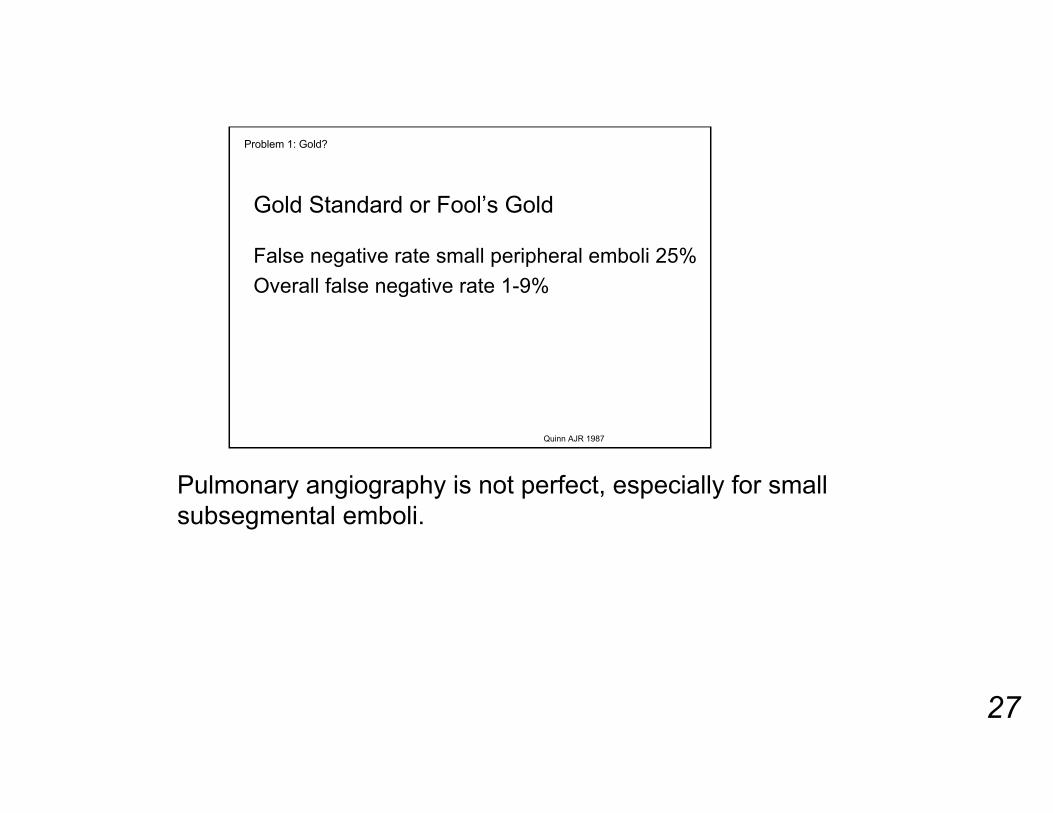
Gold Standard or Fool’s Gold
False negative rate small peripheral emboli 25%
Overall false negative rate 1-9%
Problem 1: Gold?
Quinn AJR 1987
Pulmonary angiography is not perfect, especially for smallsubsegmental emboli.
28
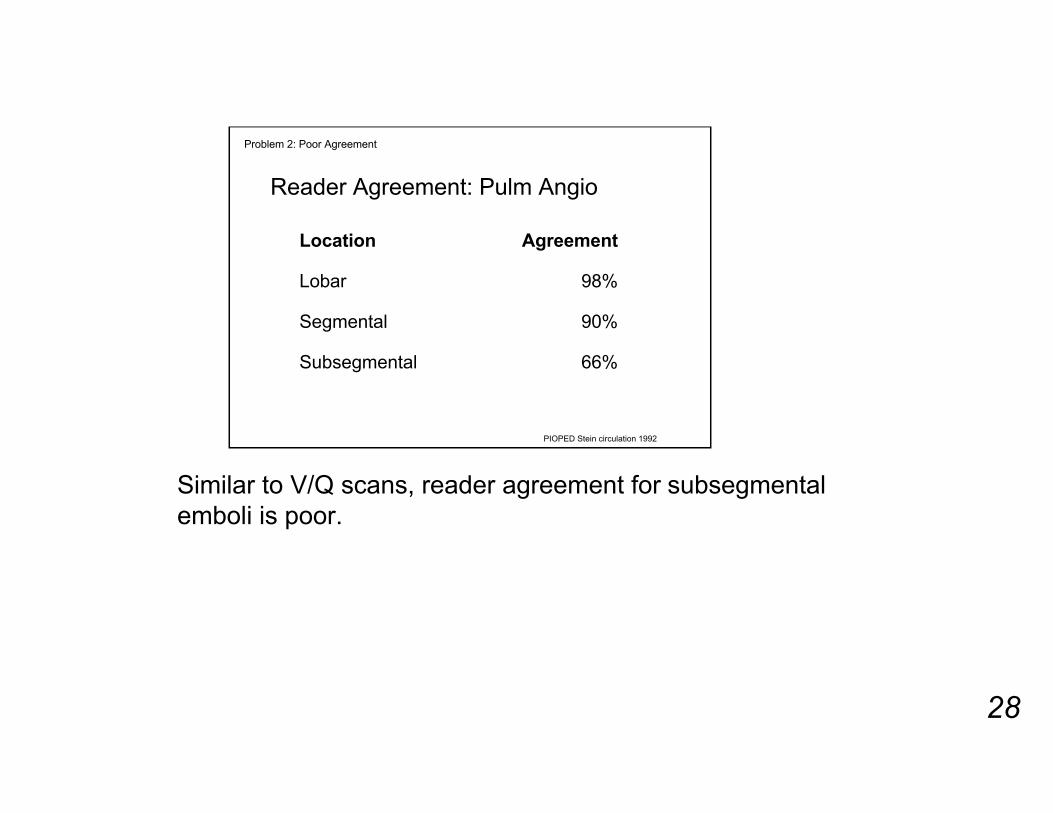
Reader Agreement: Pulm Angio
PIOPED Stein circulation 1992
Problem 2: Poor Agreement
66%Subsegmental
90%Segmental
98%Lobar
AgreementLocation
Similar to V/Q scans, reader agreement for subsegmentalemboli is poor.

29
Practice Pattern
Problem 3: Frequently not done
Henschke Chest 1995Sustman AJR 1982
Pulm Angio
71 (14%)
Pulm Angio
50 (12%)
525 (81%)
unresolved
434 (72%)
unresolved
V/Q scanV/Q scan
650 suspected600 suspected
…1995…1982
Over the years little has changed in physician’s practicepatterns. Pulmonary angiography is a rarely utilized modality.
30
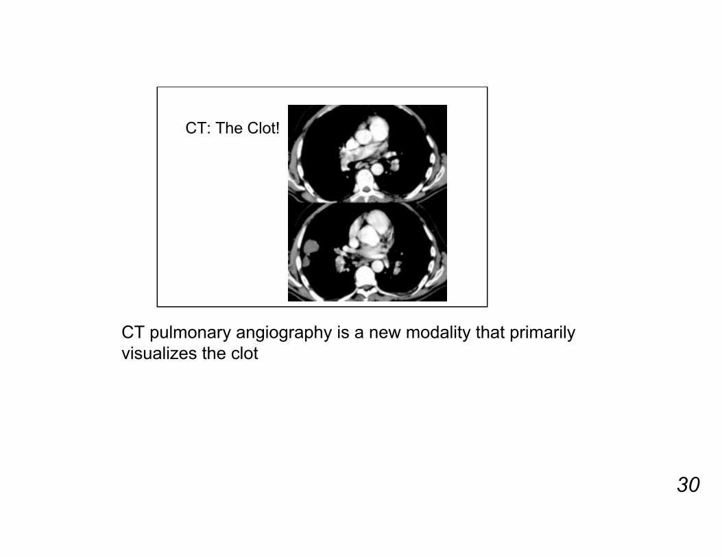
CT: The Clot!
CT pulmonary angiography is a new modality that primarilyvisualizes the clot
31
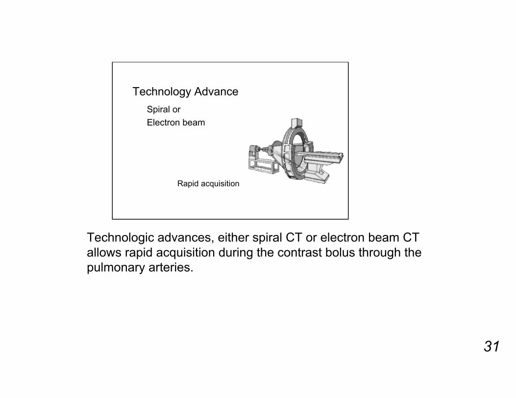
Technology Advance
Spiral or
Electron beam
Rapid acquisition
Technologic advances, either spiral CT or electron beam CTallows rapid acquisition during the contrast bolus through thepulmonary arteries.
32
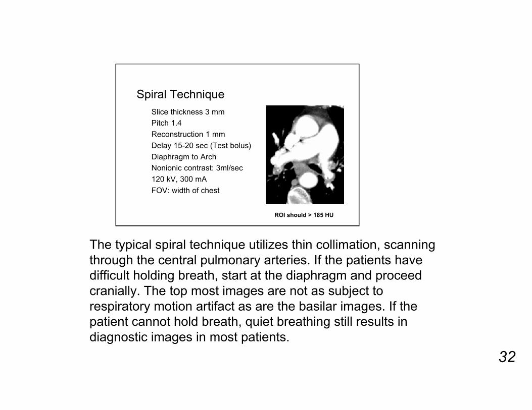
Spiral TechniqueSlice thickness 3 mm
Pitch 1.4
Reconstruction 1 mm
Delay 15-20 sec (Test bolus)
Diaphragm to Arch
Nonionic contrast: 3ml/sec
120 kV, 300 mA
FOV: width of chest
ROI should > 185 HU
The typical spiral technique utilizes thin collimation, scanningthrough the central pulmonary arteries. If the patients havedifficult holding breath, start at the diaphragm and proceedcranially. The top most images are not as subject torespiratory motion artifact as are the basilar images. If thepatient cannot hold breath, quiet breathing still results indiagnostic images in most patients.
33
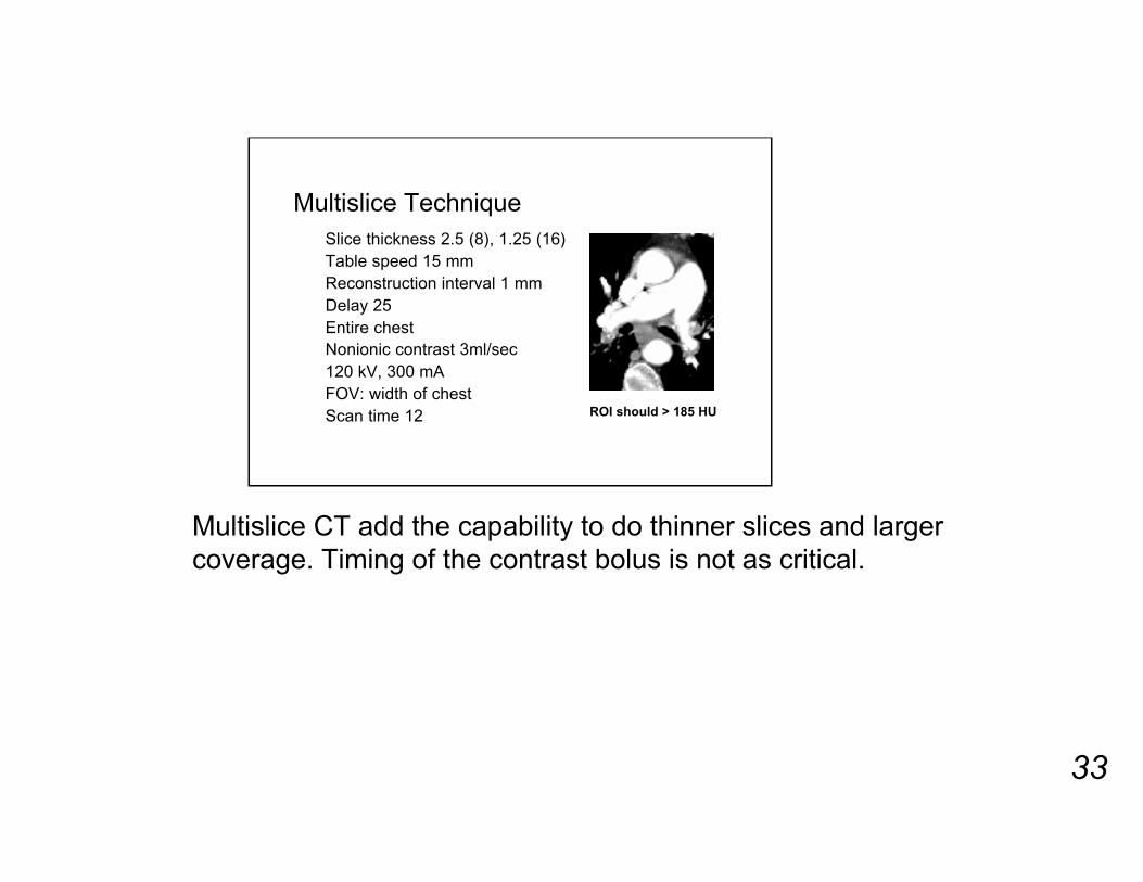
Multislice TechniqueSlice thickness 2.5 (8), 1.25 (16)Table speed 15 mmReconstruction interval 1 mmDelay 25Entire chestNonionic contrast 3ml/sec120 kV, 300 mAFOV: width of chestScan time 12 ROI should > 185 HU
Multislice CT add the capability to do thinner slices and largercoverage. Timing of the contrast bolus is not as critical.
34
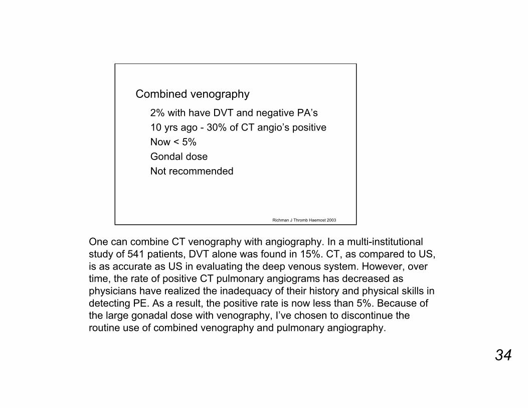
Combined venography
2% with have DVT and negative PA’s
10 yrs ago - 30% of CT angio’s positive
Now < 5%
Gondal dose
Not recommended
Richman J Thromb Haemost 2003
One can combine CT venography with angiography. In a multi-institutionalstudy of 541 patients, DVT alone was found in 15%. CT, as compared to US,is as accurate as US in evaluating the deep venous system. However, overtime, the rate of positive CT pulmonary angiograms has decreased asphysicians have realized the inadequacy of their history and physical skills indetecting PE. As a result, the positive rate is now less than 5%. Because ofthe large gonadal dose with venography, I’ve chosen to discontinue theroutine use of combined venography and pulmonary angiography.

35
Number emboli
Teigen 1993Electron CT6.8
Remy-Jardin 1992Spiral CT6.2
UPET 1970Pulm Angio8
ReferenceModalityEmboli/PT
Note the good concordance with the number of embolidetected as compared to historical studies of the number ofemboli per embolic episode.
36
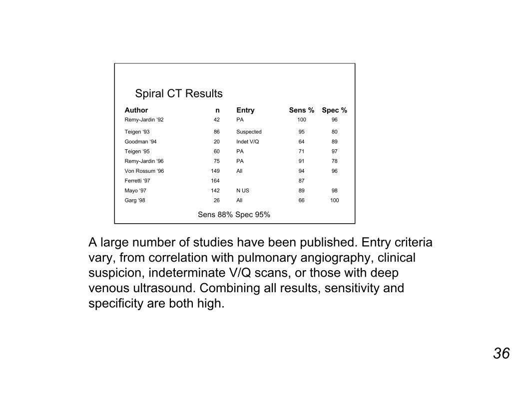
Spiral CT Results
10066All26Garg ‘98
9889N US142Mayo ‘97
87164Ferretti ‘97
9694All149Von Rossum ‘96
7891PA75Remy-Jardin ‘96
9771PA60Teigen ‘95
8964Indet V/Q20Goodman ‘94
8095Suspected86Teigen ‘93
96100PA42Remy-Jardin ‘92
Spec %Sens %EntrynAuthor
Sens 88% Spec 95%
A large number of studies have been published. Entry criteriavary, from correlation with pulmonary angiography, clinicalsuspicion, indeterminate V/Q scans, or those with deepvenous ultrasound. Combining all results, sensitivity andspecificity are both high.
37
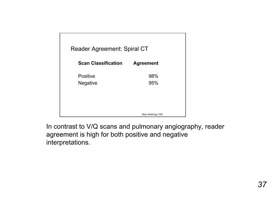
Reader Agreement: Spiral CT
95%Negative
98%Positive
AgreementScan Classification
Mayo Radiology 1997
In contrast to V/Q scans and pulmonary angiography, readeragreement is high for both positive and negativeinterpretations.
38
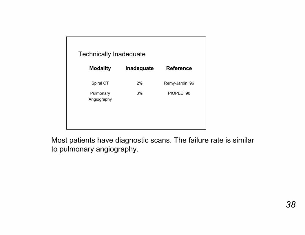
Technically Inadequate
PIOPED ‘903%Pulmonary
Angiography
Remy-Jardin ‘962%Spiral CT
ReferenceInadequateModality
Most patients have diagnostic scans. The failure rate is similarto pulmonary angiography.
39
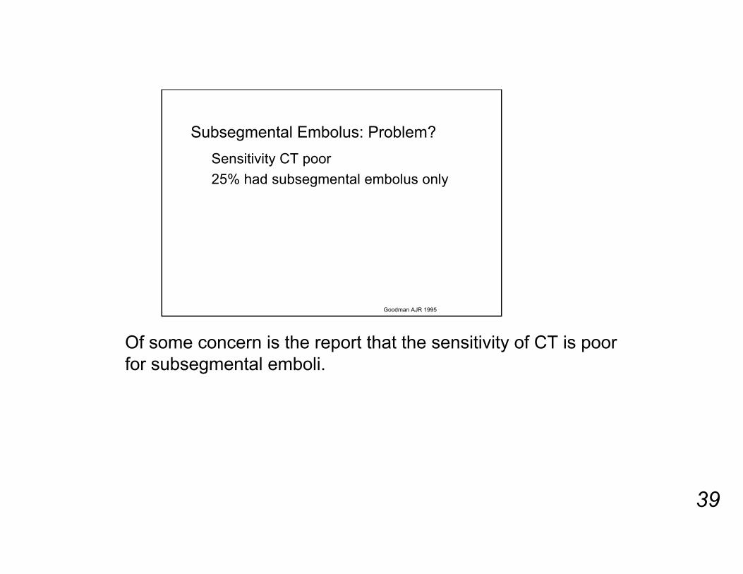
Subsegmental Embolus: Problem?
Sensitivity CT poor
25% had subsegmental embolus only
Goodman AJR 1995
Of some concern is the report that the sensitivity of CT is poorfor subsegmental emboli.
40
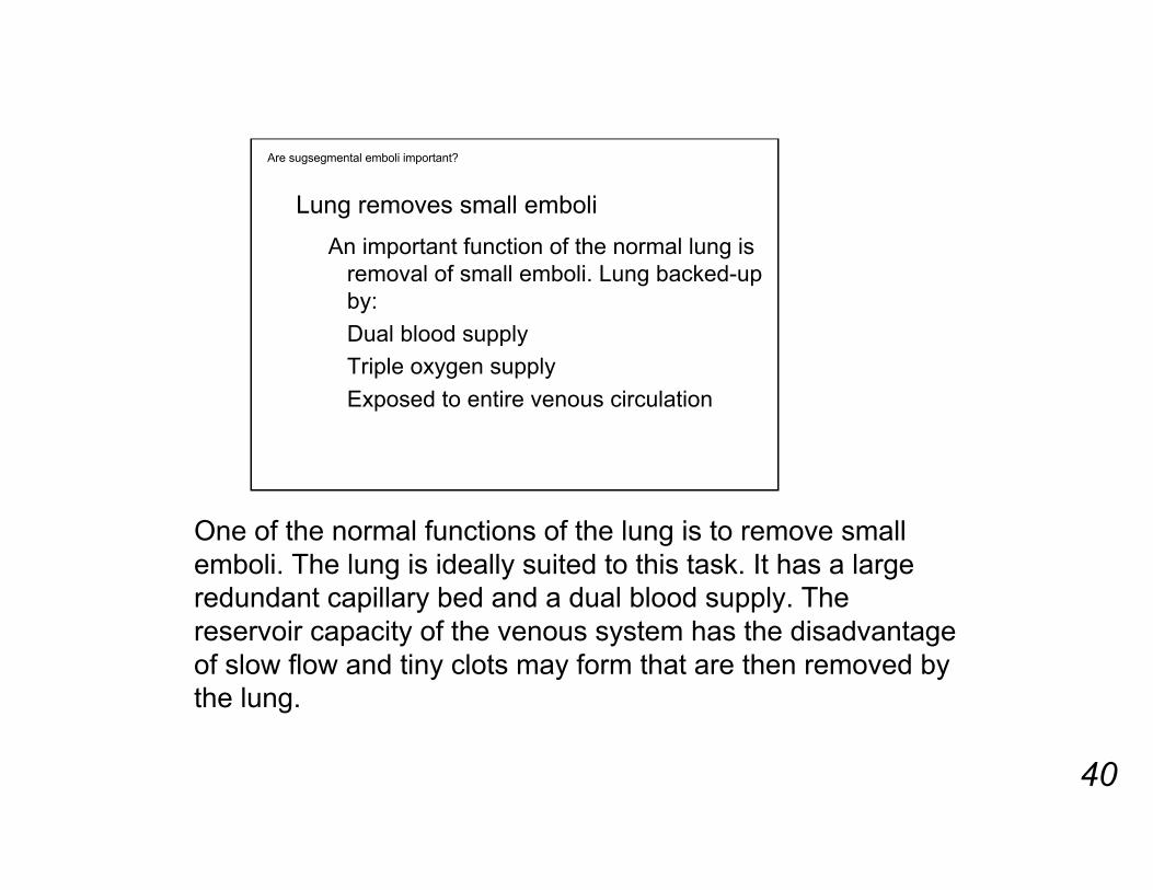
Lung removes small emboli
An important function of the normal lung isremoval of small emboli. Lung backed-upby:
Dual blood supply
Triple oxygen supply
Exposed to entire venous circulation
Are sugsegmental emboli important?
One of the normal functions of the lung is to remove smallemboli. The lung is ideally suited to this task. It has a largeredundant capillary bed and a dual blood supply. Thereservoir capacity of the venous system has the disadvantageof slow flow and tiny clots may form that are then removed bythe lung.
41
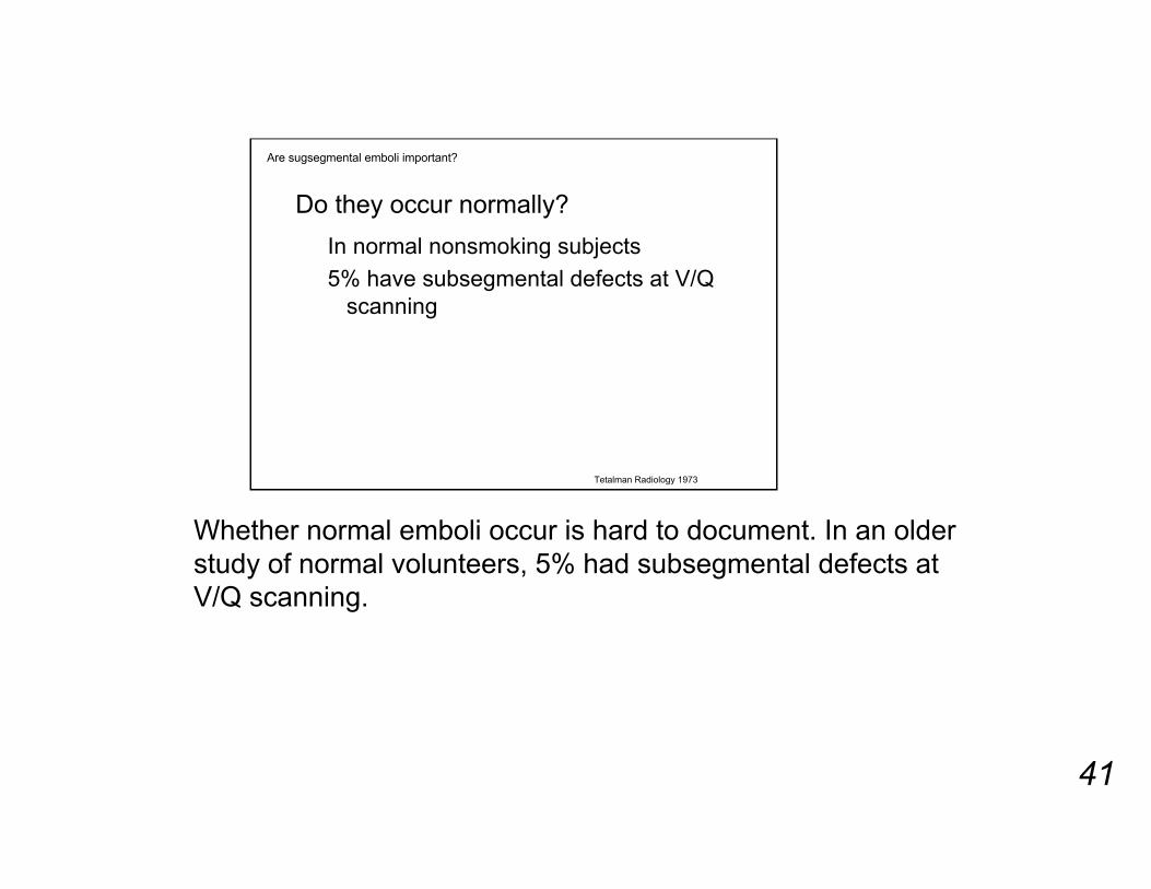
Do they occur normally?
In normal nonsmoking subjects
5% have subsegmental defects at V/Qscanning
Are sugsegmental emboli important?
Tetalman Radiology 1973
Whether normal emboli occur is hard to document. In an olderstudy of normal volunteers, 5% had subsegmental defects atV/Q scanning.
42
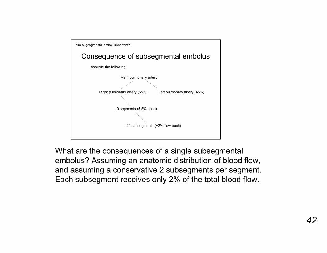
Consequence of subsegmental embolusAssume the following
Main pulmonary artery
Right pulmonary artery (55%) Left pulmonary artery (45%)
10 segments (5.5% each)
20 subsegments (~2% flow each)
Are sugsegmental emboli important?
What are the consequences of a single subsegmentalembolus? Assuming an anatomic distribution of blood flow,and assuming a conservative 2 subsegments per segment.Each subsegment receives only 2% of the total blood flow.
43
Consequence of subsegmental embolus
Is 2% a great loss?
Are sugsegmental emboli important?
Normal pulmonary reserve easily compensates for this loss. Ifmissed, these clots may have little consequence.
44
If Missed
Remember subsegmental emboli missedat angio
Outcome negative pulmonary angio
Good: PIOPED 1 yr surveillance
PE in 4 (0.6%)
Are sugsegmental emboli important?
Stein Circ 1992
For example, certainly subsegmental emboli are missed atangiography, however the outcome of patients with normalpulmonary angiography is good.
45
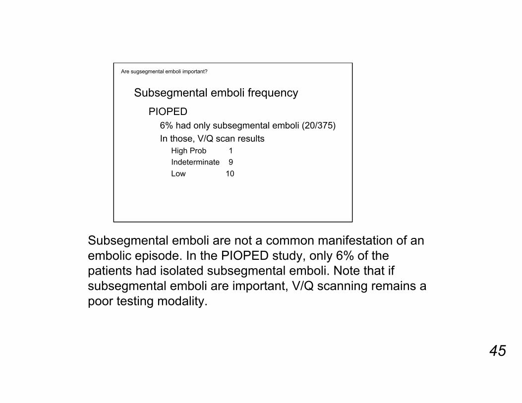
Subsegmental emboli frequency
PIOPED6% had only subsegmental emboli (20/375)
In those, V/Q scan resultsHigh Prob 1
Indeterminate 9
Low 10
Are sugsegmental emboli important?
Subsegmental emboli are not a common manifestation of anembolic episode. In the PIOPED study, only 6% of thepatients had isolated subsegmental emboli. Note that ifsubsegmental emboli are important, V/Q scanning remains apoor testing modality.
46
Subsegmental emboli
Therefore,
V/Q also not helpful and remember,
Angio has significant observer variability
Are sugsegmental emboli important?
Remember there is not a reliable test for subsegmental clots.
47
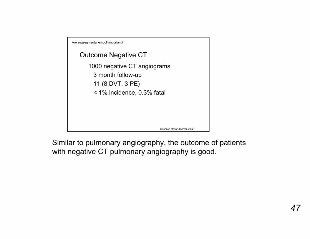
Outcome Negative CT
1000 negative CT angiograms
3 month follow-up
11 (8 DVT, 3 PE)
< 1% incidence, 0.3% fatal
Are sugsegmental emboli important?
Swensen Mayo Clin Proc 2002
Similar to pulmonary angiography, the outcome of patientswith negative CT pulmonary angiography is good.

48
Alternative diagnosis
In those without PE2/3rds additional info
Pneumonia
Cardiovascular disorders
Interstitial lung disease
Malignancy
Pleural disease
Kim Radiology 1999
Additional information
In contradistinction to V/Q scanning, CT add otherinformation. Up to 2/3rds of patients have other diagnoses atCT which may be the cause of the patient’s symptoms.

49
Additional information
In this example, the CT angiogram was normal. Focalconsolidation in the right upper lobe due to pneumonia wasthe cause of the symptoms.
50
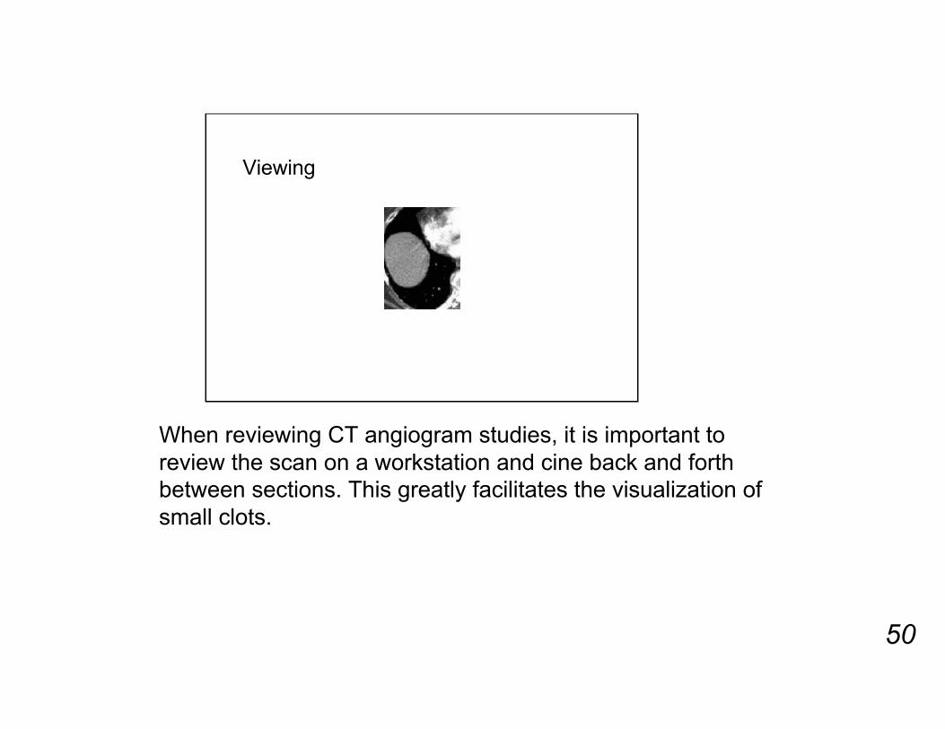
Viewing
When reviewing CT angiogram studies, it is important toreview the scan on a workstation and cine back and forthbetween sections. This greatly facilitates the visualization ofsmall clots.

51
Tips: Window/level
Various other window widths are also helpful. Here, the bonewindow better demonstrates the calcification in a chronicembolus.
52
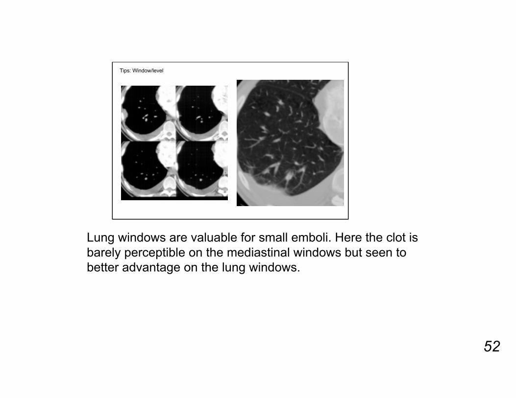
Tips: Window/level
Lung windows are valuable for small emboli. Here the clot isbarely perceptible on the mediastinal windows but seen tobetter advantage on the lung windows.
53
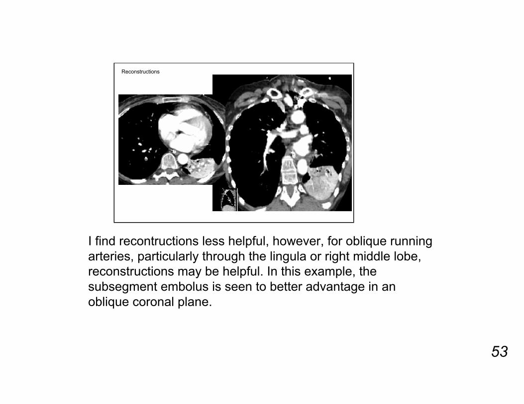
Reconstructions
I find recontructions less helpful, however, for oblique runningarteries, particularly through the lingula or right middle lobe,reconstructions may be helpful. In this example, thesubsegment embolus is seen to better advantage in anoblique coronal plane.

54
False positive clots are related to motion artifact, asymmetricfilling or normal lymph nodes. It is helpful to have a handychart showing the normal locations of lymph nodes within thehilum.
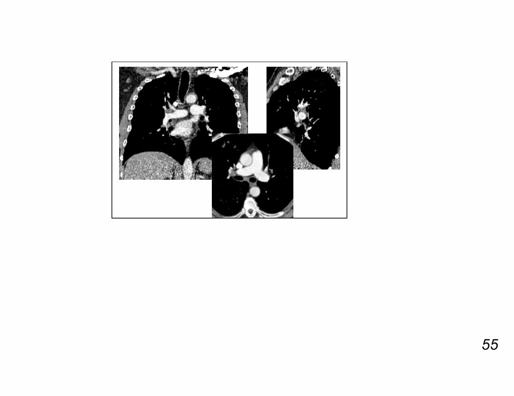
55
56
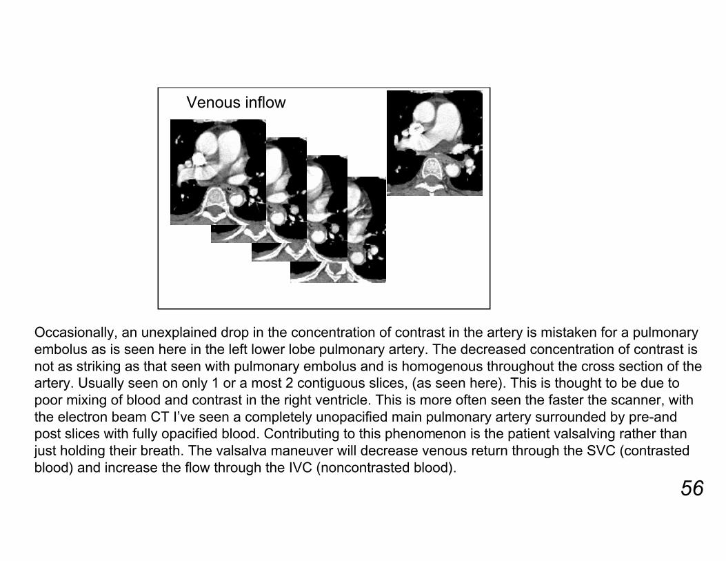
Venous inflow
Occasionally, an unexplained drop in the concentration of contrast in the artery is mistaken for a pulmonaryembolus as is seen here in the left lower lobe pulmonary artery. The decreased concentration of contrast isnot as striking as that seen with pulmonary embolus and is homogenous throughout the cross section of theartery. Usually seen on only 1 or a most 2 contiguous slices, (as seen here). This is thought to be due topoor mixing of blood and contrast in the right ventricle. This is more often seen the faster the scanner, withthe electron beam CT I’ve seen a completely unopacified main pulmonary artery surrounded by pre-andpost slices with fully opacified blood. Contributing to this phenomenon is the patient valsalving rather thanjust holding their breath. The valsalva maneuver will decrease venous return through the SVC (contrastedblood) and increase the flow through the IVC (noncontrasted blood).
57
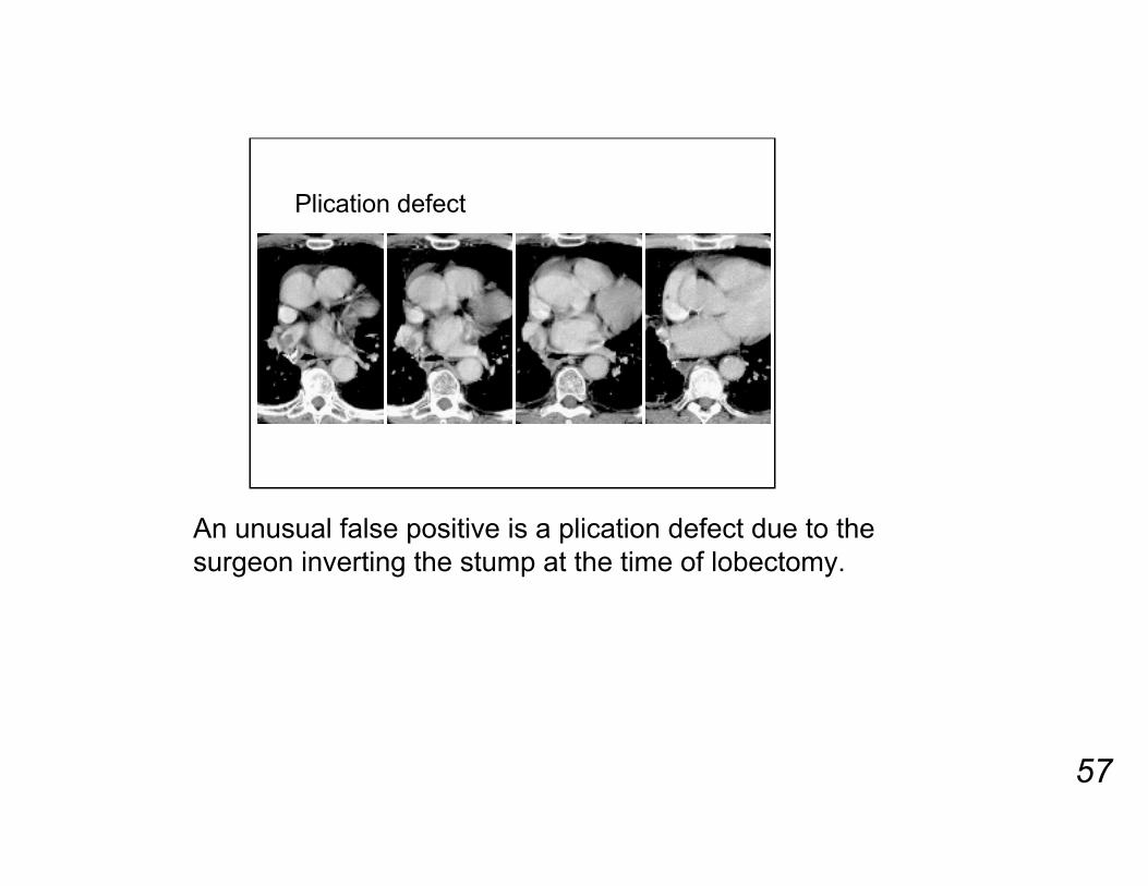
Plication defect
An unusual false positive is a plication defect due to thesurgeon inverting the stump at the time of lobectomy.
58
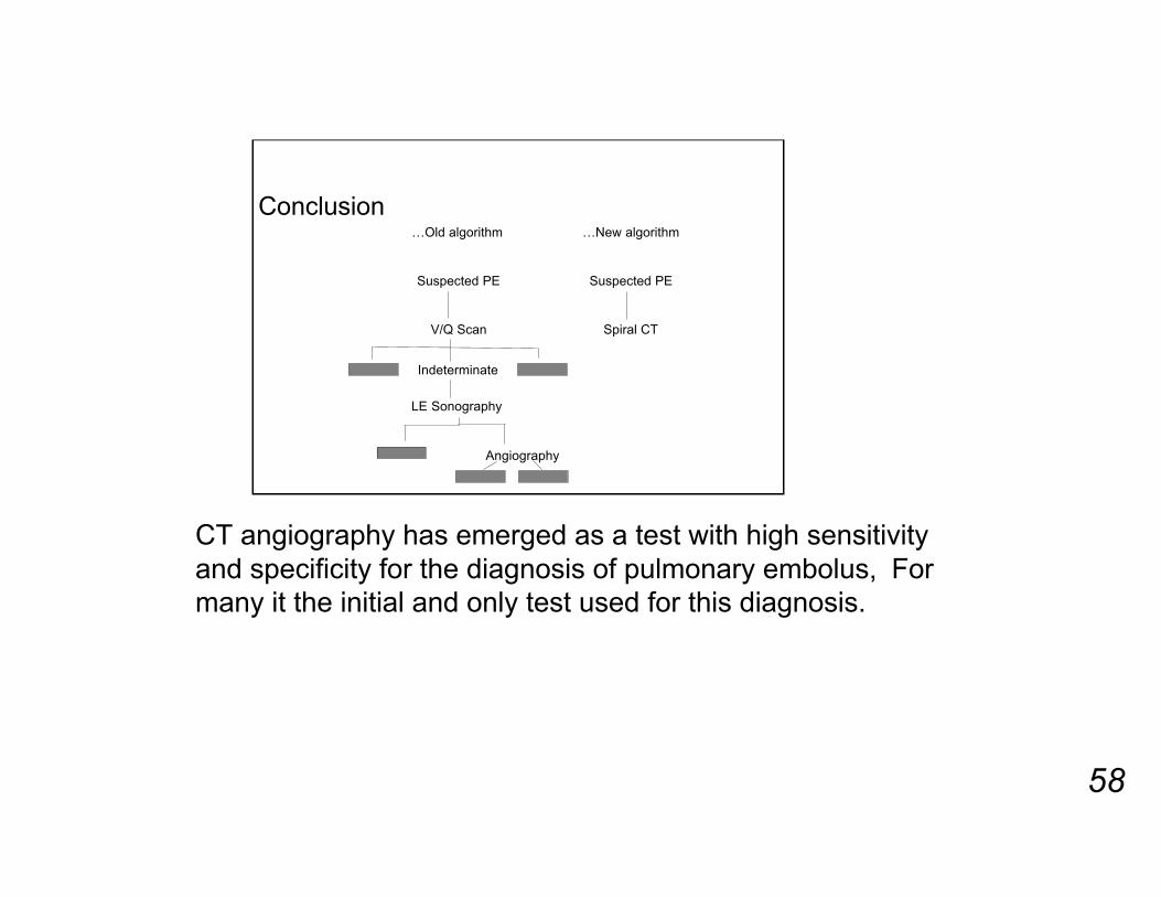
Conclusion…Old algorithm …New algorithm
Suspected PE Suspected PE
V/Q Scan Spiral CT
Indeterminate
LE Sonography
Angiography
CT angiography has emerged as a test with high sensitivityand specificity for the diagnosis of pulmonary embolus, Formany it the initial and only test used for this diagnosis.