Embed Size (px)
Citation preview
CT Protocols for Common Primary Care Diagnoses
Lacey J. McIntosh DO, MPH University of Massachusetts Medical Center UNECOM 2014 CME Program/Reunion and Alumni Weekend: Primary Care in Today’s Changing Practice Environment October 10-12, 2014 University of New England Biddeford Campus
|
|
|
|
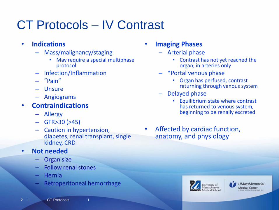
• Imaging Phases – Arterial phase
• Contrast has not yet reached the organ, in arteries only
– *Portal venous phase • Organ has perfused, contrast
returning through venous system
– Delayed phase • Equilibrium state where contrast
has returned to venous system, beginning to be renally excreted
• Affected by cardiac function,
anatomy, and physiology
CT Protocols 2
CT Protocols – IV Contrast
• Indications – Mass/malignancy/staging
• May require a special multiphase protocol
– Infection/Inflammation – “Pain” – Unsure – Angiograms
• Contraindications – Allergy – GFR>30 (>45) – Caution in hypertension,
diabetes, renal transplant, single kidney, CRD
• Not needed – Organ size – Follow renal stones – Hernia – Retroperitoneal hemorrhage
|
|
|
|
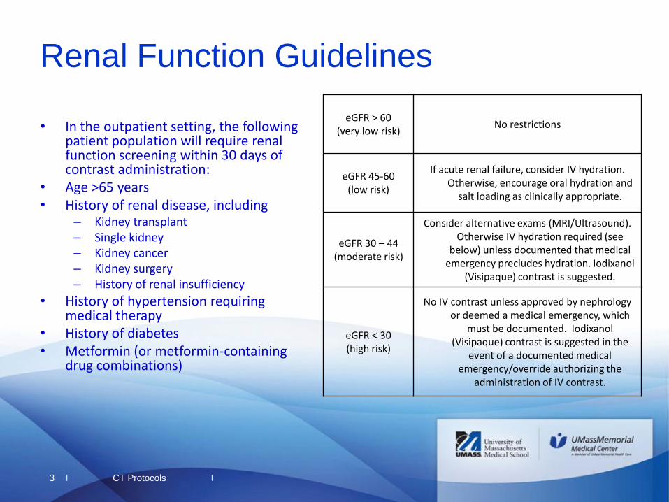
Renal Function Guidelines
• In the outpatient setting, the following patient population will require renal function screening within 30 days of contrast administration:
• Age >65 years • History of renal disease, including
– Kidney transplant – Single kidney – Kidney cancer – Kidney surgery – History of renal insufficiency
• History of hypertension requiring medical therapy
• History of diabetes • Metformin (or metformin-containing
drug combinations)
eGFR > 60 (very low risk)
No restrictions
eGFR 45-60 (low risk)
If acute renal failure, consider IV hydration. Otherwise, encourage oral hydration and
salt loading as clinically appropriate.
eGFR 30 – 44 (moderate risk)
Consider alternative exams (MRI/Ultrasound). Otherwise IV hydration required (see
below) unless documented that medical emergency precludes hydration. Iodixanol
(Visipaque) contrast is suggested.
eGFR < 30 (high risk)
No IV contrast unless approved by nephrology or deemed a medical emergency, which
must be documented. Iodixanol (Visipaque) contrast is suggested in the
event of a documented medical emergency/override authorizing the
administration of IV contrast.
CT Protocols 3
|
|
|
|
4
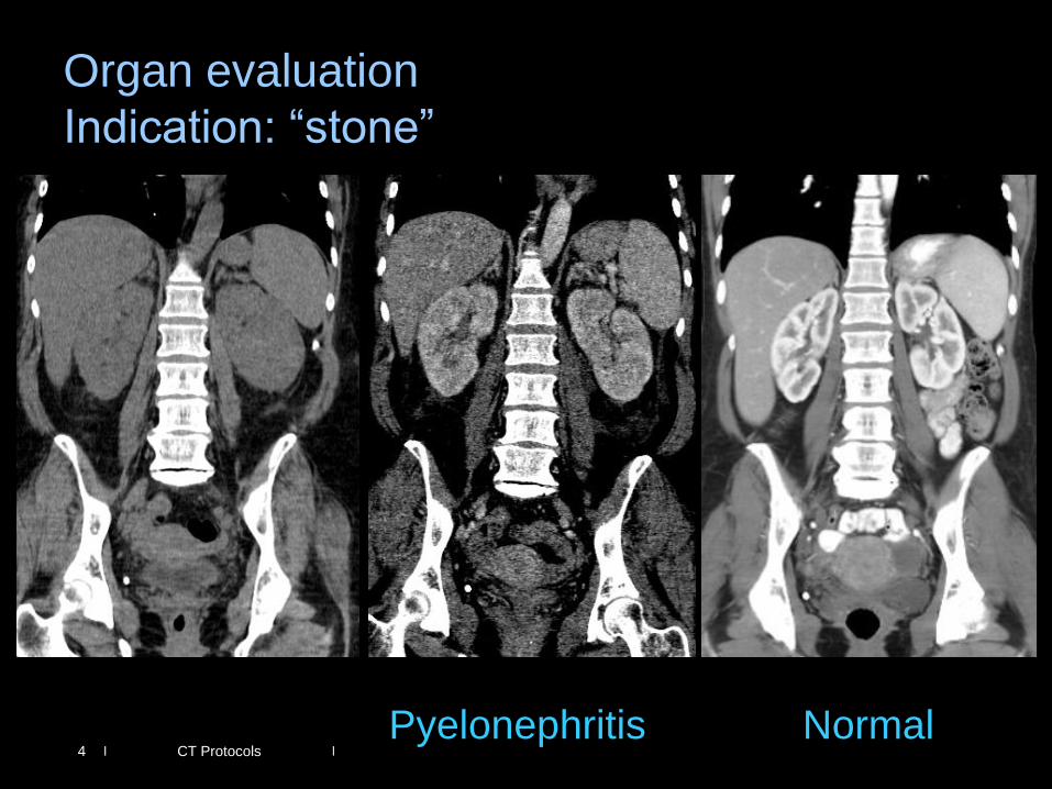
Organ evaluation
Indication: “stone”
Pyelonephritis Normal CT Protocols
|
|
|
|
5
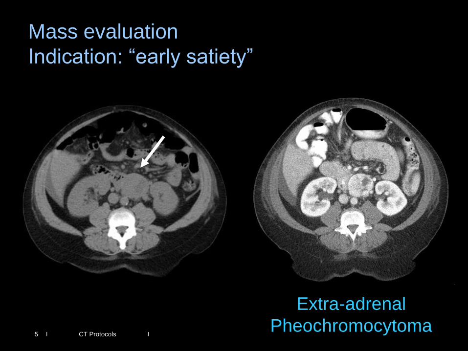
Mass evaluation
Indication: “early satiety”
Extra-adrenal
Pheochromocytoma CT Protocols
|
|
|
|
• Bladder Contrast – Fistula – Bladder wall integrity
• Rectal Contrast – Fistula – Post surgical – Penetrating trauma
• “Size Matters”
• Things are different in the ER
setting
6
CT Protocols – Oral Contrast
• Indications - Body – Mostly for us to identify bowel
• From other structures • Evaluate wall
– Volumen
– Functional – Evaluate gastric bypass – Post surgical is a must!
• Gastrograffin
• Contraindications – Intolerance – Will obscure your finding
• Not needed – Angiograms – Organ specific exams
CT Protocols
|
|
|
|
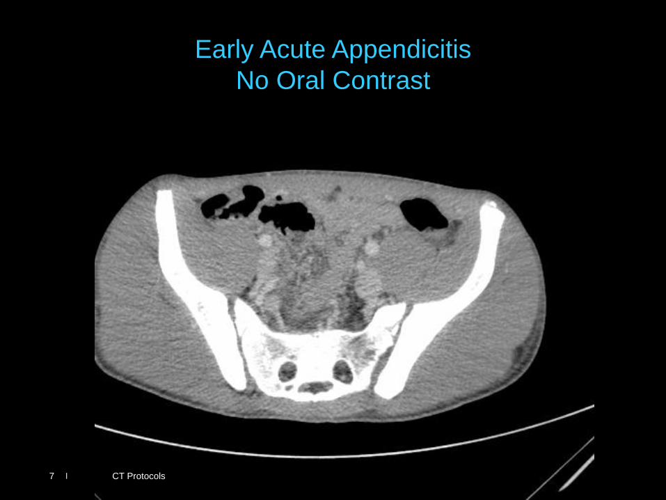
Early Acute Appendicitis
No Oral Contrast
CT Protocols 7
|
|
|
|
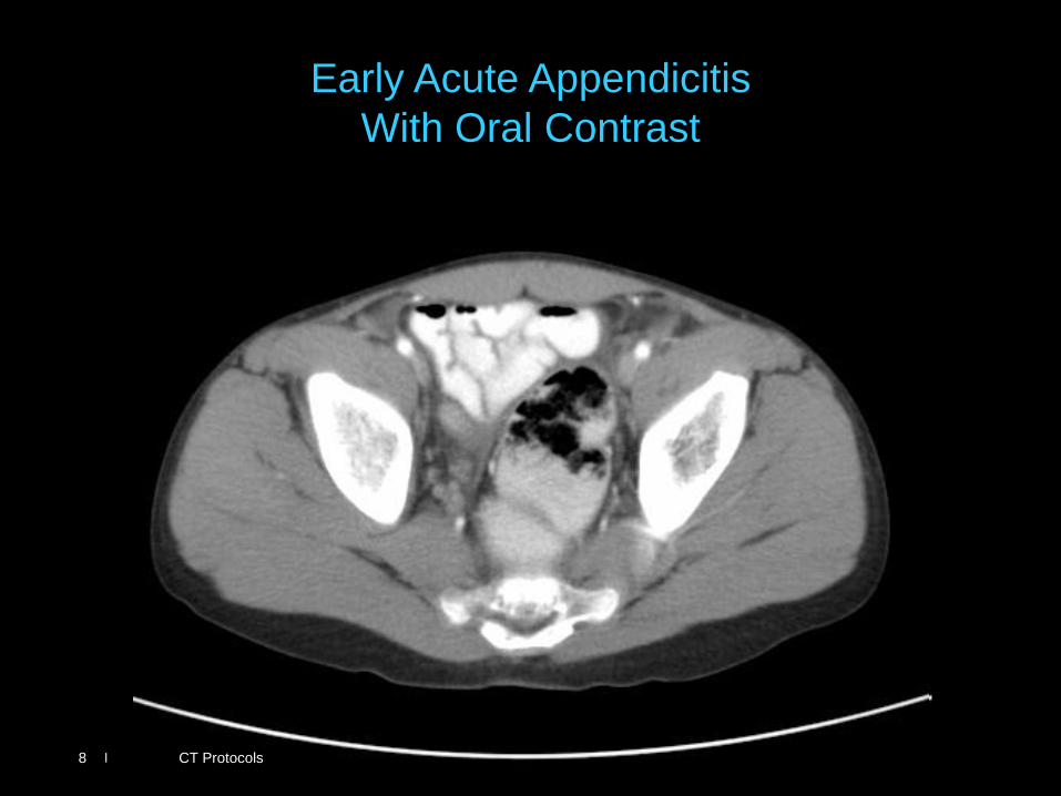
Early Acute Appendicitis
With Oral Contrast
CT Protocols 8
|
|
|
|
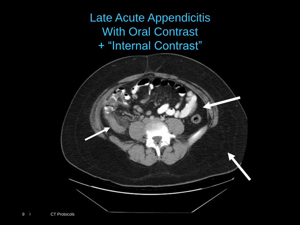
Late Acute Appendicitis
With Oral Contrast
+ “Internal Contrast”
CT Protocols 9
|
|
|
|
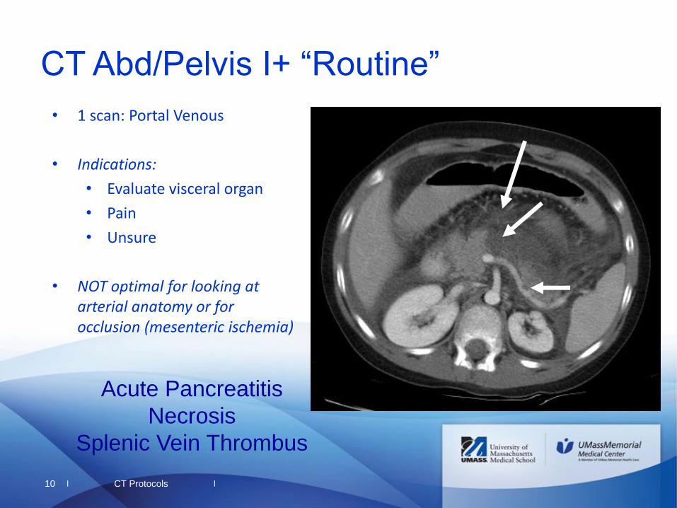
10
CT Abd/Pelvis I+ “Routine”
• 1 scan: Portal Venous
• Indications:
• Evaluate visceral organ
• Pain
• Unsure
• NOT optimal for looking at arterial anatomy or for occlusion (mesenteric ischemia)
Acute Pancreatitis
Necrosis
Splenic Vein Thrombus
CT Protocols
|
|
|
|
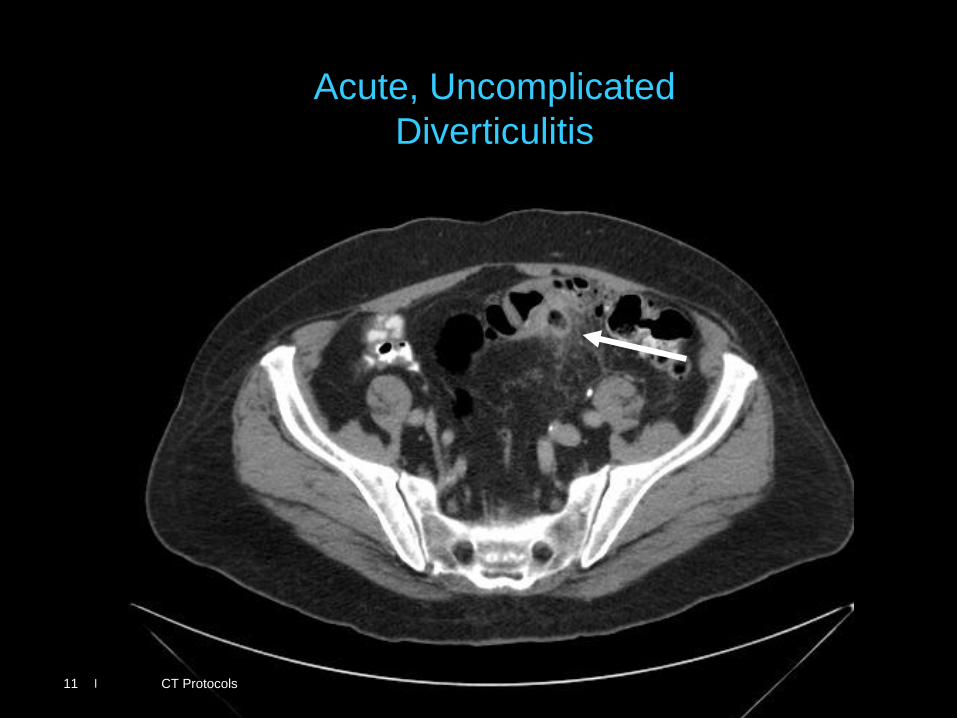
Acute, Uncomplicated
Diverticulitis
CT Protocols 11
|
|
|
|
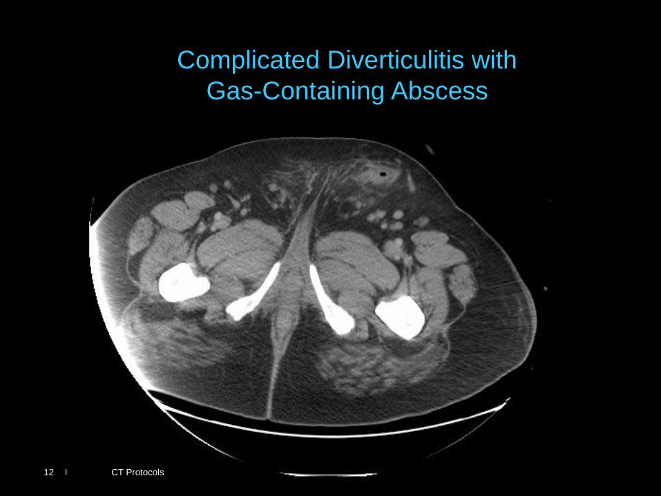
Complicated Diverticulitis with
Gas-Containing Abscess
CT Protocols 12
|
|
|
|
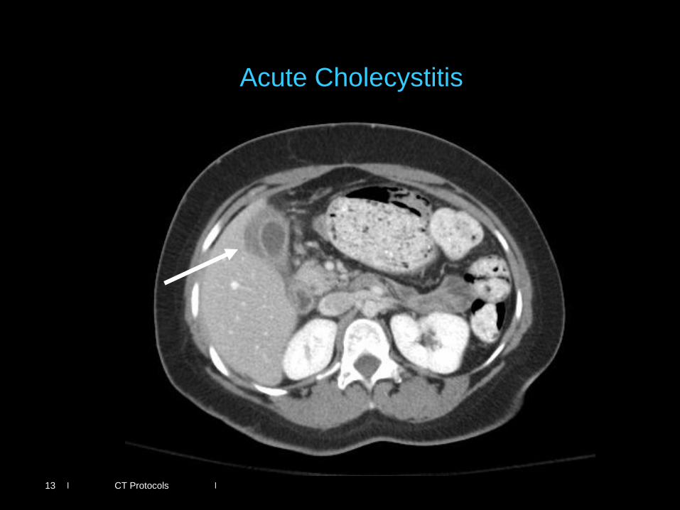
Acute Cholecystitis
CT Protocols 13
|
|
|
|
14
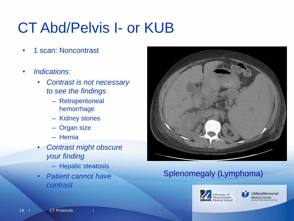
CT Abd/Pelvis I- or KUB
• 1 scan: Noncontrast
• Indications:
• Contrast is not necessary
to see the findings
– Retroperitoneal
hemorrhage
– Kidney stones
– Organ size
– Hernia
• Contrast might obscure
your finding
– Hepatic steatosis
• Patient cannot have
contrast
CT Protocols
Splenomegaly (Lymphoma)
|
|
|
|
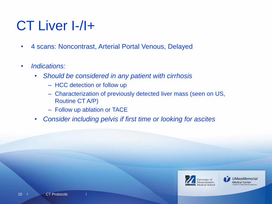
15
CT Liver I-/I+
• 4 scans: Noncontrast, Arterial Portal Venous, Delayed
• Indications:
• Should be considered in any patient with cirrhosis
– HCC detection or follow up
– Characterization of previously detected liver mass (seen on US,
Routine CT A/P)
– Follow up ablation or TACE
• Consider including pelvis if first time or looking for ascites
CT Protocols

|
|
|
|
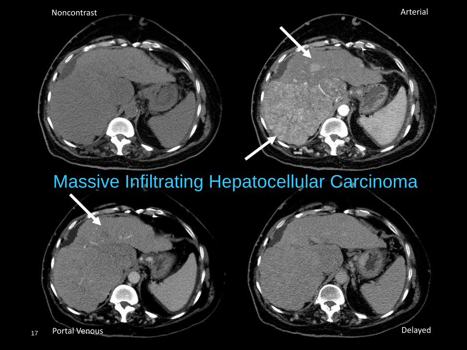
16
Hepatocellular Carcinoma
Noncontrast Arterial
Portal Venous Delayed
|
|
|
|
Massive Infiltrating Hepatocellular Carcinoma
17
Noncontrast Arterial
Portal Venous Delayed
|
|
|
|
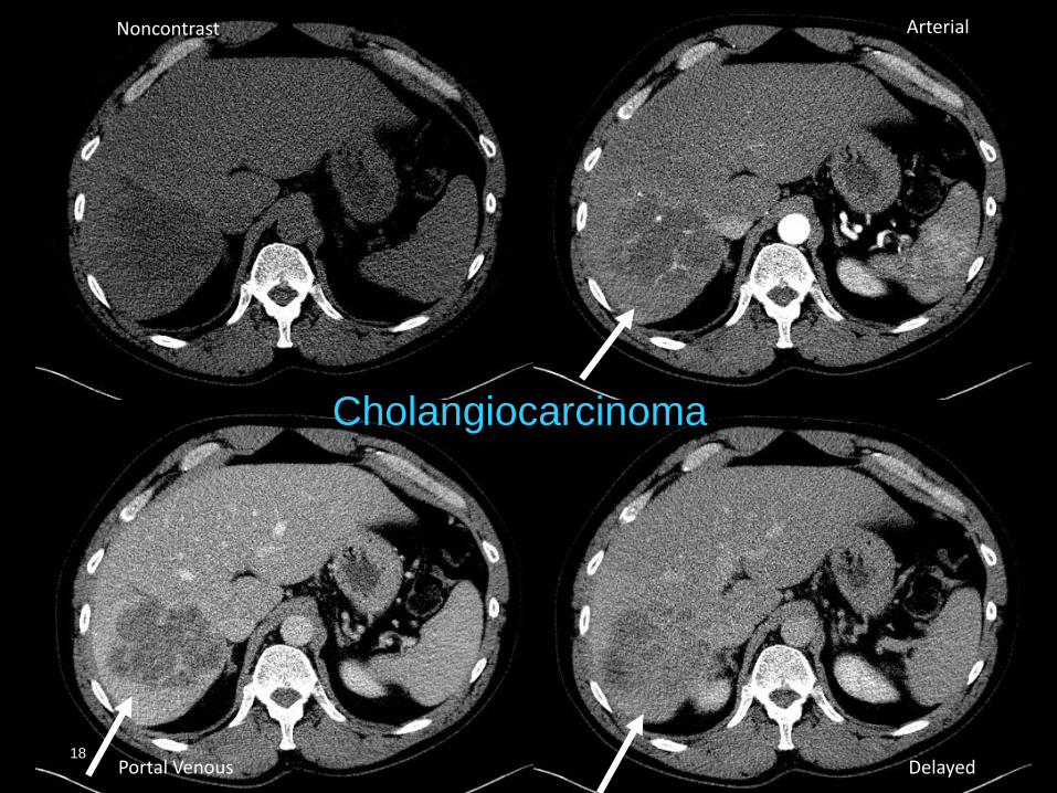
18
CT Protocols
Cholangiocarcinoma
18
Noncontrast Arterial
Portal Venous Delayed
|
|
|
|
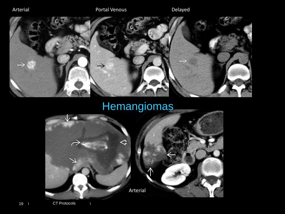
19 CT Protocols
Hemangiomas
Arterial Portal Venous Delayed
Arterial
|
|
|
|
20
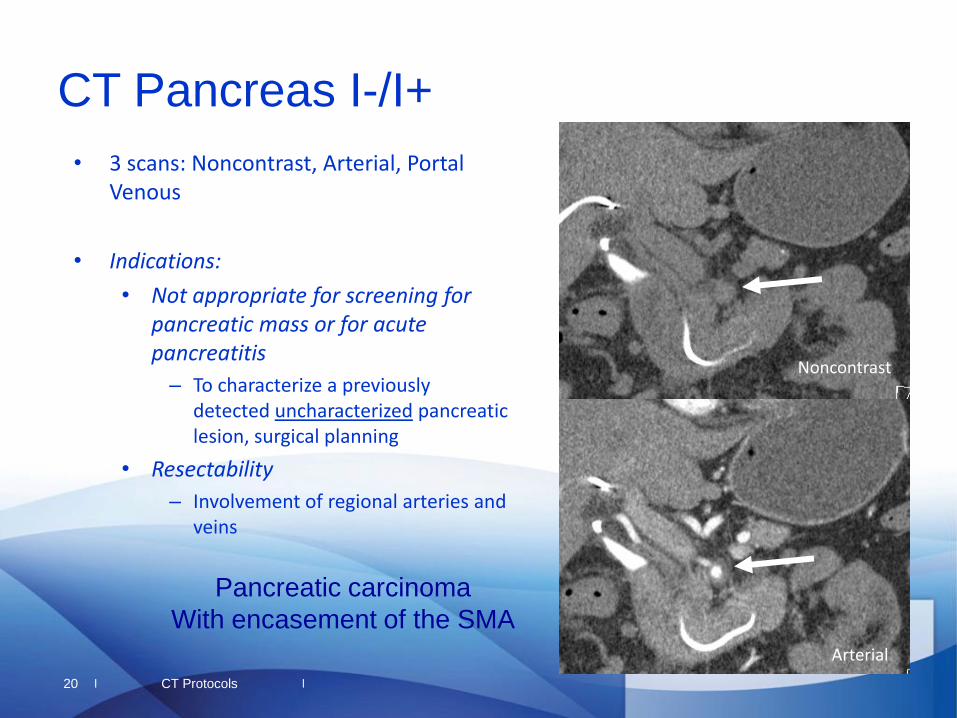
CT Pancreas I-/I+
• 3 scans: Noncontrast, Arterial, Portal Venous
• Indications:
• Not appropriate for screening for pancreatic mass or for acute pancreatitis
– To characterize a previously detected uncharacterized pancreatic lesion, surgical planning
• Resectability
– Involvement of regional arteries and veins
CT Protocols
Pancreatic carcinoma
With encasement of the SMA
Arterial
Noncontrast
|
|
|
|
21
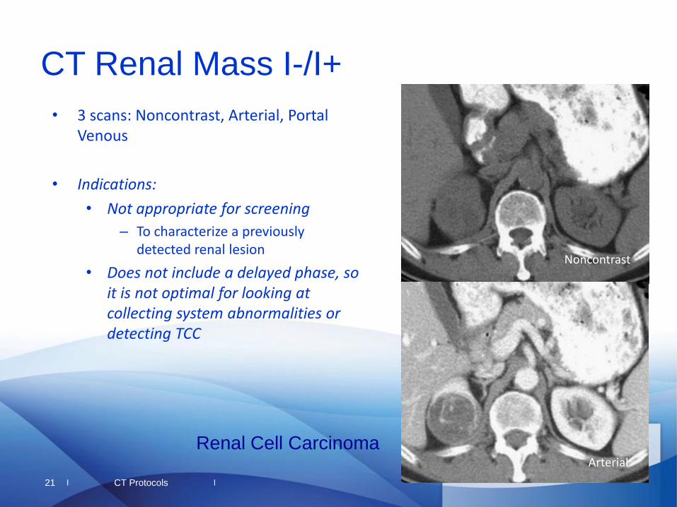
CT Renal Mass I-/I+
• 3 scans: Noncontrast, Arterial, Portal Venous
• Indications:
• Not appropriate for screening
– To characterize a previously detected renal lesion
• Does not include a delayed phase, so it is not optimal for looking at collecting system abnormalities or detecting TCC
CT Protocols
Renal Cell Carcinoma Arterial
Noncontrast
|
|
|
|
22
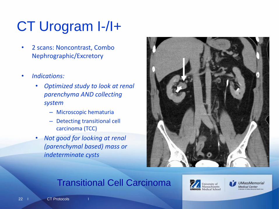
CT Urogram I-/I+
• 2 scans: Noncontrast, Combo Nephrographic/Excretory
• Indications:
• Optimized study to look at renal parenchyma AND collecting system
– Microscopic hematuria
– Detecting transitional cell carcinoma (TCC)
• Not good for looking at renal (parenchymal based) mass or indeterminate cysts
CT Protocols
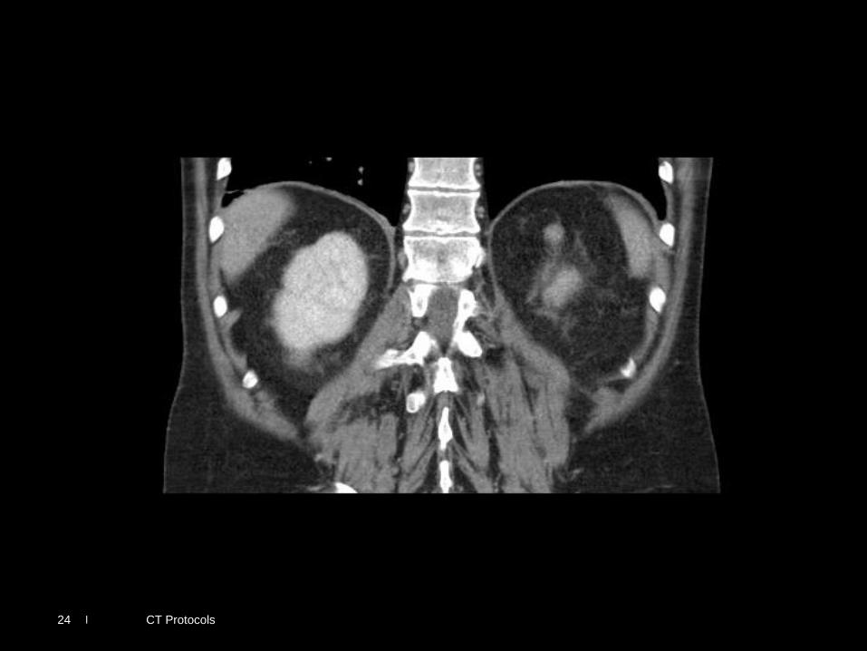
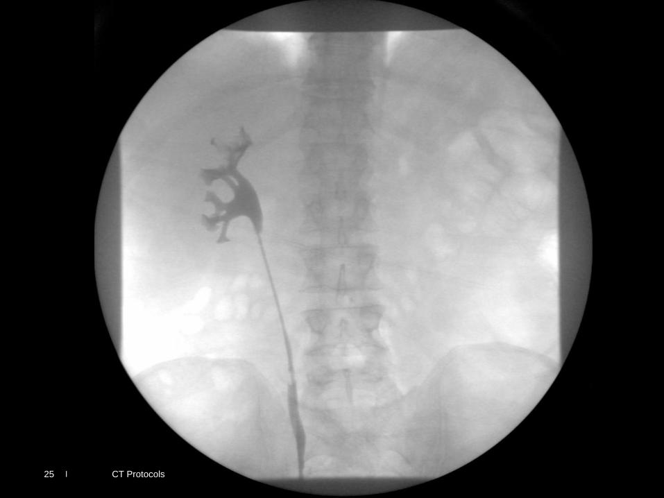
Transitional Cell Carcinoma
|
|
|
|
CT Protocols 23
|
|
|
|
CT Protocols 24
|
|
|
|
CT Protocols 25
|
|
|
|
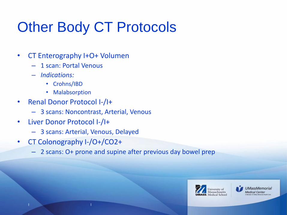
Other Body CT Protocols
• CT Enterography I+O+ Volumen – 1 scan: Portal Venous
– Indications: • Crohns/IBD
• Malabsorption
• Renal Donor Protocol I-/I+ – 3 scans: Noncontrast, Arterial, Venous
• Liver Donor Protocol I-/I+ – 3 scans: Arterial, Venous, Delayed
• CT Colonography I-/O+/CO2+ – 2 scans: O+ prone and supine after previous day bowel prep
|
|
|
|
Thoracic Imaging
27
|
|
|
|
28
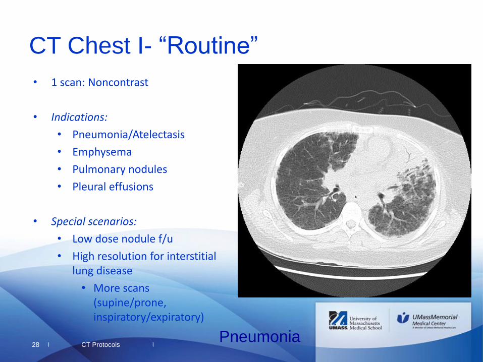
CT Chest I- “Routine”
• 1 scan: Noncontrast
• Indications:
• Pneumonia/Atelectasis
• Emphysema
• Pulmonary nodules
• Pleural effusions
• Special scenarios:
• Low dose nodule f/u
• High resolution for interstitial lung disease
• More scans (supine/prone, inspiratory/expiratory)
Pneumonia CT Protocols
|
|
|
|
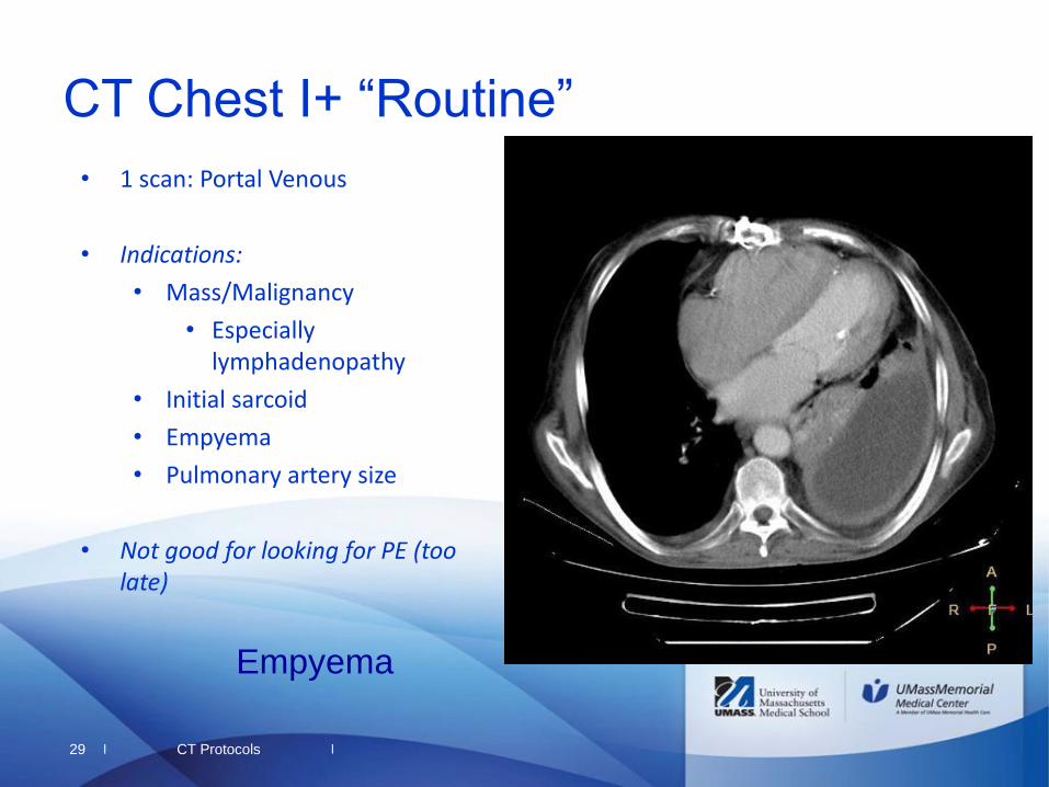
29
CT Chest I+ “Routine”
• 1 scan: Portal Venous
• Indications:
• Mass/Malignancy
• Especially lymphadenopathy
• Initial sarcoid
• Empyema
• Pulmonary artery size
• Not good for looking for PE (too late)
Empyema
CT Protocols
|
|
|
|
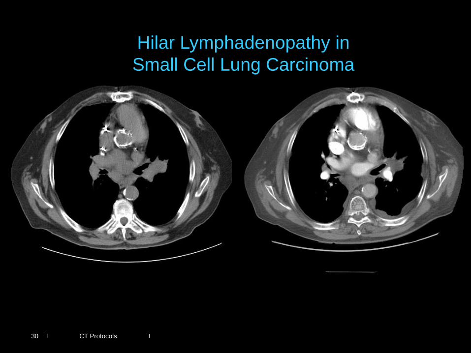
CT Protocols
Hilar Lymphadenopathy in
Small Cell Lung Carcinoma
30
|
|
|
|
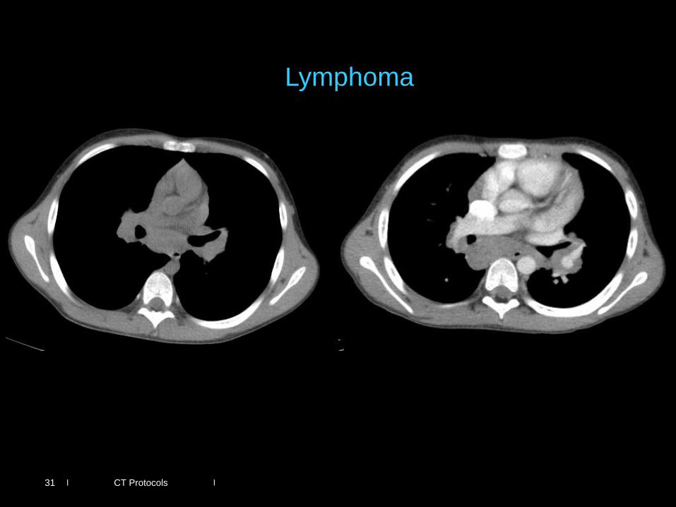
CT Protocols
Lymphoma
31
|
|
|
|
CT Protocols
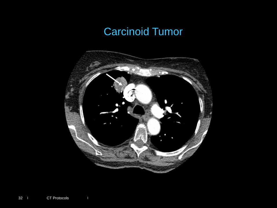
Carcinoid Tumor
32
|
|
|
|
CT Protocols
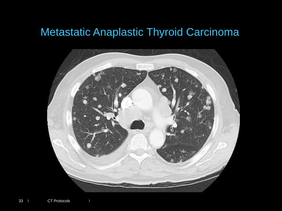
Metastatic Anaplastic Thyroid Carcinoma
33
|
|
|
|
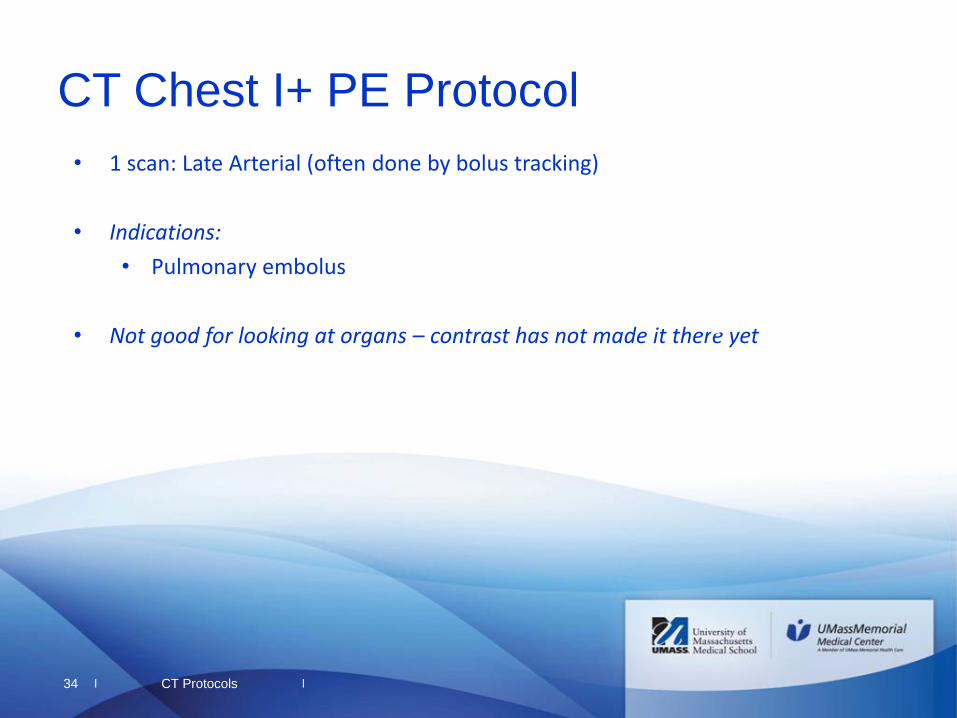
34
CT Chest I+ PE Protocol
• 1 scan: Late Arterial (often done by bolus tracking)
• Indications:
• Pulmonary embolus
• Not good for looking at organs – contrast has not made it there yet
CT Protocols
|
|
|
|
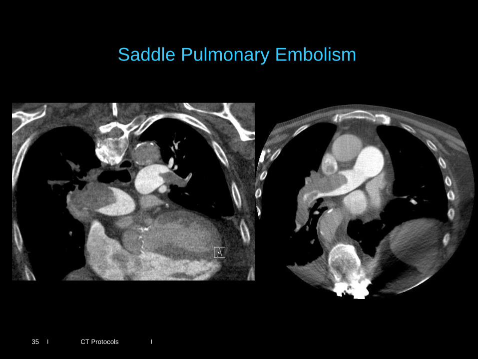
CT Protocols 35
Saddle Pulmonary Embolism
|
|
|
|
36
Other Thoracic Protocols
• Cardiac studies (depending on availability)
• May be gated
• May require beta blocker tx
• Valves
• Anatomy
• Coronary Artery Evaluation
CT Protocols
|
|
|
|
37
CT Angiograms
• No oral contrast
• CTA Aneurysm I-/I+ – 2 scans: Noncontrast, Arterial – Indications:
• Aortic aneurysm evaluation • Acute bleed (liver, bowel,
spleen, etc)
• CTA Dissection I-/I+
– 3 scans: Noncontrast, Arterial, Portal Venous
– Indications: • Aortic dissection
– Portal venous phase is included to assess organ perfusion
• CTA Stent I-/I+ – 3 scans: Noncontrast, Arterial,
Delayed – Indications:
• Evaluate endovascular repair
– Delayed phase to look for delayed leak
• CT Extremity Runoff I-/I+
– 2 scans: Noncontrast, Arterial – Indications:
• Cold limb, extremity ischemia
– Large field of view gives poor special resolution
– Usually ordered by vascular surgery
CT Protocols

|
|
|
|
Neuro
38
|
|
|
|
39
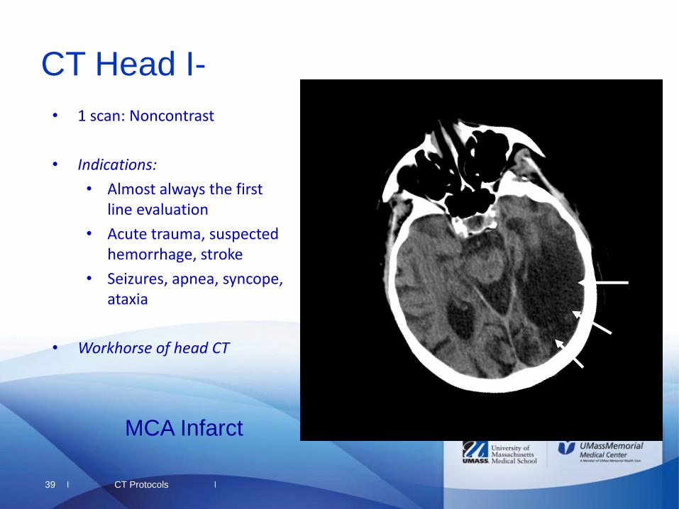
CT Head I-
CT Protocols
• 1 scan: Noncontrast
• Indications:
• Almost always the first line evaluation
• Acute trauma, suspected hemorrhage, stroke
• Seizures, apnea, syncope, ataxia
• Workhorse of head CT
MCA Infarct
|
|
|
|
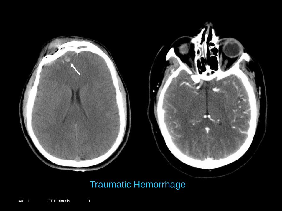
CT Protocols 40
Traumatic Hemorrhage

|
|
|
|
41 CT Protocols
Venous Sinus Thrombosis
|
|
|
|
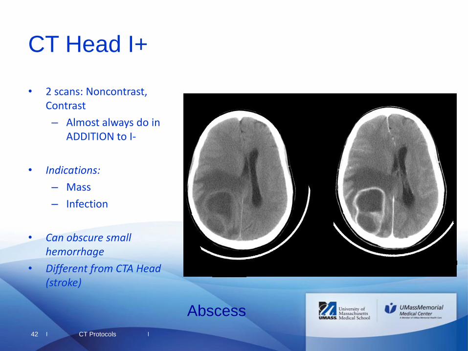
• 2 scans: Noncontrast, Contrast
– Almost always do in ADDITION to I-
• Indications:
– Mass
– Infection
• Can obscure small hemorrhage
• Different from CTA Head (stroke)
42
CT Head I+
CT Protocols
Abscess
|
|
|
|
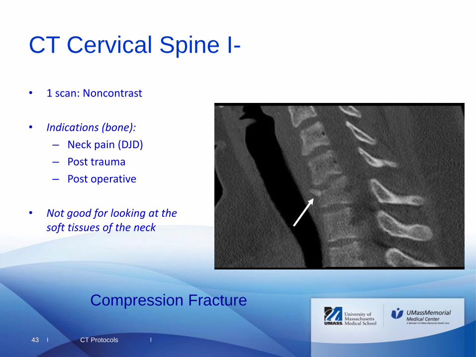
• 1 scan: Noncontrast
• Indications (bone):
– Neck pain (DJD)
– Post trauma
– Post operative
• Not good for looking at the soft tissues of the neck
43
CT Cervical Spine I-
CT Protocols
Compression Fracture
|
|
|
|
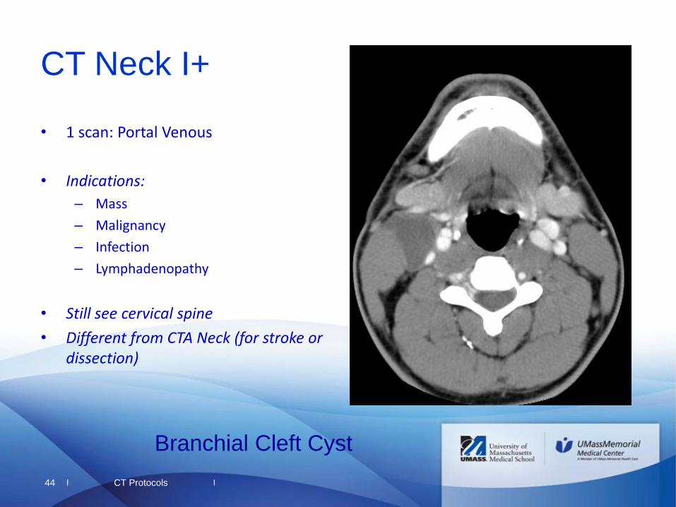
• 1 scan: Portal Venous
• Indications:
– Mass
– Malignancy
– Infection
– Lymphadenopathy
• Still see cervical spine
• Different from CTA Neck (for stroke or dissection)
44
CT Neck I+
CT Protocols
Branchial Cleft Cyst
|
|
|
|
• 2 scans: Noncontrast head; Arterial through the head and neck
• Indications:
– *Stroke
– Dissection
– Post traumatic
• Different from CT Head and Neck I+
45
CTA Head and Neck I+
CT Protocols
|
|
|
|
• CT Nasal Bones I-
– Trauma
• CT Sinus/Maxillofacial I-
– I+ if looking for infection/abscess, neoplasm
• CT Temporal Bones I-
– Hearing loss, cholestatoma, post surgical
• CT Parathyroid I+
– 4D parathyroid CT for parathyroid adenoma
46
Other Misc Neuro Exams
CT Protocols
|
|
|
|
• For bone, contrast doesn’t add much
– Only use I+ if planning to evaluate soft tissues or soft tissue component
• CT is best for bone
– If concerned for soft tissues, MRI is far superior
• Ultrasound may be a good place to start (insurance issues)
47
Musculoskeletal Protocols
CT Protocols
|
|
|
|
• Please feel free to contact me with any questions about this presentation, CT protocols, or radiology in general!
48
Thank You!
CT Protocols

|
|
|
|
• ACR Appropriateness Criteria http://www.acr.org/Quality-Safety/Appropriateness-Criteria
49
Helpful References
CT Protocols