Embed Size (px)
Citation preview
191
© 2013 The Korean Society of Pathologists/The Korean Society for CytopathologyThis is an Open Access article distributed under the terms of the Creative Commons Attribution Non-Commercial License (http://creativecommons.org/licenses/by-nc/3.0) which permits unrestricted non-commercial use, distribution, and reproduction in any medium, provided the original work is properly cited.
pISSN 1738-1843eISSN 2092-8920
EPITHELIAL ODONTOGENIC TUMORS
The pathogenetic mechanism of odontogenic tumors is close-ly related to the developmental processes of teeth. As a result, the molecular signaling mechanisms for normal enamel organs and odontogenic tumors have been closely compared. For the organogenesis of tooth germs, the reciprocal induction between odontogenic mesenchyme and enamel epithelium sequentially progresses to differentiate odontoblasts into odontogenic mes-enchyme and ameloblasts in the enamel epithelium.1,2 During these processes of tooth formation, the tumorigenesis of odon-togenic epithelium occurs in the state of immature odontogenic tissues, resulting in different histological features and variable potentials of tumor propagation. However, different epithelial odontogenic tumors can be classified into ameloblastomas, ade-nomatoid odontogenic tumors (AOTs), calcifying epithelial odontogenic tumors, odontogenic ghost cell tumors, squamous odontogenic tumors, ameloblastic fibromas, ameloblastic fibro-
odontomas, or odontomas, etc. This study was conducted to re-view the current concepts and occurrences of ameloblastomas and AOTs.
AMELOBLASTOMA
Once designated as an adamantinoma in 1885 (Malassez) but renamed to ameloblastoma in 1930 (Ivey and Churchil), amelo-blastoma is a representative benign tumor of odontogenic epi-thelium. Some authors still misuse the term adamantinoma to describe ameloblastomas, even though an adamantinoma, mean-ing “very hard,” is a rare bone tumor that differs in histology and frequency of malignancy from ameloblastomas.3 Amelo-blastomas are common, have an aggressive behavior and recur-rent course, and are rarely metastatic. However, adamantinoma-tous craniopharyngioma and adamantinoid basal cell carcinoma are terms that are still used to describe their histological charac-teristics.4
Current Concepts and Occurrence of Epithelial Odontogenic Tumors:
I. Ameloblastoma and Adenomatoid Odontogenic Tumor
Suk Keun Lee · Yeon Sook Kim1
Department of Oral Pathology, College of Dentistry, Gangneung-Wonju National University, Gangneung; 1Department of Dental Hygiene, College of Health Sciences, Cheongju University, Cheongju, Korea
Ameloblastomas and adenomatoid odontogenic tumors (AOTs) are common epithelial tumors of odontogenic origin. Ameloblastomas are clinico-pathologically classified into solid/multicystic, unicystic, desmoplastic, and peripheral types, and also divided into follicular, plexiform, acantho-matous, granular types, etc., based on their histological features. Craniopharyngiomas, derived from the remnants of Rathke’s pouch or a misplaced enamel organ, are also comparable to the odontogenic tumors. The malignant transformation of ameloblastomas results in the formation of ameloblastic carcinomas and malignant ameloblastomas depending on cytological dysplasia and metastasis, respectively. AOTs are classified into follicular, extrafollicular, and peripheral types. Ameloblastomas are common, have an aggressive behavior and recurrent course, and are rarely metastatic, while AOTs are hamartomatous benign lesions derived from the complex system of the dental lamina or its remnants. With advances in the elucidation of molecular signaling mecha-nisms in cells, the cytodifferentiation of epithelial tumor cells in ameloblastomas and AOTs can be identified using different biomarkers. Therefore, it is suggested that comprehensive pathological observation including molecular genetic information can provide a more reliable differential diag-nosis for the propagation and prognosis of ameloblastomas and AOTs. This study aimed to re-view the current concepts of ameloblastomas and AOTs and to discuss their clinico-pathological features relevant to tumorigenesis and prognosis.
Key Words: Ameloblastoma; Craniopharyngioma; Adenomatoid odontogenic tumor
Received: March 19, 2013Accepted: April 25, 2013
Corresponding AuthorSuk Keun Lee, D.D.S., Ph.D.Department of Oral Pathology, College of Dentistry, Gangneung-Wonju National University, 123 7 Jukheongil, Gangneung 210-702, KoreaTel: +82-33-640-2228Fax: +82-33-642-6410E-mail: [email protected]
The Korean Journal of Pathology 2013; 47: 191-202http://dx.doi.org/10.4132/KoreanJPathol.2013.47.3.191
▒ REVIEW ▒
http://www.koreanjpathol.org http://dx.doi.org/10.4132/KoreanJPathol.2013.47.3.191
192 • Lee SK, et al.
Characteristics of ameloblastoma
Ameloblastoma is a slow growing odontogenic epithelial tu-mor of the jaw and accounts for about 1% of all oral tumors and about 18% of odontogenic tumors. It is primarily seen in adults in the third to fifth decades of life, with almost equal sex predi-lection.5 Radiographically, it appears as an expansile radiolucen-cy with thinned and perforated cortices, frequently causing root resorption. These features are common in giant cell tumors, an-eurysmal bone cysts, and renal cell carcinoma metastasis, thus a definitive diagnosis can be made through histopathology.
A total of 5,213 cases of ameloblastomas are found in litera-ture, including 3,677 cases of ameloblastomas available in liter-ature from 1960 to 1993,5 340 cases of ameloblastomas in the Malaysian population (1993 through 2008),6 and 1,196 cases of ameloblastomas diagnosed during the years of 1993 to 2009 in Chulalongkorn University, Bangkok.7 These studies revealed the mean age of patients as 36.1 years, with 2,709 (51.9%) patients being male, and mandibular tumors (81.7%) outnumbering maxillary tumors. In the variants of ameloblastomas, approxi-mately 10.7% were unicystic ameloblastomas and 1.8% were peripheral ameloblastomas.
In Korea, about 500 cases of ameloblastomas were sporadi-cally reported, and among them, 452 cases were available to be statistically analyzed.8-10 The ratio of males to females was about 57:43, and the average age was approximately 33.8 years. The most frequent site of involvement was around the mandibular molar area (57.7%). The tumors were composed of convention-al ameloblastomas (48.9%), unicystic ameloblastomas (25.3%), and peripheral ameloblastomas (3.1%). The recurrence rate for conventional ameloblastomas (17.1%) was significantly higher than for the unicystic type (9.1%). According to the histology, the acanthomatous, plexiform, and follicular patterns of the conventional ameloblastomas had similar recurrence rates of 16.2%, 15.9%, and 12.7%, respectively.
Molecular mechanisms of ameloblastoma
Every cellular changes, including proliferation, differentiation, senescence, tumorigenesis, etc., occur through the activation or inactivation of related molecular signaling pathways. The over-expression or underexpression of important signaling molecules may play an important role in the tumorigenesis of ameloblas-tomas. First of all, several proteins expressed in the enamel epi-thelium during the early stage of tooth formation, including amelogenin,11 tuftelin,12 and ameloblastin,13,14 are specifically expressed in tumor cells of ameloblastomas, but enamelin and amelotin expressed in the mineralizing stage of tooth formation
are rarely positive in tumor cells of ameloblastomas.15,16 Different proteins functioning in the signaling of the prolif-
eration and differentiation of enamel epithelium and odontogen-ic tumors have been identified by many authors. The cellular and stromal proteins of syndecan-1,17 perlecan, α-dystroglycan, integrin β1,18 CD10, and osteopontin19 are overexpressed in ameloblastomas that exhibit locally invasive behavior and a high risk of recurrence. Solid ameloblastomas show an intense expression of fibronectin at the epithelial-mesenchymal inter-face, whereas desmoplastic ameloblastomas reveal no immuno-expression of fibronectin at this site. Ameloblastomas present a stronger immunoreaction of tenascin than AOTs, especially at the epithelial-mesenchymal interface, while AOTs and desmo-plastic ameloblastomas exhibit an intense labeling for type I collagen. These expression patterns of matrix proteins agree with the more locally invasive behavior of ameloblastomas in comparison to AOTs.20
The local invasiveness of ameloblastoma is also related to the expressions of metalloproteinase (MMP)-2 and MMP-9. The overexpression of MMP-9 in ameloblastomas is possibly modu-lated by the unmethylation of the gene and may influence the aggressive behavior and high recurrence rates of ameloblasto-ma.21
The facts that receptor activator of nuclear factor κB ligand (RANKL) and MMP-9 are expressed in ameloblastomas may suggest that ameloblastoma cells have the potential to induce osteoclastogenesis, resulting in the rapid destruction of bone marrow.22,23 The higher expression rates of interleukin (IL)-1α and IL-6 are associated with tumor size in ameloblastomas. There-fore, it is suggested that the IL-1α and IL-6 cytokines play a role in the aggressive behavior of ameloblastomas by increasing bone resorption.24
The overexpression of cyclin D1, a member of G1 cyclins con-trolling the cell-cycle transit from the G1 to S phase, is found in some plexiform ameloblastomas but is relatively mild in AOTs. The peripheral columnar and central stellate reticulum-like cells of ameloblastomas exhibit a dominant immunoreaction of cyclin D1, which is almost non-existent in the squamous and granular cells of ameloblastomas.25
Midkine is a heparin-binding growth factor overexpressed in various human cancers and is expressed in ameloblastomas and ameloblastic carcinomas. This midkine expression in the major-ity of ameloblastomas may suggest a role of the protein in tu-mor development, progression, and behavior.26
Epithelial growth factor receptor expression had no statistical significance for local recurrence, patient age, or tumor size, wher-
http://www.koreanjpathol.orghttp://dx.doi.org/10.4132/KoreanJPathol.2013.47.3.191
Ameloblastoma and Adenomatoid Odontogenic Tumor • 193
eas a significant relation existed between CD10 expression and the Ki-67 labeling index, which is significantly related to the recurrence of ameloblastomas.27
The overexpression of p53 and murine double minute 2 (MD M2) are associated with the pathogenesis and oncogenesis of ameloblastomas.28 The overexpression of p-mTOR, p-4E-BP1, and p-p70S6K in the nucleus also are related to the inva-siveness of ameloblastomas.29
The transforming growth factor-β (TGF-β)/SMAD signaling pathway is commonly activated in ameloblastomas, AOTs, and calcifying cystic odontogenic tumors. Meanwhile, the TGF-β/SM AD immunoreaction is significantly reduced in ameloblas-tomas in comparison to AOTs and calcifying cystic odontogenic tumors. These changes may lead to the more aggressive biologi-cal behavior of ameloblastomas through increased cell prolifera-tion and reduced apoptosis and differentiation.30
WNT proteins (except WNT-10b) are heterogeneously ex-pressed in different types of ameloblastomas, indicating that WNT expression is closely related to tumor cell differentiation and invasion.31 However, the WNT-related bone-forming genes of WDR5 and runt-related transcription factor 2 (RUNX2) are down-regulated in ameloblastomas.32
Alterations in the Sonic Hedgehog signaling pathway, includ-ing protein patched homolog (PTCH) gene mutations, have been associated with the pathogenesis of some odontogenic tu-mors including ameloblastomas and are closely related to ne-void basal cell carcinoma syndrome through the genetic altera-tion of PTCH. Therefore, ameloblastoma diagnosis possibly warrants a search for associated cutaneous basal cell carcinomas and other benign and malignant tumors related to nevoid basal cell carcinoma syndrome.33-35
There appears to be no difference in the expression of E-cad-herin or β-catenin between tooth germs and solid and unicystic ameloblastomas. The expression of these molecules seems to be mainly related to the process of cell differentiation.36
Rho GTPases that regulate the cell cycle, shape, polarization, invasion, migration, and apoptosis are overexpressed in amelo-blastomas. Therefore, it is suggested that these GTPases play a role in the neoplastic epithelial cell phenotype determination of ameloblastomas (polarized or non-polarized), as well as in vari-ant (solid or unicystic) and subtype (follicular or plexiform) de-termination.37
On the other hand, tumor necrosis factor-related apoptosis-inducing ligand (TRAIL) and TRAIL receptor are diffusely ex-pressed in ameloblastomas and are possibly involved in the neo-plastic transformation of odontogenic epithelium. FAS (CD95),
FAS ligand, and caspase-3 are also expressed in ameloblasto-mas.38 These expressions may suggest some intrinsic regulation of neoplastic cell proliferation and death in ameloblastomas, thus explaining their slow growth and inability to metastasize.39
Histological types of ameloblastoma
Histologically, ameloblastomas are classified into follicular, plexiform, acanthomatous, and granular cell types. Less com-mon histologic variants are clear cell, desmoplastic, basal cell, papilliferous, and keratoameloblastoma types. Generally, one-third of ameloblastomas are plexiform, one-third are follicular, and other variants such as acanthomatous types usually occur in older patients.
Follicular type
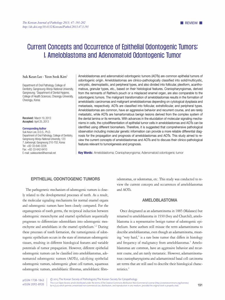
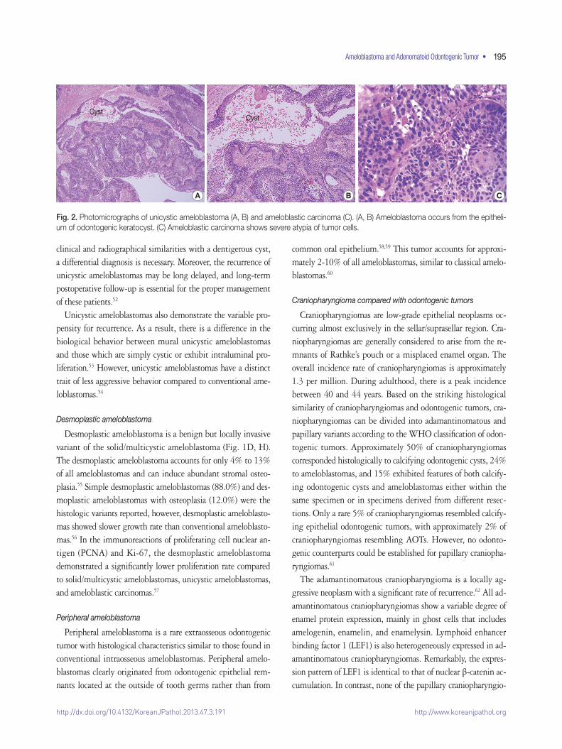
The follicular type of ameloblastoma frequently forms vari-ous sizes of dental follicle structures and possesses an outer ar-rangement of columnar or palisaded ameloblast-like cells and inner zone of triangular-shaped cells resembling the stellate re-ticulum in the bell stage of the tooth germ (Fig. 1A, E). Histo-pathology shows cells that have a tendency for nuclei to move away from the basement membrane. This process is referred to as “reverse polarization.” The central cells sometimes degener-ate to form central microcysts.
The follicular type of ameloblastomas grow and usually form multicystic nodules, resulting in multilocular tumor masses with frequent recurrence compared to the other types of amelo-blastomas.40
Plexiform type
The plexiform type contains epithelium that proliferates in a “fish net pattern,” or in a “cord-like fashion anastomosing each other,” hence the name “plexiform.” There are layers of cells be-tween the proliferating epithelium with well-formed desmo-somal junctions, simulating spindle cell layers (Fig. 1B, F). Plexi-form ameloblastomas with multiple cyst formation are rare and benign odontogenic tumors which may reach grotesque propor-tions, affecting a large region of the jaw.41
Acanthomatous type
Acanthomatous ameloblastoma is the extremely rare variant exhibiting solid epithelial cell nests with peripheral palisading ameloblastic cells and central squamous cell differentiation (Fig. 1C, G).42 The acanthomatous ameloblastoma masquerades as a squamous cell carcinoma and also appears as a “hybrid” amelo-blastoma admixed with a pronounced desmoplastic pattern.43
http://www.koreanjpathol.org http://dx.doi.org/10.4132/KoreanJPathol.2013.47.3.191
194 • Lee SK, et al.
Granular cell type
Granular cell ameloblastoma is a variant of ameloblastoma, where the tumor cells located in the central portion of the folli-cles have granular eosinophilic cytoplasm and the peripheral tumor cells resemble ameloblasts. The granular cells are a tran-sitional or matured phase in the life cycle of ameloblastomas, starting with normal stellate reticulum-like cells, leading to a production of granules and finally resulting in degeneration and the formation of cystic areas, differing from the aggressive tu-mor nature reported previously.44 However, the granular cell type is rare and accounts for only 4% of ameloblastomas.45
Others
Keratoameloblastoma is a very rare ameloblastoma variant defined by extensive squamous metaplasia and keratinization. Hemangiomatous ameloblastoma is also described as a rare am-eloblastic variant.46
Basal cell ameloblastoma is also a very rare variant of amelo-blastoma resembling basaloid squamous cell carcinoma but pos-sessing the pathological features of conventional ameloblastoma.47
Adenoid ameloblastoma is a rare variant in this category and can cause problems in diagnosis due to the presence of areas re-sembling AOT and the occurrence of varying degrees of denti-noid formation.48
Clinico-pathological classification of ameloblastoma
According to the World Health Organization (WHO), ame-loblastomas are classified into the following types depending on the origin of tumorigenesis: solid/multicystic, extraosseous/pe-ripheral, desmoplastic, and unicystic. The common solid/mul-
ticystic ameloblastomas arise from enamel epithelial rests in jaw bone, while unicystic ameloblastomas arise from the epithelium of odontogenic cysts. Desmoplastic ameloblastomas exhibit ac-tive stromal proliferation, while peripheral ameloblastomas arise from dental lamina rests and oral mucosa epithelium. These tu-mor types differ in biological behavior and rate of recurrence. Therefore, each type of ameloblastoma requires different forms of treatment.49 Additionally craniopharyngiomas arising from the rests of Rathke’s pouch epithelium are comparable to ame-loblastomas.
Solid/multicystic ameloblastoma (intraosseous ameloblastoma,
central ameloblastoma, conventional ameloblastoma)
Conventional ameloblastomas are the most common and prevalent among odontogenic tumors. Clinico-radiographically, they present as intraosseous lesions which exhibit slow, painless swelling or expansion of the jaw, with multilocular expansile radiolucency most frequently found in the mandibular molar/ramus area. Depending on the histological features, the conven-tional ameloblastoma is also called a solid/multicystic amelo-blastoma, in contrast to the unicystic ameloblastoma.50
Unicystic ameloblastoma (mural ameloblastoma)
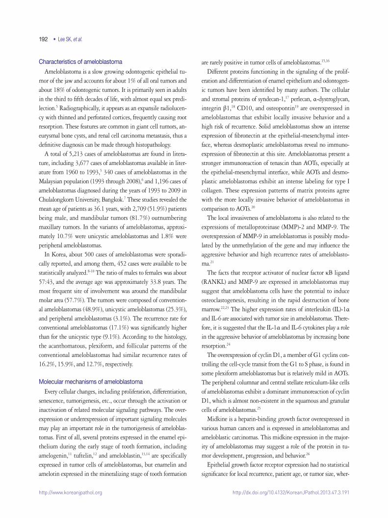
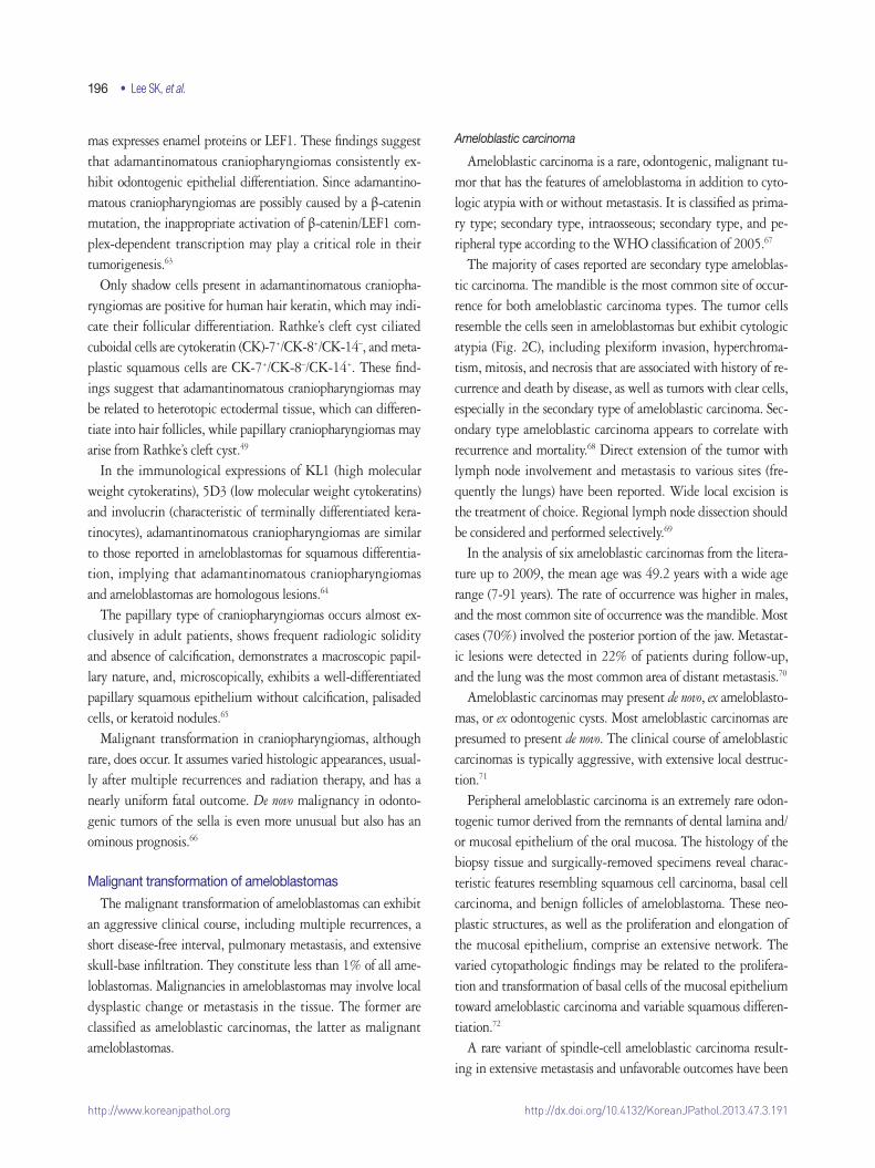
Unicystic ameloblastomas refer to the cystic lesions that ex-hibit clinical, radiographic, or gross features of a mandibular cyst but upon histologic examination show typical ameloblas-toma epithelium lining part of the cyst cavity, with or without luminal and/or mural tumor growth (Fig. 2A, B).51 The unicys-tic ameloblastoma is believed to be less aggressive than the con-ventional ameloblastoma. Since this tumor shows considerable
A
E
B
F
C
G
D
H
Fig. 1. Photomicrographs of the different types of ameloblastoma. (A, E) Follicular type. (B, F) Plexiform type. (C, G) Acanthomatous type. (D, H) Desmoplastic type. Panels (E), (F), (G), and (H) are the magnifications of panels (A), (B), (C), and (D), respectively.
http://www.koreanjpathol.orghttp://dx.doi.org/10.4132/KoreanJPathol.2013.47.3.191
Ameloblastoma and Adenomatoid Odontogenic Tumor • 195
CystCyst
A B C
Fig. 2. Photomicrographs of unicystic ameloblastoma (A, B) and ameloblastic carcinoma (C). (A, B) Ameloblastoma occurs from the epitheli-um of odontogenic keratocyst. (C) Ameloblastic carcinoma shows severe atypia of tumor cells.
clinical and radiographical similarities with a dentigerous cyst, a differential diagnosis is necessary. Moreover, the recurrence of unicystic ameloblastomas may be long delayed, and long-term postoperative follow-up is essential for the proper management of these patients.52
Unicystic ameloblastomas also demonstrate the variable pro-pensity for recurrence. As a result, there is a difference in the biological behavior between mural unicystic ameloblastomas and those which are simply cystic or exhibit intraluminal pro-liferation.53 However, unicystic ameloblastomas have a distinct trait of less aggressive behavior compared to conventional ame-loblastomas.54
Desmoplastic ameloblastoma
Desmoplastic ameloblastoma is a benign but locally invasive variant of the solid/multicystic ameloblastoma (Fig. 1D, H). The desmoplastic ameloblastoma accounts for only 4% to 13% of all ameloblastomas and can induce abundant stromal osteo-plasia.55 Simple desmoplastic ameloblastomas (88.0%) and des-moplastic ameloblastomas with osteoplasia (12.0%) were the histologic variants reported, however, desmoplastic ameloblasto-mas showed slower growth rate than conventional ameloblasto-mas.56 In the immunoreactions of proliferating cell nuclear an-tigen (PCNA) and Ki-67, the desmoplastic ameloblastoma demonstrated a significantly lower proliferation rate compared to solid/multicystic ameloblastomas, unicystic ameloblastomas, and ameloblastic carcinomas.57
Peripheral ameloblastoma
Peripheral ameloblastoma is a rare extraosseous odontogenic tumor with histological characteristics similar to those found in conventional intraosseous ameloblastomas. Peripheral amelo-blastomas clearly originated from odontogenic epithelial rem-nants located at the outside of tooth germs rather than from
common oral epithelium.58,59 This tumor accounts for approxi-mately 2-10% of all ameloblastomas, similar to classical amelo-blastomas.60
Craniopharyngioma compared with odontogenic tumors
Craniopharyngiomas are low-grade epithelial neoplasms oc-curring almost exclusively in the sellar/suprasellar region. Cra-niopharyngiomas are generally considered to arise from the re-mnants of Rathke’s pouch or a misplaced enamel organ. The overall incidence rate of craniopharyngiomas is approximately 1.3 per million. During adulthood, there is a peak incidence between 40 and 44 years. Based on the striking histological similarity of craniopharyngiomas and odontogenic tumors, cra-niopharyngiomas can be divided into adamantinomatous and papillary variants according to the WHO classification of odon-togenic tumors. Approximately 50% of craniopharyngiomas corresponded histologically to calcifying odontogenic cysts, 24% to ameloblastomas, and 15% exhibited features of both calcify-ing odontogenic cysts and ameloblastomas either within the same specimen or in specimens derived from different resec-tions. Only a rare 5% of craniopharyngiomas resembled calcify-ing epithelial odontogenic tumors, with approximately 2% of craniopharyngiomas resembling AOTs. However, no odonto-genic counterparts could be established for papillary craniopha-ryngiomas.61
The adamantinomatous craniopharyngioma is a locally ag-gressive neoplasm with a significant rate of recurrence.62 All ad-amantinomatous craniopharyngiomas show a variable degree of enamel protein expression, mainly in ghost cells that includes amelogenin, enamelin, and enamelysin. Lymphoid enhancer binding factor 1 (LEF1) is also heterogeneously expressed in ad-amantinomatous craniopharyngiomas. Remarkably, the expres-sion pattern of LEF1 is identical to that of nuclear β-catenin ac-cumulation. In contrast, none of the papillary craniopharyngio-
http://www.koreanjpathol.org http://dx.doi.org/10.4132/KoreanJPathol.2013.47.3.191
196 • Lee SK, et al.
mas expresses enamel proteins or LEF1. These findings suggest that adamantinomatous craniopharyngiomas consistently ex-hibit odontogenic epithelial differentiation. Since adamantino-matous craniopharyngiomas are possibly caused by a β-catenin mutation, the inappropriate activation of β-catenin/LEF1 com-plex-dependent transcription may play a critical role in their tumorigenesis.63
Only shadow cells present in adamantinomatous craniopha-ryngiomas are positive for human hair keratin, which may indi-cate their follicular differentiation. Rathke’s cleft cyst ciliated cuboidal cells are cytokeratin (CK)-7+/CK-8+/CK-14–, and meta-plastic squamous cells are CK-7+/CK-8–/CK-14+. These find-ings suggest that adamantinomatous craniopharyngiomas may be related to heterotopic ectodermal tissue, which can differen-tiate into hair follicles, while papillary craniopharyngiomas may arise from Rathke’s cleft cyst.49
In the immunological expressions of KL1 (high molecular weight cytokeratins), 5D3 (low molecular weight cytokeratins) and involucrin (characteristic of terminally differentiated kera-tinocytes), adamantinomatous craniopharyngiomas are similar to those reported in ameloblastomas for squamous differentia-tion, implying that adamantinomatous craniopharyngiomas and ameloblastomas are homologous lesions.64
The papillary type of craniopharyngiomas occurs almost ex-clusively in adult patients, shows frequent radiologic solidity and absence of calcification, demonstrates a macroscopic papil-lary nature, and, microscopically, exhibits a well-differentiated papillary squamous epithelium without calcification, palisaded cells, or keratoid nodules.65
Malignant transformation in craniopharyngiomas, although rare, does occur. It assumes varied histologic appearances, usual-ly after multiple recurrences and radiation therapy, and has a nearly uniform fatal outcome. De novo malignancy in odonto-genic tumors of the sella is even more unusual but also has an ominous prognosis.66
Malignant transformation of ameloblastomas
The malignant transformation of ameloblastomas can exhibit an aggressive clinical course, including multiple recurrences, a short disease-free interval, pulmonary metastasis, and extensive skull-base infiltration. They constitute less than 1% of all ame-loblastomas. Malignancies in ameloblastomas may involve local dysplastic change or metastasis in the tissue. The former are classified as ameloblastic carcinomas, the latter as malignant ameloblastomas.
Ameloblastic carcinoma
Ameloblastic carcinoma is a rare, odontogenic, malignant tu-mor that has the features of ameloblastoma in addition to cyto-logic atypia with or without metastasis. It is classified as prima-ry type; secondary type, intraosseous; secondary type, and pe-ripheral type according to the WHO classification of 2005.67
The majority of cases reported are secondary type ameloblas-tic carcinoma. The mandible is the most common site of occur-rence for both ameloblastic carcinoma types. The tumor cells resemble the cells seen in ameloblastomas but exhibit cytologic atypia (Fig. 2C), including plexiform invasion, hyperchroma-tism, mitosis, and necrosis that are associated with history of re-currence and death by disease, as well as tumors with clear cells, especially in the secondary type of ameloblastic carcinoma. Sec-ondary type ameloblastic carcinoma appears to correlate with recurrence and mortality.68 Direct extension of the tumor with lymph node involvement and metastasis to various sites (fre-quently the lungs) have been reported. Wide local excision is the treatment of choice. Regional lymph node dissection should be considered and performed selectively.69
In the analysis of six ameloblastic carcinomas from the litera-ture up to 2009, the mean age was 49.2 years with a wide age range (7-91 years). The rate of occurrence was higher in males, and the most common site of occurrence was the mandible. Most cases (70%) involved the posterior portion of the jaw. Metastat-ic lesions were detected in 22% of patients during follow-up, and the lung was the most common area of distant metastasis.70
Ameloblastic carcinomas may present de novo, ex ameloblasto-mas, or ex odontogenic cysts. Most ameloblastic carcinomas are presumed to present de novo. The clinical course of ameloblastic carcinomas is typically aggressive, with extensive local destruc-tion.71
Peripheral ameloblastic carcinoma is an extremely rare odon-togenic tumor derived from the remnants of dental lamina and/or mucosal epithelium of the oral mucosa. The histology of the biopsy tissue and surgically-removed specimens reveal charac-teristic features resembling squamous cell carcinoma, basal cell carcinoma, and benign follicles of ameloblastoma. These neo-plastic structures, as well as the proliferation and elongation of the mucosal epithelium, comprise an extensive network. The varied cytopathologic findings may be related to the prolifera-tion and transformation of basal cells of the mucosal epithelium toward ameloblastic carcinoma and variable squamous differen-tiation.72
A rare variant of spindle-cell ameloblastic carcinoma result-ing in extensive metastasis and unfavorable outcomes have been
http://www.koreanjpathol.orghttp://dx.doi.org/10.4132/KoreanJPathol.2013.47.3.191
Ameloblastoma and Adenomatoid Odontogenic Tumor • 197
reported in about seven cases in the literature.73 Ultrastructural and immunohistochemical examinations also show the spindle-cell component of the tumor to be epithelial in character.73
In genome analysis, the CpG methylation of p16 (cyclin-de-pendent kinase inhibitor 2A) is observed in all ameloblastic car-cinoma samples, but only one ameloblastoma specimen exhibits the mutation. Therefore, it is presumed that p16 alteration may play a role in the malignant progression of ameloblastic carci-noma.74
Malignant ameloblastoma
The WHO defines malignant ameloblastoma as a lesion ex-hibiting features of an ameloblastoma in primary and metastat-ic growths. The WHO classification emphasizes metastasis as a diagnostic criterion but is rather vague in defining its histopa-thologic aspects. It is advocated that the term malignant amelo-blastoma be reserved for those lesions that, in spite of a seem-ingly innocuous histology, have produced metastatic growth. The WHO classification should be modified to include amelo-blastic carcinoma as a diagnostic term for lesions that combine features of an ameloblastoma with a less-differentiated histo-morphology.75
It is not possible to distinguish conventional intraosseous ameloblastomas from malignant ameloblastomas according to histopathologic features. It is necessary to pay special attention, especially in elderly patients, and to carry out further clinical, radiological, and pathohistological diagnostic procedures, such as immunohistochemical analysis.76 When metastases occur, al-though uncommon, lungs (71%) constitute the most frequent site involved, followed by cervical lymph nodes (28%). The fe-male-to-male ratio is about 1:1.1. Primary tumor is diagnosed in about 28% of cases at ages ≤20 years, with a maxilla-to-man-dible ratio of approximately 1:5.2. The mean disease-free inter-val and survival for pulmonary metastasis is about 14.4 years and 3 years, respectively, and about 13 years and 6 years for cer-vical metastasis, respectively.76 So far, less than 50 cases of ame-loblastoma with metastases have been reported.77
ADENOMATOID ODONTOGENIC TUMOR
Characteristics of adenomatoid odontogenic tumor
AOT is an uncommon, hamartomatous, benign, epithelial lesion of odontogenic origin that was first described by Drie-baldt in 1907 as a pseudo-adenoameloblastoma. The current WHO classification of odontogenic tumors defines AOT as be-ing composed of odontogenic epithelium in a variety of histoar-
chitectural patterns, embedded in mature connective tissue stro-ma, and characterized by slow but progressive growth.78
The introduction of the name “AOT” has resulted in simpler and fruitful surgical management like enucleation and curet-tage with no reports of recurrence. In the past, a similar lesion with the diagnosis of adeno-ameloblastoma resulted in unneces-sary mutilating surgery.79
Compared to ameloblastomas, AOT is a benign, nonaggres-sive tumor with limited growth and no tendency of recurrence. This is often misdiagnosed as an odontogenic cyst and accounts for about 1% to 9% of all odontogenic tumors. It is predomi-nantly found in young and female patients, is more often locat-ed in the maxilla and is typically associated with an unerupted permanent tooth. There are three variants of AOT—follicular, extrafollicular, and peripheral.80
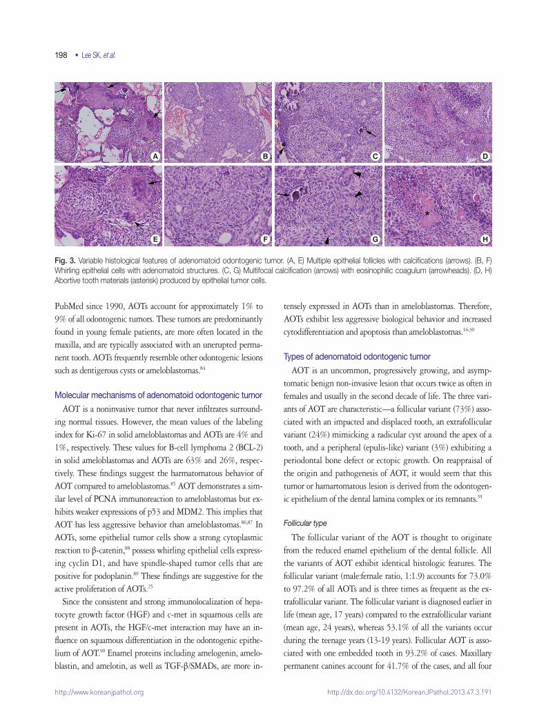
Among 15 cases of AOT, the anterior maxilla was the most common site (66.6%), and radiographically, most cases showed a unilocular radiolucency with well-defined borders (57.1%). Histologically, most cases exhibited a predominantly solid grow-th pattern (46.7%) or a similar proportion of solid and cribri-form patterns (46.7%) (Fig. 3A, B, E, F). Eosinophilic amor-phous material (“tumor droplets”) was found in all cases (100%) (Fig. 3C, D, G, H). Most tumors contained duct-like spaces (93.3%) (Fig. 3B, C, F, G) and convoluted structures (60.0%), whereas a minor proportion of the cases presented with calcify-ing epithelial odontogenic tumor-like areas (26.7%). Variable amounts of calcified materials were found in most AOTs (80.0 %) (Fig. 3A, D, E, H), whereas osteodentin and perivascular hyalinization were seen only rarely (6.7% for each). About five (33.3%) cases had areas mimicking a dentigerous cyst and were mostly diagnosed in females (80.0%).81
AOT has numerous dispersed or clustered radiopaque foci compared to the presence of calcification in calcifying cystic odontogenic tumors showing a thin radiopaque line and dis-crete radiopaque foci. Therefore, radiolucency with numerous radiopaque foci (particularly when the radiolucency surrounds a portion of the root or entire tooth) is suggestive of an AOT ra-ther than a calcifying cystic odontogenic tumor.82
In a review of 272 AOTs with special emphasis on radiologi-cal features, the patient age at time of diagnosis ranged from 3 years to 82 years (mean, 18.4 years). The maxilla-to-mandible ratio was 1.7:1. Small opacities were present in 77% of the le-sions and were associated with expansion of the cortex. The sig-nificant radiological features in patients aged 30 years and above were root resorption and lesions crossing the midline.83
According to all reports regarding AOTs and cited in
http://www.koreanjpathol.org http://dx.doi.org/10.4132/KoreanJPathol.2013.47.3.191
198 • Lee SK, et al.
PubMed since 1990, AOTs account for approximately 1% to 9% of all odontogenic tumors. These tumors are predominantly found in young female patients, are more often located in the maxilla, and are typically associated with an unerupted perma-nent tooth. AOTs frequently resemble other odontogenic lesions such as dentigerous cysts or ameloblastomas.84
Molecular mechanisms of adenomatoid odontogenic tumor
AOT is a noninvasive tumor that never infiltrates surround-ing normal tissues. However, the mean values of the labeling index for Ki-67 in solid ameloblastomas and AOTs are 4% and 1%, respectively. These values for B-cell lymphoma 2 (BCL-2) in solid ameloblastomas and AOTs are 63% and 26%, respec-tively. These findings suggest the harmatomatous behavior of AOT compared to ameloblastomas.85 AOT demonstrates a sim-ilar level of PCNA immunoreaction to ameloblastomas but ex-hibits weaker expressions of p53 and MDM2. This implies that AOT has less aggressive behavior than ameloblastomas.86,87 In AOTs, some epithelial tumor cells show a strong cytoplasmic reaction to β-catenin,88 possess whirling epithelial cells express-ing cyclin D1, and have spindle-shaped tumor cells that are positive for podoplanin.89 These findings are suggestive for the active proliferation of AOTs.25
Since the consistent and strong immunolocalization of hepa-tocyte growth factor (HGF) and c-met in squamous cells are present in AOTs, the HGF/c-met interaction may have an in-fluence on squamous differentiation in the odontogenic epithe-lium of AOT.90 Enamel proteins including amelogenin, amelo-blastin, and amelotin, as well as TGF-β/SMADs, are more in-
tensely expressed in AOTs than in ameloblastomas. Therefore, AOTs exhibit less aggressive biological behavior and increased cytodifferentiation and apoptosis than ameloblastomas.16,30
Types of adenomatoid odontogenic tumor
AOT is an uncommon, progressively growing, and asymp-tomatic benign non-invasive lesion that occurs twice as often in females and usually in the second decade of life. The three vari-ants of AOT are characteristic—a follicular variant (73%) asso-ciated with an impacted and displaced tooth, an extrafollicular variant (24%) mimicking a radicular cyst around the apex of a tooth, and a peripheral (epulis-like) variant (3%) exhibiting a periodontal bone defect or ectopic growth. On reappraisal of the origin and pathogenesis of AOT, it would seem that this tumor or hamartomatous lesion is derived from the odontogen-ic epithelium of the dental lamina complex or its remnants.91
Follicular type
The follicular variant of the AOT is thought to originate from the reduced enamel epithelium of the dental follicle. All the variants of AOT exhibit identical histologic features. The follicular variant (male:female ratio, 1:1.9) accounts for 73.0% to 97.2% of all AOTs and is three times as frequent as the ex-trafollicular variant. The follicular variant is diagnosed earlier in life (mean age, 17 years) compared to the extrafollicular variant (mean age, 24 years), whereas 53.1% of all the variants occur during the teenage years (13-19 years). Follicular AOT is asso-ciated with one embedded tooth in 93.2% of cases. Maxillary permanent canines account for 41.7% of the cases, and all four
A
E
B
F
C
G
D
H
Fig. 3. Variable histological features of adenomatoid odontogenic tumor. (A, E) Multiple epithelial follicles with calcifications (arrows). (B, F) Whirling epithelial cells with adenomatoid structures. (C, G) Multifocal calcification (arrows) with eosinophilic coagulum (arrowheads). (D, H) Abortive tooth materials (asterisk) produced by epithelial tumor cells.
http://www.koreanjpathol.orghttp://dx.doi.org/10.4132/KoreanJPathol.2013.47.3.191
Ameloblastoma and Adenomatoid Odontogenic Tumor • 199
canines are involved in 60.1% of AOT-associated cases.92
Extrafollicular type
Compared to the follicular variant of AOT, the origin of the extrafollicular variant remains unclear. However, the available reviews in the literature suggest that some extrafollicular AOTs may arise as a secondary phenomenon within pre-existing odon-togenic cysts or cystic tumors. For example, the tumor may originate from the epithelial lining of an odontogenic cyst or unicystic ameloblastoma.93,94 A rare subvariant of the extrafol-licular type of AOT may radiographically mimic periapical dis-eases, which is initially suspicious of small periapical pathology. This subvariant is very rare, with only 12 cases reported in the literature.95
Peripheral type
Peripheral AOT occurring far distant from tooth germ struc-tures is rarely encountered. So far, there have been only 14 re-ported cases of peripheral AOTs. A marked female predomi-nance was apparent in peripheral AOTs, and approximately 90% of the peripheral AOTs occurred in the maxilla,96 showing a striking tendency to occur in the anterior maxilla. The tumor primarily manifested in the incisor and can involve the maxil-lary antrum.97
SUMMARY
Ameloblastomas and AOTs are benign epithelial tumors of odontogenic origin. The former demonstrates an aggressive be-havior and frequent recurrence, while the latter exhibits limited growth and no tendency of recurrence. The differential diagno-sis between ameloblastomas and AOTs is essential. Due to the local invasive growth and the possible potential for the malig-nant transformation of ameloblastomas, it is recommended by WHO that ameloblastomas be classified into solid/multicystic, unicystic, desmoplastic, and peripheral ameloblastomas accord-ing to their clinicopathological features rather than the previous histological types including follicular, plexiform, acanthoma-tous, granular types, etc. AOTs are also classified into follicular, extrafollicular, and peripheral types, even though all types ex-hibit similar histological features. With advance in the elucida-tion of molecular signaling mechanisms in cells, the cytodiffer-entiation of epithelial tumor cells in ameloblastomas and AOTs can be identified using different biomarkers. Therefore, it is suggested that comprehensive pathological observation includ-ing molecular genetic information can provide a more reliable
differential diagnosis for the propagation and prognosis of ame-loblastomas and AOTs.
Conflicts of InterestNo potential conflict of interest relevant to this article was
reported.
REFERENCES
1. Sarkar L, Cobourne M, Naylor S, Smalley M, Dale T, Sharpe PT. Wnt/Shh interactions regulate ectodermal boundary formation during mammalian tooth development. Proc Natl Acad Sci U S A 2000; 97: 4520-4.
2.ThesleffI,VaahtokariA,KettunenP,AbergT.Epithelial-mesenchy-mal signaling during tooth development. Connect Tissue Res 1995; 32: 9-15.
3.KimJY,KangGH,ChiJG.Adamantinomaoftibiawithpredomi-nantfeaturesoffibrousdysplasia:acasereport.JKoreanMedSci1996; 11: 444-8.
4.KatoN,EndoY,TamuraG,MotoyamaT.Ameloblastomawithbas-alcellcarcinoma-likefeatureemergingasanasalpolyp.PatholInt1999; 49: 747-51.
5.ReichartPA,PhilipsenHP,SonnerS.Ameloblastoma:biologicalprofileof3677cases.EurJCancerBOralOncol1995;31B:86-99.
6.SiarCH,LauSH,NgKH.Ameloblastomaofthejaws:aretrospec-tiveanalysisof340casesinaMalaysianpopulation.JOralMaxillo-fac Surg 2012; 70: 608-15.
7.DhanuthaiK,ChantarangsuS,RojanawatsirivejS,et al. Ameloblas-toma:amulticentricstudy.OralSurgOralMedOralPatholOralRadiol 2012; 113: 782-8.
8.HongJ,YunPY,ChungIH,et al.Long-termfollowuponrecurrenceof305ameloblastomacases.IntJOralMaxillofacSurg2007;36:283-8.
9.DoscherJC,KramerJM,FantasiaJE.Alargecalcifyinglesionofthemaxillainachild.JAmDentAssoc2011;142:1026-30.
10.LimJ,AhnH,MinS,HongSD,LeeJI,HongSP.Oligonucleotidemicroarrayanalysisofameloblastomacomparedwithdentigerouscyst.JOralPatholMed2006;35:278-85.
11.SneadML,LuoW,HsuDD,MelroseRJ,LauEC,StenmanG.Hu-manameloblastomatumorsexpresstheamelogeningene.OralSurgOralMedOralPathol1992;74:64-72.
12.DeutschD,FermonE,LustmannJ,et al.TuftelinmRNAisexpressedin a human ameloblastoma tumor. Connect Tissue Res 1998; 39: 177-84.
13.LeeSK,KimSM,LeeYJ,YamadaKM,YamadaY,ChiJG.Thestruc-tureoftheratameloblastingeneanditsexpressioninamelogene-sis. Mol Cells 2003; 15: 216-25.
http://www.koreanjpathol.org http://dx.doi.org/10.4132/KoreanJPathol.2013.47.3.191
200 • Lee SK, et al.
14.LeeSK,KrebsbachPH,MatsukiY,NanciA,YamadaKM,YamadaY.Ameloblastinexpressioninratincisorsandhumantoothgerms.IntJDevBiol1996;40:1141-50.
15.SakuT,OkabeH,ShimokawaH.Immunohistochemicaldemon-strationofenamelproteinsinodontogenictumors.JOralPatholMed 1992; 21: 113-9.
16.CriveliniMM,FelipiniRC,MiyaharaGI,deSousaSC.Expressionof odontogenic ameloblast-associated protein, amelotin, ameloblas-tin, and amelogenin in odontogenic tumors: immunohistochemical analysisandpathogeneticconsiderations.JOralPatholMed2012;41: 272-80.
17.Al-OtaibiO,KhounganianR,AnilS,RajendranR.Syndecan-1(CD138)surfaceexpressionmarkscelltypeanddifferentiationiname-loblastoma, keratocystic odontogenic tumor, and dentigerous cyst. JOralPatholMed2013;42:186-93.
18.MishraM,NaikVV,KaleAD,AnkolaAV,PilliGS.Perlecan(base-ment membrane heparan sulfate proteoglycan) and its role in oral malignancies:anoverview.IndianJDentRes2011;22:823-6.
19.MasloubSM,Abdel-AzimAM,ElhamidES.CD10andosteopontinexpressionindentigerouscystandameloblastoma.DiagnPathol2011; 6: 44.
20.deMedeirosAM,NonakaCF,GalvãoHC,deSouzaLB,FreitasRdeA.Expressionofextracellularmatrixproteinsinameloblastomasandadenomatoidodontogenictumors.EurArchOtorhinolaryngol2010; 267: 303-10.
21.FariasLC,GomesCC,RodriguesMC,et al.Epigeneticregulationofmatrixmetalloproteinaseexpressioninameloblastoma.BMCClinPathol 2012; 12: 11.
22.QianY,HuangHZ.TheroleofRANKLandMMP-9inthebonere-sorptioncausedbyameloblastoma.JOralPatholMed2010;39:592-8.
23.TekkesinMS,MutluS,OlgacV.TheroleofRANK/RANKL/OPGsignallingpathwaysinosteoclastogenesisinodontogenickerato-cysts,radicularcysts,andameloblastomas.HeadNeckPathol2011;5: 248-53.
24.SengüvenB,OygürT.Investigationofinterleukin-1alphaandin-terleukin-6expressionandinterleukin-1alphagenepolymorphisminkeratocysticodontogenictumorsandameloblastomas.MedOralPatolOralCirBucal2011;16:e467-72.
25.KumarH,VandanaR,KumarG.Immunohistochemicalexpressionof cyclin D1 in ameloblastomas and adenomatoid odontogenic tu-mors.JOralMaxillofacPathol2011;15:283-7.
26.ScheperMA,DuarteEC,IntapaC,et al.Expressionofmidkineinameloblastomasanditscorrelationwithclinicopathologicparame-ters.OralSurgOralMedOralPatholOralRadiol2012;114:497-502.
27.Abdel-AzizA,AminMM.EGFR,CD10andproliferationmarkerKi67expressioninameloblastoma:possibleroleinlocalrecurrence.
Diagn Pathol 2012; 7: 14.28.Sharifi-SistaniN,ZartabH,BabakoohiS,et al.Immunohistochemi-calcomparisonoftheexpressionofp53andMDM2proteinsinam-eloblastomasandkeratocysticodontogenictumors.JCraniofacSurg2011; 22: 1652-6.
29.LiN,ZhongM,SongM.ExpressionofphosphorylatedmTORandits regulatory protein is related to biological behaviors of amelo-blastoma.IntJClinExpPathol2012;5:660-7.
30.KarathanasiV,TosiosKI,NikitakisNG,et al.TGF-beta1,Smad-2/-3,Smad-1/-5/-8,andSmad-4signalingfactorsareexpressedinamelo-blastomas, adenomatoid odontogenic tumors, and calcifying cystic odontogenictumors:animmunohistochemicalstudy.JOralPatholMed 2013; 42: 415-23.
31.SukarawanW,SimmonsD,SuggsC,LongK,WrightJT.WNT5Aexpressioninameloblastomaanditsrolesinregulatingenamelepi-theliumtumorigenicbehaviors.AmJPathol2010;176:461-71.
32.SathiGA,TsujigiwaH,ItoS,et al.OsteogenicgenesrelatedtothecanonicWNTpathwayaredown-regulatedinameloblastoma.OralSurgOralMedOralPatholOralRadiol2012;114:771-7.
33.PontiG,PastorinoL,PollioA,et al. Ameloblastoma: a neglected cri-terionfornevoidbasalcellcarcinoma(Gorlin)syndrome.FamCan-cer 2012; 11: 411-8.
34.ParkHR,HanYI,SolMY,LeeSK.Nevoidbasalcellcarcinomasyn-drome:reportofacase.KoreanJPathol1995;29:263-7.
35.SongKY,ChoiYH,KimMK,LeeKK,HamEK.Pathologicalanaly-sisofthebasalcellcarcinoma.KoreanJPathol1994;28:160-7.
36.AlvesPereiraKM,doAmaralBA,dosSantosBR,GalvãoHC,Freit-asRdeA,deSouzaLB.ImmunohistochemicalexpressionofE-cad-herinandbeta-catenininameloblastomasandtoothgerms.OralSurgOralMedOralPatholOralRadiolEndod2010;109:425-31.
37.ModoloF,BizMT,deSousaSM,FachinelliRdeL,CremaVO.Im-munohistochemicalexpressionofRhoGTPasesinameloblastomas.JOralPatholMed2012;41:400-7.
38.KumamotoH,KimiK,OoyaK.Immunohistochemicalanalysisofapoptosis-relatedfactors(Fas,Fasligand,caspase-3andsingle-stran-dedDNA)inameloblastomas.JOralPatholMed2001;30:596-602.
39.RizzardiC,LeocataP,VenturaL,et al. Apoptosis-related factors (TRAIL,DR4,DR5,DcR1,DcR2,apoptoticcells)andproliferativeactivity in ameloblastomas. Anticancer Res 2009; 29: 1137-42.
40.ShahidiS,BronooshP,DaneshbodY.Follicularameloblastomapre-sentingasasinonasaltumor.IranRedCrescentMedJ2012;14:113-6.
41.Castro-SilvaII,IsraelMS,LimaGS,deQueirozChavesLourençoS.Difficultiesinthediagnosisofplexiformameloblastoma.OralMax-illofac Surg 2012; 16: 115-8.
42.BansalM,ChaturvediTP,BansalR,KumarM.Acanthomatousam-eloblastomaofanteriormaxilla.JIndianSocPedodPrevDent2010;
http://www.koreanjpathol.orghttp://dx.doi.org/10.4132/KoreanJPathol.2013.47.3.191
Ameloblastoma and Adenomatoid Odontogenic Tumor • 201
28: 209-11.43.AhujaA,MathurS,IyerVK.Acanthomatousameloblastomamas-
querading as a squamous cell carcinoma. Cytopathology 2012 Apr 25[Epub].http://dx.doi.org/10.1111/j.1365-2303.2012.00978.x.
44.GuptaS,GrewalH,SahK.Granularcellameloblastomashowingdesmoplasia. Ann Saudi Med 2012; 32: 537-40.
45.BansalA,BhatnagarA,SaxenaS.Metastasizinggranularcellame-loblastoma.JOralMaxillofacPathol2012;16:122-4.
46.SharmaVK,VermaSK,GoyalL,ChaudharyPK.Hemangiomatousameloblastomainmaxilla:areportofaveryrarecase.DentResJ(Isfahan)2012;9:345-9.
47.IdeF,ShimoyamaT,HorieN.Basaloidsquamouscellcarcinomaversusbasalcellameloblastoma.OralOncol1998;34:154-5.
48.SaxenaK,JoseM,ChatraLK,SequieraJ.Adenoidameloblastomawithdentinoid.JOralMaxillofacPathol2012;16:272-6.
49.TateyamaH,TadaT,OkabeM,TakahashiE,EimotoT.Differentkeratinprofilesincraniopharyngiomasubtypesandameloblasto-mas. Pathol Res Pract 2001; 197: 735-42.
50.KimTJ,LeeYS,KimBK,LeeKY.Ameloblastomaassociatedwithdentinogenicghostcelltumor:acasereport.KoreanJPathol2006;40: 297-302.
51.ChaudharyZ,SangwanV,PalUS,SharmaP.Unicysticameloblas-toma:adiagnosticdilemma.NatlJMaxillofacSurg2011;2:89-92.
52.GuptaN,SaxenaS,RathodVC,AggarwalP.Unicysticameloblas-tomaofthemandible.JOralMaxillofacPathol2011;15:228-31.
53.DeMeloWM,Pereira-SantosD,SonodaCK,Pereira-FreitasSA,deMouraWL,dePauloCravinhosJC.Largeunicysticameloblastomaofthemandible:managementguidedbybiologicalbehavior.JCra-niofac Surg 2012; 23: e499-502.
54.KimJ,ChoiIJ.Ameloblastomaarisinginodontogeniccysts:reportof5casesanditshistologiccharacteristics.KoreanJPathol1986;20:435-41.
55.CervelliD,MarianettiTM,BonielloR,et al.Giantneglecteddesmo-plasticameloblastoma:reconstructionwithfreefibulaflap.JCra-niofac Surg 2012; 23: e171-4.
56.EffiomOA,OdukoyaO.Desmoplasticameloblastoma:analysisof17Nigeriancases.OralSurgOralMedOralPatholOralRadiolEn-dod 2011; 111: e27-31.
57.Bologna-MolinaR,Mosqueda-TaylorA,Molina-FrecheroN,Mori-EstevezAD,Sánchez-AcuñaG.ComparisonofthevalueofPCNAandKi-67asmarkersofcellproliferationinameloblastictumors.MedOralPatolOralCirBucal2013;18:e174-9.
58.KatoH,OtaY,SasakiM,et al.Peripheralameloblastomaofthelow-er molar gingiva: a case report and immunohistochemical study. TokaiJExpClinMed2012;37:30-4.
59.IdeF,MishimaK,MiyazakiY,SaitoI,KusamaK.Peripheralamelo-
blastomain-situ:anevidentialfactofsurfaceepitheliumorigin.OralSurgOralMedOralPatholOralRadiolEndod2009;108:763-7.
60.BeenaVT,ChoudharyK,HeeraR,RajeevR,SivakumarR,Vidhya-dharanK.Peripheralameloblastoma:acasereportandreviewofliterature. Case Rep Dent 2012; 2012: 571509.
61.PaulusW,StöckelC,KraussJ,SörensenN,RoggendorfW.Odonto-genic classification of craniopharyngiomas: a clinicopathological studyof54cases.Histopathology1997;30:172-6.
62.GiangasperoF,BurgerPC,OsborneDR,SteinRB.Suprasellarpap-illarysquamousepithelioma(“papillarycraniopharyngioma”).AmJSurgPathol1984;8:57-64.
63. Sekine S, Takata T, Shibata T, et al.ExpressionofenamelproteinsandLEF1inadamantinomatouscraniopharyngioma:evidenceforitsodontogenicepithelialdifferentiation.Histopathology2004;45:573-9.
64.BadgerKV,GardnerDG.Therelationshipofadamantinomatouscraniopharyngiomatoghostcellameloblastomaofthejaws:ahis-topathologicandimmunohistochemicalstudy.JOralPatholMed1997; 26: 349-55.
65.ZoicasF,SchöflC.Craniopharyngiomainadults.FrontEndocrinol(Lausanne)2012;3:46.
66.RodriguezFJ,ScheithauerBW,TsunodaS,KovacsK,VidalS,Piep-grasDG.Thespectrumofmalignancyincraniopharyngioma.AmJSurg Pathol 2007; 31: 1020-8.
67.HorváthA,HorváthE,PopşorS.Mandibularameloblasticcarcino-mainayoungpatient.RomJMorpholEmbryol2012;53:179-83.
68.CasarotoAR,ToledoGL,FilhoJL,SoaresCT,CapelariMM,LaraVS.Ameloblasticcarcinoma,primarytype:casereport,immuno-histochemicalanalysisandliteraturereview.AnticancerRes2012;32: 1515-25.
69.BediRS,ChughA,PasrichaN.Ameloblasticcarcinomaofmaxilla.NatlJMaxillofacSurg2012;3:70-4.
70.YoonHJ,HongSP,LeeJI,LeeSS,HongSD.Ameloblasticcarcino-ma:ananalysisof6caseswithreviewoftheliterature.OralSurgOralMedOralPatholOralRadiolEndod2009;108:904-13.
71.PundirS,SaxenaS,RathodV,AggrawalP.Ameloblasticcarcinoma:secondarydedifferentiatedcarcinomaofthemandible:reportofarareentitywithabriefreview.JOralMaxillofacPathol2011;15:201-4.
72.FujitaS,AnamiM,SatohN,et al. Cytopathologic features of secon-dary peripheral ameloblastic carcinoma: a case report. Diagn Cyto-pathol 2011; 39: 354-8.
73.KawauchiS,HayatsuY,TakahashiM,et al. Spindle-cell ameloblas-ticcarcinoma:acasereportwithimmunohistochemical,ultrastruc-tural,andcomparativegenomichybridizationanalyses.OncolRep2003; 10: 31-4.
http://www.koreanjpathol.org http://dx.doi.org/10.4132/KoreanJPathol.2013.47.3.191
202 • Lee SK, et al.
74.KhojastehA,KhodayariA,RahimiF,et al.Hypermethylationofp16 tumor-suppressor gene in ameloblastic carcinoma, ameloblas-toma,anddentalfollicles.JOralMaxillofacSurg2013;71:62-5.
75.SlootwegPJ,MüllerH.Malignantameloblastomaorameloblasticcarcinoma.OralSurgOralMedOralPathol1984;57:168-76.
76.DissanayakeRK,JayasooriyaPR,SiriwardenaDJ,TilakaratneWM.Reviewofmetastasizing(malignant)ameloblastoma(METAM):patternofmetastasisandtreatment.OralSurgOralMedOralPatholOralRadiolEndod2011;111:734-41.
77.LaiH,WangJ.Benignmetastasizingameloblastomaormalignantameloblastoma?JCraniofacSurg2011;22:995-7.
78.BaskaranP,MisraS,KumarMS,MithraR.Adenomatoidodonto-genictumor:areportoftwocaseswithhistopathologycorrelation.JClinImagingSci2011;1:64.
79.VasudevanK,KumarS,Vijayasamundeeswari,VigneswariS.Ade-nomatoid odontogenic tumor, an uncommon tumor. Contemp Clin Dent 2012; 3: 245-7.
80.SharmaN,PassiS,KumarVV.Adenomatoidodontogenictumor:as an unusual mandibular manifestation. Contemp Clin Dent 2012; 3(Suppl1):S29-32.
81.deMatosFR,NonakaCF,PintoLP,deSouzaLB,deAlmeidaFreit-as R. Adenomatoid odontogenic tumor: retrospective study of 15 caseswithemphasisonhistopathologicfeatures.HeadNeckPathol2012; 6: 430-7.
82.ChindasombatjaroenJ,PoomsawatS,KakimotoN,ShimamotoH.Calcifying cystic odontogenic tumor and adenomatoid odontogen-ictumor:radiographicevaluation.OralSurgOralMedOralPatholOralRadiol2012;114:796-803.
83.BeckerT,BuchnerA,KaffeI.Criticalevaluationoftheradiologicaland clinical features of adenomatoid odontogenic tumour. Dento-maxillofacRadiol2012;41:533-40.
84.HandschelJG,DepprichRA,ZimmermannAC,BraunsteinS,Kü-blerNR.Adenomatoidodontogenictumorofthemandible:reviewoftheliteratureandreportofararecase.HeadFaceMed2005;1:3.
85.RazaviSM,TabatabaieSH,HoseiniAT,HoseiniET,KhabazianA.AcomparativeimmunohistochemicalstudyofKi-67andBcl-2expres-sion in solid ameloblastoma and adenomatoid odontogenic tumor. DentResJ(Isfahan)2012;9:192-7.
86.SalehinejadJ,Zare-MahmoodabadiR,SaghafiS,et al.Immunohis-
tochemical detection of p53 and PCNA in ameloblastoma and ade-nomatoidodontogenictumor.JOralSci2011;53:213-7.
87.KrishnaA,KaveriH,NaveenKumarRK,KumaraswamyKL,Shy-lajaS,MurthyS.OverexpressionofMDM2proteininameloblasto-masascomparedtoadenomatoidodontogenictumor.JCancerResTher 2012; 8: 232-7.
88.HarnetJC,PedeutourF,RaybaudH,AmbrosettiD,FabasT,Lom-bardiT.Immunohistologicalfeaturesinadenomatoidodontogenictumor:reviewoftheliteratureandfirstexpressionandmutationalanalysisofbeta-catenininthisunusuallesionofthejaws.JOralMaxillofacSurg2013;71:706-13.
89.TsunekiM,MaruyamaS,YamazakiM,ChengJ,SakuT.Podoplaninexpressionprofilescharacteristicofodontogenictumor-specifictis-sue architectures. Pathol Res Pract 2012; 208: 140-6.
90.PoomsawatS,PunyasinghJ,VejchapipatP,LarbcharoensubN.Co-expressionofhepatocytegrowthfactorandc-metinepithelialodon-togenictumors.ActaHistochem2012;114:400-5.
91.PhilipsenHP,SammanN,OrmistonIW,WuPC,ReichartPA.Vari-antsoftheadenomatoidodontogenictumorwithanoteontumororigin.JOralPatholMed1992;21:348-52.
92.PhilipsenHP,ReichartPA,ZhangKH,NikaiH,YuQX.Adenoma-toidodontogenictumor:biologicprofilebasedon499cases.JOralPathol Med 1991; 20: 149-58.
93.JivanV,AltiniM,MeerS,MahomedF.Adenomatoidodontogenictumor(AOT)originatinginaunicysticameloblastoma:acasere-port.HeadNeckPathol2007;1:146-9.
94.PrasadG,NairP,ThomasS,GharoteH,SinghN,BhambalA.Ex-trafollicularadenomatoidodontogenictumour.BMJCaseRep2011;2011.http://dx.doi.org/10.1136/bcr.03.2011.3963.
95.AvinashTejasviML,PrashanthShenaiK,ChatraL.Atypicalcaseofperiapicaladenomatoidodontogenictumour.JMaxillofacOralSurg2010; 9: 99-101.
96.IdeF,MishimaK,SaitoI,KusamaK.Rareperipheralodontogenictumors:reportof5casesandcomprehensivereviewofthelitera-ture.OralSurgOralMedOralPatholOralRadiolEndod2008;106:e22-8.
97.SandhuSV,NarangRS,JawandaM,RaiS.Adenomatoidodonto-genictumorassociatedwithdentigerouscystofthemaxillaryan-trum:arareentity.JOralMaxillofacPathol2010;14:24-8.