Embed Size (px)
Citation preview
Current ConCepts in the ManageMent of tuberCulosis
Mayo Clin Proc. • April 2011;86(4):348-361 • doi:10.4065/mcp.2010.0820 • www.mayoclinicproceedings.com348
For personal use. Mass reproduce only with permission from Mayo Clinic Proceedingsa .
syMposiuM on antiMiCrobial therapy
From the Division of Infectious Diseases (I.G.S.) and Division of Primary Care Internal Medicine (M.L.W.), Mayo Clinic, Rochester, MN.
Address correspondence to Irene G. Sia, MD, MSc, Division of Infectious Diseases, 200 First St SW, Rochester, MN 55905 ([email protected]). In-dividual reprints of this article and a bound reprint of the entire Symposium on Antimicrobial Therapy will be available for purchase from our Web site www.mayoclinicproceedings.com.
© 2011 Mayo Foundation for Medical Education and Research
Irene G. Sia, MD, MSc, and Mark L. Wieland, MD, MPH
Current Concepts in the Management of Tuberculosis
Effective medical therapy for tuberculosis (TB) has existed for more than half a century, yet TB remains
among the most pressing public health issues of our day. Tuberculosis is, in part, a disease of poverty.1 The fact that it remains the eighth leading cause of death in the world speaks to the challenges facing practitioners and public health officials as they try to control a disease that is so en-twined in the cultural and economic fabric of society. Chal-lenges to effective solutions include lack of access to diag-nosis and treatment, the frequent coexistence of epidemics of TB and human immunodeficiency virus (HIV), and the
On completion of this article, readers should be able to (1) identify patients at high risk of tuberculosis; (2) accurately and efficiently diagnose active tuberculosis on the basis of clinical presentation, laboratory results, and microbiological tests; and (3) prescribe first-line therapy for latent and active uncomplicated pulmonary tuberculosis.
Tuberculosis (TB) poses a serious threat to public health through-out the world but disproportionately afflicts low-income nations. Persons in close contact with a patient with active pulmonary TB and those from endemic regions of the world are at highest risk of primary infection, whereas patients with compromised immune systems are at highest risk of reactivation of latent TB infection (LTBI). Tuberculosis can affect any organ system. Clinical mani-festations vary accordingly but often include fever, night sweats, and weight loss. Positive results on either a tuberculin skin test or an interferon-γ release assay in the absence of active TB establish a diagnosis of LTBI. A combination of epidemiological, clinical, ra-diographic, microbiological, and histopathologic features is used to establish the diagnosis of active TB. Patients with suspected active pulmonary TB should submit 3 sputum specimens for acid-fast bacilli smears and culture, with nucleic acid amplification testing performed on at least 1 specimen. For patients with LTBI, treatment with isoniazid for 9 months is preferred. Patients with active TB should be treated with multiple agents to achieve bac-terial clearance, to reduce the risk of transmission, and to prevent the emergence of drug resistance. Directly observed therapy is recommended for the treatment of active TB. Health care profes-sionals should collaborate, when possible, with local and state public health departments to care for patients with TB. Patients with drug-resistant TB or coinfection with human immunodeficien-cy virus should be treated in collaboration with TB specialists. Public health measures to prevent the spread of TB include appro-priate respiratory isolation of patients with active pulmonary TB, contact investigation, and reduction of the LTBI burden.
Mayo Clin Proc. 2011;86(4):348-361
AFB = acid-fast bacilli; BCG = bacille Calmette-Guérin; CFP-10 = cul-ture filtrate protein 10; DOT = directly observed therapy; EMB = ethambutol; ESAT-6 = early-secreted antigenic target 6; HIV = human immuno deficiency virus; IFN-γ = interferon-γ; IGRA = IFN-γ release assay; INH = isoniazid; LTBI = latent TBI; MDR-TB = multidrug-resistant TB; NAA = nucleic acid amplification; PZA = pyrazinamide; RIF = rifampin; TB = tuberculosis; TBI = TB infection; TST = tuberculin skin test; XDR-TB = extensively drug-resistant TB
increasing prevalence of multidrug-resistant TB (MDR-TB).2 Although a chasm in disease burden exists between resource-rich and poor regions, an increasingly mobile and connected global community has ensured that TB remains highly relevant to practitioners throughout the world. This review highlights key principles in the management of TB. For purposes of definition, TB infection (TBI) occurs when a susceptible person inhales droplets containing Mycobac-terium tuberculosis nuclei that travel through the respirato-ry tract to the alveoli. In most patients, an immune response limits propagation of TBI, resulting in an asymptomatic, noninfectious, localized infection that may remain in the body for many years. These patients have positive immuno-logic test results for M tuberculosis and carry a diagnosis of latent TBI (LTBI). A constellation of clinical, radiographic, microbiological, and histopathologic hallmarks are used to diagnose active TB disease.3
EPIDEMIOLOGY
An estimated one-third of the world’s population is infect-ed with TB.4 Tuberculosis accounted for 1.3 million deaths in 2007, and the prevalence of active disease is estimated at 13.7 million (206 per 100,000 persons).5 Incident cases of active TB are highest (≥100 cases per 100,000 persons) in sub-Saharan Africa, India and Central Asia, parts of East-ern Europe, Southeast Asia, and Micronesia. Intermediate incidence rates (26-100 per 100,000 persons) are observed in Central and South America, China, and northern Africa. Low rates (<25 per 100,000 persons) occur in the United States, Canada, Australia, Western Europe, and Japan.5,6
While absolute numbers have been on the rise, the prevalence of TB in relationship to population has trend-ed downward during the past 15 years, and global public

Current ConCepts in the ManageMent of tuberCulosis
Mayo Clin Proc. • April 2011;86(4):348-361 • doi:10.4065/mcp.2010.0820 • www.mayoclinicproceedings.com 349
For personal use. Mass reproduce only with permission from Mayo Clinic Proceedingsa .
health efforts have averted an estimated 6 million deaths during this time.2 Nevertheless, the emergence of drug re-sistance coupled with the persistence of HIV and global poverty have thwarted a more substantive break in the TB epidemic. Indeed, 0.5 million cases of MDR-TB, in which the infecting organism is resistant to at least isoni-azid (INH) and rifampin (RIF), were reported in 2007, and 55 countries reported at least 1 case of extensively drug-resistant TB (XDR-TB),5 in which the organism is resistant to at least INH, RIF, fluoroquinolones, and either amino-glycosides or capreomycin, or both. The magnitude of the problem is particularly overwhelming in parts of the Rus-sian Federation and Central Asia, where the proportion of MDR-TB among incident TB cases ranged from 12% to 28% between 1994 and 2009, compared with 0% to 3% in the United States.7
As in the rest of the world, the incidence of TBI in the United States has declined during the past decade, but this decline has been much less pronounced among foreign-born Americans. More than half of active TB cases in the United States currently occur in foreign-born individuals,5,8 and most cases result from reactivation of LTBI.9,10 The ef-fect of global migration on TB has been seen throughout the developed world, most dramatically in London, where cases of active TB increased by 50% between 1999 and 2009, mostly among foreign-born individuals.11
Sociodemographic risk factors for TBI include recent residence in an endemic region of the world, low socioeco-nomic position, being a member of a racial or ethnic minor-ity (in the United States), homelessness, residency or em-ployment at high-risk facilities (eg, correctional facilities, homeless shelters, skilled nursing facilities), and employ-ment as a health care worker caring for patients with TB.
TRANSMISSION
Tuberculosis is transmitted through droplet aerosolization by an individual with active pulmonary disease. The high-est risk of transmission occurs among patients with cavi-tary or positive acid-fast bacilli (AFB) smears12; however, patients with negative smears but positive cultures may still transmit the disease.13
Host factors dramatically influence which of those ex-posed to TB are most likely to contract primary disease or progress to active disease. Those with greater susceptibility include persons with immune systems that have been com-promised through either diseases, such as HIV infection and hematologic and reticuloendothelial malignancies, or through immunosuppressive medications, such as corti-costeroids, tumor necrosis factor α inhibitors, calcineurin inhibitors, and cytotoxic chemotherapeutic agents. Fur-thermore, patients with chronic diseases, such as diabetes,
chronic kidney disease, and silicosis, are at elevated risk. Fi-nally, age younger than 4 years, long-term malnutrition, and substance abuse are independent risk factors for disease.3
CLINICAL MANIFESTATIONS
Pulmonary TB Primary Pulmonary TB. Symptoms occurring around the time of inoculation are referred to as primary pulmo-nary TB. Symptoms are generally mild and include low-grade fever.14,15 Two-thirds of persons with primary pulmo-nary TB remain asymptomatic. Physical examination find-ings are generally unremarkable, and the most common radiographic finding is hilar adenopathy.16 Less common radiographic findings include pulmonary infiltrates in the mid and lower lung field. Reactivation TB. Approximately 90% of TB cases among adults can be attributed to reactivation TB. Symp-toms present insidiously, most commonly with fever, cough, weight loss, fatigue, and night sweats. Less com-mon symptoms include chest pain, dyspnea, and hemopty-sis. Physical examination findings are nonspecific and may include rales or signs of pleural effusion (eg, dullness to percussion). Chest radiography demonstrates infiltrates in the apical-posterior segment of the upper lobes, and up to 20% of these infiltrates are associated with cavities char-acterized by air-fluid levels. Although not specific for TB, apical computed tomographic findings may show a “tree in bud” morphology manifested by centrilobular lesions, nodules, and branching linear densities.17,18 Among the roughly 15% of patients who present without upper lung field infiltrates, a variety of radiographic findings have been described, including lower lung infiltrates (especially superior segments), nodules, effusions, and hilar adenopa-thy. Finally, up to 5% of patients with active pulmonary disease may have normal findings on chest radiography.19,20 This is particularly worth noting among patients coinfected with HIV, who are more likely to have atypical (eg, less predisposition for upper lobes) or normal findings on chest radiography.21
Endobronchial TB. Endobronchial TB develops as the direct extension of TB from a pulmonary parenchy-mal source or sputum inoculation into the bronchial tree.22 Symptoms may include barking cough with sputum pro-duction, and examination may reveal rhonchi and wheez-ing23,24; the wheezing may lead to misdiagnosis of asthma.25 Diagnosis and response to therapy may be assessed through bronchoscopy.26
ExTraPulmonary TBExtrapulmonary TB accounts for roughly 15% of TB cases among immunocompetent hosts27 and for 50% to
Current ConCepts in the ManageMent of tuberCulosis
Mayo Clin Proc. • April 2011;86(4):348-361 • doi:10.4065/mcp.2010.0820 • www.mayoclinicproceedings.com350
For personal use. Mass reproduce only with permission from Mayo Clinic Proceedingsa .
70% of cases that occur in the context of coinfection with HIV.28-30 In low-incidence countries, immigrants from endemic countries are much more likely to present with extrapulmonary TB.31-33 As a rule, TB can present in any organ system; therefore, vigilance and examination for extrapulmonary disease are important for all persons be-ing evaluated for TBI. A summary of the most common presentations of extrapulmonary TB follows (see also Table 1). Tuberculous Lymphadenitis. Up to 40% of extrapul-monary TB cases are attributable to tuberculous lymph-adenitis.27 It presents most commonly in the cervical lymph nodes, followed by the mediastinal and axillary nodes.34,35 A typical presenting symptom is long-term, unilateral, nontender lymphadenopathy; systemic symptoms are of-ten absent.36 On examination, the node is typically matted and adherent to surrounding structures.36,37 If tuberculous lymphadenitis is clinically suspected, fine-needle aspira-tion should be pursued, followed by lymph node biopsy if the aspiration is nondiagnostic.38,39
Pleural TB. Accounting for roughly 4% of all TB cas-es, pleural TB is the second leading cause of extrapulmo-nary TB.40 In addition to constitutional symptoms, patients may present with nonproductive cough and pleuritic chest pain.41 Chest radiography typically shows a unilateral effu-sion, and pleural fluid analysis shows lymphocyte-predom-inant exudative features with low glucose levels and low pH.42 Pleural fluid culture is positive in only roughly 30% of cases, whereas the combination of histology and culture from a closed pleural biopsy specimen yields a diagnosis in most cases.43
Central Nervous System TB. A devastating manifes-tation of the disease, central nervous system TB occurs in approximately 1% of all TB cases.44 Tuberculous men-ingitis is clinically heralded by a 2- to 3-week prodrome
of malaise, headache, low-grade fever, and personality changes. This prodrome is followed first by a meningitic phase that mimics bacterial meningitis (fever, nuchal ri-gidity, altered mental status) and then by a paralytic phase characterized by rapid progression to stupor, coma, seizures, paralysis, and death.45-47 Diagnosis requires a high index of suspicion, and cerebrospinal fluid analysis demonstrates elevated protein levels (100-150 mg/dL; to convert to g/L, multiply by 10), low glucose levels (<45 mg/dL; to convert to mmol/L, multiply by 0.0555), mono-nuclear pleocytosis, and an elevated cell count (100-150 cells/µL). A less common manifestation of the disease is central nervous system tuberculoma, which is character-ized by single or multiple conglomerate caseous foci within the brain that cause focal neurologic symptoms and signs of elevated intracranial pressure. Finally, spinal tuberculous arachnoiditis represents a focal inflammatory disease producing gradual encasement of the cord with associated neurologic deficits. Tuberculous Peritonitis. The most common manifesta-tion of TB in the gastrointestinal tract is tuberculous peri-tonitis.48,49 Cirrhosis and portal hypertension are associated with an increased proclivity for tuberculous peritonitis.50,51 Patients present with insidious onset of ascites (73%), ab-dominal pain (65%), weight loss (61%), and low-grade fever (59%).52 Clinically, tuberculous peritonitis may be mistak-en for ovarian carcinoma or peritoneal carcinomatosis.53,54 Unexplained lymphocytic ascites should prompt definitive diagnostic testing for peritoneal TB. Culture of tubercles ob-tained through peritoneal biopsy remains the criterion stan-dard for diagnosis. Tuberculous Pericarditis. In the developing world, tuberculous pericarditis is likely the most common cause of pericardial effusion and constrictive pericarditis55,56; however, in high-income nations, it is rare.57 Patients can present with pericardial effusion, constrictive pericarditis, or a mixed effusive and constrictive condition.58 Symp-toms are those of effusion or constriction from any cause (dyspnea, cough, orthopnea, edema) in the context of sys-temic symptoms (night sweats, low-grade fevers, weight loss).59
Skeletal TB. Skeletal TB occurs in 1% to 5% of pa-tients with TB60 and presents most commonly in the thora-columbar spine. Patients present with localized pain over the afflicted site; systemic symptoms are often absent.61 Diagnosis is confirmed through culture of specimens ob-tained through needle aspiration or biopsy.62
Miliary TB. The lymphatic and hematogenous spread of TB is referred to as miliary TB.63 Patient presenta - tion is variable, and systemic symptoms (fever, weight loss, night sweats) are common.64 When miliary TB oc-curs in the context of primary infection, patients may
TAbLe 1. Diagnosis of Common Extrapulmonary TB Manifestations
Site Diagnostic procedure
Tuberculous lymphadenitis Excisional biopsy with cultureCNS TB Characteristic CSF exam (see text for details) AFB smear and culture of CSF Polymerase chain reaction for TB of CSFPleural TB Pleural biopsy with pathology and cultureTuberculous peritonitis Laparoscopic peritoneal biopsy with cultureTuberculous pericarditis Pericardiocentesis with cultureSkeletal TB Needle biopsy and cultureGenitourinary TB Biopsy and culture of masses Culture of urineMiliary (disseminated) TB Culture of involved sites
AFB = acid-fast bacilli; CNS = central nervous system; CSF = cerebrospi-nal fluid; TB = tuberculosis.
Current ConCepts in the ManageMent of tuberCulosis
Mayo Clin Proc. • April 2011;86(4):348-361 • doi:10.4065/mcp.2010.0820 • www.mayoclinicproceedings.com 351
For personal use. Mass reproduce only with permission from Mayo Clinic Proceedingsa .
present with septic shock and acute respiratory distress syndrome.65-67
DIAGNOSIS
The diagnosis of LTBI is established by a positive result on either a tuberculin skin test (TST) or an interferon-γ (IFN-γ) release assay (IGRA), in the absence of active TB. Active TB is diagnosed on the basis of a combination of epide-miological (eg, exposure, travel to or residence in a high prevalence area, previous TB), clinical (eg, cough lasting longer than 2-3 weeks, fever, night sweats, weight loss), radiographic (eg, infiltrates, fibrosis, cavitation), microbio-logical (eg, positive sputum smear or culture), and histo-pathologic (eg, caseating granuloma) features. Patients in whom clinical suspicion for TB infections is strong on the
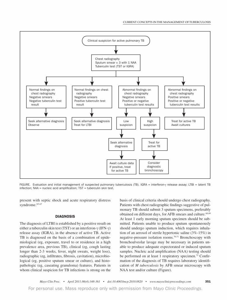
basis of clinical criteria should undergo chest radiography. Patients with chest radiographic findings suggestive of pul-monary TB should submit 3 sputum specimens, preferably obtained on different days, for AFB smears and culture.68,69 At least 1 early morning sputum specimen should be sub-mitted. Patients unable to produce sputum spontaneously should undergo sputum induction, which requires inhala-tion of an aerosol of sterile hypertonic saline (3%-15%) in negative-pressure isolation rooms.70,71 Bronchoscopy with bronchoalveolar lavage may be necessary in patients un-able to produce adequate expectorated or induced sputum samples. Nucleic acid amplification (NAA) testing should be performed on at least 1 respiratory specimen.72 Confir-mation of the diagnosis of TB requires laboratory identifi-cation of M tuberculosis by AFB smear microscopy with NAA test and/or culture (Figure).
Clinical suspicion for active pulmonary TB
Chest radiographySputum smear × 3 with 1 NAATuberculin test (TST or IGRA)
Normal findings on chest radiographyNegative smearsNegative tuberculin test result
Normal findings on chest radiographyNegative smearsPositive tuberculin test result
Abnormal findings on chest radiographyNegative smearsPositive or negative tuberculin test results
Abnormal findings on chest radiographyPositive smearsPositive or negative tuberculin test results
Seek alternative diagnosisObserve
Seek alternative diagnosisTreat for LTBI
Lowsuspicion
Highsuspicion
Treat for active TBAwait cultures
Seek alternativediagnosis
Treat foractive TB
Await culture dataIf positive, treat for active TB
Considerdiagnostic
bronchoscopy
FIGURe. evaluation and initial management of suspected pulmonary tuberculosis (Tb). IGRA = interferon-γ release assay; LTbI = latent Tb infection; NAA = nucleic acid amplification; TST = tuberculin skin test.
Current ConCepts in the ManageMent of tuberCulosis
Mayo Clin Proc. • April 2011;86(4):348-361 • doi:10.4065/mcp.2010.0820 • www.mayoclinicproceedings.com352
For personal use. Mass reproduce only with permission from Mayo Clinic Proceedingsa .
TuBErculin Skin TEST The TST, also known as the Mantoux test, requires the in-tradermal injection of 0.1 mL of 5 tuberculin units of puri-fied protein derivative into the volar surface of the forearm. The TST measures cell-mediated immunity manifesting as a delayed-type hypersensitivity to tuberculin purified protein derivative, which contains a mixture of antigens shared by several species of mycobacteria.73 The test result is recorded as the diameter of transverse induration in mil-limeters 48 to 72 hours after administration. Interpretation of the TST result varies depending on the prevalence of and the risk for progression to TB in different groups. Indura-tion between 5 and 15 mm is considered positive and may be indicative of LTBI (Table 2). The size of induration has some predictive value in that persons with TBI tend to have larger indurations74,75; however, it does not correlate with risk for progression to active TB. An increase of 10 mm or more in the skin test reaction within 2 years in persons with previously negative results (conversion) is indicative of recent M tuberculosis infection. The TST cannot dis-criminate between active TB and LTBI. Interpretation of skin-test results is the same for bacille Calmette-Guérin (BCG)–vaccinated and non-BCG–vacci-nated persons. The estimated interval between M tuberculo-sis infection and skin test reactivity (ie, skin test conversion) is 2 to 12 weeks.76,77 Therefore, those in close contact with patients who have active pulmonary TB with initial nega-tive test results should have the test repeated 8 to 12 weeks after exposure. Skin test conversion may also be due to new
delayed-type hypersensitivity after infection with nontuber-culous mycobacteria or BCG vaccination.78 The phenom-enon of reversion (ie, decrease in the size of the tuberculin reaction) may present problems in skin test interpretation when serial TSTs are performed. Therefore, TST should not be performed if a positive TST result has been documented previously or if the patient has been treated for TB. False-positive skin tests can result from BCG vaccination or infec-tion with nontuberculous mycobacteria. False-negative test results may occur because of the following technical and bi-ological limitations: presence of active TB; presence of other bacterial, fungal, and viral (eg, HIV) infections; live virus vaccination; immunosuppressive therapy; long-standing re-nal failure; malnutrition; lymphoid diseases; and age. The TST may have a booster effect on immunologic memory in patients with a history of TB, previous BCG vaccination, and exposure to nontuberculous mycobacte-ria.74,79 This results in a positive TST 1 to 4 weeks after initial negative findings on TST. Evaluation for the booster phenomenon with a second TST 1 to 4 weeks after the first test (the 2-step method) should be considered for persons from countries with a high incidence of TB, for those with a history of BCG vaccination, and (as a baseline assess-ment) for those who need periodic retesting, such as health care workers. Test interpretation is based on the induration observed with the second test. About 10% of immunocompetent persons with LTBI will develop TB disease over their lifetime, with the greatest risk for progression (5%) being in the first 2 years after infection with M tuberculosis.80 Approximately 50% of TB cases will occur within 2 years after initial infection.81 Targeted tuber-culin testing identifies persons at high risk of developing TB who would benefit from treatment for LTBI. These include persons at risk of becoming infected with M tuberculosis and those with clinical conditions associated with increased risk of progression of TB infection to active TB (Table 2). A joint statement from the American Thoracic Society and the Cen-ters for Disease Control and Prevention provides guidelines on testing and treatment of LTBI in the United States.76
inTErfEron-γ rElEaSE aSSay Serum IGRAs are in vitro tests of whole blood or mono-nuclear cells that are based on IFN-γ release after T-cell stimulation by M tuberculosis–specific proteins (eg, early-secreted antigenic target 6 [ESAT-6] and culture filtrate protein 10 [CFP-10]), which are absent from BCG vaccine strains and most nontuberculous mycobacteria. The Quan-tiFERON-TB Gold In-Tube test (Cellestis Limited, Car-negie, Victoria, Australia) is an enzyme-linked immuno-sorbent assay–based whole blood assay that measures and quantifies IFN-γ (IU/mL) released from blood collected in special tubes that are coated with M tuberculosis–specific
TAbLe 2. Interpretation of TST Results for Populations at Risk of TBa
Positive TST reaction size At-risk populations (mm)
Patients with HIV infection ≥5Patients receiving immunosuppressive therapyb Abnormal findings on chest radiography consistent with previous TB infection Persons who have come in close contact with an actively contagious patient
Patients with certain chronic conditionsc ≥10Patients with certain malignanciesd
Foreign-born persons from high-incidence regions (>25/100,000) Employees and residents of high-risk facilitiese
Healthy people at low risk of TB ≥15
a HIV = human immunodeficiency virus; TB = tuberculosis; TNF = tumor necrosis factor; TST = tuberculin skin test.
b Immunosuppressive therapies include chemotherapeutic agents, TNF-α inhibitors, and glucocorticoid therapy (>15 mg/d of prednisone equiva-lent for >1 mo).
c Diabetes, dialysis-dependent renal failure, silicosis, and being under-weight.
d Leukemia, lymphoma, and cancers of the head, neck, and lung.e Health care facilities, prisons, and homeless shelters.
Current ConCepts in the ManageMent of tuberCulosis
Mayo Clin Proc. • April 2011;86(4):348-361 • doi:10.4065/mcp.2010.0820 • www.mayoclinicproceedings.com 353
For personal use. Mass reproduce only with permission from Mayo Clinic Proceedingsa .
antigens, including ESAT-6, CFP-10, and TB7.7(p4). The T-SPOT.TB test (Oxford Immunotec Limited, Abingdon, United Kingdom) is an enzyme-linked immunospot assay that uses peripheral blood mononuclear cells incubated with mixtures of peptides containing ESAT-6 and CFP-10 to measure the number of cells secreting IFN-γ (IFN-γ-spot–forming cells). Results of IGRA are reported both qualitatively (positive, negative, indeterminate, or border-line) and quantitatively (IU/mL or IFN-γ-spot–forming cells). Reversion of IGRA test results from positive to neg-ative has been observed, particularly in those with nega-tive results on the initial TST.82 Conversion of IGRA (ie, a change in test results from negative to positive within 2 years) has not been associated with an increased risk of subsequent progression to TB disease.83
The IGRA can be used in the same setting as TST83 but has the advantage of being able to differentiate M tubercu-losis infection from previous BCG vaccination and most nontuberculous mycobacterial infections; it may also be able to discriminate true-negative responses from anergy. However, currently available IGRA, like TST, cannot distin-guish active TB from latent infection.73,82 Because ESAT-6 and CFP-10 proteins are present in Mycobacterium mari-num, Mycobacterium kansasii, and Mycobacterium szulgai, false-positive IGRA results are possible. As with negative
TST findings, negative findings on IGRA may not exclude TB infection in immunosuppressed persons. An IGRA is the preferred method of testing for groups of people who have low rates of returning to have their TST test results read and for those who have received BCG vaccine. The TST is pre-ferred for testing children younger than 5 years. For other groups being tested for LTBI, either TST or IGRA may be used.83 A summary of tests for TB is presented in Table 3.
chEST radiograPhy
Chest radiography is indicated for all persons being evalu-ated for LTBI or active TB. Pulmonary TB as a result of endogenous reactivation of latent infection classically pre-sents with infiltrates in the apical and posterior segments of the right upper lobe, the apical-posterior segment of the left upper lobe, and the superior segment of the lower lobe. Cavitation, fibrosis, and/or enlargement of the hilar and mediastinal lymph nodes may be present. In some cases, pulmonary TB may present as lobar or segmental infiltrates, lung mass, scattered fibronodular lesions (“mil-iary”), or pleural effusions.
SmEar microScoPy
Smear microscopy for the detection of AFB is the most rapid and inexpensive method for TB diagnosis. Two commonly
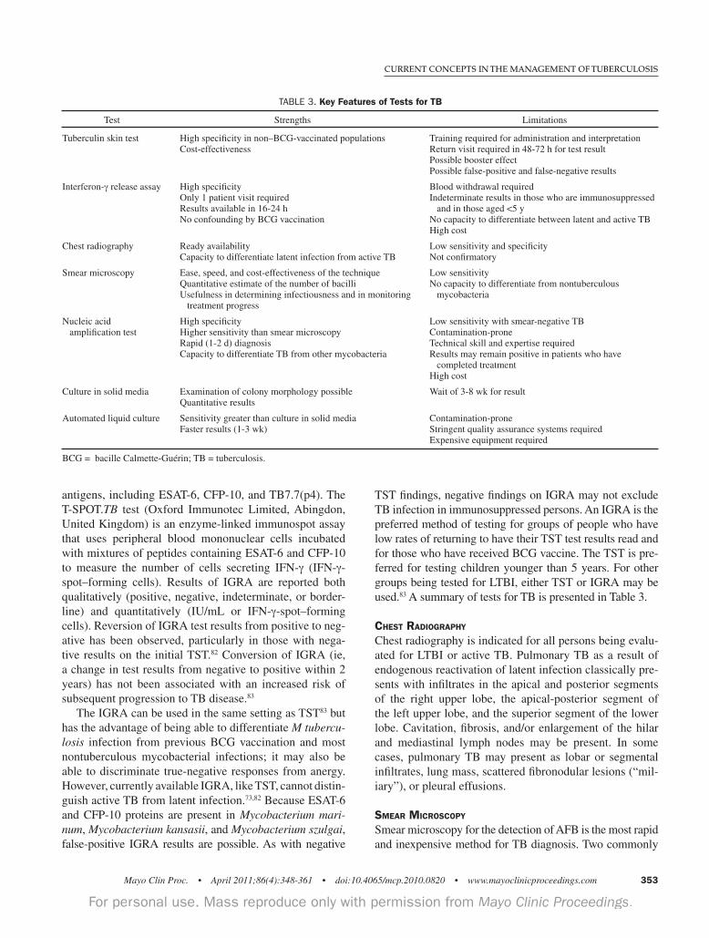
TAbLe 3. Key Features of Tests for TB
Test Strengths Limitations
Tuberculin skin test High specificity in non–BCG-vaccinated populations Training required for administration and interpretation Cost-effectiveness Return visit required in 48-72 h for test result Possible booster effect Possible false-positive and false-negative results
Interferon-γ release assay High specificity Blood withdrawal required Only 1 patient visit required Indeterminate results in those who are immunosuppressed Results available in 16-24 h and in those aged <5 y No confounding by BCG vaccination No capacity to differentiate between latent and active TB High cost
Chest radiography Ready availability Low sensitivity and specificity Capacity to differentiate latent infection from active TB Not confirmatory
Smear microscopy Ease, speed, and cost-effectiveness of the technique Low sensitivity Quantitative estimate of the number of bacilli No capacity to differentiate from nontuberculous Usefulness in determining infectiousness and in monitoring mycobacteria treatment progress
Nucleic acid High specificity Low sensitivity with smear-negative TB amplification test Higher sensitivity than smear microscopy Contamination-prone Rapid (1-2 d) diagnosis Technical skill and expertise required Capacity to differentiate TB from other mycobacteria Results may remain positive in patients who have completed treatment High cost
Culture in solid media Examination of colony morphology possible Wait of 3-8 wk for result Quantitative results
Automated liquid culture Sensitivity greater than culture in solid media Contamination-prone Faster results (1-3 wk) Stringent quality assurance systems required Expensive equipment required
BCG = bacille Calmette-Guérin; TB = tuberculosis.
Current ConCepts in the ManageMent of tuberCulosis
Mayo Clin Proc. • April 2011;86(4):348-361 • doi:10.4065/mcp.2010.0820 • www.mayoclinicproceedings.com354
For personal use. Mass reproduce only with permission from Mayo Clinic Proceedingsa .
used methods for AFB staining are the carbolfuchsin meth-ods (eg, Ziehl-Neelsen and Kinyoun methods) and the fluorochrome procedure using auramine O or auramine-rhodamine dyes. The fluorochrome method with fluores-cence microscopy is preferred because it is far more sensi-tive than the carbolfuchsin methods. The finding of AFB on respiratory specimens associated with the appropriate epi-demiological, clinical, and radiographic findings is highly suggestive of TB.
nuclEic acid amPlificaTion TEST
The NAA test is useful for the rapid detection of M tuber-culosis in respiratory specimens. The Enhanced Amplified MTD (Mycobacterium Tuberculosis Direct) test (Gen-Probe, San Diego, CA) detects M tuberculosis ribosomal RNA di-rectly from AFB smear–positive and AFB smear–negative respiratory specimens from patients with suspected TB. The Amplicor MTB (Mycobacterium Tuberculosis) test (Roche Diagnostic Systems, Branchburg, NJ) detects M tuberculosis DNA in AFB smear–positive respiratory specimens. Interpretation of NAA test results should be correlated with AFB smear results.72 Positive findings on the NAA test and a positive sputum AFB smear are strongly indicative of TB.84-86 When NAA and sputum microscopy test results are discordant, physicians should exercise their clinical judgment in deciding whether to start anti-TB treatment while culture results are awaited.72 When the clinical suspicion for TB is high, a positive NAA test result in smear-negative cases can be valuable for the early detection of TB in approximately 50% to 80% of cases.87,88 Findings on the NAA test often re-main positive after cultures become negative during therapy and can remain positive even after completion of therapy89; therefore, it should not be used for assessing infectivity or response to treatment.
culTurE
Culture remains the criterion standard for laboratory confir-mation of TB. Three types of culture media are available for the microbiological detection of M tuberculosis: egg-based (Löwenstein-Jensen), agar-based (Middlebrook 7H10 or 7H11), and liquid (Middlebrook 7H12 and other commercial broth systems). Mycobacterial growth tends to be slightly better on the egg-based medium but more rapid on the agar medium. Growth in liquid media is faster than growth on sol-id media and allows detection in 1 to 3 weeks.90 The develop-ment of automated liquid culture systems for mycobacterial growth detection, such as BACTEC 460TB and BACTEC MGIT960 (Becton Dickinson Microbiology Systems, Sparks, MD), VersaTREK Myco (Trek Diagnostic Systems, Westlake, OH), and BacT/Alert 3D (bioMérieux, Durham, NC), which are faster and more sensitive than solid media, has clearly facilitated TB diagnosis in the past decade.
nEw TEchnology
Several recently developed tests for TB, including molecu-lar drug resistance, have the potential for providing rapid diagnosis and targeted treatment.91,92 These fully automat-ed tests provide rapid drug-resistance testing for RIF and INH; however, they require sophisticated technology and are currently available only at reference laboratories.93
oThEr conSidEraTionS
At the start of LTBI therapy, baseline measurements of se-rum aspartate aminotransferase, alanine aminotransferase, and bilirubin are recommended for patients who have a his-tory of liver disease (eg, hepatitis B or C, alcoholic hepati-tis, or cirrhosis), use alcohol regularly, have risk factors for chronic liver disease, are infected with HIV, are pregnant, or have given birth within the past 3 months.76 All patients diagnosed as having LTBI should be offered voluntary HIV counseling and testing. All patients with active TB should receive counseling and be tested for HIV infection. Serologic tests for hepa-titis B and C should be performed for patients with risk factors such as injection drug use, foreign birth, and HIV infection. Susceptibility testing for INH, RIF, ethambutol (EMB), and pyrazinamide (PZA) should be performed on any initial culture that is positive. Susceptibility testing to the second-line drugs should be performed on specimens from patients who have had previous therapy, are known to have resistance to first-line drugs, are contacts of patients with drug-resistant TB, or have persistently positive cul-tures 3 months or more after starting treatment. Baseline measurements of platelet count and serum aspartate amino-transferase, alanine aminotransferase, bilirubin, alkaline phosphatase, and serum creatinine levels are recommended for all patients starting TB treatment.68
TREATMENT
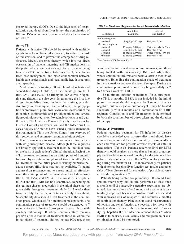
laTEnT TB infEcTion
Treatment for LTBI is recommended for persons deemed to be at relatively high risk of developing active TB (Table 2) and should be initiated only after active TB has been excluded by clinical and radiographic evaluations. Failure to rule out TB may result in inadequate treatment and de-velopment of drug resistance. For most patients, treatment with INH for 9 months is preferred (Table 4).76,94 Pyridox-ine supplementation (25 mg/d) to INH is recommended for patients at an increased risk of neuropathy, including those with preexisting peripheral neuropathy, nutritional deficiency, diabetes mellitus, HIV infection, renal failure, alcoholism, or thyroid disease and those who are preg-nant or breast-feeding. Intermittent treatment (ie, a twice-weekly regimen) should only be performed as directly
Current ConCepts in the ManageMent of tuberCulosis
Mayo Clin Proc. • April 2011;86(4):348-361 • doi:10.4065/mcp.2010.0820 • www.mayoclinicproceedings.com 355
For personal use. Mass reproduce only with permission from Mayo Clinic Proceedingsa .
TAbLe 4. Treatment Regimens for Latent Tuberculosis Infection
Adult dose Interval Medication (maximum) and duration
Preferred regimen Isoniazid 5 mg/kg (300 mg) Daily for 9 moAlternative regimens Isoniazid 15 mg/kg (900 mg) Twice weekly for 9 mo Isoniazid 5 mg/kg (300 mg) Daily for 6 mo Isoniazid 15 mg/kg (900 mg) Twice weekly for 6 mo Rifampin 10 mg/kg (600 mg) Daily for 4 mo
Data from MMWR Recomm Rep.76
observed therapy (DOT). Due to the high rates of hospi-talization and death from liver injury, the combination of RIF and PZA is no longer recommended for the treatment of LTBI.94
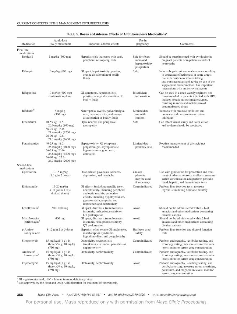
acTivE TBPatients with active TB should be treated with multiple agents to achieve bacterial clearance, to reduce the risk of transmission, and to prevent the emergence of drug re-sistance. Directly observed therapy, which involves direct observation of patients ingesting anti-TB medications, is the preferred management strategy for all patients being treated for TB. For treatment to be successful, patient-cen-tered case management and close collaboration between health care professionals and local public health programs are imperative. Medications for treating TB are classified as first- and second-line drugs (Table 5). First-line drugs are INH, RIF, EMB, and PZA. The rifamycin derivatives rifapen-tine and rifabutin are also considered among the first-line drugs. Second-line drugs include the aminoglycosides streptomycin, kanamycin, and amikacin; the polypep-tide capreomycin; p-aminosalicylic acid; cycloserine; the thioamides ethionamide and prothionamide; and several fluoroquinolones (eg, moxifloxacin, levofloxacin and gati-floxacin). The American Thoracic Society, the Centers for Disease Control and Prevention, and the Infectious Dis-eases Society of America have issued a joint statement on the treatment of TB in the United States.68 An overview of this guideline and summary recommendations follow. Four treatment regimens are recommended for patients with drug-susceptible disease. Although these regimens are broadly applicable, treatment must be individualized on the basis of each patient’s clinical situation. Each of the 4 TB treatment regimens has an initial phase of 2 months followed by a continuation phase of 4 or 7 months (Table 6). Treatment in the initial phase is usually empirical be-cause susceptibility data may not be available. To guard against drug resistance and to ensure maximal effective-ness, the initial phase of treatment should include 4 drugs (INH, RIF, PZA, and EMB). If the isolate is susceptible to INH and RIF, EMB can be discontinued. Depending on the regimen chosen, medication in the initial phase may be given daily throughout treatment, daily for 2 weeks then twice weekly thereafter, or 3 times weekly throughout. Susceptibility data should direct treatment in the continu-ation phase, which lasts for 4 months in most patients. The continuation phase of treatment should be extended to 7 months for the following 3 groups of patients: those with cavitary pulmonary TB whose sputum culture remains positive after 2 months of treatment; those in whom the initial phase of treatment did not include PZA (eg, those
who have severe liver disease or are pregnant); and those being treated with once-weekly INH and rifapentine whose sputum culture remains positive after 2 months of treatment. Extending the continuation phase of treatment in these situations reduces the rate of relapse. During the continuation phase, medications may be given daily or 2 to 3 times a week with DOT. The minimum duration of treatment for culture-posi-tive TB is 6 months. If PZA is not included in the initial phase, treatment should be given for 9 months. Smear-negative, culture-negative pulmonary TB may be treated successfully with 4 months of a combination INH-RIF regimen. Completion of anti-TB treatment is determined by both the total number of doses taken and the duration of therapy.
follow-uP EvaluaTion
Patients receiving treatment for TB infection or disease should be counseled about adverse effects and should have clinical evaluations at least once monthly to assess adher-ence and evaluate for possible adverse effects of anti-TB medications (Table 5). Patients receiving INH for LTBI therapy should be given no more than a 1-month drug sup-ply and should be monitored monthly for drug-induced he-patotoxicity or other adverse effects.95 Laboratory monitor-ing during treatment for LTBI is indicated only for patients with abnormal baseline liver function test results and other risks of liver disease and for evaluation of possible adverse effects during treatment.76
Patients being treated for pulmonary TB should have sputum microscopy and culture performed at least once a month until 2 consecutive negative specimens are ob-tained. Sputum culture after 2 months of treatment is par-ticularly important because a positive result is associated with increased risk of relapse96-99 and requires 7 months of continuation therapy. Platelet counts and measurements of hepatic and renal function are necessary for those with baseline abnormalities or those at increased risk of toxic-ity (eg, hepatitis B or C infection, alcohol abuse).68 When EMB is to be used, visual acuity and red-green color dis-crimination should be monitored.
Current ConCepts in the ManageMent of tuberCulosis
Mayo Clin Proc. • April 2011;86(4):348-361 • doi:10.4065/mcp.2010.0820 • www.mayoclinicproceedings.com356
For personal use. Mass reproduce only with permission from Mayo Clinic Proceedingsa .
TAbLe 5. Doses and Adverse Effects of Antituberculosis Medicationsa
Adult dose Use in Medication (daily maximum) Important adverse effects pregnancy Comments
First-line medications Isoniazid 5 mg/kg (300 mg) Hepatitis (risk increases with age), Safe for fetus; Should be supplemented with pyridoxine in peripheral neuropathy, rash increased pregnant patients or in patients at risk of hepatotoxicity neuropathy postpartum
Rifampin 10 mg/kg (600 mg) GI upset, hepatotoxicity, pruritus, Safe Induces hepatic microsomal enzymes, resulting orange discoloration of bodily in decreased effectiveness of some drugs; fluids use with caution in women taking oral contraceptives and advise on use of the supplement barrier method; has important interactions with antiretroviral agents
Rifapentine 10 mg/kg (600 mg) GI symptoms, hepatotoxicity, Insufficient Can be used in a once-weekly regimen; not continuation phase pruritus, orange discoloration of information recommended in patients infected with HIV; bodily fluids induces hepatic microsomal enzymes, resulting in increased metabolism of coadministered drugs
Rifabutinb 5 mg/kg Neutropenia, uveitis, polyarthralgia, Limited data; Interacts with protease inhibitors and (300 mg) rash, hepatotoxicity, and orange use with nonnucleoside reverse transcriptase discoloration of bodily fluids caution inhibitors
Ethambutol 40-55 kg: 14.5- Optic neuritis and peripheral Safe Can affect visual acuity and color vision 20.0 mg/kg (800 mg) neuropathy and so these should be monitored 56-75 kg: 16.0- 21.4 mg/kg (1200 mg) 76-90 kg: 17.8- 21.1 mg/kg (1600 mg) Pyrazinamide 40-55 kg: 18.2- Hepatotoxicity, GI symptoms, Limited data; Routine measurement of uric acid not 25.0 mg/kg (1000 mg) polyarthralgia, asymptomatic probably safe recommended 56-75 kg: 20.0- hyperuricemia, gout, rash, 26.8 mg/kg (1500 mg) dermatitis 76-90 kg: 22.2- 26.3 mg/kg (2000 mg) Second-line medications Cycloserine 10-15 mg/kg Dose-related psychosis, seizures, Crosses Use with pyridoxine for prevention and treat- (1.0 g in 2 doses) depression, and headache placenta; ment of adverse neurotoxic effects; measure may be used serum concentration and perform periodic if necessary renal, hepatic, and hematologic tests
Ethionamide 15-20 mg/kg GI effects, including metallic taste; Contraindicated Perform liver function tests, measure (1.0 g/d in 1 or 2 neurotoxicity, including peripheral thyroid-stimulating hormone monthly divided doses) and optic neuritis; endocrine effects, including hypothyroidism, gynecomastia, alopecia, and impotence; and hepatoxicity Levofloxacinb 500-1000 mg GI upset, dizziness, tremulousness, Avoid Should not be administered within 2 h of insomnia, rash, photosensitivity, antacids and other medications containing QT prolongation divalent cations Moxifloxacin/ 400 mg GI upset, dizziness, tremulousness, Avoid Should not be administered within 2 h of gatifloxacinb insomnia, rash, photosensitivity, antacids and other medications containing QT prolongation divalent cations
p-Amino- 8-12 g in 2 or 3 doses Hepatitis, often severe GI intolerance, Has been used Perform liver function and thyroid function salicylic acid malabsorption syndrome, safely tests hypothyroidism, and coagulopathy
Streptomycin 15 mg/kg/d (1 g); in Ototoxicity, neurotoxicity Contraindicated Perform audiography, vestibular testing, and those >59 y, 10 mg/kg (weakness, circumoral paresthesia), Romberg testing; measure serum creatinine (750 mg) nephrotoxicity levels; monitor serum drug concentration
Amikacin/ 15 mg/kg/d (1 g); in Ototoxicity, nephrotoxicity Contraindicated Perform audiography, vestibular testing, and kanamycinb those >59 y, 10 mg/kg Romberg testing; measure serum creatinine (750 mg) levels; monitor serum drug concentration
Capreomycin 15 mg/kg/d (1 g); in Ototoxicity, nephrotoxicity Avoid Perform audiography, Romberg testing, and those >59 y, 10 mg/kg vestibular testing; measure serum creatinine, (750 mg) potassium, and magnesium levels; monitor serum drug concentration a GI = gastrointestinal; HIV = human immunodeficiency virus.b Not approved by the Food and Drug Administration for treatment of tuberculosis.
Current ConCepts in the ManageMent of tuberCulosis
Mayo Clin Proc. • April 2011;86(4):348-361 • doi:10.4065/mcp.2010.0820 • www.mayoclinicproceedings.com 357
For personal use. Mass reproduce only with permission from Mayo Clinic Proceedingsa .
Follow-up chest radiography after 2 months and at the completion of treatment is optional. Expert consultation should be sought for the management of patients who de-velop substantial adverse effects and require alternative treatment regimens.100-103
TREATMENT IN SPECIAL SITUATIONS
infEcTion wiTh hivIn general, the treatment of LTBI and TB in HIV-infected adults is the same as in adults not infected with HIV, with a few exceptions. Treatment for TB can be complicated by the interaction between rifamycins, antiretroviral agents, and other anti-infective drugs prescribed for opportunistic infections. In persons receiving antiretroviral therapy, RIF should be avoided or used with caution. Rifabutin, which has fewer problematic drug interactions, may be substitut-ed for RIF. During the continuation phase of treatment, the INH-rifapentine regimen should never be used because of the high relapse rate. Concurrent initiation of anti-TB and antiretroviral therapy may cause increased adverse effects and para-doxical reactions in patients not already receiving treat-ment. The term immune reconstitution inflammatory syn-drome is used to describe this paradoxical reaction, which presents as worsening clinical (eg, high fever, weight
loss, increased lymphadenopathy) and radiographic (eg, increased pulmonary infiltrates) manifestations of TB resulting from the immune reconstitution achieved by an-tiretroviral therapy.104-107 The mechanism for these para-doxical reactions is unclear but appears to be immune me-diated: lower CD4 counts in patients with HIV infection seem to be associated with a higher risk of developing immune reconstitution inflammatory syndrome.104,108 For these reasons, antiretroviral therapy should be delayed for 2 to 8 weeks after starting anti-TB therapy.109 Treatment of HIV-related TB is complex and is best managed by those with expertise in both HIV infection and TB.
ExTraPulmonary TBThe same basic principles for the treatment of pulmonary TB apply to extrapulmonary TB. For TB at any site, a treat-ment course of 6 to 9 months with regimens that include INH and RIF is recommended; the single exception is men-ingitis, for which 9 to 12 months of treatment is recom-mended. The addition of corticosteroids to anti-TB treat-ment is recommended for patients with TB of the pericar-dium and central nervous system, including the meninges.
PrEgnancy and BrEaST-fEEding
For the treatment of TB in pregnant women, the initial regi-men should be INH, RIF, and EMB for at least 9 months.
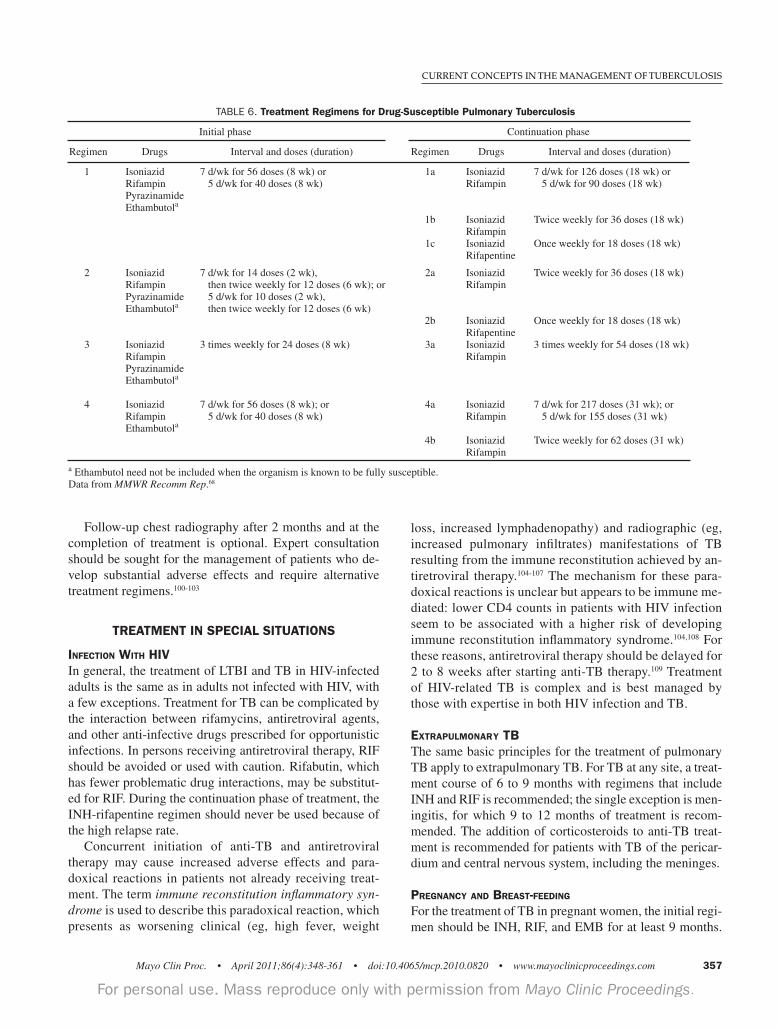
TAbLe 6. Treatment Regimens for Drug-Susceptible Pulmonary Tuberculosis
Initial phase Continuation phase
Regimen Drugs Interval and doses (duration) Regimen Drugs Interval and doses (duration)
1 Isoniazid 7 d/wk for 56 doses (8 wk) or 1a Isoniazid 7 d/wk for 126 doses (18 wk) or Rifampin 5 d/wk for 40 doses (8 wk) Rifampin 5 d/wk for 90 doses (18 wk) Pyrazinamide Ethambutola
1b Isoniazid Twice weekly for 36 doses (18 wk) Rifampin 1c Isoniazid Once weekly for 18 doses (18 wk) Rifapentine 2 Isoniazid 7 d/wk for 14 doses (2 wk), 2a Isoniazid Twice weekly for 36 doses (18 wk) Rifampin then twice weekly for 12 doses (6 wk); or Rifampin Pyrazinamide 5 d/wk for 10 doses (2 wk), Ethambutola then twice weekly for 12 doses (6 wk) 2b Isoniazid Once weekly for 18 doses (18 wk) Rifapentine 3 Isoniazid 3 times weekly for 24 doses (8 wk) 3a Isoniazid 3 times weekly for 54 doses (18 wk) Rifampin Rifampin Pyrazinamide Ethambutola 4 Isoniazid 7 d/wk for 56 doses (8 wk); or 4a Isoniazid 7 d/wk for 217 doses (31 wk); or Rifampin 5 d/wk for 40 doses (8 wk) Rifampin 5 d/wk for 155 doses (31 wk) Ethambutola 4b Isoniazid Twice weekly for 62 doses (31 wk) Rifampin
a Ethambutol need not be included when the organism is known to be fully susceptible.Data from MMWR Recomm Rep.68
Current ConCepts in the ManageMent of tuberCulosis
Mayo Clin Proc. • April 2011;86(4):348-361 • doi:10.4065/mcp.2010.0820 • www.mayoclinicproceedings.com358
For personal use. Mass reproduce only with permission from Mayo Clinic Proceedingsa .
Although teratogenicity data for PZA are limited, it is probably safe to use in pregnancy. Breast-feeding should not be discouraged for women receiving anti-TB treat-ment. Pyridoxine supplementation (25 mg/d) is recom-mended for all pregnant and breast-feeding women taking INH.
drug-rESiSTanT TBTreatment of drug-resistant TB (resistance of the organ-ism to 1 first-line drug) has become even more complex, difficult, and expensive with the emergence of MDR-TB and XDR-TB. Patients with drug-resistant TB may acquire further drug resistance and are at high risk of treatment failure.110,111 These patients should receive prompt expert consultation. General guidelines for treating patients with drug-resistant TB include using multiple (4-6) drugs, in-cluding an injectable agent to which the organism is sus-ceptible. Treatment includes second-line drugs that have many adverse effects and are more expensive and less effective than first-line drugs. Careful supervision under DOT is mandatory to ensure adherence to therapy. Treat-ment is extended to 24 months after culture conversion, with posttreatment follow-up for 24 months. Case series reveal a possible role for surgical therapy in select cases of MDR-TB and XDR-TB.112
CONTROL AND ELIMINATION
Active TB is frequently treated in the outpatient setting. Patients with active TB should be managed in conjunction with local public health offices and TB control agencies to coordinate visits and maximize adherence. When ac-tive TB is suspected, patients should wear a simple sur-gical mask when they are out of the house or near other people. Initial treatment in the hospital may be appropriate for patients who are acutely ill, those with substantial comorbid illness, or infectious patients who are nonad-herent to therapy.68 These patients should be admitted to a negative-pressure isolation room with at least 6 air ex-changes per hour.3 Health care professionals should wear a fit-tested N95 mask; those who have not been fit-tested or are unable to use an N95 mask should use a powered air-purifying respirator. Simple surgical masks are more effective than N95 masks at preventing extrusion of respi-ratory droplets; therefore, patients with TB should wear a simple surgical mask when they are not in their isola-tion room. Patients may be removed from isolation when clinical improvement is seen on effective therapy and when 3 consecutive sputum samples on separate days are AFB smear–negative.3 If patients with active pulmonary TB are medically stable for discharge but are not yet AFB
smear–negative, they may be discharged only if a defini-tive plan for outpatient therapy has been coordinated with the local TB control agency, if no children younger than 4 years or no immunocompromised persons live with them, and if they agree to leave their home only for medical appointments.3 The preferred mechanism of therapy for both inpatients and outpatients is DOT.113,114
Treatment of patients with active TB is the top public health priority for TB control, followed by contact inves-tigation of all persons who came into close contact with these patients before initiation of therapy. Such contact in-vestigation should be carried out on all patients with con-firmed active pulmonary TB and on selected patients with suspected pulmonary TB before testing is complete.115 Pa-tients with extrapulmonary TB are generally not infectious; therefore, contact investigation is not indicated. The third priority for TB elimination and control is to reduce the population-based burden of LTBI through tar-geted testing and treatment. This strategy is particularly im-portant in low-incidence nations, where most TB cases arise from reactivation of LTBI. In high-incidence, resource-poor settings, this strategy is rarely feasible. Testing for LTBI is indicated to detect patients at risk of new infection or at risk of reactivation of LTBI due to underlying medical condi-tions. Persons at risk of new infection include contacts of patients with active pulmonary TB, employees at facilities with a high risk of exposure (eg, prisons, health care facili-ties, homeless shelters), and those who have recently immi-grated (ie, within the past 5 years) from regions of the world where TB is endemic. Persons at highest risk of reactiva-tion include those taking immunosuppressive medications (ie, chemotherapy, tumor necrosis factor α inhibitors) and those with HIV infection, hematologic malignancy, silicosis, dialysis-dependent renal failure, or changes on chest radiog-raphy consistent with previous TB.76
CONCLUSION
Tuberculosis remains a devastating disease throughout the world. Efforts to eradicate it have been thwarted by pover-ty, lack of health care access, drug resistance, immunosup-pressed populations (eg, HIV-infected persons), and global migration. Effective management requires prompt recog-nition using a combination of clinical, radiographic, mi-crobiological, and histopathologic hallmarks and initiation of appropriate multidrug therapy. In addition to effective treatment of patients with active TB, public health man-agement strategies include contact investigation and testing of persons who came into close contact with patients with active TB before initiation of therapy and reduction of the population-based burden of LTBI through targeted testing and treatment.
Current ConCepts in the ManageMent of tuberCulosis
Mayo Clin Proc. • April 2011;86(4):348-361 • doi:10.4065/mcp.2010.0820 • www.mayoclinicproceedings.com 359
For personal use. Mass reproduce only with permission from Mayo Clinic Proceedingsa .
REFERENCES 1. Spence DP, Hotchkiss J, Williams CS, Davies PD. Tuberculosis and poverty. BMJ. 1993;307:759-761. 2. Lonnroth K, Castro KG, Chakaya JM, et al. Tuberculosis con-trol and elimination 2010-50: cure, care, and social development. Lancet. 2010;375:1814-1829. 3. Jensen PA, Lambert LA, Iademarco MF, Ridzon R. Guidelines for preventing the transmission of Mycobacterium tuberculosis in health-care set-tings, 2005. MMWR Recomm Rep. 2005;54(RR-17):1-141. 4. Lonnroth K, Raviglione M. Global epidemiology of tuberculosis: pros-pects for control. Semin Respir Crit Care Med. 2008;29:481-491. 5. World Health Organization. Global Tuberculosis Control: Epidemi-ology, Strategy, Financing: WHO Report 2009. Geneva, Switzerland: World Health Organization; 2009. 6. World Health Organization. Global Tuberculosis Control: A Short Update to the 2009 Report. Geneva, Switzerland: World Health Organization; 2009. 7. World Health Organization. Multidrug and Extensively Drug-Resistant TB (M/XDR-TB): 2010 Global Report on Surveillance and Response. Geneva, Switzerland: World Health Organization; 2010. 8. Centers for Disease Control and Prevention (CDC). Reported Tubercu-losis in the United States, 2009. Atlanta, GA: US Department of Health and Human Services, CDC; 2010. http://www.cdc.gov/tb/statistics/reports/2009 /pdf/report2009.pdf. Accessed February 22, 2011. 9. Geng E, Kreiswirth B, Driver C, et al. Changes in the transmission of tuberculosis in New York City from 1990 to 1999. N Engl J Med. 2002;346: 1453-1458. 10. Cain KP, Benoit SR, Winston CA, Mac Kenzie WR. Tuberculosis among foreign-born persons in the United States. JAMA. 2008;300:405-412. 11. Zumla A. The white plague returns to London–with a vengeance. Lan-cet. 2011;377:10-11. 12. Loudon RG, Spohn SK. Cough frequency and infectivity in patients with pulmonary tuberculosis. Am Rev Respir Dis. 1969;99:109-111. 13. Tostmann A, Kik SV, Kalisvaart NA, et al. Tuberculosis transmission by patients with smear-negative pulmonary tuberculosis in a large cohort in the Netherlands. Clin Infect Dis. 2008;47:1135-1142. 14. Poulsen A. Some clinical features of tuberculosis; 1: incubation period. Acta Tuberc Pneumol Scand. 1950;24(3-4):311-346. 15. Poulsen A. Some clinical features of tuberculosis [concl]. Acta Tuberc Pneumol Scand. 1957;33(1-2):37-92. 16. Krysl J, Korzeniewska-Kosela M, Muller NL, FitzGerald JM. Radio-logic features of pulmonary tuberculosis: an assessment of 188 cases. Can As-soc Radiol J. 1994;45:101-107. 17. Lee KS, Song KS, Lim TH, Kim PN, Kim IY, Lee BH. Adult-onset pulmonary tuberculosis: findings on chest radiographs and CT scans. AJR Am J Roentgenol. 1993;160:753-758. 18. Im JG, Itoh H, Shim YS, et al. Pulmonary tuberculosis: CT findings–early active disease and sequential change with antituberculous therapy. Radi-ology. 1993;186:653-660. 19. Marciniuk DD, McNab BD, Martin WT, Hoeppner VH. Detection of pulmonary tuberculosis in patients with a normal chest radiograph. Chest. 1999;115:445-452. 20. Pepper T, Joseph P, Mwenya C, et al. Normal chest radiography in pul-monary tuberculosis: implications for obtaining respiratory specimen cultures. Int J Tuberc Lung Dis. 2008;12:397-403. 21. Greenberg SD, Frager D, Suster B, Walker S, Stavropoulos C, Roth-pearl A. Active pulmonary tuberculosis in patients with AIDS: spectrum of radiographic findings (including a normal appearance). Radiology. 1994; 193:115-119. 22. Rikimaru T. Endobronchial tuberculosis. Expert Rev Anti Infect Ther. 2004;2:245-251. 23. Lee JH, Park SS, Lee DH, Shin DH, Yang SC, Yoo BM. Endobron-chial tuberculosis: clinical and bronchoscopic features in 121 cases. Chest. 1992;102:990-994. 24. Van den Brande PM, Van de Mierop F, Verbeken EK, Demedts M. Clin-ical spectrum of endobronchial tuberculosis in elderly patients. Arch Intern Med. 1990;150:2105-2108. 25. Rikimaru T, Kinosita M, Yano H, et al. Diagnostic features and thera-peutic outcome of erosive and ulcerous endobronchial tuberculosis. Int J Tu-berc Lung Dis. 1998;2:558-562.
26. Chung HS, Lee JH. Bronchoscopic assessment of the evolution of endo-bronchial tuberculosis. Chest. 2000;117:385-392. 27. Peto HM, Pratt RH, Harrington TA, LoBue PA, Armstrong LR. Epide-miology of extrapulmonary tuberculosis in the United States, 1993-2006. Clin Infect Dis. 2009;49:1350-1357. 28. Raviglione MC, Narain JP, Kochi A. HIV-associated tuberculosis in developing countries: clinical features, diagnosis, and treatment. Bull World Health Organ. 1992;70:515-526. 29. Haas DW, Des Prez RM. Tuberculosis and acquired immunodefi-ciency syndrome: a historical perspective on recent developments. Am J Med. 1994;96:439-450. 30. Jones BE, Young SM, Antoniskis D, Davidson PT, Kramer F, Barnes PF. Relationship of the manifestations of tuberculosis to CD4 cell counts in patients with human immunodeficiency virus infection. Am Rev Respir Dis. 1993;148:1292-1297. 31. te Beek LA, van der Werf MJ, Richter C, Borgdorff MW. Extrapulmo-nary tuberculosis by nationality, The Netherlands, 1993-2001. Emerg Infect Dis. 2006;12:1375-1382. 32. Kempainen R, Nelson K, Williams DN, Hedemark L. Mycobacte-rium tuberculosis disease in Somali immigrants in Minnesota. Chest. 2001; 119:176-180. 33. Rieder HL, Snider DE Jr, Cauthen GM. Extrapulmonary tuberculosis in the United States. Am Rev Respir Dis. 1990;141:347-351. 34. Thompson MM, Underwood MJ, Sayers RD, Dookeran KA, Bell PR. Peripheral tuberculous lymphadenopathy: a review of 67 cases. Br J Surg. 1992;79:763-764. 35. Geldmacher H, Taube C, Kroeger C, Magnussen H, Kirsten DK. As-sessment of lymph node tuberculosis in northern Germany: a clinical review. Chest. 2002;121:1177-1182. 36. Dandapat MC, Mishra BM, Dash SP, Kar PK. Peripheral lymph node tuberculosis: a review of 80 cases. Br J Surg. 1990;77:911-912. 37. Jones PG, Campbell PE. Tuberculous lymphadenitis in childhood: the significance of anonymous mycobacteria. Br J Surg. 1962;50:302-314. 38. Ellison E, Lapuerta P, Martin SE. Fine needle aspiration diagnosis of mycobacterial lymphadenitis: Sensitivity and predictive value in the United States. Acta Cytol. 1999;43:153-157. 39. Lee KC, Tami TA, Lalwani AK, Schecter G. Contemporary manage-ment of cervical tuberculosis. Laryngoscope. 1992;102:60-64. 40. Baumann MH, Nolan R, Petrini M, Lee YC, Light RW, Schneider E. Pleural tuberculosis in the United States: incidence and drug resistance. Chest. 2007;131:1125-1132. 41. Berger HW, Mejia E. Tuberculous pleurisy. Chest. 1973;63:88-92. 42. Valdes L, Alvarez D, San Jose E, et al. Tuberculous pleurisy: a study of 254 patients. Arch Intern Med. 1998;158:2017-2021. 43. Gopi A, Madhavan SM, Sharma SK, Sahn SA. Diagnosis and treatment of tuberculous pleural effusion in 2006. Chest. 2007;131:880-889. 44. Phypers M, Harris T, Power C. CNS tuberculosis: a longitudinal anal-ysis of epidemiological and clinical features. Int J Tuberc Lung Dis. 2006; 10:99-103. 45. Sutlas PN, Unal A, Forta H, Senol S, Kirbas D. Tuberculous meningitis in adults: review of 61 cases. Infection. 2003;31:387-391. 46. Kent SJ, Crowe SM, Yung A, Lucas CR, Mijch AM. Tuberculous men-ingitis: a 30-year review. Clin Infect Dis. 1993;17:987-994. 47. Hinman AR. Tuberculous meningitis at Cleveland Metropolitan Gen-eral Hospital 1959 to 1963. Am Rev Respir Dis. 1967;95:670-673. 48. al Karawi MA, Mohamed AE, Yasawy MI, et al. Protean manifestation of gastrointestinal tuberculosis: report on 130 patients. J Clin Gastroenterol. 1995;20:225-232. 49. Sheldon CD, Probert CS, Cock H, et al. Incidence of abdominal tubercu-losis in Bangladeshi migrants in east London. Tuber Lung Dis. 1993;74:12-15. 50. Aguado JM, Pons F, Casafont F, San Miguel G, Valle R. Tuberculous peritonitis: a study comparing cirrhotic and noncirrhotic patients. J Clin Gas-troenterol. 1990;12:550-554. 51. Shakil AO, Korula J, Kanel GC, Murray NG, Reynolds TB. Diagnostic features of tuberculous peritonitis in the absence and presence of chronic liver disease: a case control study. Am J Med. 1996;100:179-185. 52. Sanai FM, Bzeizi KI. Systematic review: tuberculous peritonitis–pre-senting features, diagnostic strategies and treatment. Aliment Pharmacol Ther. 2005;22:685-700.
Current ConCepts in the ManageMent of tuberCulosis
Mayo Clin Proc. • April 2011;86(4):348-361 • doi:10.4065/mcp.2010.0820 • www.mayoclinicproceedings.com360
For personal use. Mass reproduce only with permission from Mayo Clinic Proceedingsa .
53. Bilgin T, Karabay A, Dolar E, Develioglu OH. Peritoneal tuberculo-sis with pelvic abdominal mass, ascites and elevated CA 125 mimicking advanced ovarian carcinoma: a series of 10 cases. Int J Gynecol Cancer. 2001;11:290-294. 54. Rodriguez E, Pombo F. Peritoneal tuberculosis versus peritoneal carcinomatosis: distinction based on CT findings. J Comput Assist Tomogr. 1996;20:269-272. 55. Jain S, Sharma N, Varma S, Rajwanshi A, Verma JS, Sharma BK. Pro-file of cardiac tamponade in the medical emergency ward of a North Indian hospital. Can J Cardiol. 1999;15:671-675. 56. Reuter H, Burgess LJ, Doubell AF. Epidemiology of pericardial ef-fusions at a large academic hospital in South Africa. Epidemiol Infect. 2005;133:393-399. 57. Cameron J, Oesterle SN, Baldwin JC, Hancock EW. The etiologic spec-trum of constrictive pericarditis. Am Heart J. 1987;113:354-360. 58. Mayosi BM, Wiysonge CS, Ntsekhe M, et al. Clinical characteristics and initial management of patients with tuberculous pericarditis in the HIV era: the Investigation of the Management of Pericarditis in Africa (IMPI Af-rica) registry. BMC Infect Dis. 2006;6:2. 59. Fowler NO, Manitsas GT. Infectious pericarditis. Prog Cardiovasc Dis. 1973;16:323-336. 60. Mehta JB, Dutt A, Harvill L, Mathews KM. Epidemiology of extrapul-monary tuberculosis: a comparative analysis with pre-AIDS era. Chest. 1991; 99:1134-1138. 61. Hodgson SP, Ormerod LP. Ten-year experience of bone and joint tuber-culosis in Blackburn 1978-1987. J R Coll Surg Edinb. 1990;35:259-262. 62. Mondal A. Cytological diagnosis of vertebral tuberculosis with fine-needle aspiration biopsy. J Bone Joint Surg Am. 1994;76:181-184. 63. Sharma SK, Mohan A, Sharma A, Mitra DK. Miliary tuberculosis: new insights into an old disease. Lancet Infect Dis. 2005;5:415-430. 64. Kim JH, Langston AA, Gallis HA. Miliary tuberculosis: epidemi-ology, clinical manifestations, diagnosis, and outcome. Rev Infect Dis. 1990;12:583-590. 65. Ahuja SS, Ahuja SK, Phelps KR, Thelmo W, Hill AR. Hemodynamic confirmation of septic shock in disseminated tuberculosis. Crit Care Med. 1992; 20:901-903. 66. Gachot B, Wolff M, Clair B, Regnier B. Severe tuberculosis in pa-tients with human immunodeficiency virus infection. Intensive Care Med. 1990;16:491-493. 67. Piqueras AR, Marruecos L, Artigas A, Rodriguez C. Miliary tubercu-losis and adult respiratory distress syndrome. Intensive Care Med. 1987;13: 175-182. 68. American Thoracic Society; Centers for Disease Control and Preven-tion (CDC); Infectious Diseases Society of America. Treatment of tuberculo-sis [published correction appears in MMWR Recomm Rep. 2005;53(51):1203. Dosage error in article text]. MMWR Recomm Rep. 2003;52(RR-11):1-77. 69. American Thoracic Society. Diagnostic standards and classification of tuberculosis in adults and children.. Am J Respir Crit Care Med. 2000;161(4, pt 1):1376-1395. 70. Shinnick TM, Good RC. Diagnostic mycobacteriology laboratory prac-tices. Clin Infect Dis. 1995;21(2):291-299. 71. Francis J. Curry National Tuberculosis Center. Sputum induction. In: Tu-berculosis Infection Control: A Practical Manual for Preventing TB. San Fran-cisco, CA: University of California; 2007:73-86. http://www.nationaltbcenter .edu/TB_IC/. Acessed February 22, 2011. 72. Centers for Disease Control and Prevention (CDC). Updated guide-lines for the use of nucleic acid amplification tests in the diagnosis of tubercu-losis. MMWR Morb Mortal Wkly Rep. 2009;58(1):7-10. 73. Mack U, Migliori G, Sester M, et al; TBNET. LTBI: latent tuberculosis infection or lasting immune responses to M. tuberculosis? A TBNET consen-sus statement. Eur Respir J. 2009;33(5):956-973. 74. Moreno S, Blazquez R, Novoa A, et al. The effect of BCG vaccination on tuberculin reactivity and the booster effect among hospital employees. Arch Intern Med. 2001;161:1760-1765. 75. Menzies R, Vissandjee B, Amyot D. Factors associated with tuber-culin reactivity among the foreign-born in Montreal. Am Rev Respir Dis. 1992;146:752-756. 76. American Thoracic Society. Targeted tuberculin testing and treatment of latent tuberculosis infection. MMWR Recomm Rep. 2000;49(RR-6):1-51.
77. Huebner RE, Schein MF, Bass JB Jr. The tuberculin skin test. Clin In-fect Dis. 1993;17:968-975. 78. Menzies D. Interpretation of repeated tuberculin tests: boosting, con-version, and reversion. Am J Respir Crit Care Med. 1999;159:15-21. 79. Menzies R, Vissandjee B, Rocher I, St Germain Y. The booster effect in two-step tuberculin testing among young adults in Montreal. Ann Intern Med. 1994;120:190-198. 80. Centers for Disease Control and Prevention (CDC). Core Curriculum on Tuberculosis: What the Clinician Should Know. 4th ed. Atlanta, GA: Cen-ters for Disease Control and Prevention; 2000. 81. Blower SM, McLean AR, Porco TC, et al. The intrinsic transmission dynamics of tuberculosis epidemics. Nat Med. 1995;1(8):815-821. 82. Menzies D, Pai M, Comstock G. Meta-analysis: new tests for the diag-nosis of latent tuberculosis infection: areas of uncertainty and recommenda-tions for research. Ann Intern Med. 2007;146:340-354. 83. Mazurek GH, Jereb J, Vernon A, LoBue P, Goldberg S, Castro K. Up-dated guidelines for using Interferon Gamma Release Assays to detect Myco-bacterium tuberculosis infection—United States, 2010. MMWR Recomm Rep. 2010;59:1-25. 84. Laraque F, Griggs A, Slopen M, Munsiff SS. Performance of nucleic acid amplification tests for diagnosis of tuberculosis in a large urban setting. Clin Infect Dis. 2009;49:46-54. 85. Campos M, Quartin A, Mendes E, et al. Feasibility of shortening re-spiratory isolation with a single sputum nucleic acid amplification test. Am J Respir Crit Care Med. 2008;178:300-305. 86. Catanzaro A, Perry S, Clarridge JE, et al. The role of clinical suspicion in evaluating a new diagnostic test for active tuberculosis: results of a multi-center prospective trial. JAMA. 2000;283:639-645. 87. Moore DF, Guzman JA, Mikhail LT. Reduction in turnaround time for laboratory diagnosis of pulmonary tuberculosis by routine use of a nucleic acid amplification test. Diagn Microbiol Infect Dis. 2005;52:247- 254. 88. Guerra RL, Hooper NM, Baker JF, et al. Use of the amplified Mycobac-terium tuberculosis direct test in a public health laboratory: test performance and impact on clinical care. Chest. 2007;132:946-951. 89. Schluger NW, Rom WN. Current approaches to the diagnosis of active pulmonary tuberculosis. Am J Respir Crit Care Med. 1994;149:264-267. 90. Cruciani M, Scarparo C, Malena M, Bosco O, Serpelloni G, Men-goli C. Meta-analysis of BACTEC MGIT 960 and BACTEC 460 TB, with or without solid media, for detection of mycobacteria. J Clin Microbiol. 2004;42:2321-2325. 91. Pai M, Minion J, Sohn H, Zwerling A, Perkins M. Novel and improved technologies for tuberculosis diagnosis: progress and challenges. Clin Chest Med. 2009;2009;30(4):701-716. 92. Boehme CC, Nabeta P, Hillemann D, et al. Rapid molecular detection of tuberculosis and rifampin resistance. N Engl J Med. 2010;363(11):1005- 1015. 93. Small PM, Pai M. Tuberculosis diagnosis–time for a game change [edi-torial]. N Engl J Med. 2010;363:1070-1071. 94. Centers for Disease Control and Prevention (CDC); American Thoracic Society. Update: adverse event data and revised American Thoracic Society/CDC recommendations against the use of rifampin and pyrazinamide for treat-ment of latent tuberculosis infection–United States, 2003. MMWR Morb Mor-tal Wkly Rep. 2003;52(31):735-739. 95. Centers for Disease Control and Prevention (CDC). Severe isoniazid-associated liver injuries among persons being treated for latent tuberculo-sis infection–United States, 2004-2008. MMWR Morb Mortal Wkly Rep. 2010;59:224-229. 96. Zierski M, Bek E, Long MW, Snider DE Jr. Short-course (6-month) cooperative tuberculosis study in Poland: results 30 months after completion of treatment. Am Rev Respir Dis. 1981;124:249-251. 97. Hong Kong Chest Service/Tuberculosis Research Centre, Madras/Brit-ish Medical Research Council. A controlled trial of a 2-month, 3-month, and 12-month regimens of chemotherapy for sputum smear-negative pulmonary tuberculosis: the results at 30 months. Am Rev Respir Dis. 1981;124:138- 142. 98. Mitchison DA. Assessment of new sterilizing drugs for treating pul-monary tuberculosis by culture at 2 months. Am Rev Respir Dis. 1993;147: 1062-1063.
Current ConCepts in the ManageMent of tuberCulosis
Mayo Clin Proc. • April 2011;86(4):348-361 • doi:10.4065/mcp.2010.0820 • www.mayoclinicproceedings.com 361
For personal use. Mass reproduce only with permission from Mayo Clinic Proceedingsa .
99. Benator D, Bhattacharya M, Bozeman L, et al. Rifapentine and isoni-azid once a week versus rifampicin and isoniazid twice a week for treatment of drug-susceptible pulmonary tuberculosis in HIV-negative patients: a ran-domised clinical trial. Lancet. 2002;360:528-534. 100. Francis J. Curry National Tuberculosis Center. http://www.nationaltbcenter .edu. Accessed February 22, 2011. 101. Heartland National Tuberculosis Center. http://www.heartlandntbc.org/. Accessed February 22, 2011. 102. New Jersey Medical School Global Tuberculosis Institute. http://www .umdnj.edu/globaltb/home.htm. Accessed February 22, 2011. 103. Southeastern National Tuberculosis Center. http://sntc.medicine.ufl.edu/. Accessed February 22, 2011. 104. Murdoch DM, Venter WD, Feldman C, Van Rie A. Incidence and risk factors for the immune reconstitution inflammatory syndrome in HIV patients in South Africa: a prospective study. AIDS. 2008;22(5):601-610. 105. French MA, Lenzo N, John M, et al. Immune restoration disease after the treatment of immunodeficient HIV-infected patients with highly active an-tiretroviral therapy. HIV Med. 2000;1(2):107-115. 106. Narita M, Ashkin D, Hollender ES, Pitchenik AE. Paradoxical worsen-ing of tuberculosis following antiretroviral therapy in patients with AIDS. Am J Respir Crit Care Med. 1998;158:157-161. 107. Wendel KA, Alwood KS, Gachuhi R, Chaisson RE, Bishai WR, Ster-ling TR. Paradoxical worsening of tuberculosis in HIV-infected persons. Chest. 2001;120:193-197. 108. Manabe YC, Campbell JD, Sydnor E, Moore RD. Immune reconstitu-tion inflammatory syndrome: risk factors and treatment implications. J Acquir Immune Defic Syndr. 2007;46(4):456-462.
The Symposium on Antimicrobial Therapy will continue in the May issue.
This activity was designated for 1 AMA PRA Category 1 Credit(s).™
The contributions to the Symposium on Antimicrobial Therapy are now a CME activity. For CME credit, see the link on our Web site at mayoclinicproceedings.com.
109. Kaplan JE, Benson C, Holmes KH, Brooks JT, Pau A, Masur H. Guidelines for prevention and treatment of opportunistic infections in HIV-infected adults and adolescents: recommendations from CDC, the National Institutes of Health, and the HIV Medicine Association of the In - fectious Diseases Society of America. MMWR Recomm Rep. 2009;58(RR-4): 1-207. 110. Shah NS, Pratt R, Armstrong L, Robison V, Castro KG, Cegielski JP. Extensively drug-resistant tuberculosis in the United States, 1993-2007. JAMA. 2008;300:2153-2160. 111. Kwon YS, Kim YH, Suh GY, et al. Treatment outcomes for HIV-unin-fected patients with multidrug-resistant and extensively drug-resistant tubercu-losis. Clin Infect Dis. 2008;47:496-502. 112. Kang MW, Kim HK, Choi YS, et al. Surgical treatment for multidrug-resistant and extensive drug-resistant tuberculosis. Ann Thorac Surg. 2010; 89:1597-1602. 113. Weis SE, Slocum PC, Blais FX, et al. The effect of directly observed therapy on the rates of drug resistance and relapse in tuberculosis. N Engl J Med. 1994;330:1179-1184. 114. Chaulk CP, Kazandjian VA. Directly observed therapy for treatment completion of pulmonary tuberculosis: consensus statement of the Public Health Tuberculosis Guidelines Panel [published correction appears in JAMA. 1998;280(2):134]. JAMA. 1998;279(12):943-948. 115. National Tuberculosis Controllers Association; Centers for Disease Controll and Prevention (CDC). Guidelines for the investigation of contacts of persons with infectious tuberculosis: recommendations from the Nation-al Tuberculosis Controllers Association and CDC. MMWR Recomm Rep. 2005;54(RR-15):1-47.