Embed Size (px)
Citation preview
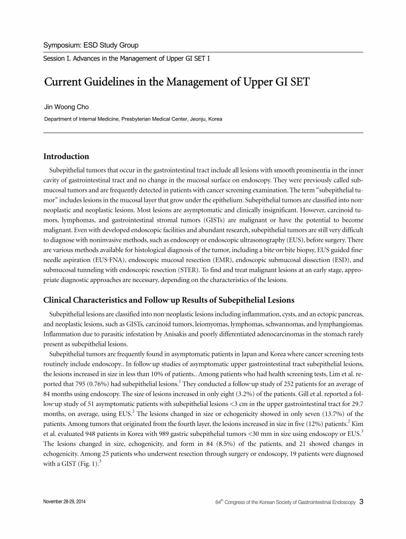
November 28-29, 2014 64th Congress of the Korean Society of Gastrointestinal Endoscopy 3
Current Guidelines in the Management of Upper GI SET
Jin Woong Cho
Department of Internal Medicine, Presbyterian Medical Center, Jeonju, Korea
Symposium: ESD Study Group
Session I. Advances in the Management of Upper GI SET I
Introduction
Subepithelial tumors that occur in the gastrointestinal tract include all lesions with smooth prominentia in the inner
cavity of gastrointestinal tract and no change in the mucosal surface on endoscopy. They were previously called sub-
mucosal tumors and are frequently detected in patients with cancer screening examination. The term “subepithelial tu-
mor” includes lesions in the mucosal layer that grow under the epithelium. Subepithelial tumors are classified into non‐neoplastic and neoplastic lesions. Most lesions are asymptomatic and clinically insignificant. However, carcinoid tu-
mors, lymphomas, and gastrointestinal stromal tumors (GISTs) are malignant or have the potential to become
malignant. Even with developed endoscopic facilities and abundant research, subepithelial tumors are still very difficult
to diagnose with noninvasive methods, such as endoscopy or endoscopic ultrasonography (EUS), before surgery. There
are various methods available for histological diagnosis of the tumor, including a bite‐on‐bite biopsy, EUS guided fine‐needle aspiration (EUS‐FNA), endoscopic mucosal resection (EMR), endoscopic submucosal dissection (ESD), and
submucosal tunneling with endoscopic resection (STER). To find and treat malignant lesions at an early stage, appro-
priate diagnostic approaches are necessary, depending on the characteristics of the lesions.
Clinical Characteristics and Follow‐up Results of Subepithelial Lesions
Subepithelial lesions are classified into non‐neoplastic lesions including inflammation, cysts, and an ectopic pancreas,
and neoplastic lesions, such as GISTs, carcinoid tumors, leiomyomas, lymphomas, schwannomas, and lymphangiomas.
Inflammation due to parasitic infestation by Anisakis and poorly differentiated adenocarcinomas in the stomach rarely
present as subepithelial lesions.
Subepithelial tumors are frequently found in asymptomatic patients in Japan and Korea where cancer screening tests
routinely include endoscopy.. In follow‐up studies of asymptomatic upper gastrointestinal tract subepithelial lesions,
the lesions increased in size in less than 10% of patients.. Among patients who had health screening tests, Lim et al. re-
ported that 795 (0.76%) had subepithelial lesions.1 They conducted a follow‐up study of 252 patients for an average of
84 months using endoscopy. The size of lesions increased in only eight (3.2%) of the patients. Gill et al. reported a fol-
low‐up study of 51 asymptomatic patients with subepithelial lesions <3 cm in the upper gastrointestinal tract for 29.7
months, on average, using EUS.2 The lesions changed in size or echogenicity showed in only seven (13.7%) of the
patients. Among tumors that originated from the fourth layer, the lesions increased in size in five (12%) patients.2 Kim
et al. evaluated 948 patients in Korea with 989 gastric subepithelial tumors <30 mm in size using endoscopy or EUS.3
The lesions changed in size, echogenicity, and form in 84 (8.5%) of the patients, and 21 showed changes in
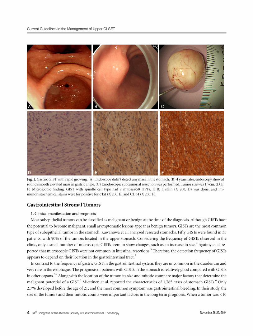
echogenicity. Among 25 patients who underwent resection through surgery or endoscopy, 19 patients were diagnosed
with a GIST (Fig. 1).3
Current Guidelines in the Management of Upper GI SET
4 64th Congress of the Korean Society of Gastrointestinal Endoscopy November 28-29, 2014
Fig. 1. Gastric GIST with rapid growing. (A) Endoscopy didn’t detect any mass in the stomach. (B) 4 years later, endoscopy showedround smooth elevated mass in gastric angle. (C) Enodoscopic subtumorial resection was performed. Tumor size was 1.7cm. (D, E,F) Microscopic finding. GIST with spindle cell type had 7 mitoses/50 HPFs. H & E stain (X 200, D) was done, and im-munohistochemical stains were for positive for c‐kit (X 200, E) and CD34 (X 200, F).
Gastrointestinal Stromal Tumors
1. Clinical manifestation and prognosis
Most subepithelial tumors can be classified as malignant or benign at the time of the diagnosis. Although GISTs have
the potential to become malignant, small asymptomatic lesions appear as benign tumors. GISTs are the most common
type of subepithelial tumor in the stomach. Kawanowa et al. analyzed resected stomachs. Fifty GISTs were found in 35
patients, with 90% of the tumors located in the upper stomach. Considering the frequency of GISTs observed in the
clinic, only a small number of microscopic GISTs seem to show changes, such as an increase in size.4 Agaimy et al. re-
ported that microscopic GISTs were not common in intestinal resections.5 Therefore, the detection frequency of GISTs
appears to depend on their location in the gastrointestinal tract.5
In contrast to the frequency of gastric GIST in the gastrointestinal system, they are uncommon in the duodenum and
very rare in the esophagus. The prognosis of patients with GISTs in the stomach is relatively good compared with GISTs
in other organs.6,7 Along with the location of the tumor, its size and mitotic count are major factors that determine the
malignant potential of a GIST.6 Miettinen et al. reported the characteristics of 1,765 cases of stomach GISTs.6 Only
2.7% developed before the age of 21, and the most common symptom was gastrointestinal bleeding. In their study, the
size of the tumors and their mitotic counts were important factors in the long‐term prognosis. When a tumor was <10
A B C
D E F
Jin Woong Cho
November 28-29, 2014 64th Congress of the Korean Society of Gastrointestinal Endoscopy 5
cm or the mitotic count was <5/50 HPFs, only 2-3% were metastasized. However, in tumors >10 cm and in those with
a mitotic count >5/50 HPFs, 86% were metastasized.8 All GISTs that occurred in the intestines had more than a moder-
ate possibility of metastasis when they were >5 cm or had >5 mitoses/50 HPFs. In tumors <5 cm with a mitotic count
<5/50 HPFs, the intestinal GISTs had a low possibility of metastasis.6
Other than the size of the tumor and the mitotic count, mucosal disruption, necrosis, high cellularity, and tumor
rupture are additional pathological factors that affect GIST malignancy.9,10 Trupiano et al. divided 77 GIST patients into
an adverse outcome group and a nonadverse outcome group.11 They suggested that the following were associated with
malignancy: a tumor larger than 7 cm, high cellularity, mucosal invasion, a high nuclear grade, more than 5 mitoses/50
HPFs, a mixed cell type, the existence of a myxoid background, and the absence of stromal hyalinization. In their study,
the sensitivity was 100%, and the specificity was 92%, showing a high predictive value.
Nilsson et al. analyzeded 288 GIST patients based on a risk classification system proposed in 2002.12 There were only
two (1.2%) tumor‐related deaths among the patients classified as having a very low risk, a low risk, or an intermediate
risk. In contrast, among those classified as having high‐risk and malignant tumors, there were 63% and 83% of tumor‐related deaths, respectively.12
2. Treatment approach
An National Comprehensive Cancer Network (NCCN) Report in 2010 suggested different treatments for gastro-
intestinal tract GISTs with malignant potential, depending on their location, size, and mitotic counts. According to the
report, if the GIST was >2 cm or <2 cm with symptoms, it should be removed. It noted that GISTs <2 cm and found by
accident should first undergo EUS‐FNA or an abdomino/pelvis computed tomography with contrast enhancement
(CECT). If the EUS revealed a high‐risk tumor (irregular border, cystic space, ulceration, echogenic foci, and hetero-
geneity), it should be surgically removed. If there was no high risk, the GIST should be followed up at 6-12 months
intervals.13,14 In 2014, the European Society for Medical Oncology (ESMO) group proposed that histologically diag-
nosed small GISTs should be removed.15 They based this proposal on previous studies showing that tumors <2 cm with
<5 mitoses/50 HPFs are classified as very low risk, although they can metastasize in very rare cases.10,16
3. Endoscopy and EUS
According to an earlier study, gastric GISTs located in the gastroesophageal junction or fundus or those that exhibit
coagulation necrosis, ulceration, or mucosal invasion have a poor prognosis, whereas those located in the antrum have
a good prognosis.8 Another study reported that GISTs with irregular borders or tumorous ulcers on endoscopy have the
potential to become malignant.17
The accuracy of EUS in diagnosing subepithelial lesions is relatively low (46-48%). According to Hwang et al., masses
present in the third and fourth layers are more difficult to diagnose.18,19 EUS is the most accurate test to distinguish the
layer where a lesion is located.14 Its internal echo pattern is also very useful for deciding the course of treatment.14 High‐risk lesions on EUS are those with irregular borders and internal heterogeneity, including anechoic aresa (necrosis) and
echogenic foci (bleeding), heterogeneous enhancement, and regional lymph node enlargement.17,20
Diagnostic approach to tumors located in the mucosal and submucosal layer
Most lesions classified as subepithelial are located in the mucosal and submucosal layers and are asymptomatic.
However, malignant adenocarcinomas, lymphomas, or tumors with metastasis can result in symptoms. Patients with
anisakis infestation in the stomach present with upper abdominal pain in both the acute and chronic phases.

Current Guidelines in the Management of Upper GI SET
6 64th Congress of the Korean Society of Gastrointestinal Endoscopy November 28-29, 2014
Subepithelial lesions can be diagnosed by endoscope and EUS, although the accuracy of the diagnosis depends on the
skill of the endoscopist. In some lesions, EMR or EUS‐FNA may be helpful in the diagnosis. According to a recom-
mendation by the American Society of Gastrointestinal Endoscopy (ASGE) in 2013, symptomatic lesions or lesions that
increase in size should be resected using EMR or ESD.21 For asymptomatic lesions or those that do not change in size,
lipomas, vascular lesions, or cysts can be diagnosed only by using EUS and other type of lesions should be evaluated
histologically after the following: EUS‐FNA, a bite‐on‐bite biopsy, an EUS core biopsy, the unroofing technique, a single
‐incision needle‐knife (SINK) biopsy, submucosal endoscopy with core biopsy, or a jumbo biopsy. If not enough tissues
are acquired, EMR, ESD, or a gastrofiberscopic follow‐up should be performed.
In 2011, the guideline of European Society of Gastrointestinal Endoscopy (ESGE) suggested that a bite‐on‐bite biopsy
should first be conducted, followed by an endoscopic resection. Patients with suspected lymphomas, neuroendocrine
tumors, or extrinsic tumors on EUS should be managed with EUS‐guided FNA or a biopsy.22
Approach to tumors located in the muscularis propria layer
GISTs, leiomyomas, and schwannomas are located in the fourth layer on EUS. Occasionally, an ectopic pancreas may
invade the third and fourth layer. GISTs are most common in the stomach, and leiomyomas are frequent in the
esophagus. In terms of the diagnostic approach, it is very important to distinguish a GIST from other types of tumors,
precisely judge the malignant potential of the tumor, and decide how to treat it. The accuracy of the diagnosis was re-
ported to be less than 50% when using only EUS. Invasive tests, such as EUS‐FNA, were reported to have a diagnostic
yield of over 90%. However, it is difficult to measure and interpret the level of mitoses in a GIST using FNA or various
biopsy techniques because the cellularity and the mitotic count of GISTs is different according to the site within the
tumor. The fixation time and the type of fixative can also affect measurements of mitoses.11
1. Tumors less than 2 cm
Miettinen et al. reported the long‐term prognosis of GISTs.6 Tumors <2 cm were not metastasized if the number of
mitoses was less than 5/50 HPFs. However, GISTs with a mitotic count greater than 6/50 HPFs showed a high level of
metastasis in all gastrointestinal tract tumors, except the stomach. Japan Gastroenterological Endoscopy Society (JGES)
in 2013 recommended the use of EUS, EUS‐FNA, CT with contrast enhancement (CT‐CE), and surgery for gastric sub-
epithelial tumors <2 cm suggestive of malignancy (an irregular border or a tumorous ulcer) on endoscopy.17 It recom-
mended that lesions not considered malignant should be followed up every year or two using endoscopy or EUS. In cas-
es of asymptomatic subepithelial lesions <2 cm in the gastrointestinal tract that showed no changes in size, the ASGE
suggested that a histological diagnosis should be attempted using various methods.15 If this approach failed, it suggested
doing an endoscopic follow‐up. The ESMO and the ESGE suggested performing EUS three months after the detection
of subepithelial tumors <2 cm in the esophagus, stomach, and duodenum, followed by a yearly follow‐up thereafter.21,22
If the lesions increased in size or became symptomatic, they should be removed.21,22
2. Tumors larger than 2 cm
Although GISTs between 2 and 5 cm with mitoses less than 5/50 HPFs were reported to have a low possibility (less
than 10%) of metastasis, the risk of metastasis increased to 16‐73% when the mitotic count increased.6 The ASGE sug-
gested STER or curative resection via surgery when the tumors between 2 and 4 cm is symptomatic, or increase in size.15
According to the ESGE, laparoscopic wedge resection was the best treatment method. As the diagnostic accuracy and
evaluation of mitoses are limited with EUS‐FNA and biopsies, the ESGE recommended these only for GISTs with a high
Jin Woong Cho
November 28-29, 2014 64th Congress of the Korean Society of Gastrointestinal Endoscopy 7
risk in surgery, tumors located in the cardia or esophagus, or unresectable GISTs.22 The JGES also recommended surgi-
cal resection for gastric subepithelial lesions between 2 and 5 cm.17 In 2008, the Japan Society of Clinical Oncology
(JSCO) suggested that tumors greater than 5 cm were an indication for surgery.23 It recommended EUS, EUS‐FNA, or
CECT for gastric lesions of 2-5 cm. In addition, it stated that if a biopsy result indicated that the lesion was not a GIST,
it should choose a treatment method according to the types of disease. If endoscopy and CECT (necrosis, hemorrhage,
irregularity of the margin, abundant blood flow) did not suggest malignancy, it advised that the lesion could be oper-
ated upon using endoscopic surgery or periodically observed.
Conclusion
Subepithelial lesions include various neoplastic and non‐neoplastic tumors. Most of these are benign. Malignant tu-
mors can be distinguished with endoscopy and EUS. Although GISTs have the potential to become malignant, Small
(<2 cm) asymptomatic tumors usually have benign clinical course. GIST is the most common subepithelial tumor to
occur in the stomach. Although various methods are employed to diagnose GISTs, the risk of GIST metastasis cannot
be accurately predicted before lesions are completely resected.
Recently, new endoscopic diagnostic methods and treatment techniques have been developed that allow the diagnosis
and resection of lesions located in the muscularis propria, without any complications. These endoscopic methods have
different guidelines depending on regions where they are performed as the surgeons have different level of experience.
The role of endoscopy has expanded to include not only the diagnosis of subepithelial lesions but also their treatment.
References
1. Lim YJ, Son HJ, Lee JS, et al. Clinical course of subepithelial lesions detected on upper gastrointestinal endoscopy. World J
Gastroenterol 2010;16:439‐444.
2. Gill KR1, Camellini L, Conigliaro R, et al. The natural history of upper gastrointestinal subepithelial tumors: a multicenter endoscopic
ultrasound survey. J Clin Gastroenterol 2009;43:723‐726.
3. Kim MY, Jung HY, Choi KD, et al. Natural history of asymptomatic small gastric subepithelial tumors. J Clin Gastroenterol
2011;45:330‐336.
4. Kawanowa K, Sakuma Y, Sakurai S, et al. High incidence of microscopic gastrointestinal stromal tumors in the stomach. Hum Pathol
2006;37:1527‐1535.
5. Agaimy A1, Wünsch PH, Dirnhofer S, Bihl MP, Terracciano LM, Tornillo L. Microscopic gastrointestinal stromal tumors in esoph-
ageal and intestinal surgical resection specimens: a clinicopathologic, immunohistochemical, and molecular study of 19 lesions. Am J
Surg Pathol 2008;32:867‐873.
6. Miettinen M, Lasota J. Gastrointestinal stromal tumor. Semin Diagn Pathol 2006;23:70‐83.
7. Joensuu H. Risk stratification of patients diagnosed with gastrointestinal stromal tumor. Hum Pathol 2008;39:1411‐1419.
8. Miettinen M, Sobin LH, Lasota J. Gastrointestinal stromal tumor of Stomach. Am J Surg Pathol 2005;29:52‐68.
9. Tryggvason G, Gíslason HG, Magnússon MK, Jónasson JG. Gastrointestinal stromal tumors in Iceland, 1990‐2003: the icelandic GIST
study, a population‐based incidence and pathologic risk stratification study. Int J Cancer 2005;117:289‐293.
10. Fletcher CD, Berman JJ, Corless C, et al. Diagnosis of gastrointestinal stromal tumors: A consensus approach. Hum Pathol
2002;33:459‐465. Review
11. Trupiano JK1, Stewart RE, Misick C, Appelman HD, Goldblum JR. Gastric stromal tumors: a clinicopathologic study of 77 cases with
correlation of features with nonaggressive and aggressive clinical behaviors. Am J Surg Pathol 2002;26:705‐714.
12. Nilsson B, Bümming P, Meis‐Kindblom JM, et al. Gastrointestinal stromal tumors: the incidence, prevalence, clinical course, and prog-
nostication in the preimatinib mesylate era‐‐a population‐based study in western Sweden. Cancer 2005;103:821‐829.
13. Demetri GD, von Mehren M, Antonescu CR, et al. NCCN Task Force report: update on the management of patients with gastro-
intestinal stromal tumors. J Natl Compr Canc Netw 2010;8(Suppl 2):1S‐41S.
Current Guidelines in the Management of Upper GI SET
8 64th Congress of the Korean Society of Gastrointestinal Endoscopy November 28-29, 2014
14. Lachter J, Bishara N, Rahimi E, Shiller M, Cohen H, Reshef R. EUS clarifies the natural history and ideal management of GISTs.
Hepatogastroenterology 2008;55:1653‐1656.
15. Khashab MA, Pasricha PJ. Conquering the third space: challenges and opportunities for diagnostic and therapeutic endoscopy.
Gastrointest Endosc 2013;77:146‐148.
16. Meesters B1, Pauwels PA, Pijnenburg AM, Vlasveld LT, Repelaer van Driel OJ. Metastasis in a benign duodenal stromal tumour. Eur J
Surg Oncol 1998;24:334‐335.
17. Nishida T1, Kawai N, Yamaguchi S, Nishida Y. Submucosal tumors: comprehensive guide for the diagnosis and therapy of gastro-
intestinal submucosal tumors. Dig Endosc 2013;25:479‐489.
18. Hwang JH, Saunders MD, Rulyak SJ, Shaw S, Nietsch H, Kimmey MB. A prospective study comparing endoscopy and EUS in the eval-
uation of GI subepithelial masses. Gastrointest Endosc 2005;62:202‐208.
19. Karaca C, Turner BG, Cizginer S, Forcione D, Brugge W. Accuracy of EUS in the evaluation of small gastric subepithelial lesions.
Gastrointest Endosc 2010;71:722‐727.
20. Palazzo L1, Landi B, Cellier C, Cuillerier E, Roseau G, Barbier JP. Endosonographic features predictive of benign and malignant gas-
trointestinal stromal cell tumours Gut. 2000;46:88‐92.
21. ESMO / European Sarcoma Network Working Group. Gastrointestinal stromal tumors: ESMO Clinical Practice Guidelines for diag-
nosis, treatment and follow‐up. Ann Oncol 2014;25(Suppl 3):21S‐26S.
22. Dumonceau JM, Polkowski M, Larghi A, et al. ; European Society of Gastrointestinal Endoscopy. Indications, results, and clinical im-
pact of endoscopic ultrasound (EUS)‐guided sampling in gastroenterology: European Society of Gastrointestinal Endoscopy (ESGE)
Clinical Guideline. Endoscopy 2011;43:897‐912.
23. Nishida T, Hirota S, Yanagisawa A, et al. ; GIST Guideline Subcommittee. Clinical practice guidelines for gastrointestinal stromal tu-
mor (GIST) in Japan: English version. Int J Clin Oncol 2008;13:416‐430.