Embed Size (px)
Citation preview
MOLECULAR AND CELLULAR BIOLOGY, Nov. 2010, p. 5406–5420 Vol. 30, No. 220270-7306/10/$12.00 doi:10.1128/MCB.00217-10Copyright © 2010, American Society for Microbiology. All Rights Reserved.
Cyclic AMP Controls mTOR through Regulation of the DynamicInteraction between Rheb and Phosphodiesterase 4D�
Hyun Wook Kim,1 Sang Hoon Ha,1† Mi Nam Lee,1 Elaine Huston,2 Do-Hyung Kim,3 Sung Key Jang,1
Pann-Ghill Suh,1 Miles D. Houslay,2 and Sung Ho Ryu1,4,5*Division of Molecular and Life Sciences, Pohang University of Science and Technology, Pohang 790-784, South Korea1;
Molecular Pharmacology Group, Wolfson Link and Davidson Buildings, Institute of Neuroscience and Psychology,University of Glasgow, University Avenue, Glasgow G12 8QQ, Scotland, United Kingdom2; Department of
Biochemistry, Molecular Biology and Biophysics, University of Minnesota, 6-155 Jackson Hall,321 Church Street SE, Minneapolis, Minnesota 554553; Division of Integrative Biosciences andBiotechnology, Pohang University of Science and Technology, Pohang 790-784, South Korea4;
and School of Interdisciplinary Bioscience and Bioengineering, Pohang University ofScience and Technology, Pohang 790-784, South Korea5
Received 23 February 2010/Returned for modification 5 April 2010/Accepted 29 August 2010
The mammalian target of rapamycin complex 1 (mTORC1) is a molecular hub that regulates proteinsynthesis in response to a number of extracellular stimuli. Cyclic AMP (cAMP) is considered to be animportant second messenger that controls mTOR; however, the signaling components of this pathway have notyet been elucidated. Here, we identify cAMP phosphodiesterase 4D (PDE4D) as a binding partner of Rheb thatacts as a cAMP-specific negative regulator of mTORC1. Under basal conditions, PDE4D binds Rheb in anoncatalytic manner that does not require its cAMP-hydrolyzing activity and thereby inhibits the ability ofRheb to activate mTORC1. However, elevated cAMP levels disrupt the interaction of PDE4D with Rheb andincrease the interaction between Rheb and mTOR. This enhanced Rheb-mTOR interaction induces theactivation of mTORC1 and cap-dependent translation, a cellular function of mTORC1. Taken together, ourresults suggest a novel regulatory mechanism for mTORC1 in which the cAMP-determined dynamic interac-tion between Rheb and PDE4D provides a key, unique regulatory event. We also propose a new role for PDE4as a molecular transducer for cAMP signaling.
Cyclic AMP (cAMP) is a second messenger that is involved inintracellular signaling in response to a number of membrane-impermeable hormones (61, 80). cAMP plays a fundamental rolein a multitude of cellular processes, including gene transcription,cell adhesion, and ion channel gating (9, 81, 90). cAMP levels aredelicately regulated by the coordinated control of its rate of syn-thesis via adenylyl cyclase activity and its rate of degradation via alarge family of cAMP-hydrolyzing phosphodiesterases (PDEs) (9,16, 31, 49). Of these PDEs, the cAMP-specific PDE4 family iswidely expressed and is the current therapeutic target of selectiveinhibitors for the treatment of inflammatory diseases, such asasthma and chronic obstructive pulmonary disease, as well asdepression and cognitive deficits (31, 34). Four gene familiesencode the large family of PDE4 isoforms, which have similarcatalytic activities but distinct cellular functions. These differencesare due to differences in specific intracellular targeting and sig-naling complex formation with various binding partners, whichgenerate the temporal and spatial dynamics of cAMP levels (19,31, 32, 57, 89).
Members of the phosphodiesterase 4D (PDE4D) subfamily arewidely expressed (17, 32, 33), and the functional roles of specific
PDE4 isoforms are intimately connected with their ability to in-teract with specific binding partners, such as the scaffold proteinsRACK1 (7, 94), myomegalin (87), �-arrestin (5, 50), AKAPs (20,55, 56, 75, 82), DISC1 (58), Spectrin (18), and Ndel (15). It is nowgenerally accepted that distinct PDE4 isoforms establish the com-partmentalization of cAMP signaling in cells by shaping cAMPgradients around themselves and bound proteins, thereby con-trolling the function of cAMP effectors in these complexes (19, 31,57, 63, 89). However, it is also accepted that PDE4 isoforms canundergo conformational changes in response to posttranslationalmodifications (1, 6, 28, 46), sequestration to scaffolds (94), andbinding to inhibitors and substrates (32, 74, 85). Here, we uncovera novel functional role of a PDE4 isoform as a cAMP effectorrather than through simply terminating cAMP signaling viacAMP hydrolysis.
mTOR interacts with Raptor to form mTOR complex 1(mTORC1), which plays an essential role in protein synthesisin mammals in response to various signals, including insulin,nutrients, amino acids, and cellular energy status (37, 39, 67,91). The best-characterized downstream effectors of mTORC1are the two translational regulators S6 kinase 1 (S6K1) and 4Ebinding protein 1 (4EBP1) (11, 12, 25). In response to up-stream signals, mTORC1 directly phosphorylates S6K1 and4EBP1, which induces translation initiation (24, 30, 53). Al-though mTOR recognizes various environmental cues andeach signal can regulate mTOR activity, the precise molecularmechanisms of how diverse signals control mTOR remain un-clear. Indeed, even cAMP has been identified as an activator of
* Corresponding author. Mailing address: Division of Molecular andLife Sciences, Pohang University of Science and Technology, Pohang,Kyungbook 790-784, South Korea. Phone: 82-54-279-2292. Fax: 82-54-279-0645. E-mail: [email protected].
† Present address: Department of Chemical and Systems Biology,Stanford University School of Medicine, Stanford, CA 94305.
� Published ahead of print on 13 September 2010.
5406
Dow
nloa
ded
from
http
s://j
ourn
als.
asm
.org
/jour
nal/m
cb o
n 18
Oct
ober
202
1 by
58.
124.
48.1
51.
mTORC1, although the details of the mechanism of mTORC1regulation by cAMP are not well understood (43, 78).
Several upstream regulators of mTOR have been identified(23, 38, 65, 66, 86). Rheb, a member of the Ras-related smallGTPases, is one of the best-characterized upstream activatorsof mTORC1 (2, 68, 76, 93). Like the other small GTPases, theactivity of Rheb is regulated by guanine nucleotide bindingstatus. Conversely, the best-characterized negative regulator ofmTOR is the tuberous sclerosis complex (TSC1/TSC2), whichhas GTPase-activating protein (GAP) activity toward Rheb. Anumber of environmental signals, such as insulin, nutrients,and cellular energy status, are recognized by the TSC complex,which controls the guanine nucleotide binding status of Rheband thereby regulates mTOR activity (10, 23, 38, 51, 84, 96). Inaddition, phosphatidic acid, phospholipase D, PRAS40, andRag GTPase have been identified as mTOR regulators thatrespond to specific signals (21, 26, 65, 66, 79, 86). However,there is still no clear relationship between cAMP signalingcomponents and mTOR regulators.
In this study, we identified cAMP-specific PDE4D as a novelRheb binding partner that serves as a sensor for cAMP signal-ing. This allows cAMP, through PDE4D, to release Rheb forthe activation of mTORC1. This novel mechanism suggeststhat cAMP signals are transduced to mTORC1, and then tocap-dependent translation, through a novel pathway involvingthe dynamic interaction between PDE4D and Rheb.
MATERIALS AND METHODS
Antibodies and materials. Anti-Rheb (C19) antibodies were purchased fromSanta Cruz Biotechnology (Santa Cruz, CA), and anti-mTOR-phospho-PS6K1(pS6K1) (Thr389), -S6K1, -p4EBP1 (Thr37/46), -4EBP1, -pERK (Thr202/Tyr204), -extracellular signal-regulated kinase (anti-ERK), -pAKT (Ser473), and-AKT antibodies and rapamycin were obtained from Cell Signaling (Beverly,MA). 3-[(3-Cholamidopropyl)-dimethylammonio]-1-propanesulfonate (CHAPS),H89, forskolin, cAMP, cyclic GMP (cGMP), GTP�S, GDP�S, isoproterenol, andanti-FLAG monoclonal antibodies were purchased from Sigma (St. Louis, MO).Compound C and PD98059 were purchased from Merck (Darmstadt, Germany).Anti-PDE4D polyclonal antibodies were made as previously reported (8). Anti-hemagglutinin (HA) 12CA5 antibodies were harvested from the supernatants ofhybridoma cell lines (44). Protein A-Sepharose and protein G-Sepharose beads werepurchased from RepliGen (Needham, MA) and Pierce (Rockford, IL), respectively.7-Methyl-GTP Sepharose 4B was purchased from GE (Buckinghamshire, UnitedKingdom). Dulbecco’s modified Eagle’s medium (DMEM), Roswell Park MemorialInstitute (RPMI) 1640 medium, and Lipofectamine were obtained from Invitrogen(Carlsbad, CA). Recombinant 4EBP1 was purchased from Stratagene (GardenGrove, CA). Horseradish peroxidase-conjugated goat anti-mouse IgA, IgM, and IgGand peroxidase-conjugated goat anti-rabbit IgG were purchased from Kierkegaardand Perry Laboratories (Gaithersburg, MD). Peroxidase-conjugated donkey anti-goat IgG antibodies were obtained from Santa Cruz Biotechnology. An enhancedchemiluminescence kit was purchased from Amersham Biosciences International(Buckinghamshire, United Kingdom).
Plasmids and RNA interference. The HA-tagged Rheb clone was kindly pro-vided by Ariel F. Castro (Indiana University School of Medicine). Myc-mTORand HA-Raptor were kindly provided by David M. Sabatini (MassachusettsInstitute of Technology). Myc-S6K, FLAG-TSC1, and FLAG-TSC2 were kindlyprovided by John Blenis (Harvard Medical School). The mammalian expressionvectors for PDE4D1, PDE4D2, and PDE4D5 were constructed as previouslyreported (8). Green fluorescent protein (GFP)-Rheb, His-Rheb, and glutathioneS-transferase (GST)-Rheb were constructed as previously reported (44). Thefull-length coding region of Raptor obtained by PCR was subcloned into theN-terminal pFlag-CMV2 vector with EcoRI and BamHI. The full-length codingregions of PDE4D1 and PDE4D2 obtained by PCR were subcloned into theN-terminal pFlag-CMV2 vector with EcoRI and BamHI. The full-length codingregion of PDE4D5 obtained by PCR was subcloned into the N-terminal pFlag-CMV2 vector with HindIII and EcoRI. The catalytic region of PDE4D5 obtainedby PCR was subcloned into the N-terminal pFlag-CMV2 vector with EcoRI and
BamHI. To introduce the D556A mutation into PDE4D5, FLAG-PDE4D5 wasPCR amplified by using the following oligomers: sense oligomer 5�-AAA CTCTGA ACT AGC GCT GAT GTA CAA TG-3� and antisense oligomer 5�-CATTGT ACA TCA GCG CTA GTT CAG AGT TT-3�. DNA fragments wereligated into the pFlag-CMV2 vector previously digested with HindIII and EcoRI.To construct the GST fusion PDE4D5 fragments, each fragment of PDE4D5obtained by PCR was subcloned into the pGEX-4T-1 vector cut with EcoRI andBamHI. The small interfering RNAs (siRNAs) for PDE4D were located in theregion of the PDE4D5 transcript that codes for residues 456 to 461 (5�-AAGAACUUGCCUUGAUGUACA-3�) and were purchased from Dharmacon (50).The small hairpin RNA (shRNA) for Rheb was constructed in the pLKO shRNAvector. The target sequence for Rheb1 was 5�-GAGGACACTGGGAATATATTC-3�. The bicistronic reporter pRMF with the c-myc internal ribosome entrysite (IRES) was made as previously reported (41).
Cell culture and transfection. HEK293 cells and TSC1�/� and TSC1�/� mouseembryo fibroblasts (MEFs) were maintained in DMEM containing 10% fetal bovineserum (FBS) (Cambrex, Walkersville, MD). HeLa and Ovcar3 cells were maintainedin DMEM containing 10% FBS (Gibco, Carlsbad, CA). SK-OV3 and T47D cellswere maintained in RPMI medium containing 10% FBS (Cambrex, Walkersville,MD). Transfection was performed by using Lipofectamine according to the manu-facturer’s instructions. Cells were allowed to express the recombinant proteins or toknock down the target proteins by siRNA for 24 h after transfection and were thendeprived of serum for an additional 24 h. The cells were subjected to Western blotor coimmunoprecipitation analysis.
Sample preparation and Western blot analysis. After harvesting of theHEK293 cells, total extracts were prepared by sonication in ice-cold lysisbuffer (40 mM HEPES [pH 7.5], 120 mM NaCl, 1 mM EDTA, 10 mMpyrophosphate, 10 mM glycerophosphate, 50 mM NaF, 1.5 mM Na3VO4,0.5% CHAPS, 1 mM phenylmethylsulfonyl fluoride [PMSF], 5 mM MgCl2,and protease inhibitor cocktail). The prepared cell extracts were spun at14,000 rpm for 15 min, and the supernatant was subjected to Western blot orcoimmunoprecipitation analysis.
Quantification of cAMP. A cAMP-measuring kit was purchased from Neu-ronex (Pohang, South Korea). The cAMP concentration in HEK293 cells wasmeasured by using a [3H]cAMP competition assay for evaluating interactionswith cAMP binding proteins according to the manufacturer’s instructions.
Coimmunoprecipitation. The cell extract (1 mg) was incubated with 2 �g ofthe indicated antibodies and protein A-Sepharose beads or protein G-Sepharosebeads. After 5 h of incubation at 4°C, the resulting pellets were washed four timeswith ice-cold lysis buffer, subjected to SDS-PAGE, and immunoblotted with therespective antibodies.
In vitro binding assay. The mapping of the Rheb binding site on PDE4D5 wasperformed by incubating equal amounts of GST-PDE4D fragments with 200 ngof purified His-tagged Rheb. After 4 h of incubation at 4°C, the resulting pelletswere washed four times with ice-cold lysis buffer, subjected to SDS-PAGE, andimmunoblotted with anti-Rheb antibodies.
Rheb nucleotide binding assay. To analyze the GTP and GDP loading status ofRheb, recombinant HA-Rheb was transfected into HEK293 cells, and cells wereincubated in 0.5 mCi 32Pi for 4 h. These cells were harvested in buffer A (50 mMHEPES [pH 7.4], 500 mM NaCl, 10 mM MgCl2, 1 mg/ml bovine serum albumin[BSA], 1 mM dithiothreitol [DTT], 1% Triton X-100, and protease inhibitors).HA-tagged Rheb was immunoprecipitated with an anti-HA antibody. Immunopre-cipitates were washed twice each with both buffer A and buffer B (50 mM HEPES[pH 7.4], 100 mM NaCl, 10 mM MgCl2, 0.1% Triton X-100, and protease inhibitors).GTP and GDP bound to Rheb were released with 20 �l Rheb elution buffer (5 mMEDTA, 0.2% SDS, 5 mM DTT, 0.5 mM GDP, and 0.5 mM GTP) at 68°C for 20 minand then resolved by thin-layer chromatography on polyethyleneimine (PEI) cellu-lose plates with 0.75 M KH2PO4 (pH 3.4). The amount of radioactive GTP and GDPwas quantified with Multi Gauge software (Fuji).
In vitro kinase assay for mTORC1 activity. Recombinant Myc-mTOR andFLAG-Raptor were transfected into HEK293 cells and then immunoprecipitated byusing an anti-FLAG antibody, as previously described (40). Purified 4EBP1 was usedas a substrate for the in vitro kinase assays, and the activities were measured with ananti-phospho-4EBP1 antibody (Thr37/46). The kinase assay was performed withkinase buffer containing 25 mM HEPES (pH 7.4), 50 mM KCl, 10 mM MgCl2, 4 mMMnCl2, 20% glycerol, 2 mM DTT, 0.1 mM ATP, and 0.25 �g 4EBP1 from theimmunoprecipitate, which was incubated for 10 min at 37°C.
m7-GTP binding assay. Cell extracts were incubated with 10 �l of m7-GTPSepharose beads at 4°C for 4 h, and the beads were then washed four times withice-cold lysis buffer. The resulting pellets were subjected to SDS-PAGE andimmunoblotted with anti-4EBP1 and anti-eIF-4E antibodies (69).
Translation assay with HEK293 cells. Translation was assayed by luciferasereporter activity. With the pRMF reporter, luciferase activities were mea-
VOL. 30, 2010 PDE4D NEGATIVELY REGULATES mTORC1 5407
Dow
nloa
ded
from
http
s://j
ourn
als.
asm
.org
/jour
nal/m
cb o
n 18
Oct
ober
202
1 by
58.
124.
48.1
51.
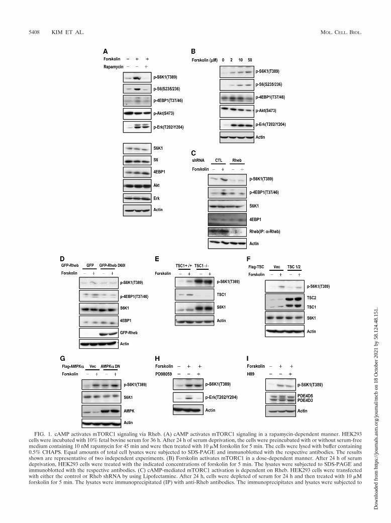
FIG. 1. cAMP activates mTORC1 signaling via Rheb. (A) cAMP activates mTORC1 signaling in a rapamycin-dependent manner. HEK293cells were incubated with 10% fetal bovine serum for 36 h. After 24 h of serum deprivation, the cells were preincubated with or without serum-freemedium containing 10 nM rapamycin for 45 min and were then treated with 10 �M forskolin for 5 min. The cells were lysed with buffer containing0.5% CHAPS. Equal amounts of total cell lysates were subjected to SDS-PAGE and immunoblotted with the respective antibodies. The resultsshown are representative of two independent experiments. (B) Forskolin activates mTORC1 in a dose-dependent manner. After 24 h of serumdeprivation, HEK293 cells were treated with the indicated concentrations of forskolin for 5 min. The lysates were subjected to SDS-PAGE andimmunoblotted with the respective antibodies. (C) cAMP-mediated mTORC1 activation is dependent on Rheb. HEK293 cells were transfectedwith either the control or Rheb shRNA by using Lipofectamine. After 24 h, cells were depleted of serum for 24 h and then treated with 10 �Mforskolin for 5 min. The lysates were immunoprecipitated (IP) with anti-Rheb antibodies. The immunoprecipitates and lysates were subjected to
5408 KIM ET AL. MOL. CELL. BIOL.
Dow
nloa
ded
from
http
s://j
ourn
als.
asm
.org
/jour
nal/m
cb o
n 18
Oct
ober
202
1 by
58.
124.
48.1
51.
sured by using a dual-luciferase reporter assay system (Labsystems, Ramsey,MN). Equal amounts of extract were used to assay the cap-dependent trans-lation of Renilla luciferase (R-Luc) or IRES-dependent translation of fireflyluciferase (F-Luc). Cap-dependent translation was calculated by normalizingthe R-Luc activity to the F-Luc activity as described previously (13, 47).
RESULTS
cAMP activates mTORC1 through Rheb. To examine therelationship between cAMP and mTORC1 and determinewhether cAMP increases mTORC1 activity, we investigatedcAMP-dependent mTORC1 activation and the requirement ofRheb in cAMP-mediated mTORC1 signaling. As shown in Fig.1A, we confirmed (42, 78), using forskolin, which is a directactivator of adenylyl cyclase, that cAMP augmented the phos-phorylation of S6K1 (T389) and 4EBP1 (T37/46), the best-characterized downstream effectors of mTORC1 (Fig. 1A).However, in these cells, cAMP did not alter the phosphoryla-tion of Akt (S473), a downstream effector of mTORC2 (Fig.1A). Furthermore, cAMP-mediated mTORC1 activation wasreduced by treatment with rapamycin, an mTORC1 inhibitor.However, rapamycin had no effect on either Akt or Erk (T202/Y204) phosphorylation. Despite this result, we found thatmTORC1 activity was enhanced by increasing concentrationsof the adenylyl cyclase activator forskolin (Fig. 1B). Indeed,mTORC1 activity was increased by cAMP-elevating agonists,such as isoproterenol and epinephrine (data not shown).
Next, we evaluated whether cAMP-mediated mTORC1 ac-tivation requires Rheb activity. Indeed, we found that the ac-tivation of mTORC1 by cAMP decreased when Rheb wassilenced (Fig. 1C). Furthermore, the overexpression of Rheb-D60I, a GDP-bound dominant negative form, diminished thelevels of phospho-S6K1 and phospho-4EBP1 (Fig. 1D).
We investigated whether TSC, the GAP for Rheb, has aneffect on cAMP-mediated mTORC1 activation. As shown inFig. 1E, treatment with forskolin elevated the levels of phos-pho-S6K1 in TSC1�/� MEFs (Fig. 1E). However, both theexpression and phosphorylation levels of S6K1 were highlyelevated in TSC�/� MEFs, while mTORC1 activity in TSC�/�
MEFs was not affected by forskolin (Fig. 1E). Indeed, thecoexpression of TSC1 and TSC2 had little effect on cAMP-
mediated mTORC1 activation (Fig. 1F). These results suggestthat TSC does not provide a main pathway for cAMP-medi-ated mTORC1 regulation.
It was reported previously that cAMP can regulate a number ofsignaling pathways, including AMP-activated protein kinase(AMPK) and mitogen-activated protein kinase (MAPK), whichcontrol mTORC1 (36, 77, 95). To examine the relationship be-tween cAMP and known pathways that control mTORC1, wechecked cAMP-dependent mTORC1 activation under conditionswhere either the AMPK or the MAPK pathways were inhibited.The overexpression of the dominant negative form of recombi-nant AMPK�1 increased basal phosphorylation levels of S6K1,while forskolin treatment elicited an increase in phospho-S6K1levels similar to those seen for transfections with the vector con-trol (Fig. 1G). Furthermore, PD98059, a MAPK inhibitor, did notaffect cAMP-mediated mTORC1 regulation (Fig. 1H). These re-sults indicate that cAMP-mediated mTORC1 activation requiresother distinct mechanisms in addition to TSC, AMPK, andMAPK pathways.
Protein kinase A (PKA) is a major target of cAMP (62), andcertain reports have shown that cAMP-mediated mTORC1signaling might be affected by PKA (78). Therefore, we inves-tigated whether cAMP-mediated mTORC1 activation re-quired PKA in our experimental system. However, we foundthat H89, a PKA inhibitor, did not have a significant effect onmTORC1 activity (Fig. 1I). These results suggest that thecAMP signal that we observed is linked to mTOR throughRheb but not through either TSC or PKA.
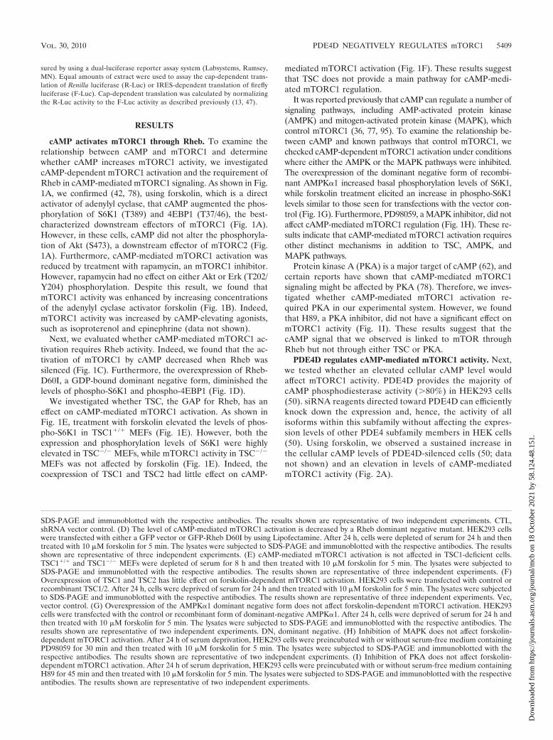
PDE4D regulates cAMP-mediated mTORC1 activity. Next,we tested whether an elevated cellular cAMP level wouldaffect mTORC1 activity. PDE4D provides the majority ofcAMP phosphodiesterase activity (�80%) in HEK293 cells(50). siRNA reagents directed toward PDE4D can efficientlyknock down the expression and, hence, the activity of allisoforms within this subfamily without affecting the expres-sion levels of other PDE4 subfamily members in HEK cells(50). Using forskolin, we observed a sustained increase inthe cellular cAMP levels of PDE4D-silenced cells (50; datanot shown) and an elevation in levels of cAMP-mediatedmTORC1 activity (Fig. 2A).
SDS-PAGE and immunoblotted with the respective antibodies. The results shown are representative of two independent experiments. CTL,shRNA vector control. (D) The level of cAMP-mediated mTORC1 activation is decreased by a Rheb dominant negative mutant. HEK293 cellswere transfected with either a GFP vector or GFP-Rheb D60I by using Lipofectamine. After 24 h, cells were depleted of serum for 24 h and thentreated with 10 �M forskolin for 5 min. The lysates were subjected to SDS-PAGE and immunoblotted with the respective antibodies. The resultsshown are representative of three independent experiments. (E) cAMP-mediated mTORC1 activation is not affected in TSC1-deficient cells.TSC1�/� and TSC1�/� MEFs were depleted of serum for 8 h and then treated with 10 �M forskolin for 5 min. The lysates were subjected toSDS-PAGE and immunoblotted with the respective antibodies. The results shown are representative of three independent experiments. (F)Overexpression of TSC1 and TSC2 has little effect on forskolin-dependent mTORC1 activation. HEK293 cells were transfected with control orrecombinant TSC1/2. After 24 h, cells were deprived of serum for 24 h and then treated with 10 �M forskolin for 5 min. The lysates were subjectedto SDS-PAGE and immunoblotted with the respective antibodies. The results shown are representative of three independent experiments. Vec,vector control. (G) Overexpression of the AMPK�1 dominant negative form does not affect forskolin-dependent mTORC1 activation. HEK293cells were transfected with the control or recombinant form of dominant-negative AMPK�1. After 24 h, cells were deprived of serum for 24 h andthen treated with 10 �M forskolin for 5 min. The lysates were subjected to SDS-PAGE and immunoblotted with the respective antibodies. Theresults shown are representative of two independent experiments. DN, dominant negative. (H) Inhibition of MAPK does not affect forskolin-dependent mTORC1 activation. After 24 h of serum deprivation, HEK293 cells were preincubated with or without serum-free medium containingPD98059 for 30 min and then treated with 10 �M forskolin for 5 min. The lysates were subjected to SDS-PAGE and immunoblotted with therespective antibodies. The results shown are representative of two independent experiments. (I) Inhibition of PKA does not affect forskolin-dependent mTORC1 activation. After 24 h of serum deprivation, HEK293 cells were preincubated with or without serum-free medium containingH89 for 45 min and then treated with 10 �M forskolin for 5 min. The lysates were subjected to SDS-PAGE and immunoblotted with the respectiveantibodies. The results shown are representative of two independent experiments.
VOL. 30, 2010 PDE4D NEGATIVELY REGULATES mTORC1 5409
Dow
nloa
ded
from
http
s://j
ourn
als.
asm
.org
/jour
nal/m
cb o
n 18
Oct
ober
202
1 by
58.
124.
48.1
51.
Since the PDE4D5 isoform is the major species present inHEK293 cells (50), we analyzed whether the overexpression ofrecombinant forms of either wild-type (wt) PDE4D5 or a cata-lytically inactive PDE4D5 mutant (D556A-PDE4D5) would af-fect mTORC1 activity. The overexpression of wt PDE4D5 but notD556A-PDE4D5 rapidly reduced intracellular cAMP levels (Fig.2B) and PKA activity (50, 60). However, although wt PDE4D5overexpression decreased cAMP-mediated mTORC1 activity(Fig. 2C), unexpectedly, we found that D556A-PDE4D5 also re-duced mTORC1 activation (Fig. 2C). Note that D556A-PDE4D5has no observable PDE activity and does not decrease intracel-lular cyclic AMP levels in HEK293 cells (Fig. 2B) (4, 50), sinceAsp556 provides a critical component of the catalytic active site(32). These results suggest that PDE4D5 may have a negative rolein regulating mTORC1 activation that is independent of its en-zymatic activity.
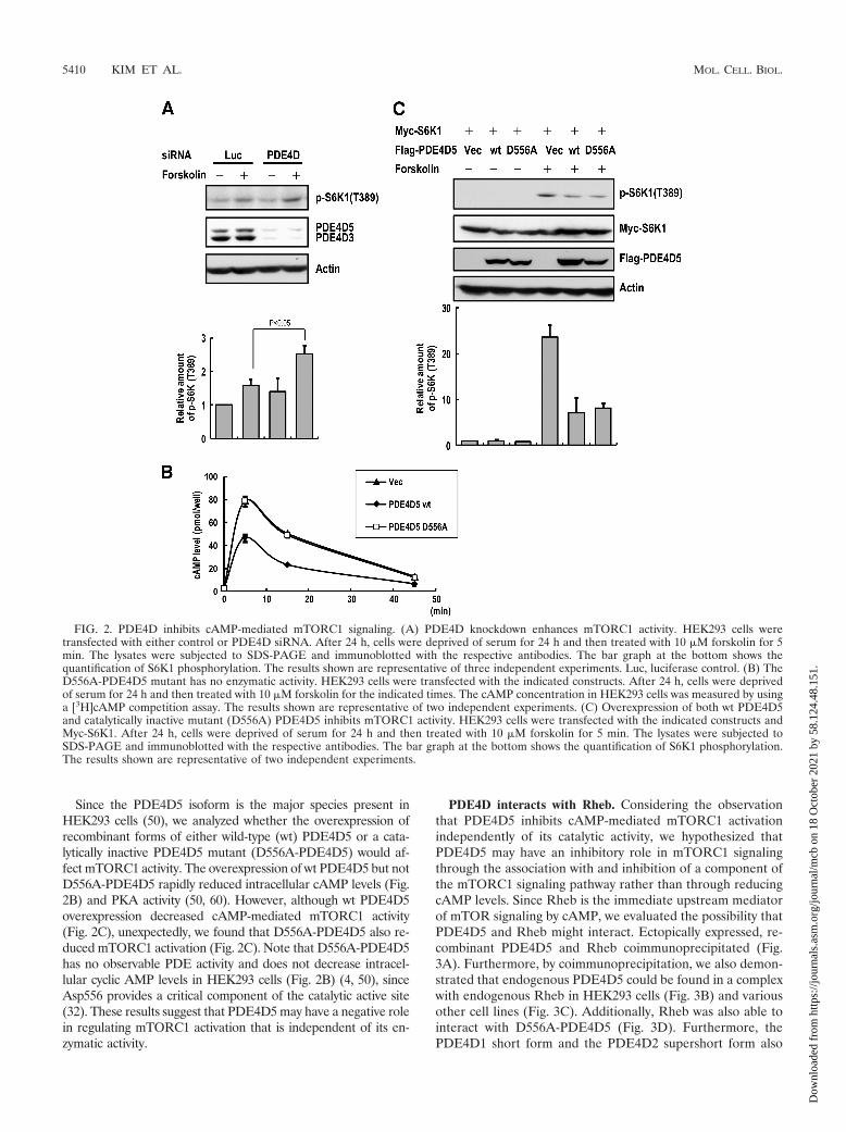
PDE4D interacts with Rheb. Considering the observationthat PDE4D5 inhibits cAMP-mediated mTORC1 activationindependently of its catalytic activity, we hypothesized thatPDE4D5 may have an inhibitory role in mTORC1 signalingthrough the association with and inhibition of a component ofthe mTORC1 signaling pathway rather than through reducingcAMP levels. Since Rheb is the immediate upstream mediatorof mTOR signaling by cAMP, we evaluated the possibility thatPDE4D5 and Rheb might interact. Ectopically expressed, re-combinant PDE4D5 and Rheb coimmunoprecipitated (Fig.3A). Furthermore, by coimmunoprecipitation, we also demon-strated that endogenous PDE4D5 could be found in a complexwith endogenous Rheb in HEK293 cells (Fig. 3B) and variousother cell lines (Fig. 3C). Additionally, Rheb was also able tointeract with D556A-PDE4D5 (Fig. 3D). Furthermore, thePDE4D1 short form and the PDE4D2 supershort form also
FIG. 2. PDE4D inhibits cAMP-mediated mTORC1 signaling. (A) PDE4D knockdown enhances mTORC1 activity. HEK293 cells weretransfected with either control or PDE4D siRNA. After 24 h, cells were deprived of serum for 24 h and then treated with 10 �M forskolin for 5min. The lysates were subjected to SDS-PAGE and immunoblotted with the respective antibodies. The bar graph at the bottom shows thequantification of S6K1 phosphorylation. The results shown are representative of three independent experiments. Luc, luciferase control. (B) TheD556A-PDE4D5 mutant has no enzymatic activity. HEK293 cells were transfected with the indicated constructs. After 24 h, cells were deprivedof serum for 24 h and then treated with 10 �M forskolin for the indicated times. The cAMP concentration in HEK293 cells was measured by usinga [3H]cAMP competition assay. The results shown are representative of two independent experiments. (C) Overexpression of both wt PDE4D5and catalytically inactive mutant (D556A) PDE4D5 inhibits mTORC1 activity. HEK293 cells were transfected with the indicated constructs andMyc-S6K1. After 24 h, cells were deprived of serum for 24 h and then treated with 10 �M forskolin for 5 min. The lysates were subjected toSDS-PAGE and immunoblotted with the respective antibodies. The bar graph at the bottom shows the quantification of S6K1 phosphorylation.The results shown are representative of two independent experiments.
5410 KIM ET AL. MOL. CELL. BIOL.
Dow
nloa
ded
from
http
s://j
ourn
als.
asm
.org
/jour
nal/m
cb o
n 18
Oct
ober
202
1 by
58.
124.
48.1
51.
coimmunoprecipitated with Rheb (Fig. 3E), suggesting thatthis binding is not an isoform-specific property but is associ-ated with regions common to all isoforms, which differ only intheir N-terminal regions.
Because Rheb is a GTPase, we investigated whether theassociation with and inhibition by PDE4D extended to othermembers of the GTPase superfamily or was binding specific forRheb. Thus, we analyzed the interaction of PDE4D5 withseveral related small GTPases and the interaction of Rheb withother PDE isotypes. Although PDE4D5 immunoprecipitatedwith Rheb, it did not immunoprecipitate with either Ras orRap2A (Fig. 3F), and Rheb did not interact with PDE5A (Fig.3G). These results suggest that PDE4D interacts only withspecific GTPases.
cAMP inhibits the interaction between Rheb and PDE4D.To identify the region of PDE4D responsible for interactionswith Rheb, we generated glutathione S-transferase (GST) fu-sions of PDE4D fragments (Fig. 4A). Using purified His-Rheband the GST-PDE4D fragments, we found that the catalyticdomain of PDE4D (F2) is responsible for binding to Rheb(Fig. 4B). Because the catalytic domain of PDE4D is also thebinding region for cAMP, we tested whether elevated cAMPlevels affected the association and inhibition of Rheb byPDE4D5. As shown in Fig. 4C, the Rheb-PDE4D interactionwas decreased upon the addition of cAMP in vitro. The cata-lytic activity of PDE4D is not involved in the dynamic interac-tion with Rheb, since the addition of cAMP similarly caused adissociation of Rheb from GST-F2-D556A (Fig. 4C). This in-
FIG. 3. PDE4D5 specifically interacts with Rheb. (A) Interaction be-tween recombinant PDE4D5 and Rheb. HEK293 cells were transfectedwith wild-type FLAG-PDE4D5 and HA-Rheb. After 36 h, the cell lysateswere immunoprecipitated with either anti-HA or anti-FLAG antibodies.Both PDE4D5-bound Rheb and Rheb-bound PDE4D5 were analyzed byanti-HA immunoblotting and anti-FLAG immunoblotting, respectively.The results shown are representative of two independent experiments.(B) Confirmation of the interaction between endogenous PDE4D5 andRheb. HEK293 cells were immunoprecipitated with Rheb-specific or con-trol goat IgG antibodies. Coimmunoprecipitated PDE4D5 was analyzedby anti-PDE4D antibodies. The results shown are representative of twoindependent experiments. (C) Rheb interacts with PDE4D5 in variouscell lines. Each cell lysate was immunoprecipitated with Rheb-specific orcontrol goat IgG antibodies. Coimmunoprecipitated PDE4D5 was ana-lyzed by immunoblotting with anti-PDE4D antibodies. (D) Interactionbetween the PDE4D5 catalytically inactive mutant and Rheb. HEK293cells transfected with wt PDE4D5 or D556A-PDE4D5 were immunopre-cipitated with anti-FLAG antibodies. Coimmunoprecipitated recombi-nant PDE4D5 was analyzed by anti-FLAG antibodies. The results shownare representative of two independent experiments. (E) Rheb interactswith all of the PDE4D splicing variants. HEK293 cells were transfectedwith the indicated constructs. After 36 h, the cell lysates were immuno-precipitated with anti-FLAG antibody. Coimmunoprecipitated Rheb wasanalyzed by anti-Rheb antibodies. The results shown are representative oftwo independent experiments. (F) PDE4D5 interacts with Rheb but notother small GTPases. HEK293 cells were transfected with the indicatedconstructs. After 36 h, the cell lysates were immunoprecipitated withanti-HA antibodies. Coimmunoprecipitated PDE4D5 was analyzed byanti-FLAG antibodies. The results shown are representative of two inde-pendent experiments. (G) Rheb interacts with PDE4D5 but not PDE5A.HEK293 cells were transfected with the indicated constructs. After 36 h,the cell lysates were immunoprecipitated with anti-HA antibodies. Coim-munoprecipitated PDE4D5 and mPDE5A were analyzed by anti-FLAGantibodies. The results shown are representative of two independent ex-periments.
VOL. 30, 2010 PDE4D NEGATIVELY REGULATES mTORC1 5411
Dow
nloa
ded
from
http
s://j
ourn
als.
asm
.org
/jour
nal/m
cb o
n 18
Oct
ober
202
1 by
58.
124.
48.1
51.
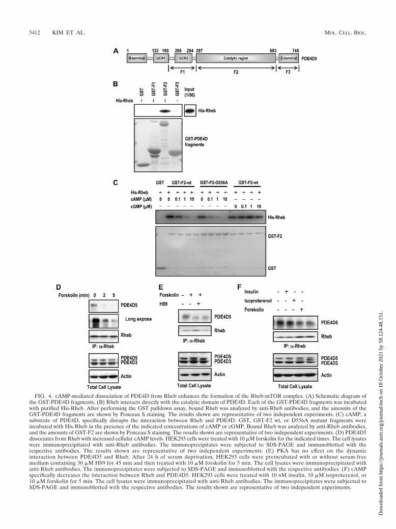
FIG. 4. cAMP-mediated dissociation of PDE4D from Rheb enhances the formation of the Rheb-mTOR complex. (A) Schematic diagram ofthe GST-PDE4D fragments. (B) Rheb interacts directly with the catalytic domain of PDE4D. Each of the GST-PDE4D fragments was incubatedwith purified His-Rheb. After performing the GST pulldown assay, bound Rheb was analyzed by anti-Rheb antibodies, and the amounts of theGST-PDE4D fragments are shown by Ponceau S staining. The results shown are representative of two independent experiments. (C) cAMP, asubstrate of PDE4D, specifically disrupts the interaction between Rheb and PDE4D. GST, GST-F2 wt, or D556A mutant fragments wereincubated with His-Rheb in the presence of the indicated concentrations of cAMP or cGMP. Bound Rheb was analyzed by anti-Rheb antibodies,and the amounts of GST-F2 are shown by Ponceau S staining. The results shown are representative of two independent experiments. (D) PDE4D5dissociates from Rheb with increased cellular cAMP levels. HEK293 cells were treated with 10 �M forskolin for the indicated times. The cell lysateswere immunoprecipitated with anti-Rheb antibodies. The immunoprecipitates were subjected to SDS-PAGE and immunoblotted with therespective antibodies. The results shown are representative of two independent experiments. (E) PKA has no effect on the dynamicinteraction between PDE4D5 and Rheb. After 24 h of serum deprivation, HEK293 cells were preincubated with or without serum-freemedium containing 30 �M H89 for 45 min and then treated with 10 �M forskolin for 5 min. The cell lysates were immunoprecipitated withanti-Rheb antibodies. The immunoprecipitates were subjected to SDS-PAGE and immunoblotted with the respective antibodies. (F) cAMPspecifically decreases the interaction between Rheb and PDE4D5. HEK293 cells were treated with 10 nM insulin, 10 �M isoproterenol, or10 �M forskolin for 5 min. The cell lysates were immunoprecipitated with anti-Rheb antibodies. The immunoprecipitates were subjected toSDS-PAGE and immunoblotted with the respective antibodies. The results shown are representative of two independent experiments.
5412 KIM ET AL. MOL. CELL. BIOL.
Dow
nloa
ded
from
http
s://j
ourn
als.
asm
.org
/jour
nal/m
cb o
n 18
Oct
ober
202
1 by
58.
124.
48.1
51.
teraction was not affected by the addition of cGMP (Fig. 4C),which neither is hydrolyzed by nor binds to PDE4 (32).
To confirm the dynamic interaction, we tested whethercAMP regulates the interaction between PDE4D and Rheb ina cellular system. The interaction of endogenous PDE4D withRheb was strongly reduced under conditions of elevated cAMPcellular levels, which was achieved by treatment with the ad-enylyl cyclase activator forskolin (Fig. 4D). The PKA inhibitorH89 had no effect on the ability of the elevation of cAMP levelsto disrupt the interaction between PDE4D5 and Rheb, whichcorrelated with the mTORC1 activity (Fig. 4E). We also ob-
served a similar result when isoproterenol, a �-adrenergic re-ceptor agonist that increases cAMP levels, was used to chal-lenge these cells (Fig. 4F). The interaction was not affected byinsulin treatment (Fig. 4F).
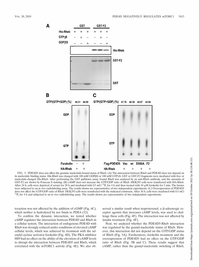
Next, we analyzed whether the PDE4D5-Rheb interactionwas regulated by the guanyl-nucleotide status of Rheb. How-ever, this interaction did not depend on the GTP/GDP statusof Rheb (Fig. 5A). Furthermore, forskolin treatment and theoverexpression of PDE4D5 had no effect on the GTP/GDPratio of Rheb (Fig. 5B and C). These results suggest thatcAMP, rather than the guanyl-nucleotide switching of Rheb,
FIG. 5. PDE4D5 does not affect the guanine nucleotide-bound status of Rheb. (A) The interaction between Rheb and PDE4D does not depend onits nucleotide binding status. His-Rheb was charged with 100 mM GDP�S or 100 mM GTP�S. GST or GST-F2 fragments were incubated with free ornucleotide-charged His-Rheb. After performing the GST pulldown assay, bound Rheb was analyzed by an anti-Rheb antibody, and the amounts ofGST-F2 are shown by Ponceau S staining. (B) cAMP does not increase the GTP/GDP ratio of Rheb. HEK293 cells were transfected with HA-Rheb.After 24 h, cells were deprived of serum for 20 h and incubated with 0.5 mCi 32Pi for 4 h and then treated with 10 �M forskolin for 5 min. The lysateswere subjected to an in vivo radiolabeling assay. The results shown are representative of two independent experiments. (C) Overexpression of PDE4D5does not affect the GTP/GDP ratio of Rheb. HEK293 cells were transfected with the indicated constructs. After 36 h, cells were incubated with 0.5 mCi32Pi for 4 h and subjected to an in vivo radiolabeling assay. The results shown are representative of two independent experiments.
VOL. 30, 2010 PDE4D NEGATIVELY REGULATES mTORC1 5413
Dow
nloa
ded
from
http
s://j
ourn
als.
asm
.org
/jour
nal/m
cb o
n 18
Oct
ober
202
1 by
58.
124.
48.1
51.
5414 KIM ET AL. MOL. CELL. BIOL.
Dow
nloa
ded
from
http
s://j
ourn
als.
asm
.org
/jour
nal/m
cb o
n 18
Oct
ober
202
1 by
58.
124.
48.1
51.
may be a direct regulator of the dynamic interaction betweenPDE4D and Rheb.
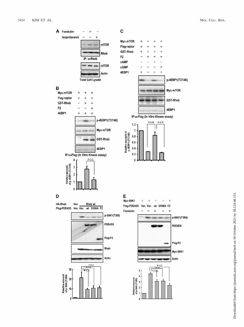
PDE4D negatively regulates mTORC1. Interestingly, forsko-lin treatment enhanced the interaction between Rheb andmTOR, which was inversely correlated with the reduction inthe Rheb-PDE4D interaction (Fig. 6A). Since the PDE4D-Rheb interaction was regulated by cAMP and PDE4D inhib-ited cAMP-induced mTORC1 activity, we next determinedwhether the interaction of PDE4D with Rheb affected Rheb-stimulated mTORC1 activity. We first analyzed in vitromTORC1 kinase activity using purified GST-Rheb and thePDE4D catalytic domain (F2-PDE4D). The results showedthat the increase in mTORC1 kinase activity induced by GST-Rheb was inhibited by the addition of F2-PDE4D (Fig. 6B).This inhibition was recovered upon the addition of cAMP but
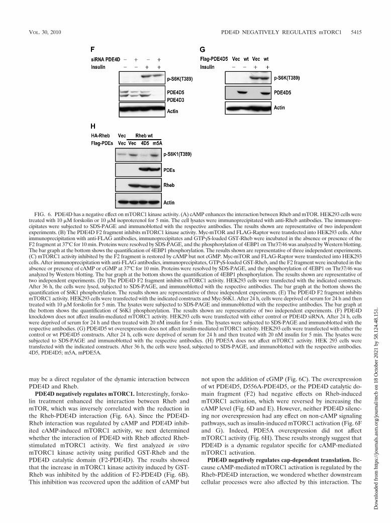
not upon the addition of cGMP (Fig. 6C). The overexpressionof wt PDE4D5, D556A-PDE4D5, or the PDE4D catalytic do-main fragment (F2) had negative effects on Rheb-inducedmTORC1 activation, which were reversed by increasing thecAMP level (Fig. 6D and E). However, neither PDE4D silenc-ing nor overexpression had any effect on non-cAMP signalingpathways, such as insulin-induced mTORC1 activation (Fig. 6Fand G). Indeed, PDE5A overexpression did not affectmTORC1 activity (Fig. 6H). These results strongly suggest thatPDE4D is a dynamic regulator specific for cAMP-mediatedmTORC1 activation.
PDE4D negatively regulates cap-dependent translation. Be-cause cAMP-mediated mTORC1 activation is regulated by theRheb-PDE4D interaction, we wondered whether downstreamcellular processes were also affected by this interaction. The
FIG. 6. PDE4D has a negative effect on mTORC1 kinase activity. (A) cAMP enhances the interaction between Rheb and mTOR. HEK293 cells weretreated with 10 �M forskolin or 10 �M isoproterenol for 5 min. The cell lysates were immunoprecipitated with anti-Rheb antibodies. The immunopre-cipitates were subjected to SDS-PAGE and immunoblotted with the respective antibodies. The results shown are representative of two independentexperiments. (B) The PDE4D F2 fragment inhibits mTORC1 kinase activity. Myc-mTOR and FLAG-Raptor were transfected into HEK293 cells. Afterimmunoprecipitation with anti-FLAG antibodies, immunoprecipitates and GTP�S-loaded GST-Rheb were incubated in the absence or presence of theF2 fragment at 37°C for 10 min. Proteins were resolved by SDS-PAGE, and the phosphorylation of 4EBP1 on Thr37/46 was analyzed by Western blotting.The bar graph at the bottom shows the quantification of 4EBP1 phosphorylation. The results shown are representative of three independent experiments.(C) mTORC1 activity inhibited by the F2 fragment is restored by cAMP but not cGMP. Myc-mTOR and FLAG-Raptor were transfected into HEK293cells. After immunoprecipitation with anti-FLAG antibodies, immunoprecipitates, GTP�S-loaded GST-Rheb, and the F2 fragment were incubated in theabsence or presence of cAMP or cGMP at 37°C for 10 min. Proteins were resolved by SDS-PAGE, and the phosphorylation of 4EBP1 on Thr37/46 wasanalyzed by Western blotting. The bar graph at the bottom shows the quantification of 4EBP1 phosphorylation. The results shown are representative oftwo independent experiments. (D) The PDE4D F2 fragment inhibits mTORC1 activity. HEK293 cells were transfected with the indicated constructs.After 36 h, the cells were lysed, subjected to SDS-PAGE, and immunoblotted with the respective antibodies. The bar graph at the bottom shows thequantification of S6K1 phosphorylation. The results shown are representative of three independent experiments. (E) The PDE4D F2 fragment inhibitsmTORC1 activity. HEK293 cells were transfected with the indicated constructs and Myc-S6K1. After 24 h, cells were deprived of serum for 24 h and thentreated with 10 �M forskolin for 5 min. The lysates were subjected to SDS-PAGE and immunoblotted with the respective antibodies. The bar graph atthe bottom shows the quantification of S6K1 phosphorylation. The results shown are representative of two independent experiments. (F) PDE4Dknockdown does not affect insulin-mediated mTORC1 activity. HEK293 cells were transfected with either control or PDE4D siRNA. After 24 h, cellswere deprived of serum for 24 h and then treated with 20 nM insulin for 5 min. The lysates were subjected to SDS-PAGE and immunoblotted with therespective antibodies. (G) PDE4D5 wt overexpression does not affect insulin-mediated mTORC1 activity. HEK293 cells were transfected with either thecontrol or wt PDE4D5 constructs. After 24 h, cells were deprived of serum for 24 h and then treated with 20 nM insulin for 5 min. The lysates weresubjected to SDS-PAGE and immunoblotted with the respective antibodies. (H) PDE5A does not affect mTORC1 activity. HEK 293 cells weretransfected with the indicated constructs. After 36 h, the cells were lysed, subjected to SDS-PAGE, and immunoblotted with the respective antibodies.4D5, PDE4D5; m5A, mPDE5A.
VOL. 30, 2010 PDE4D NEGATIVELY REGULATES mTORC1 5415
Dow
nloa
ded
from
http
s://j
ourn
als.
asm
.org
/jour
nal/m
cb o
n 18
Oct
ober
202
1 by
58.
124.
48.1
51.
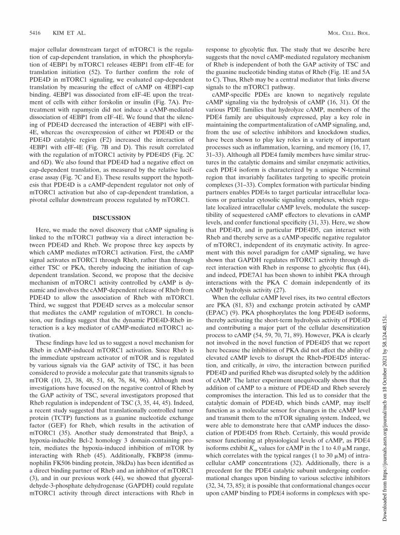
major cellular downstream target of mTORC1 is the regula-tion of cap-dependent translation, in which the phosphoryla-tion of 4EBP1 by mTORC1 releases 4EBP1 from eIF-4E fortranslation initiation (52). To further confirm the role ofPDE4D in mTORC1 signaling, we evaluated cap-dependenttranslation by measuring the effect of cAMP on 4EBP1-capbinding. 4EBP1 was dissociated from eIF-4E upon the treat-ment of cells with either forskolin or insulin (Fig. 7A). Pre-treatment with rapamycin did not induce a cAMP-mediateddissociation of 4EBP1 from eIF-4E. We found that the silenc-ing of PDE4D decreased the interaction of 4EBP1 with eIF-4E, whereas the overexpression of either wt PDE4D or thePDE4D catalytic region (F2) increased the interaction of4EBP1 with eIF-4E (Fig. 7B and D). This result correlatedwith the regulation of mTORC1 activity by PDE4D5 (Fig. 2Cand 6D). We also found that PDE4D had a negative effect oncap-dependent translation, as measured by the relative lucif-erase assay (Fig. 7C and E). These results support the hypoth-esis that PDE4D is a cAMP-dependent regulator not only ofmTORC1 activation but also of cap-dependent translation, apivotal cellular downstream process regulated by mTORC1.
DISCUSSION
Here, we made the novel discovery that cAMP signaling islinked to the mTORC1 pathway via a direct interaction be-tween PDE4D and Rheb. We propose three key aspects bywhich cAMP mediates mTORC1 activation. First, the cAMPsignal activates mTORC1 through Rheb, rather than througheither TSC or PKA, thereby inducing the initiation of cap-dependent translation. Second, we propose that the decisivemechanism of mTORC1 activity controlled by cAMP is dy-namic and involves the cAMP-dependent release of Rheb fromPDE4D to allow the association of Rheb with mTORC1.Third, we suggest that PDE4D serves as a molecular sensorthat mediates the cAMP regulation of mTORC1. In conclu-sion, our findings suggest that the dynamic PDE4D-Rheb in-teraction is a key mediator of cAMP-mediated mTORC1 ac-tivation.
These findings have led us to suggest a novel mechanism forRheb in cAMP-induced mTORC1 activation. Since Rheb isthe immediate upstream activator of mTOR and is regulatedby various signals via the GAP activity of TSC, it has beenconsidered to provide a molecular gate that transmits signals tomTOR (10, 23, 38, 48, 51, 68, 76, 84, 96). Although mostinvestigations have focused on the negative control of Rheb bythe GAP activity of TSC, several investigators proposed thatRheb regulation is independent of TSC (3, 35, 44, 45). Indeed,a recent study suggested that translationally controlled tumorprotein (TCTP) functions as a guanine nucleotide exchangefactor (GEF) for Rheb, which results in the activation ofmTORC1 (35). Another study demonstrated that Bnip3, ahypoxia-inducible Bcl-2 homology 3 domain-containing pro-tein, mediates the hypoxia-induced inhibition of mTOR byinteracting with Rheb (45). Additionally, FKBP38 (immu-nophilin FK506 binding protein, 38kDa) has been identified asa direct binding partner of Rheb and an inhibitor of mTORC1(3), and in our previous work (44), we showed that glyceral-dehyde-3-phosphate dehydrogenase (GAPDH) could regulatemTORC1 activity through direct interactions with Rheb in
response to glycolytic flux. The study that we describe heresuggests that the novel cAMP-mediated regulatory mechanismof Rheb is independent of both the GAP activity of TSC andthe guanine nucleotide binding status of Rheb (Fig. 1E and 5Ato C). Thus, Rheb may be a central mediator that links diversesignals to the mTORC1 pathway.
cAMP-specific PDEs are known to negatively regulatecAMP signaling via the hydrolysis of cAMP (16, 31). Of thevarious PDE families that hydrolyze cAMP, members of thePDE4 family are ubiquitously expressed, play a key role inmaintaining the compartmentalization of cAMP signaling, and,from the use of selective inhibitors and knockdown studies,have been shown to play key roles in a variety of importantprocesses such as inflammation, learning, and memory (16, 17,31–33). Although all PDE4 family members have similar struc-tures in the catalytic domains and similar enzymatic activities,each PDE4 isoform is characterized by a unique N-terminalregion that invariably facilitates targeting to specific proteincomplexes (31–33). Complex formation with particular bindingpartners enables PDE4s to target particular intracellular loca-tions or particular cytosolic signaling complexes, which regu-late localized intracellular cAMP levels, modulate the suscep-tibility of sequestered cAMP effectors to elevations in cAMPlevels, and confer functional specificity (31, 33). Here, we showthat PDE4D, and in particular PDE4D5, can interact withRheb and thereby serve as a cAMP-specific negative regulatorof mTORC1, independent of its enzymatic activity. In agree-ment with this novel paradigm for cAMP signaling, we haveshown that GAPDH regulates mTORC1 activity through di-rect interaction with Rheb in response to glycolytic flux (44),and indeed, PDE7A1 has been shown to inhibit PKA throughinteractions with the PKA C domain independently of itscAMP hydrolysis activity (27).
When the cellular cAMP level rises, its two central effectorsare PKA (81, 83) and exchange protein activated by cAMP(EPAC) (9). PKA phosphorylates the long PDE4D isoforms,thereby activating the short-term hydrolysis activity of PDE4Dand contributing a major part of the cellular desensitizationprocess to cAMP (54, 59, 70, 71, 89). However, PKA is clearlynot involved in the novel function of PDE4D5 that we reporthere because the inhibition of PKA did not affect the ability ofelevated cAMP levels to disrupt the Rheb-PDE4D5 interac-tion, and critically, in vitro, the interaction between purifiedPDE4D and purified Rheb was disrupted solely by the additionof cAMP. The latter experiment unequivocally shows that theaddition of cAMP to a mixture of PDE4D and Rheb severelycompromises the interaction. This led us to consider that thecatalytic domain of PDE4D, which binds cAMP, may itselffunction as a molecular sensor for changes in the cAMP leveland transmit them to the mTOR signaling system. Indeed, wewere able to demonstrate here that cAMP induces the disso-ciation of PDE4D5 from Rheb. Certainly, this would providesensor functioning at physiological levels of cAMP, as PDE4isoforms exhibit Km values for cAMP in the 1 to 4.0 �M range,which correlates with the typical ranges (1 to 30 �M) of intra-cellular cAMP concentrations (32). Additionally, there is aprecedent for the PDE4 catalytic subunit undergoing confor-mational changes upon binding to various selective inhibitors(32, 34, 73, 85); it is possible that conformational changes occurupon cAMP binding to PDE4 isoforms in complexes with spe-
5416 KIM ET AL. MOL. CELL. BIOL.
Dow
nloa
ded
from
http
s://j
ourn
als.
asm
.org
/jour
nal/m
cb o
n 18
Oct
ober
202
1 by
58.
124.
48.1
51.
FIG. 7. PDE4D has a negative effect on cap-dependent translation efficiency. (A) cAMP decreases the interaction between 4EBP1 and the capstructure. After 24 h of serum deprivation, HEK293 cells were preincubated with or without serum-free medium containing 10 nM rapamycin for45 min and were then treated with 10 �M forskolin or 20 nM insulin for 5 min. The cell lysates were incubated with 10 �l of m7-GTP Sepharosebeads for 4 h. The resulting beads were subjected to SDS-PAGE and immunoblotted with the respective antibodies. The results shown arerepresentative of two independent experiments. (B) PDE4D knockdown disrupts the interaction between 4EBP1 and the cap structure. HEK293cells were transfected with either control or PDE4D siRNA. After 24 h, cells were deprived of serum for 24 h and then treated with 10 �M forskolinfor 5 min. After the pulldown assay was performed, the resulting beads were subjected to SDS-PAGE and immunoblotted with the respectiveantibodies. The results shown are representative of two independent experiments. (C) PDE4D knockdown enhances cap-dependent translation.HEK293 cells were transfected with either control or PDE4D siRNA. After 24 h, cells were deprived of serum for 24 h and then treated with 10�M forskolin for 36 h. The resulting lysates were measured by the dual-luciferase reporter assay. The results shown are representative of twoindependent experiments. NT, no treatment. (D) PDE4D5 overexpression enhances the interaction between 4EBP1 and the cap structure.HEK293 cells were transfected with the indicated constructs. After 36 h, the cell lysates were incubated with 10 �l of m7-GTP Sepharose beadsfor 4 h. The resulting beads were subjected to SDS-PAGE and immunoblotted with the respective antibodies. The results shown are representativeof two independent experiments. (E) PDE4D5 overexpression inhibits cap-dependent translation. HEK293 cells were transfected with theindicated constructs. After 36 h, the resulting lysates were measured by the dual-luciferase reporter assay. The results shown are representativeof two independent experiments.
VOL. 30, 2010 PDE4D NEGATIVELY REGULATES mTORC1 5417
Dow
nloa
ded
from
http
s://j
ourn
als.
asm
.org
/jour
nal/m
cb o
n 18
Oct
ober
202
1 by
58.
124.
48.1
51.
cific partner proteins. All of the PDE4 crystal structures re-ported to date have focused on the core catalytic unit withvarious bound inhibitors (88). However, there are no crystalstructures of either full-length PDE4 or the enzyme in complexwith proteins that sequester it, and thus, we have no knowledgeof structural changes from such approaches. However, bindingstudies of inhibitors, phosphorylation states, and the binding ofpartner proteins have all identified various marked changes inactivity, thermostability, and inhibitor binding that are consis-tent with profound conformational changes (32, 33, 73). In-deed, there is a large amount of literature indicating thatPDE4 isoforms can adopt multiple conformational states (32,34, 73). We suggest here that the release of Rheb from PDE4Dmight thus be dynamically controlled in cells by cAMP binding.Moreover, we also observed that isoproterenol, a �-adrenergicreceptor agonist that increases cAMP levels, induced the dis-sociation of PDE4D5 from Rheb in cells. These results implythat the regulatory mechanism of Rheb by PDE4D5 may con-sist of the binding of cAMP to the PDE4D catalytic site, whichthen displaces Rheb from PDE4D5. Whether this occursthough competition between Rheb and cAMP for binding toPDE4D5 or through an allosteric conformational change re-mains to be determined. It is interesting that the RACK1signaling scaffold binds to PDE4D5 not only at a site within itsunique N-terminal domain but also at the catalytic domain,where it abuts the cAMP binding site (7) and causes a confor-mational change that is detectable through an altered bindingof the inhibitor rolipram to this enzyme (94). A crystal struc-ture of the complex of PDE4D and Rheb may provide criticalevidence for this hypothesis, although this will be a challengingendeavor, since to date, the full-length PDE4 structure has notbeen obtained due to the propensity of the full-length enzymeto aggregate.
It was shown previously that the major function of cAMP isto modulate the transcription of target genes through thePKA-CREB pathway (64, 72). However, there have been a fewreports showing the cAMP-mediated regulation of translation;for example, it was shown previously that cAMP induces themRNA translation of tyrosine hydroxylase in dopaminergicneurons (14, 22, 92). Here, we show that cAMP also has apositive effect on cap-dependent translation via the mTORC1pathway. As a general rule, the regulation of translation pro-vides cells with the flexibility to rapidly respond to environ-mental alterations under certain conditions, such as apoptosis,that require immediate changes in protein levels (29). If cAMPregulates both transcription and translation, cells may havediverse and rapid responses for various environmental condi-tions. Further studies are needed to identify the environmentalconditions that induce cAMP-mediated cap-dependent trans-lation.
mTORC1 is a molecular hub that receives various signalsfrom the extracellular environment to modulate protein syn-thesis. Cells may have developed multiple ways to regulatemTORC1 signaling in response to diverse environmental cues.The best-characterized upstream regulators of mTORC1 areTSC and Rheb. In addition to regulation by TSC-Rheb,PRAS40 and Rag GTPase have been identified as mTORC1regulators in response to insulin and amino acids, respectively(65, 66, 86). Here, we have identified a novel function forPDE4D in cAMP-mediated mTORC1 activation. According to
our model, PDE4D has dual roles as a specific sensor of cAMPand a cAMP-hydrolyzing enzyme. When the cellular cAMPlevel rises, PDE4D recognizes the increased level and dissoci-ates from Rheb, thereby enhancing the interaction betweenRheb and mTOR. Simultaneously, free PDE4D may functionas a negative-feedback regulator via the hydrolysis of cAMPthat surrounds mTORC1. Thus, cells may have an advancedmechanism that allows mTORC1 to immediately respond tothe cAMP signal via the dual function of PDE4D. The possiblenegative regulation of PDE4D by Rheb binding under basalconditions and of the cAMP-induced activation of PDE4Dmay be interesting for future studies aimed at elucidating ad-ditional details of the cross talk between the cAMP and mTORpathways.
ACKNOWLEDGMENTS
We thank A. F. Castro for providing the Rheb constructs and D. M.Sabatini for the Myc-mTOR and HA-Raptor constructs. We alsothank J. Blenis for providing the Myc-S6K, FLAG-TSC1, and FLAG-TSC2 constructs. We thank D. J. Kwiatkowski for the TSC1 MEFs.
This work was supported by grant FRP08B1-160 of the 21C Fron-tier Functional Proteomics Project and by grant KPF-2008-005-C00036 of the Global Research Network Program from the KoreanMinistry of Education, Science, and Technology. Work in the lab-oratory of M.D.H. was supported by grants from the Medical Re-search Council (United Kingdom) (grant G0600765) and the Fon-dation Leducg (grant 06CVD02).
We declare that no conflict of interests exists.
REFERENCES
1. Alvarez, R., C. Sette, D. Yang, R. M. Eglen, R. Wilhelm, E. R. Shelton, andM. Conti. 1995. Activation and selective inhibition of a cyclic AMP-specificphosphodiesterase, PDE-4D3. Mol. Pharmacol. 48:616–622.
2. Aspuria, P. J., and F. Tamanoi. 2004. The Rheb family of GTP-bindingproteins. Cell. Signal. 16:1105–1112.
3. Bai, X., D. Ma, A. Liu, X. Shen, Q. J. Wang, Y. Liu, and Y. Jiang. 2007. Rhebactivates mTOR by antagonizing its endogenous inhibitor, FKBP38. Science318:977–980.
4. Baillie, G. S., E. Huston, G. Scotland, M. Hodgkin, I. Gall, A. H. Peden, C.MacKenzie, E. S. Houslay, R. Currie, T. R. Pettitt, A. R. Walmsley, M. J.Wakelam, J. Warwicker, and M. D. Houslay. 2002. TAPAS-1, a novel mi-crodomain within the unique N-terminal region of the PDE4A1 cAMP-specific phosphodiesterase that allows rapid, Ca2�-triggered membrane as-sociation with selectivity for interaction with phosphatidic acid. J. Biol.Chem. 277:28298–28309.
5. Baillie, G. S., A. Sood, I. McPhee, I. Gall, S. J. Perry, R. J. Lefkowitz, andM. D. Houslay. 2003. Beta-arrestin-mediated PDE4 cAMP phosphodiester-ase recruitment regulates beta-adrenoceptor switching from Gs to Gi. Proc.Natl. Acad. Sci. U. S. A. 100:940–945.
6. Beard, M. B., A. E. Olsen, R. E. Jones, S. Erdogan, M. D. Houslay, and G. B.Bolger. 2000. UCR1 and UCR2 domains unique to the cAMP-specific phos-phodiesterase family form a discrete module via electrostatic interactions.J. Biol. Chem. 275:10349–10358.
7. Bolger, G. B., G. S. Baillie, X. Li, M. J. Lynch, P. Herzyk, A. Mohamed, L. H.Mitchell, A. McCahill, C. Hundsrucker, E. Klussmann, D. R. Adams, andM. D. Houslay. 2006. Scanning peptide array analyses identify overlappingbinding sites for the signalling scaffold proteins, beta-arrestin and RACK1, incAMP-specific phosphodiesterase PDE4D5. Biochem. J. 398:23–36.
8. Bolger, G. B., S. Erdogan, R. E. Jones, K. Loughney, G. Scotland, R. Hoff-mann, I. Wilkinson, C. Farrell, and M. D. Houslay. 1997. Characterization offive different proteins produced by alternatively spliced mRNAs from thehuman cAMP-specific phosphodiesterase PDE4D gene. Biochem. J. 328(Pt.2):539–548.
9. Bos, J. L. 2006. Epac proteins: multi-purpose cAMP targets. Trends Bio-chem. Sci. 31:680–686.
10. Brugarolas, J., K. Lei, R. L. Hurley, B. D. Manning, J. H. Reiling, E. Hafen,L. A. Witters, L. W. Ellisen, and W. G. Kaelin, Jr. 2004. Regulation ofmTOR function in response to hypoxia by REDD1 and the TSC1/TSC2tumor suppressor complex. Genes Dev. 18:2893–2904.
11. Brunn, G. J., C. C. Hudson, A. Sekulic, J. M. Williams, H. Hosoi, P. J.Houghton, J. C. Lawrence, Jr., and R. T. Abraham. 1997. Phosphorylation ofthe translational repressor PHAS-I by the mammalian target of rapamycin.Science 277:99–101.
12. Burnett, P. E., R. K. Barrow, N. A. Cohen, S. H. Snyder, and D. M. Sabatini.
5418 KIM ET AL. MOL. CELL. BIOL.
Dow
nloa
ded
from
http
s://j
ourn
als.
asm
.org
/jour
nal/m
cb o
n 18
Oct
ober
202
1 by
58.
124.
48.1
51.
1998. RAFT1 phosphorylation of the translational regulators p70 S6 kinaseand 4E-BP1. Proc. Natl. Acad. Sci. U. S. A. 95:1432–1437.
13. Chappell, S. A., J. P. LeQuesne, F. E. Paulin, M. L. deSchoolmeester, M.Stoneley, R. L. Soutar, S. H. Ralston, M. H. Helfrich, and A. E. Willis. 2000.A mutation in the c-myc-IRES leads to enhanced internal ribosome entry inmultiple myeloma: a novel mechanism of oncogene de-regulation. Oncogene19:4437–4440.
14. Chen, X., L. Xu, P. Radcliffe, B. Sun, and A. W. Tank. 2008. Activation oftyrosine hydroxylase mRNA translation by cAMP in midbrain dopaminergicneurons. Mol. Pharmacol. 73:1816–1828.
15. Collins, D. M., H. Murdoch, A. J. Dunlop, E. Charych, G. S. Baillie, Q.Wang, F. W. Herberg, N. Brandon, A. Prinz, and M. D. Houslay. 2008. Ndel1alters its conformation by sequestering cAMP-specific phosphodiesterase-4D3 (PDE4D3) in a manner that is dynamically regulated through proteinkinase A (PKA). Cell. Signal. 20:2356–2369.
16. Conti, M., and J. Beavo. 2007. Biochemistry and physiology of cyclic nucle-otide phosphodiesterases: essential components in cyclic nucleotide signal-ing. Annu. Rev. Biochem. 76:481–511.
17. Conti, M., W. Richter, C. Mehats, G. Livera, J. Y. Park, and C. Jin. 2003.Cyclic AMP-specific PDE4 phosphodiesterases as critical components ofcyclic AMP signaling. J. Biol. Chem. 278:5493–5496.
18. Creighton, J., B. Zhu, M. Alexeyev, and T. Stevens. 2008. Spectrin-anchoredphosphodiesterase 4D4 restricts cAMP from disrupting microtubules andinducing endothelial cell gap formation. J. Cell Sci. 121:110–119.
19. Di Benedetto, G., A. Zoccarato, V. Lissandron, A. Terrin, X. Li, M. D.Houslay, G. S. Baillie, and M. Zaccolo. 2008. Protein kinase A type I andtype II define distinct intracellular signaling compartments. Circ. Res. 103:836–844.
20. Dodge, K. L., S. Khouangsathiene, M. S. Kapiloff, R. Mouton, E. V. Hill,M. D. Houslay, L. K. Langeberg, and J. D. Scott. 2001. mAKAP assemblesa protein kinase A/PDE4 phosphodiesterase cAMP signaling module.EMBO J. 20:1921–1930.
21. Fang, Y., M. Vilella-Bach, R. Bachmann, A. Flanigan, and J. Chen. 2001.Phosphatidic acid-mediated mitogenic activation of mTOR signaling. Sci-ence 294:1942–1945.
22. Fuller, S. J., C. J. Gaitanaki, and P. H. Sugden. 1990. Effects of cat-echolamines on protein synthesis in cardiac myocytes and perfused heartsisolated from adult rats. Stimulation of translation is mediated through thealpha 1-adrenoceptor. Biochem. J. 266:727–736.
23. Garami, A., F. J. Zwartkruis, T. Nobukuni, M. Joaquin, M. Roccio, H.Stocker, S. C. Kozma, E. Hafen, J. L. Bos, and G. Thomas. 2003. Insulinactivation of Rheb, a mediator of mTOR/S6K/4E-BP signaling, is inhibitedby TSC1 and 2. Mol. Cell 11:1457–1466.
24. Gingras, A. C., B. Raught, and N. Sonenberg. 2004. mTOR signaling totranslation. Curr. Top. Microbiol. Immunol. 279:169–197.
25. Gingras, A. C., B. Raught, and N. Sonenberg. 2001. Regulation of translationinitiation by FRAP/mTOR. Genes Dev. 15:807–826.
26. Ha, S. H., D. H. Kim, I. S. Kim, J. H. Kim, M. N. Lee, H. J. Lee, S. K. Jang,P. G. Suh, and S. H. Ryu. 2006. PLD2 forms a functional complex withmTOR/raptor to transduce mitogenic signals. Cell. Signal. 18:2283–2291.
27. Han, P., P. Sonati, C. Rubin, and T. Michaeli. 2006. PDE7A1, a cAMP-specific phosphodiesterase, inhibits cAMP-dependent protein kinase by adirect interaction with C. J. Biol. Chem. 281:15050–15057.
28. Hoffmann, R., I. R. Wilkinson, J. F. McCallum, P. Engels, and M. D.Houslay. 1998. cAMP-specific phosphodiesterase HSPDE4D3 mutantswhich mimic activation and changes in rolipram inhibition triggered byprotein kinase A phosphorylation of Ser-54: generation of a molecularmodel. Biochem. J. 333(Pt. 1):139–149.
29. Holcik, M., and N. Sonenberg. 2005. Translational control in stress andapoptosis. Nat. Rev. Mol. Cell Biol. 6:318–327.
30. Holz, M. K., and J. Blenis. 2005. Identification of S6 kinase 1 as a novelmammalian target of rapamycin (mTOR)-phosphorylating kinase. J. Biol.Chem. 280:26089–26093.
31. Houslay, M. D. 2009. Underpinning compartmentalised cAMP signallingthrough targeted cAMP breakdown. Trends Biochem. Sci. 35:91–100.
32. Houslay, M. D., and D. R. Adams. 2003. PDE4 cAMP phosphodiesterases:modular enzymes that orchestrate signalling cross-talk, desensitization andcompartmentalization. Biochem. J. 370:1–18.
33. Houslay, M. D., G. S. Baillie, and D. H. Maurice. 2007. cAMP-specificphosphodiesterase-4 enzymes in the cardiovascular system: a molecular tool-box for generating compartmentalized cAMP signaling. Circ. Res. 100:950–966.
34. Houslay, M. D., P. Schafer, and K. Y. Zhang. 2005. Keynote review: phos-phodiesterase-4 as a therapeutic target. Drug Discov. Today 10:1503–1519.
35. Hsu, Y. C., J. J. Chern, Y. Cai, M. Liu, and K. W. Choi. 2007. DrosophilaTCTP is essential for growth and proliferation through regulation of dRhebGTPase. Nature 445:785–788.
36. Hutchinson, D. S., E. Chernogubova, O. S. Dallner, B. Cannon, and T.Bengtsson. 2005. Beta-adrenoceptors, but not alpha-adrenoceptors, stimu-late AMP-activated protein kinase in brown adipocytes independently ofuncoupling protein-1. Diabetologia 48:2386–2395.
37. Inoki, K., and K. L. Guan. 2006. Complexity of the TOR signaling network.Trends Cell Biol. 16:206–212.
38. Inoki, K., T. Zhu, and K. L. Guan. 2003. TSC2 mediates cellular energyresponse to control cell growth and survival. Cell 115:577–590.
39. Jacinto, E., and M. N. Hall. 2003. Tor signalling in bugs, brain and brawn.Nat. Rev. Mol. Cell Biol. 4:117–126.
40. Kim, D. H., D. D. Sarbassov, S. M. Ali, J. E. King, R. R. Latek, H. Erdju-ment-Bromage, P. Tempst, and D. M. Sabatini. 2002. mTOR interacts withraptor to form a nutrient-sensitive complex that signals to the cell growthmachinery. Cell 110:163–175.
41. Kim, J. H., K. Y. Paek, K. Choi, T. D. Kim, B. Hahm, K. T. Kim, and S. K.Jang. 2003. Heterogeneous nuclear ribonucleoprotein C modulates transla-tion of c-myc mRNA in a cell cycle phase-dependent manner. Mol. Cell.Biol. 23:708–720.
42. Kwon, G., C. A. Marshall, K. L. Pappan, M. S. Remedi, and M. L. McDaniel.2004. Signaling elements involved in the metabolic regulation of mTOR bynutrients, incretins, and growth factors in islets. Diabetes 53(Suppl. 3):S225–S232.
43. Lecureuil, C., S. Tesseraud, E. Kara, N. Martinat, A. Sow, I. Fontaine, C.Gauthier, E. Reiter, F. Guillou, and P. Crepieux. 2005. Follicle-stimulatinghormone activates p70 ribosomal protein S6 kinase by protein kinase A-me-diated dephosphorylation of Thr 421/Ser 424 in primary Sertoli cells. Mol.Endocrinol. 19:1812–1820.
44. Lee, M. N., S. H. Ha, J. Kim, A. Koh, C. S. Lee, J. H. Kim, H. Jeon, D. H.Kim, P. G. Suh, and S. H. Ryu. 2009. Glycolytic flux signals to mTORthrough glyceraldehyde-3-phosphate dehydrogenase-mediated regulation ofRheb. Mol. Cell. Biol. 29:3991–4001.
45. Li, Y., Y. Wang, E. Kim, P. Beemiller, C. Y. Wang, J. Swanson, M. You, andK. L. Guan. 2007. Bnip3 mediates the hypoxia-induced inhibition on mam-malian target of rapamycin by interacting with Rheb. J. Biol. Chem. 282:35803–35813.
46. Lim, J., G. Pahlke, and M. Conti. 1999. Activation of the cAMP-specificphosphodiesterase PDE4D3 by phosphorylation. Identification and functionof an inhibitory domain. J. Biol. Chem. 274:19677–19685.
47. Ling, J., S. J. Morley, and J. A. Traugh. 2005. Inhibition of cap-dependenttranslation via phosphorylation of eIF4G by protein kinase Pak2. EMBO J.24:4094–4105.
48. Long, X., Y. Lin, S. Ortiz-Vega, K. Yonezawa, and J. Avruch. 2005. Rhebbinds and regulates the mTOR kinase. Curr. Biol. 15:702–713.
49. Lugnier, C. 2006. Cyclic nucleotide phosphodiesterase (PDE) superfamily: anew target for the development of specific therapeutic agents. Pharmacol.Ther. 109:366–398.
50. Lynch, M. J., G. S. Baillie, A. Mohamed, X. Li, C. Maisonneuve, E.Klussmann, G. van Heeke, and M. D. Houslay. 2005. RNA silencing iden-tifies PDE4D5 as the functionally relevant cAMP phosphodiesterase inter-acting with beta arrestin to control the protein kinase A/AKAP79-mediatedswitching of the beta2-adrenergic receptor to activation of ERK inHEK293B2 cells. J. Biol. Chem. 280:33178–33189.
51. Ma, L., Z. Chen, H. Erdjument-Bromage, P. Tempst, and P. P. Pandolfi.2005. Phosphorylation and functional inactivation of TSC2 by Erk: implica-tions for tuberous sclerosis and cancer pathogenesis. Cell 121:179–193.
52. Ma, X. M., and J. Blenis. 2009. Molecular mechanisms of mTOR-mediatedtranslational control. Nat. Rev. Mol. Cell Biol. 10:307–318.
53. Ma, X. M., S. O. Yoon, C. J. Richardson, K. Julich, and J. Blenis. 2008.SKAR links pre-mRNA splicing to mTOR/S6K1-mediated enhanced trans-lation efficiency of spliced mRNAs. Cell 133:303–313.
54. MacKenzie, S. J., G. S. Baillie, I. McPhee, C. MacKenzie, R. Seamons, T.McSorley, J. Millen, M. B. Beard, G. van Heeke, and M. D. Houslay. 2002.Long PDE4 cAMP specific phosphodiesterases are activated by proteinkinase A-mediated phosphorylation of a single serine residue in upstreamconserved region 1 (UCR1). Br. J. Pharmacol. 136:421–433.
55. McCahill, A., T. McSorley, E. Huston, E. V. Hill, M. J. Lynch, I. Gall, G.Keryer, B. Lygren, K. Tasken, G. van Heeke, and M. D. Houslay. 2005. Inresting COS1 cells a dominant negative approach shows that specific, an-chored PDE4 cAMP phosphodiesterase isoforms gate the activation, bybasal cyclic AMP production, of AKAP-tethered protein kinase A type IIlocated in the centrosomal region. Cell. Signal. 17:1158–1173.
56. McSorley, T., E. Stefan, V. Henn, B. Wiesner, G. S. Baillie, M. D. Houslay,W. Rosenthal, and E. Klussmann. 2006. Spatial organisation of AKAP18 andPDE4 isoforms in renal collecting duct principal cells. Eur. J. Cell Biol.85:673–678.
57. Mongillo, M., T. McSorley, S. Evellin, A. Sood, V. Lissandron, A. Terrin, E.Huston, A. Hannawacker, M. J. Lohse, T. Pozzan, M. D. Houslay, and M.Zaccolo. 2004. Fluorescence resonance energy transfer-based analysis ofcAMP dynamics in live neonatal rat cardiac myocytes reveals distinct func-tions of compartmentalized phosphodiesterases. Circ. Res. 95:67–75.
58. Murdoch, H., S. Mackie, D. M. Collins, E. V. Hill, G. B. Bolger, E.Klussmann, D. J. Porteous, J. K. Millar, and M. D. Houslay. 2007. Isoform-selective susceptibility of DISC1/phosphodiesterase-4 complexes to dissoci-ation by elevated intracellular cAMP levels. J. Neurosci. 27:9513–9524.
59. Oki, N., S. I. Takahashi, H. Hidaka, and M. Conti. 2000. Short term feed-
VOL. 30, 2010 PDE4D NEGATIVELY REGULATES mTORC1 5419
Dow
nloa
ded
from
http
s://j
ourn
als.
asm
.org
/jour
nal/m
cb o
n 18
Oct
ober
202
1 by
58.
124.
48.1
51.
back regulation of cAMP in FRTL-5 thyroid cells. Role of PDE4D3 phos-phodiesterase activation. J. Biol. Chem. 275:10831–10837.
60. Perry, S. J., G. S. Baillie, T. A. Kohout, I. McPhee, M. M. Magiera, K. L.Ang, W. E. Miller, A. J. McLean, M. Conti, M. D. Houslay, and R. J.Lefkowitz. 2002. Targeting of cyclic AMP degradation to beta 2-adrenergicreceptors by beta-arrestins. Science 298:834–836.
61. Rall, T. W., and E. W. Sutherland. 1958. Formation of a cyclic adenineribonucleotide by tissue particles. J. Biol. Chem. 232:1065–1076.
62. Rehmann, H., A. Wittinghofer, and J. L. Bos. 2007. Capturing cyclic nucle-otides in action: snapshots from crystallographic studies. Nat. Rev. Mol. CellBiol. 8:63–73.
63. Rich, T. C., W. Xin, C. Mehats, K. A. Hassell, L. A. Piggott, X. Le, J. W.Karpen, and M. Conti. 2007. Cellular mechanisms underlying prostaglandin-induced transient cAMP signals near the plasma membrane of HEK-293cells. Am. J. Physiol. Cell Physiol. 292:C319–C331.
64. Rosenberg, D., L. Groussin, E. Jullian, K. Perlemoine, X. Bertagna, and J.Bertherat. 2002. Role of the PKA-regulated transcription factor CREB indevelopment and tumorigenesis of endocrine tissues. Ann. N. Y. Acad. Sci.968:65–74.
65. Sancak, Y., T. R. Peterson, Y. D. Shaul, R. A. Lindquist, C. C. Thoreen, L.Bar-Peled, and D. M. Sabatini. 2008. The Rag GTPases bind raptor andmediate amino acid signaling to mTORC1. Science 320:1496–1501.
66. Sancak, Y., C. C. Thoreen, T. R. Peterson, R. A. Lindquist, S. A. Kang, E.Spooner, S. A. Carr, and D. M. Sabatini. 2007. PRAS40 is an insulin-regulated inhibitor of the mTORC1 protein kinase. Mol. Cell 25:903–915.
67. Sarbassov, D. D., S. M. Ali, and D. M. Sabatini. 2005. Growing roles for themTOR pathway. Curr. Opin. Cell Biol. 17:596–603.
68. Saucedo, L. J., X. Gao, D. A. Chiarelli, L. Li, D. Pan, and B. A. Edgar. 2003.Rheb promotes cell growth as a component of the insulin/TOR signallingnetwork. Nat. Cell Biol. 5:566–571.
69. Schalm, S. S., D. C. Fingar, D. M. Sabatini, and J. Blenis. 2003. TOSmotif-mediated raptor binding regulates 4E-BP1 multisite phosphorylationand function. Curr. Biol. 13:797–806.
70. Sette, C., and M. Conti. 1996. Phosphorylation and activation of a cAMP-specific phosphodiesterase by the cAMP-dependent protein kinase. Involve-ment of serine 54 in the enzyme activation. J. Biol. Chem. 271:16526–16534.
71. Sette, C., S. Iona, and M. Conti. 1994. The short-term activation of a roli-pram-sensitive, cAMP-specific phosphodiesterase by thyroid-stimulatinghormone in thyroid FRTL-5 cells is mediated by a cAMP-dependent phos-phorylation. J. Biol. Chem. 269:9245–9252.
72. Shaywitz, A. J., and M. E. Greenberg. 1999. CREB: a stimulus-inducedtranscription factor activated by a diverse array of extracellular signals.Annu. Rev. Biochem. 68:821–861.
73. Souness, J. E., D. Aldous, and C. Sargent. 2000. Immunosuppressive andanti-inflammatory effects of cyclic AMP phosphodiesterase (PDE) type 4inhibitors. Immunopharmacology 47:127–162.
74. Souness, J. E., and S. Rao. 1997. Proposal for pharmacologically distinctconformers of PDE4 cyclic AMP phosphodiesterases. Cell. Signal. 9:227–236.
75. Stefan, E., B. Wiesner, G. S. Baillie, R. Mollajew, V. Henn, D. Lorenz, J.Furkert, K. Santamaria, P. Nedvetsky, C. Hundsrucker, M. Beyermann, E.Krause, P. Pohl, I. Gall, A. N. MacIntyre, S. Bachmann, M. D. Houslay, W.Rosenthal, and E. Klussmann. 2007. Compartmentalization of cAMP-de-pendent signaling by phosphodiesterase-4D is involved in the regulation ofvasopressin-mediated water reabsorption in renal principal cells. J. Am. Soc.Nephrol. 18:199–212.
76. Stocker, H., T. Radimerski, B. Schindelholz, F. Wittwer, P. Belawat, P.Daram, S. Breuer, G. Thomas, and E. Hafen. 2003. Rheb is an essentialregulator of S6K in controlling cell growth in Drosophila. Nat. Cell Biol.5:559–565.
77. Stork, P. J., and J. M. Schmitt. 2002. Crosstalk between cAMP and MAPkinase signaling in the regulation of cell proliferation. Trends Cell Biol.12:258–266.
78. Suh, J. M., J. H. Song, D. W. Kim, H. Kim, H. K. Chung, J. H. Hwang, J. M.Kim, E. S. Hwang, J. Chung, J. H. Han, B. Y. Cho, H. K. Ro, and M. Shong.
2003. Regulation of the phosphatidylinositol 3-kinase, Akt/protein kinase B,FRAP/mammalian target of rapamycin, and ribosomal S6 kinase 1 signalingpathways by thyroid-stimulating hormone (TSH) and stimulating type TSHreceptor antibodies in the thyroid gland. J. Biol. Chem. 278:21960–21971.
79. Sun, Y., Y. Fang, M. S. Yoon, C. Zhang, M. Roccio, F. J. Zwartkruis, M.Armstrong, H. A. Brown, and J. Chen. 2008. Phospholipase D1 is an effectorof Rheb in the mTOR pathway. Proc. Natl. Acad. Sci. U. S. A. 105:8286–8291.
80. Sutherland, E. W., and T. W. Rall. 1958. Fractionation and characterizationof a cyclic adenine ribonucleotide formed by tissue particles. J. Biol. Chem.232:1077–1091.
81. Tasken, K., and E. M. Aandahl. 2004. Localized effects of cAMP mediatedby distinct routes of protein kinase A. Physiol. Rev. 84:137–167.
82. Tasken, K. A., P. Collas, W. A. Kemmner, O. Witczak, M. Conti, and K.Tasken. 2001. Phosphodiesterase 4D and protein kinase a type II constitutea signaling unit in the centrosomal area. J. Biol. Chem. 276:21999–22002.
83. Taylor, S. S., C. Kim, C. Y. Cheng, S. H. Brown, J. Wu, and N. Kannan. 2008.Signaling through cAMP and cAMP-dependent protein kinase: diverse strat-egies for drug design. Biochim. Biophys. Acta 1784:16–26.
84. Tee, A. R., B. D. Manning, P. P. Roux, L. C. Cantley, and J. Blenis. 2003.Tuberous sclerosis complex gene products, tuberin and hamartin, controlmTOR signaling by acting as a GTPase-activating protein complex towardRheb. Curr. Biol. 13:1259–1268.
85. Terry, R., Y. F. Cheung, M. Praestegaard, G. S. Baillie, E. Huston, I. Gall,D. R. Adams, and M. D. Houslay. 2003. Occupancy of the catalytic site of thePDE4A4 cyclic AMP phosphodiesterase by rolipram triggers the dynamicredistribution of this specific isoform in living cells through a cyclic AMPindependent process. Cell. Signal. 15:955–971.
86. Vander Haar, E., S. I. Lee, S. Bandhakavi, T. J. Griffin, and D. H. Kim. 2007.Insulin signalling to mTOR mediated by the Akt/PKB substrate PRAS40.Nat. Cell Biol. 9:316–323.
87. Verde, I., G. Pahlke, M. Salanova, G. Zhang, S. Wang, D. Coletti, J. Onuffer,S. L. Jin, and M. Conti. 2001. Myomegalin is a novel protein of the Golgi/centrosome that interacts with a cyclic nucleotide phosphodiesterase. J. Biol.Chem. 276:11189–11198.
88. Wang, H., M. S. Peng, Y. Chen, J. Geng, H. Robinson, M. D. Houslay, J. Cai,and H. Ke. 2007. Structures of the four subfamilies of phosphodiesterase-4provide insight into the selectivity of their inhibitors. Biochem. J. 408:193–201.
89. Willoughby, D., G. S. Baillie, M. J. Lynch, A. Ciruela, M. D. Houslay, andD. M. Cooper. 2007. Dynamic regulation, desensitization, and cross-talk indiscrete subcellular microdomains during beta2-adrenoceptor and prostan-oid receptor cAMP signaling. J. Biol. Chem. 282:34235–34249.
90. Wong, W., and J. D. Scott. 2004. AKAP signalling complexes: focal points inspace and time. Nat. Rev. Mol. Cell Biol. 5:959–970.
91. Wullschleger, S., R. Loewith, and M. N. Hall. 2006. TOR signaling in growthand metabolism. Cell 124:471–484.
92. Xu, L., C. R. Sterling, and A. W. Tank. 2009. cAMP-mediated stimulation oftyrosine hydroxylase mRNA translation is mediated by polypyrimidine-richsequences within its 3�-untranslated region and poly(C)-binding protein 2.Mol. Pharmacol. 76:872–883.
93. Yamagata, K., L. K. Sanders, W. E. Kaufmann, W. Yee, C. A. Barnes, D.Nathans, and P. F. Worley. 1994. rheb, a growth factor- and synaptic activity-regulated gene, encodes a novel Ras-related protein. J. Biol. Chem. 269:16333–16339.
94. Yarwood, S. J., M. R. Steele, G. Scotland, M. D. Houslay, and G. B. Bolger.1999. The RACK1 signaling scaffold protein selectively interacts with thecAMP-specific phosphodiesterase PDE4D5 isoform. J. Biol. Chem. 274:14909–14917.
95. Yin, W., J. Mu, and M. J. Birnbaum. 2003. Role of AMP-activated proteinkinase in cyclic AMP-dependent lipolysis In 3T3-L1 adipocytes. J. Biol.Chem. 278:43074–43080.
96. Zhang, Y., X. Gao, L. J. Saucedo, B. Ru, B. A. Edgar, and D. Pan. 2003. Rhebis a direct target of the tuberous sclerosis tumour suppressor proteins. Nat.Cell Biol. 5:578–581.
5420 KIM ET AL. MOL. CELL. BIOL.
Dow
nloa
ded
from
http
s://j
ourn
als.
asm
.org
/jour
nal/m
cb o
n 18
Oct
ober
202
1 by
58.
124.
48.1
51.


![BMAL1 dependent regulation of the mTOR signaling pathway ... · premature aging phenotype [13], are the most striking example of this. The circadian clock controls glucose and lipid](https://img.pdfslide.net/doc/110x75/5e1bcdd291254169812809d2/bmal1-dependent-regulation-of-the-mtor-signaling-pathway-premature-aging-phenotype.jpg)


![Targeting of PI3K/AKT/mTOR pathway to inhibit T cell activation … · 2017. 8. 25. · AKT/mammalian target of rapamycin (PI3K/AKT/ mTOR) [1]. This pathway controls numerous cellular](https://img.pdfslide.net/doc/110x75/60af5eaa6ab71f4bc15363aa/targeting-of-pi3kaktmtor-pathway-to-inhibit-t-cell-activation-2017-8-25-aktmammalian.jpg)