Embed Size (px)
Citation preview
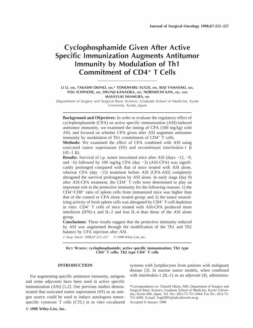
Cyclophosphamide Given After ActiveSpecific Immunization Augments Antitumor
Immunity by Modulation of Th1Commitment of CD4+ T Cells
LI LI, MD, TAKASHI OKINO, MD,* TOMOHARU SUGIE, MD, SEIJI YAMASAKI, MD,YOU ICHINOSE, MD, SHUNJI KANAOKA, MD, NORIMICHI KAN, MD, AND
MASAYUKI IMAMURA, MD
Department of Surgery and Surgical Basic Science, Graduate School of Medicine, KyotoUniversity, Kyoto, Japan
Background and Objectives:In order to evaluate the regulatory effect ofcyclophosphamide (CPA) on active specific immunization (ASI)-inducedantitumor immunity, we examined the timing of CPA (100 mg/kg) withASI, and focused on whether CPA given after ASI augments antitumorimmunity by modulation of Th1 commitment of CD4+ T cells.Methods: We examined the effect of CPA combined with ASI usingsonicated tumor supernatant (SS) and recombinant interleukin-1b(rIL-1 b).Results: Survival of i.p. tumor inoculated mice after ASI (days −12, −9,and −6) followed by 100 mg/kg CPA (day −3) (ASI-CPA) was signifi-cantly prolonged compared with that of mice treated with ASI alone,whereas CPA (day −15) treatment before ASI (CPA-ASI) completelyabrogated the survival prolongation by ASI alone. In early stage (day 0)after ASI-CPA treatment, the CD4+ T cells were determined to play animportant role in the protective immunity for the following reasons: 1) theCD4+/CD8+ ratio of spleen cells from immunized mice was higher thanthat of the control or CPA alone treated group; and 2) the tumor neutral-izing activity of fresh spleen cells was abrogated by CD4+ T-cell depletionin vitro. CD4+ T cells of mice treated with ASI-CPA produced moreinterferon (IFN)-g and IL-2 and less IL-4 than those of the ASI alonegroup.Conclusions:These results suggest that the protective immunity inducedby ASI was augmented through the modification of the Th1 and Th2balance by CPA injection after ASI.J. Surg. Oncol. 1998;67:221–227. © 1998 Wiley-Liss, Inc.
KEY WORDS: cyclophosphamide; active specific immunization; Th1 typeCD4+ T cells; Th2 type CD4+ T cells
INTRODUCTION
For augmenting specific antitumor immunity, antigensand some adjuvants have been used in active specificimmunization (ASI) [1,2]. Our previous studies demon-strated that sonicated tumor supernatant (SS) as an anti-gen source could be used to induce autologous tumor-specific cytotoxic T cells (CTL) in in vitro cocultured
systems with lymphocytes from patients with malignantdisease [3]. In murine tumor models, when combinedwith interleukin-1 (IL-1) as an adjuvant [4], administra-
*Correspondence to: Takashi Okino, MD, Department of Surgery andSurgical Basic Science, Graduate School of Medicine, Kyoto Univer-sity, Kyoto 606, Japan. Tel. No.: (81)-75-751-3444; Fax No.: (81)-75-751-4390. E-mail: [email protected] 6 January 1998
Journal of Surgical Oncology 1998;67:221–227
© 1998 Wiley-Liss, Inc.

tion of SS could include tumor-specific protective im-munity and CD4+ T cells have been revealed to be themain effectors [5].
On the other hand, ASI might not only induce an an-titumor effect but also augment tumor growth by activa-tion of certain suppressor cells during the immunizingprocess [1]. The anticancer drug cyclophosphamide(CPA) has been shown to be able to augment cell-mediated immunity through a selective reduction of hostsuppressor T cells or their precursors [6–8]. Suppressionor augmentation of host immunity by CPA is determinedby dose and timing of the administration [9,10]. Ourprevious study demonstrated that low dose CPA aug-mented the antitumor effects which were induced by IL-1in tumor bearing mice [11]. In order to evaluate the regu-latory effect of CPA on ASI-induced antitumor immu-nity, we examined the timing of CPA (100 mg/kg) withASI, and focused on whether CPA given after ASI aug-ments antitumor immunity by modulation of Th1 com-mitment of CD4+ T cells.
MATERIALS AND METHODSAnimal and Tumor Cells
Inbred male BALB/c mice weighing 20–25 g werepurchased from Japan SLC Co. Ltd. (Shizuoka, Japan)and used at 8–12 weeks of age. The tumor cell lineMOPC-104E, which is a syngeneic plasmacytoma toBALB/c mice, was maintained serially in vivo by intra-peritoneal (i.p.) passage. Tumor cells were used for ex-periments 6–8 days after inoculation.
Reagents
Recombinant human interleukin-1b (rIL-1 b) 71 sermutant was supplied by Otsuka Pharmaceutical Co. Ltd.(Tokushima, Japan). The lymphocyte activating activityof rIL-1 b is 2 × 10⁷ U/mg protein. CPA was obtainedfrom Shionogi Pharmaceutical Co. Ltd. (Osaka, Japan)and dissolved with sterile saline immediately before i.p.injection.
For in vitro negative selection of CD4+ and CD8+
T-cell subpopulations, the anti-Lyt2.2 (CD8) and anti-L3T4 (CD4) monoclonal antibody (mAb) were pur-chased from Cedarlane Laboratories Ltd. (Ontario,Canada).
Preparation of SS
Preparation of SS was described previously [12].Briefly, tumor cells were separated from ascitic fluidfollowed by red blood cell lysis with Tris-NH4Cl, andresuspended in RPMI-1640 at a concentration of 2 × 10⁷
cells/ml. After sonication for 90 sec (20 kHz, 105 W)with an ultrasonic disrupter (Tomy Seiko, Tokyo, Japan),they were centrifuged for 90 min at 15,000g. The super-natants were passed through a 0.22mm filter and storedat −80°C until use.
In Vivo Experimental Protocols
The six protocols of immunization in vivo with rIL-1b plus SS and/or CPA are shown in Figure 1. The controlgroup was treated by sterile saline 1.0 ml/mouse at thetime point; CPA (day −15) means a single CPA (100mg/kg) i.p. 15 days before tumor inoculation; CPA-ASIis CPA (day −15) treatment plus ASI immunization withrIL-1 b (1.0 mg/mouse) and SS (1.0 ml) i.p. on days 12,9, and 6 prior to tumor inoculation (i.e., days −12, −9,and −6); ASI means rIL-1b and SS treatment alone;CPA (day −3) is a single CPA i.p. 3 days before tumorinoculation; and ASI-CPA means ASI plus CPA (day −3)treatment. After these treatments, mice received i.p. in-oculation on day 0 with 1 × 10⁵ MOPC-104E tumor cells,and their survival was observed daily until day 100.
Preparation of the Spleen Cells
Spleens from treated mice were aseptically removedon day 0, minced, and passed through a No. 100 stainlesssteel mesh. After erythrocytes were lysed with 0.83%Tris-NH4Cl, the spleen cells were washed three timeswith Hank’s balanced salt solution (HBSS) and sus-pended in HBSS.
Tumor Neutralizing Assay and Cell Depletion
The antitumor effect of splenocytes was investigatedby tumor neutralizing assay. The fresh splenocytes fromtreated mice on day 0 were mixed with 5 × 10⁵ MOPC-104E tumor cells at a ratio of 30:1 and inoculated sub-cutaneously (s.c.) in a volume of 0.2 ml into the dorsumof recipient normal mice. Tumor diameter was measuredtwice per week for 21 days. For in vitro cell depletedtreatment, fresh splenocytes (1 × 10⁷/ml) were coincu-bated with anti-CD4 or anti-CD8 mAbs at 4°C for 60 minand centrifuged. The pellet was treated with Low-Tox-Mrabbit complement at ×10 dilution (Cedarlane Laborato-ries Ltd.) at 37°C for 60 min. After three washings, thesecells were used for tumor neutralizing assay in vivo.
Fig. 1. Scheme of protocols for in vivo treatments (↓) 1 × 10⁵
MOPC-104E tumor cells per mouse i.p.; () CPA (100 mg/kg) i.p.;( ) rIL-1 b (1.0 mg) and SS (1.0 ml) i.p.; sterile saline was used as acontrol.
222 Li et al.
Flow Cytometric AnalysisThe following mAbs were purchased from Pharmin-
gen (San Diego, CA): fluorescein isothiocyanate (FITC)-conjugated anti-Thy1.2 (CD3), anti-L3T4 (CD4), andanti-Ly2 (CD8). Fresh splenocytes (1 × 10⁶) were stainedfor 30 min at 4°C with corresponding mAbs. The stainedcells were washed three times with phosphate-bufferedsaline (PBS) and examined for fluorescence using aFACScan instrument (Becton Dickinson, MountainView, CA). Analysis of data was determined by counting1 × 104 viable cells.
Isolation of CD4+ T-Cell Subset From FreshSpleen Cells
Fresh splenocytes were depleted of B cells and otheradherent cells by anti-IgM panning. The non-adherentcells were collected and resuspended at 1 × 10⁷ cells/ml.They were treated with anti-CD8 mAb for 60 min at 4°Cbefore they were incubated with rabbit complement for60 min at 37°C. The final cell suspensions were washedthree times with RPMI-1640 and used for experiments asCD4+ T cells. The phenotypes of enriched CD4+ T-cellsuspensions were 96% of CD3+ and 89% of CD4+.
CD4+ T-Cells Proliferation Assay andCytokine Assay
To assess the proliferation of CD4+ T cells, enrichedCD4+ T cells (2 × 10⁵/well) from in vivo treated micewere incubated in RPMI-1640 medium supplementedwith 10% fetal bovine serum (FBS; BioWHITTAKER,Verviers, Belgium), 100mg gentamicin, and 0.2mg/mlfungizone [complete medium (CM)] at a final volume of0.2 ml in a 96-well flat-bottomed microtest plate (Nunc,Roskilde, Denmark). Three days later,3H-thymidine (1.0mCi/well) was added to each well for the last 6 hr andcells were harvested onto a 96-well UniFilter plateGFC96 by a Filtermate cell harvester (Packard, Meriden,CT). Their radioactivity was measured by a liquid scin-tillation counter (Packard) and mean count per minute(cpm) was calculated from triplicate wells.
For cytokine assay, the enriched CD4+ T cells (2 ×10⁶/ml) were incubated in CM in a 12-well culture plate(Nunc) and 5 days later their supernatants were collectedand stored at −80°C until test. The interferon (IFN)-g andIL-4 concentrations of these cultured supernatants wereanalyzed by enzyme-linked immunosorbent assay(ELISA) (Mouse Titer Screen III EIA, PerSeptive Diag-nostics, Inc., Cambridge, MA). Minimum detection lev-els of IFN-g and IL-4 were 11.7 and 1.0 pg/ml, respec-tively. The IL-2 production was analyzed by a bioassayusing an IL-2 sensitive cells line, CTLL2 (AmericanType Culture Collection, Rockville, MD).
Statistical AnalysisThe generalized Wilcoxon test and Cox-Mantel test
were used to compare survival data. Comparison of tu-
mor diameters was performed using the Student’st-test.P < 0.05 was defined as significant.
RESULTSCPA After, But Not Before, ASI Augments the
Protective Immunity Against the FollowingTumor Challenge
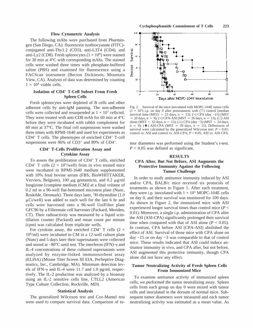
In order to study antitumor immunity induced by ASIand/or CPA, BALB/c mice received six protocols oftreatments as shown in Figure 1. After each treatment,they were i.p. inoculated with 1 × 10⁵ MOPC-104E cellson day 0, and their survival was monitored for 100 days.As shown in Figure 2, the immunized mice with ASIexperienced longer survival times than control mice (P <0.01). Moreover, a single i.p. administration of CPA afterthe ASI (ASI-CPA) significantly prolonged their survivaltime when compared with that of ASI alone (P < 0.05).In contrast, CPA before ASI (CPA-ASI) abolished theeffect of ASI. Survival of those mice with CPA alone onday −15 or on day −3 was comparable to that of controlmice. These results indicated that ASI could induce an-titumor immunity in vivo, and CPA after, but not before,ASI augmented this protective immunity, though CPAalone did not have any effect.
Tumor Neutralizing Activity of Fresh Spleen CellsFrom Immunized Mice
To examine antitumor activity of immunized spleencells, we performed the tumor neutralizing assay. Spleencells from each group on day 0 were mixed with tumorcells and inoculated in the dorsum of normal mice. Sub-sequent tumor diameters were measured and each tumorneutralizing activity was estimated as a mean value. As
Fig. 2. Survival of the mice inoculated with MOPC-104E tumor cells(1 × 10⁵) i.p. on day 0 after pretreatment with (,) control [mediansurvival time (MST)4 23 days, n4 13]; (+) CPA (day −15) (MST4 20 days, n4 9); (×) CPA-ASI (MST4 26 days, n4 13); (h) ASIalone (MST4 32 days, n4 15); (n) CPA (day −3) (MST4 24 days,n 4 9); (l) ASI-CPA (MST 4 59 days, n4 15). Differences ofsurvival were calculated by the generalized Wilcoxon test:P < 0.01:control vs. ASI and control vs. ASI-CPA;P < 0.05: ASI vs. ASI-CPA.
Cyclophosphamide Commitment of T Cells 223

shown in Figure 3, spleen cells from the mice treatedwith ASI alone exhibited higher tumor neutralizing ac-tivity than those of control group. ASI-CPA significantlyenhanced the effect of ASI, while CPA-ASI eliminatedthe antitumor activity induced by ASI. These results wereexactly compatible with survival data in Figure 2, whichconfirmed that ASI could induce the strong antitumorimmunity in vivo and CPA after ASI augmented thissystemic host defense mechanism. Next, we examinedcytotoxicity against tumor cells in vitro, but neither freshnor rIL-2 cultured spleen cells showed any direct lyticactivities (data not shown). These results suggest thatASI may not exert biological activity on effectors butmay affect the induction phase of antitumor immunity.
Phenotypic Analysis of Fresh Spleen Cells FromImmunized Mice and Subpopulation Analysis
To determine the relationship between antitumor ac-tivity and change of T-cell subsets in spleen cells ofimmunized mice, we analyzed fresh spleen cells fromeach group on day 0. After counting total cell number,spleen cells from each group were single color stainedwith anti-CD3, anti-CD4, and anti-CD8 mAbs beforeflow cytometric analysis. Data in Table I are shown asmean ± SD for three individual mice. Total cell numberand CD4+/CD8+ ratio of spleen from the mice immu-nized with ASI were significantly higher than those ofcontrol mice (P < 0.01). The mice which received ASI-CPA treatment showed a comparable CD4+/CD8+ ratioto the ASI alone group, although their total spleen cellsdecreased in number. These results suggest that ASI
treatment efficiently increased CD4+ T cells and thatCPA did not exert any effect on the CD4+/CD8+ ratioalthough it reduced the total cell number. To gain insightinto functions of CD4+ T cells, spleen cells from the miceimmunized with ASI-CPA were depleted of CD4+ orCD8+ T cells in vitro before tumor neutralizing assay.Tumor neutralizing activity induced by ASI-CPA wasabolished by depletion of CD4+ T cells, but not CD8+ Tcells (Fig. 4). These results strongly suggest that theCD4+ T cells might play an important role in this CPA-combined vaccination model.
CPA Augments the Proliferation of CD4+ T Cellsand Preferentially Increases Th1 Type Cytokines of
CD4+ T Cells
ASI-CPA significantly augments antitumor immunitycompared to ASI alone, but the CD4+/CD8+ T-cell ratiodid not change. To address the effect of CPA on CD4+ Tcells, we first examined the proliferation of CD4+ T cells.Fresh spleen cells from each treatment group were de-pleted of B cells and CD8+ T cells as described in Ma-terials and Methods, which consisted of 89% CD4+ Tcells. These CD4-enriched spleen cells were cultured inCM alone for 3 days followed by pulsation with3H-thymidine. As shown in Figure 5, CD4+ T cells fromimmunized mice with ASI-CPA exhibited apparent pro-liferative potential when compared to those from the con-trol or ASI groups.
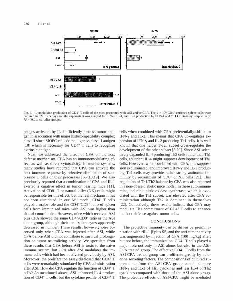
Finally, we examined the effect of cytokine profile ofCD4+ T cells. CD4+ T-enriched spleen cells were cul-tured for 5 days in CM alone and the culture supernatantswere assayed for IL-4 or IFN-g production by ELISAand for IL-2 by CTLL2 bioassay. ASI strongly inducedIL-4 production of CD4+ T cells, while CPA shifted thecytokine profile of these CD4+ T cells from IL-4 toIFN-g and IL-2, i.e., from Th2 to Th1 type (Fig. 6).
DISCUSSION
In our IL-1 plus MOPC-SS (ASI) model, preimmu-nized mice became resistant to tumorigenicity of MOPCand developed long-time survival. Moreover, this protec-tive immunity was significantly enhanced by CPA after,but not before, ASI. Subpopulation analysis showed thatCD4+ T cells in the immunized mice were involved intumor neutralizing activity. These results indicated thatCD4+ T cells seem to play a major role in this protectiveimmunity induced by ASI alone [5] or ASI combinedwith CPA, which might modulate the function of CD4+ Tcells in an unknown manner. Therefore, this study hasfocused on the analysis of CD4+ T-cell populations infresh spleen cells from immunized mice and examinedthe effect of CPA on CD4+ T cells.
IL-1 was first defined as a mediator of host inflamma-tory response and secreted mainly from mononuclearphagocytes. It is well known that IL-1 consists of two
Fig. 3. Tumor neutralizing activity of fresh spleen cells. MOPC-104E tumor cells (5 × 10⁵) were inoculated s.c. mixed with none (n,n 4 10), or 1.5 × 10⁷ spleen cells (effector to target ratio4 30) fromnormal mice (+, n4 10), CPA-ASI (×, n4 9), ASI alone (h, n 410), and ASI-CPA (l, n 4 10) treated mice, respectively. Statisticalvalues were compared by Student’st-test: *P < 0.05: ASI vs. ASI-CPA; +P < 0.01: ASI-CPA vs. other groups and ASI vs. other groups.
224 Li et al.

isoforms, i.e., IL-1a and IL-1b, and the latter exerts itsmain biological activity in the circulation. A report indi-cated that IL-1 has not only direct cytotoxicity but also anadjuvant effect with tumor vaccine [4]. The precisemechanism of this antitumor immunity remains obscure,but our previous study revealed that IL-1 modulatessome CD4+ T-cell populations because antitumor activityinduced by IL-1 and SS was abrogated by CD4+ T-celldepletion both in vivo and in vitro [5]. Although IL-1receptors are present on almost all cells, CD4+ T cellsshare their receptors selectively on Th2, but not on Th1[13,14]. In this study, CD4+ T cells from mice immu-nized with IL-1 (ASI and ASI-CPA) produced signifi-cantly more IL-4 than those from control mice, which iscompatible with the distribution of IL-1 receptors.
IL-4 is originally identified as a growth and differen-tiation factor for B cells which are responsible for hu-moral immunity and may antagonize the cell-mediatedimmune response, which is mainly involved in tumorrejection. However, among the experiments in whichvarious cytokine genes were transduced into the tumorcells, it was reported that an IL-4 producing tumor cellline could be effectively rejected by a syngeneic host andshowed therapeutic effect against established tumor[15,16]. Histologic examination suggested that this re-jection process was associated with the infiltration ofmacrophages rather than lymphoid cells [15]. Moreover,it was also reported that macrophages expressed IL-4receptors and IL-4 could activate macrophages for in-creased tumoricidal activity [17]. In this study, ASI in-creased IL-4 production of CD4+ T cells which mightcause the immunized mice to develop antitumor immu-nity. Although there is no way to explain the exact func-tion of macrophages, it is quite reasonable that macro-
TABLE I. Phenotypic Analysis of Fresh Spleen Cells From Immunized Mice by ASI and/or CPA†
Treatment groups Total no.
Nos. of staining lymphocytes per spleen (×10−5)
CD5/CD8 ratioThy1.2 (CD3) L3T4 (CD4) Ly2 (CD8)
Control 663.3 ± 169.2 212.7 ± 24.9 165.6 ± 27.3 49.3 ± 7.0 3.4 ± 0.3(32.1%)a (25.0%) (7.4%)
CPA (day −3) 158.0 ± 26.3 103.5 ± 19.2 77.8 ± 15.2 24.5 ± 5.6 3.2 ± 0.2(65.5%) (49.2%) (15.5%)
ASI 1,780.0 ± 113.7 518.4 ± 80.9 516.4 ± 63.9 89.3 ± 13.5 5.9 ± 0.6*(29.1%) (29.0%) (5.0%)
ASI-CPA 452.7 ± 78.0 166.4 ± 25.6 158.6 ± 27.5 32.1 ± 6.2 5.0 ± 0.4*(36.8%) (35.0%) (7.1%)
†In each treatment group, fresh spleen cells from three mice were analyzed by flow cytometry on day 0. Results are represented as mean ± SD.aPercentage to total number.*P < 0.01: ASI or ASI-CPA vs. control or CPA (day −3) on CD4/CD8.
Fig. 4. Tumor neutralizing activity of in vitro mAb treated freshspleen cells from ASI-CPA treated mice. Mice were inoculated s.c.with (A) 5 × 10⁵ MOPC-104E tumor cells alone, admixed with 1.5 ×10⁷ spleen cells from(B) normal mice,(C) ASI-CPA treated mice, orthose cells with(D) CD4 depletion,(E) CD8 depletion, and(F)complement alone.
Fig. 5. Proliferation of CD4+ cells from spleens of treated mice.Enriched CD4+ cells (1 × 10⁵/0.2 ml/well) were cultured in CM for 3days. After 6 hr3H-thymidine uptake, mean cpm was calculated fromtriplicate wells. *P < 0.01: others vs. ASI-CPA.
Cyclophosphamide Commitment of T Cells 225
phages activated by IL-4 efficiently process tumor anti-gen in association with major histocompatibility complexclass II since MOPC cells do not express class II antigen[18] which is necessary for CD4+ T cells to recognizeextrinsic antigen.
Next, we addressed the effect of CPA on the hostdefense mechanism. CPA has an immunomodulating ef-fect as well as direct cytotoxicity. In murine systems,many studies have reported that CPA can activate thehost immune response by selective elimination of sup-pressor T cells or their precursors [6,7,10,19]. We alsopreviously reported that a combination of CPA and IL-1exerted a curative effect in tumor bearing mice [11].Activation of CD8+ T or natural killer (NK) cells mightbe responsible for this effect, but the real mechanism hasnot been elucidated. In our ASI model, CD4+ T cellsplayed a major role and the CD4+/CD8+ ratio of spleencells from immunized mice with ASI was higher thanthat of control mice. However, mice which received ASIplus CPA showed the same CD4+/CD8+ ratio as the ASIalone group, although their total splenocytes apparentlydecreased in number. These results, however, were ob-served only when CPA was injected after ASI, whileCPA before ASI did not contribute to survival prolonga-tion or tumor neutralizing activity. We speculate fromthese results that CPA before ASI is toxic to the naiveimmune system, but CPA after ASI modulates the im-mune cells which had been activated previously by ASI.Moreover, the proliferation assay disclosed that CD4+ Tcells were remarkably stimulated by CPA administrationafter ASI. How did CPA regulate the function of CD4+ Tcells? As mentioned above, ASI enhanced IL-4 produc-tion of CD4+ T cells, but the cytokine profile of CD4+ T
cells when combined with CPA preferentially shifted toIFN-g and IL-2. This means that CPA up-regulates ex-pansion of IFN-g and IL-2 producing Th1 cells. It is wellknown that one helper T-cell subset cross-regulates thedevelopment of the other subset [8,20]. Since ASI selec-tively expanded IL-4 producing Th2 cells rather than Th1cells, abundant IL-4 might suppress development of Th1cells. However, when combined with CPA, this suppres-sion is eliminated, and improved IFN-g and IL-2 produc-ing Th1 cells may provide rather strong antitumor im-munity by recruitment of CD8+ or NK cells [21]. Thisregulation of Th1/Th2 balance by CPA was also reportedin a non-obese diabetic mice model. In these autoimmunemice, inducible nitric oxidase synthetase, which is asso-ciated with the Th1 subset, was elevated after CPA ad-ministration although Th2 is dominant in themselves[22]. Collectively, these results indicate that CPA maymodulate Th1 commitment of CD4+ T cells to enhancethe host defense against tumor cells.
CONCLUSIONS
The protective immunity can be driven by preimmu-nization with rIL-1b plus SS, and the anti-tumor activitywas augmented by injection of CPA (100 mg/kg) after,but not before, the immunization. CD4+ T cells played amajor role not only in ASI alone, but also in the ASI-CPA treated group. The effective CD4+ T cells from theASI-CPA treated group can proliferate greatly by auto-crine secreting factors. The compositions of cultured su-pernatants from the ASI-CPA group contained moreIFN-g and IL-2 of Th1 cytokines and less IL-4 of Th2cytokines compared with those of the ASI alone group.The protective effects of ASI-CPA might be mediated
Fig. 6. Lymphokine production of CD4+ T cells of the mice pretreated with ASI and/or CPA. The 2 × 10⁶ CD4+-enriched spleen cells werecultured in CM for 5 days and the supernatant was assayed for IFN-g, IL-4, and IL-2 production by ELISA and CTLL2 bioassay, respectively.*P < 0.01: vs. other groups.
226 Li et al.
through modification of the cytokine balance betweenthe Th1 and Th2 subset of CD4+ T cells.
ACKNOWLEDGMENTS
We thank the staff at the Animal Experiment Centerand the Radioisotope Research Center, Kyoto University,where a part of this study was performed. We also thankMs. Aiko Tanaka for her assistance. We appreciateOtsuka Pharmaceutical Co. Ltd. (Tokushima, Japan) forproviding rIL-1 b.
REFERENCES1. Hook DS, Foshag LJ, Nizze AS, et al.: Suppressor cell activity in
a randomized trial of patients receiving active specific immuno-therapy with melanoma cell vaccine and low dosages of cyclo-phosphamide. Cancer Res 1990;50:5358–5364.
2. Mitchell MS, Kan-Mitchell J, Kempf RA, et al.: Active specificimmunotherapy for melanoma: Phase I trial of allogeneic lysatesand a novel adjuvant. Cancer Res 1988;48:5883–5893.
3. Okino T, Kan N, Nakanishi M, et al.: The therapeutic effect ofOK-432 combined adoptive immunotherapy against liver metas-tases from breast cancer. J Cancer Res Clin Oncol 1990;116:197–202.
4. McCune CS, Marquis DM: Interleukin 1 as an adjuvant for activespecific immunotherapy in a murine tumor model. Cancer Res1990;50:1212–1215.
5. Moriguchi Y, Kan N, Okino T, et al.: A new model of activespecific immunotherapy using interleukin-1 and sonicated tumorsupernatant in murine tumor system. J Surg Oncol 1996;62:78–85.
6. Glaser M: Regulation of specific cell-mediated cytotoxic responseagainst SV-40 induced tumor associated antigens by depletion ofsuppressor T cell with cyclophosphamide in mice. J Exp Med1979;149:774–779.
7. Milton JD, Carpenter CB, Addison IE: Depressed T-cell reactivityand suppressor activity of lymphoid cells from cyclophospha-mide-treated mice. Cell Immunol 1976;24:308–317.
8. Le-Gros G, Ben-Sasson SZ, Sedar R, et al.: Generation of inter-leukin-4 (IL-4)-producing cells in vivo and in vitro: IL-2 and IL-4are required for in vitro generation of IL-4 producing cells. J ExpMed 1982;172:921–929.
9. Turk JL, Parker D: Effect of cyclophosphamide on immunologicalcontrol mechanisms. Immunol Rev 1982;65:99–113.
10. Mokyr MB, Hengst JC, Dray S: Role of anti-tumor immunity incyclophosphamide-induced rejection of subcutaneous non-palpable MOPC-315 tumors. Cancer Res 1982;42:974–979.
11. Harada T, Kan N, Ichinose Y, et al.: The synergistic antitumoreffect of recombinant interleukin-1 and low-dose of cyclophos-phamide in tumor-bearing mice. J Surg Oncol 1994;56:39–45.
12. Kan N, Ohgaki K, Inamoto T, Kodama H: Anti-tumor and thera-peutic effects of spleen cells from tumor-bearing mice culturedwith T cell growth factor and soluble tumor extract. Cancer Im-munol Immunother 1984;18:215–222.
13. Lacey DL, Erdman JM: IL-1 and IL-4 modulate IL-1 receptorexpression in a murine T cell line. J Immunol 1990;145:4145–4153.
14. Taylor-Robinson AW, Phillips RS: Expression of the IL-1 recep-tor discriminates Th2 from Th1 cloned CD4+ T cells specific forPlasmodium chabaudi.Immunology 1994;81:216–221.
15. Golumbek PT, Lazenby AJ, Levitsky HI, et al.: Treatment ofestablished renal cancer by tumor cells engineered to secrete in-terleukin-4. Science 1991;254:713–716.
16. Tepper RI, Pattengale PK, Leder P: Murine interleukin-4 displayspotent anti-tumor activity in vivo. Cell 1989;57:503–512.
17. Crawford RM, Finbloom DS, Ohara J, et al.: B cell stimulatoryfactor-1 (interleukin 4) activated macrophages for increased tu-moricidal activity and expression of Ia antigens. J Immunol 1987;169:135–141.
18. Sugie T, Kubota H, Sato M, et al.: NK1+CD4−CD8−a b T cells inthe peritoneal cavity. J Immunol 1996;157:3925–3935.
19. North RJ: Cyclophosphamide-facilitated adoptive immunotherapyof an established tumor depends on elimination of tumor-inducedsuppressor T cells. J Exp Med 1982;155:1063–1074.
20. Swain SL, Weimberg AD, English M, Huston G: IL-4 directs thedevelopment of Th2-like helper effectors. J Immunol 1990;145:3796–3806.
21. Lee KY, Goedegebuure PS, Linehan DC, Eberlein TJ: Immuno-regulatory effects of CD4+ T helper subset in human melanoma.Surgery 1995;117:365–372.
22. Rothe H, Faust A, Shade U, et al.: Cyclophosphamide treatment offemale non-obese diabetic mice causes enhanced expression ofinducible nitric oxide synthase and interferon-gamma, but not ofinterleukin-4. Diabetologia 1994;37:1154–1158.
Cyclophosphamide Commitment of T Cells 227


![PROCYTOX® [Cyclophosphamide] - Baxter](https://img.pdfslide.net/doc/110x75/62038fe2da24ad121e4adae9/procytox-cyclophosphamide-baxter.jpg)