Embed Size (px)
Citation preview
Cysteine Protease-Binding Protein Family 6 Mediates the Traffickingof Amylases to Phagosomes in the Enteric Protozoan Entamoebahistolytica
Atsushi Furukawa,a,b* Kumiko Nakada-Tsukui,a Tomoyoshi Nozakia,c
Department of Parasitology, National Institute of Infectious Diseases, Toyama, Tokyo, Japana; Department of Parasitology, Gunma University Graduate School of Medicine,Maebashi, Japanb; Graduate School of Life and Environmental Sciences, University of Tsukuba, Tsukuba, Ibaraki, Japanc
Phagocytosis plays a pivotal role in nutrient acquisition and evasion from the host defense systems in Entamoeba histolytica, theintestinal protozoan parasite that causes amoebiasis. We previously reported that E. histolytica possesses a unique class of a hy-drolase receptor family, designated the cysteine protease-binding protein family (CPBF), that is involved in trafficking of hydro-lases to lysosomes and phagosomes, and we have also reported that CPBF1 and CPBF8 bind to cysteine proteases or �-hexo-saminidase �-subunit and lysozymes, respectively. In this study, we showed by immunoprecipitation that CPBF6, one of themost highly expressed CPBF proteins, specifically binds to �-amylase and �-amylase. We also found that CPBF6 is localized inlysosomes, based on immunofluorescence imaging. Immunoblot and proteome analyses of the isolated phagosomes showed thatCPBF6 mediates transport of amylases to phagosomes. We also demonstrated that the carboxyl-terminal cytosolic region ofCPBF6 is engaged in the regulation of the trafficking of CPBF6 to phagosomes. Our proteome analysis of phagosomes also re-vealed new potential phagosomal proteins.
Phagocytosis and phagosome biogenesis play indispensable andpivotal roles in the pathogenesis of Entamoeba histolytica. E.
histolytica trophozoites ingest and digest microorganisms in thelarge intestine, for the acquisition of nutrients (1), and also hostcells (2) during tissue invasion, for the creation of a parasiticniche. Phagocytosis also plays a role in the evasion from host im-mune systems (3). It has been demonstrated that the levels of invitro and in vivo virulence of clinical and laboratory strains corre-late well with the ability for phagocytosis (4–7). In addition,phagosomes contain a panel of proteins that have been implicatedin pathogenesis, such as cysteine proteases (CPs) (8), amoebapores (9), and galactose-/N-acetylgalactosamine-specific lectin(10, 11). Therefore, an understanding of the molecular mecha-nisms of phagosome biogenesis and trafficking, together with thecharacterization of “uncharacterized hypothetical” proteins inphagosomes, should further uncover the links between phagocy-tosis and pathogenicity.
Recently, the molecular mechanisms of phagocytosis havestarted to be unveiled. For example, an unconventional myosin,myosin IB, was shown to be involved in cytoskeleton rearrange-ment during phagocytosis (12). It was also shown that phospha-tidylinositol signaling is involved in phagocytosis. The time- andposition-specific accumulations of phosphatidylinositol-3-phos-phate (PI3P) and phosphatidylinositol (3,4,5)-trisphosphate(PIP3) (13–15) on the phagosomal membrane were demon-strated. Surface Ca2�-binding kinase (C2PK) was shown to beinvolved in the initiation of phagocytosis (16). C2PK localizes inclose proximity to the plasma membrane through phosphatidyl-serine in the presence of Ca2� and thereby recruits E. histolyticaCaBP1 (EhCaBP1) and actin to the phagocytic cup during eryth-rophagocytosis. Conditional suppression of C2PK expression andoverexpression of a mutant form demonstrated its role in the ini-tiation of the formation of the phagocytic cup. Surface transmem-brane kinase (TMK96) and p21-activated kinase (PAK) also havebeen shown to play important roles in phagocytosis of human
erythrocytes (17, 18). TMK96 was identified in the proteome ofisolated phagosomes and plays an important role in the clearanceof dead host cells. PAK was shown to be highly concentrated in thenascent pseudopod in motile trophozoites and appears to be aregulatory element controlling pseudopod extension and cell po-larity. Overproduction of the carboxyl-terminal kinase domain ofPAK causes multiple pseudopod formation and enhanced eryth-rophagocytosis.
Although the mechanisms of initiation of phagocytosis havebeen vigorously investigated, it is only recently that scientists havestarted to understand the fundamental mechanisms of how hy-drolases are selectively transported to phagosomes, leading to or-ganelle maturation. Transport and activation of hydrolytic en-zymes are tightly regulated. Otherwise, they are deleterious to thecell. In yeast and mammals, trafficking of major hydrolytic en-zymes is mediated by the specific cargo receptors mannose-6-phosphate receptor (M6PR) and Vps10/sortilin. M6PR regulatesthe trafficking of M6P-modified lysosomal hydrolases, includingcathepsin L and �-hexosaminidase, while Vps10p/sortilin regu-lates the trafficking of carboxypeptidase Y in yeast or cathepsin Din mammals. E. histolytica appears to lack a Vps10/sortilin ho-
Received 30 August 2012 Returned for modification 4 October 2012Accepted 10 December 2012
Published ahead of print 18 March 2013
Editor: J. F. Urban, Jr.
Address correspondence to Tomoyoshi Nozaki, [email protected].
* Present address: Atsushi Furukawa, Laboratory of Bimolecular Science, Faculty ofPharmaceutical Sciences, Hokkaido University, Japan.
Supplemental material for this article may be found at http://dx.doi.org/10.1128/IAI.00915-12.
Copyright © 2013, American Society for Microbiology. All Rights Reserved.
doi:10.1128/IAI.00915-12
1820 iai.asm.org Infection and Immunity p. 1820–1829 May 2013 Volume 81 Number 5
on June 3, 2018 by guesthttp://iai.asm
.org/D
ownloaded from
molog. Furthermore, immuno-affinity pulldown of a putativeM6PR (EHI_096320) failed to coprecipitate any cargo protein,suggesting that putative M6PRs are not likely receptors/carriers oflysosomal hydrolases (K. Nakada-Tsukui, unpublished data). Werecently discovered a novel class of a single-transmembrane car-rier/receptor family, designated the cysteine protease-bindingprotein family (CPBF), that specifically binds to various lysosomalhydrolytic enzymes and regulates their trafficking in E. histolytica(19, 20). CPBF consists of 11 proteins (CPBF1 to -11) with signif-icant mutual identity and structural conservation: the signal pep-tide at the amino terminus, a single transmembrane domain closeto the carboxyl terminus, and the Yxx� motif at the carboxylterminus. CPBF1 was initially discovered as a receptor/carrier forone of three major cysteine proteases, EhCP-A5 (20). We furtherdemonstrated that CPBF1 is essential for the processing and lyso-somal transport of EhCP-A5. On the other hand, CPBF8 binds to�-hexosaminidase �-subunit and lysozymes and transports themto phagosomes (19). We further showed that repression of CPBF8by gene silencing decreased degradation of the Gram-positive ba-cillus Clostridium perfringens and in vitro cytopathic activityagainst mammalian cells.
In the present study, we have characterized another member ofthe most highly expressed CPBF genes among the family, CPBF6.We show that CPBF6 is distributed to lysosomes in steady stateand transported to phagosomes upon phagocytosis. We have fur-ther identified cargo proteins, by affinity immunoprecipitation ofCPBF6, followed by liquid chromatography-tandem mass spec-trometry (LC-MS/MS) analysis, to be �-amylase and �-amylase.We also confirmed the role of CPBF6 in phagosomal transport of�-amylase and �-amylase by repression of CPBF6, based on West-ern blotting and proteome analyses. In addition, this proteomeanalysis shows a new potential for phagosomal proteins. Finally,we show that the carboxyl-terminal region of CPBF6 plays a cru-cial role in phagosomal localization.
MATERIALS AND METHODSMicroorganisms and cultivation. Trophozoites of E. histolytica HM-1:IMSS Cl-6 and G3 strains (21, 22) were cultured axenically at 35°C in 13-by 100-mm screw-cap Pyrex glass tubes or 25-cm2 plastic culture flasks inBI-S-33 medium as previously described (23, 24). Chinese hamster ovary(CHO) cells were grown in F-12 medium (Invitrogen, CA) supplementedwith 10% fetal bovine serum on a 10-cm-diameter tissue culture dish(Iwaki, Tokyo, Japan) under 5% CO2 at 37°C.
Plasmid construction and production of E. histolytica transfor-mants. The protein-coding region of CPBF6 was amplified by PCR fromcDNA using the sense and antisense oligonucleotides 5=-GCGAGATCTATGTCAATGAGTTGTCTGACTT-3= and 5=-CAGCGAGATCTAGAAAGGTCATGATAACCA-3= (BglII restriction sites are underlined). The am-plified PCR product was digested with BglII and ligated into BglII-digested pEhExHA (25), to produce pEhEx-CPBF6-HA. The plasmid toproduce the mutant form of CPBF6 that lacks a 43-amino-acid (aa)-longserine-rich region (SRR, aa 815 to 857; SSDSSSSSSPSASSTQPSTSSSAPNPSGESSNNTEDNNGTKIG; pEhEx-CPBF6�SRR-HA) was constructedas follows. A DNA fragment was amplified by PCR using pEhEx-CPBF6-HA as a template and the sense and antisense oligonucleotides5=-TGGATTATTTTTGGTGTTGTTCTCTTCTTG-3= and 5=-TGGTATTTTTTCAAGTTTCATCAATATTTC-3=. The PCR product was treatedwith the BKL kit (TaKaRa, Shiga, Japan) and self-ligated to producepEhEx-CPBF6�SRR-HA. The resulting plasmid carries a mutant CPBF6protein that lacks the SRR. pEhEx-CPBF6�cytosol-HA, which lacks a15-aa-long cytosolic carboxyl-terminal region after the transmembraneregion, and CPBF6�NNGYHDLS-HA, which lacks an 8-aa-long cytosolic
carboxyl-terminal region, were constructed as follows. The protein-cod-ing region of CPBF6�cytosol and CPBF6�NNGYHDLS were amplifiedby PCR by using the oligonucleotides 5=-GCGAGATCTATGTCAATGAGTTGTCTGACT-3= and 5=-GCGAGATCTTATGAAACCAATAATAAAGGC-3= (for CPBF6�cytosol) and 5=-GGAAGATCTATGTCAATGAGTTGTCTGACT-3= and 5=-GGAAGATCTCTTTTTACGATATATTATGAA-3= (for CPBF6�NNGYHDLS) (BglII restriction sites are underlined).respectively. The amplified PCR products were digested with BglIIand ligated into BglII-digested pEhExHA to produce pEhEx-CPBF6�cytosol-HA and CPBF6�NNGYHDLS-HA, respectively. Thetransformants that expressed full-length or truncated forms ofCPBF6-HA were established by transfection of the wild-type HM1:IMSSCl6 strain with pEhEx-CPBF6-HA, pEhEx-CPBF6�SRR-HA, pEhEx-CPBF6�cytosol-HA, or CPBF6�NNGYHDLS-HA by liposome-medi-ated transfection, as previously described (26). For gene silencing ofCPBF6, the 420-bp-long 5= end of the CPBF6 protein-coding region wasamplified by PCR from cDNA using the oligonucleotides 5=-CGCAGGCCTATGTCAATGAGTTGTCTGACT-3= and 5=-GCAGAGCTCATAAGCACCAGGGGCAGAATA-3= (StuI and SacI restriction sites are under-lined). The PCR-amplified DNA fragment was digested with StuI and SacIand ligated into StuI- and SacI-digested pSAP2-Gunma, to producepSAP2-CPBF6. The CPBF6 gene-silenced strain was established by trans-fection of the G3 strain with pSAP2-CPBF6 as described above. We alsoestablished a control strain by transfection of the G3 strain with pSAP2-Gunma.
Antibodies. Anti-�-amylase and anti-�-amylase antibodies wereraised against a mixture of the peptides DIPLEEFDRLKSKG (aa 45 to 58)and GAHFVENHDENRAV (aa 319 to 332) for �-amylase and RIKKHLNKIRSDLT (aa 547 to 560) and LADEIDGHKSLTAN (aa 607 to 620) for�-amylase. Pyridine nucleotide transhydrogenase (EhPNT) antibody waspreviously described (27). Anti-hemagglutinin (HA) 11MO mousemonoclonal antibody was purchased from Berkeley Antibody (Berkeley,CA). Alexa Fluor anti-mouse and anti-rabbit IgG and horseradish perox-idase-conjugated goat anti-mouse and anti-rabbit IgG were purchasedfrom Invitrogen.
Immunofluorescene assay. For the staining of lysosomes, amoebaewere incubated in the BI-S-33 medium containing LysoTracker RedDND-99 (Invitrogen; diluted 1:500) at 35°C. CHO cells grown in F-12medium supplemented with 10% fetal bovine serum were incubated with10 �M CellTracker Blue (Invitrogen) at 37°C for 1 h. After washing withBI-S-33 medium three times, CellTracker-labeled CHO cells were addedto the 8-mm-diameter wells of an 8-well glass slide (Thermo Scientific,Rockford, IL) containing 5 � 104 E. histolytica trophozoites, and the mix-ture was further incubated at 35°C in the BI-S-33 medium. After incuba-tion, cells were fixed with 3.7% paraformaldehyde–phosphate-bufferedsaline (PBS) for 10 min and permeabilized with 0.2% saponin–PBS for 10min at ambient temperature. The cells were then allowed to react withanti-HA 11MO mouse monoclonal antibody (diluted 1:1,000) and AlexaFluor-488 anti-mouse secondary antibody (1:1,000). The samples wereexamined on a Carl-Zeiss (Thornwood, NY) LSM510 confocal laser-scan-ning microscope. Images were further analyzed using LSM510 software.We defined CPBF6-positive vacuoles based on criteria described else-where (19).
Immunoprecipitation. Approximately 3 � 106 trophozoites ofstrains expressing CPBF6-HA, CPBF6�SRR, CPBF6�cytosol, orCPBF6�NNGYHDLS were lysed in 2 ml of lysis buffer (50 mM Tris-HCl[pH 7.5], 150 mM NaCl, 1% Triton X-100 [Tokyo Kasei, Tokyo, Japan],0.5 mg/ml E-64 [Sigma-Aldrich, St. Louis, MO], and Complete mini pro-tease inhibitor cocktail [Roche, Basel, Switzerland]). Approximately 10mg of the lysate was incubated with protein G-Sepharose beads (50 �l ofan 80% slurry; Amersham Biosciences, Uppsala, Sweden) in 2 ml of lysisbuffer at 4°C for 90 min, and centrifuged at 800 � g at 4°C for 3 min toremove proteins that were nonspecifically bound to the protein G-Sep-harose beads. The precleared lysate was mixed with 90 �l of anti-HAantibody conjugated to agarose (50% slurry; Sigma-Aldrich) and incu-
Traffic of Lysosomal Amylases in E. histolytica
May 2013 Volume 81 Number 5 iai.asm.org 1821
on June 3, 2018 by guesthttp://iai.asm
.org/D
ownloaded from
bated at 4°C for 3.5 h. The agarose beads were collected by centrifugationat 800 � g at 4°C for 3 min and washed four times with wash buffer (50mM Tris-HCl [pH 7.5], 150 mM NaCl, 1% Triton X-100). The agarosebeads were then incubated with 180 �l of HA peptide (20 �g/ml) at 4°Covernight to dissociate proteins from the beads. The eluate was applied toSDS-PAGE gels, and silver stained, immunoblotting, activity assays, andprotein sequencing were performed.
Protein digestion, LC-MS/MS), and database search for proteinidentification. For identification of phagosomal proteins, we carried outtwo different digestion methods for LC-MS/MS analysis. Briefly, for thesolution method, a 50-�g equivalent of the protein solution was extractedwith 300 �l methanol–75 �l chloroform–225 �l water. After the upperaqueous layer was discarded, 325 �l of methanol was added to the organicphase. The sample was vitiated and centrifuged for 5 min. The pellet wasair dried and resuspended in ammonium bicarbonate, reduced, alkylated,and digested with 1 �g trypsin (Promega, Madison, WI) overnight atroom temperature. The sample was acidified to 5% with acetic acid andthen subjected to LC-MS/MS. For the in-gel digestion method, a 10-�gequivalent of the solution was subjected to SDS-PAGE until the dye frontmigrated 1 cm. An approximately 1-cm2 piece was excised from the gel,transferred to a siliconized tube, and destained in 200 �l of 50% methanolovernight. The gel pieces were dehydrated in acetonitrile, rehydrated in 30�l of 10 mM dithiothreitol in 0.1 M ammonium bicarbonate, and reducedwith dithiothreitol at room temperature for 0.5 h. The dithiothreitol so-lution was removed and the sample alkylated in 30 �l 50 mM iodoacet-amide in 0.1 M ammonium bicarbonate at room temperature for 0.5 h.The reagent was removed, and the gel pieces were dehydrated in 100 �lacetonitrile. The acetonitrile was removed, and the gel pieces were rehy-drated in 100 �l 0.1 M ammonium bicarbonate. The pieces were dehy-drated in 100 �l acetonitrile, the acetonitrile was removed, and the pieceswere completely dried by vacuum centrifugation. The gel pieces wererehydrated in 20 ng/�l trypsin in 50 mM ammonium bicarbonate on icefor 10 min. Any excess enzyme solution was removed, and 20 �l of 50 mMammonium bicarbonate was added. The sample was digested overnight at37°C, and the peptides that formed were extracted from the polyacryl-amide in two 30-�l aliquots of 50% acetonitrile–5% formic acid. Theseextracts were combined and evaporated to 15 �l for MS analysis.
The LC-MS/MS system consisted of a Thermo Electron OrbitrapVelos ETD mass spectrometer system with a Protana nanospray ionsource interfaced with a self-packed 8-cm by 75-�m (inner diameter)Phenomenex Jupiter 10-�m C18 reversed-phase capillary column. Ap-proximately 2-�g equivalents of each sample were injected, and the pep-
tides were eluted from the column with an acetonitrile– 0.1 M acetic acidgradient at a flow rate of 0.5 �l/min over 2 h. The nanospray ion sourcewas operated at 2.5 kV. The digest was analyzed using the double-playcapability of the instrument, acquiring full scan mass spectra to determinepeptide molecular weights and product ion spectra to identify amino acidsequences in sequential scans. This mode of analysis produces approxi-mately 30,000 CAD spectra of ions that range in abundance over severalorders of magnitude.
The data were analyzed by database searches using the Sequest searchalgorithm against the E. histolytica genome database at the J. Craig VenterInstitute (http://www.jcvi.org/). The identified proteins were consideredphagosomal when two unique peptides were detected.
Transcriptomic analysis. Expression analysis was performed by usinga custom-made E. histolytica array from Affymetrix, Inc. (Santa Clara,CA), as previously described (28–30). Labeled cRNA for hybridizationwas prepared from 5 �g of total RNA, according to the protocol of themanufacturer. Hybridization and scanning were also performed accord-ing to the protocols of the manufacturer. Raw probe intensities were gen-erated by using the GeneChip operating software (GCOS) and a Gene-Titan instrument. The averages of triplicate raw probe intensities arereported.
Phagosome purification. Phagosome purification was performed aspreviously described (19, 31, 32). Briefly, amoebae were incubated withcarboxylated latex beads for 1 h, and after brief centrifugation, the cellpellet was resuspended and mechanically homogenized by using a Douncehomogenizer. The phagosomes were isolated by ultracentrifugation on asucrose gradient.
Enzymatic assay. The amylase assay was performed by using theEnzChek amylase assay kit (Invitrogen). Briefly, the amoeba lysate, cul-ture supernatant, or phagosomal fraction was mixed with 200 �g/ml sub-strate solution containing BODIPY FL-conjugated DQ starch in 100 �l of0.1 M morpholinepropanesulfonic acid (MOPS; pH 6.9). The fluores-cence was measured at excitation and emission wavelengths of 485 and530 nm, respectively, at 25°C for 60 min.
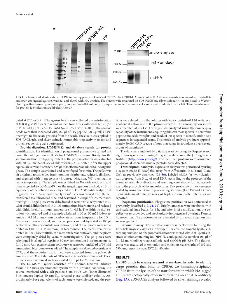
RESULTSCPBF6 binds to �-amylase and �-amylase. In order to identifycargo proteins that bind to CPBF6, we immunoprecipitatedCPBF6 from the lysates of the transformant in which HA-taggedCPBF6 was ectopically expressed, by using an anti-HA antibody(Fig. 1A). SDS-PAGE analysis followed by silver staining revealed
FIG 1 Isolation and identification of CPBF6-binding proteins. Lysates of CPBF6-HA, CPBF8-HA, and control (HA) transformants were mixed with anti-HA-antibody-conjugated agarose, washed, and eluted with HA peptide. The eluates were separated on SDS-PAGE and silver stained (A) or subjected to Westernblotting with anti-�-amylase, anti-�-amylase, and anti-HA antibody (B). Apparent molecular masses of standards are indicated on the left. Three bands excisedfor protein identification are labeled (A to C).
Furukawa et al.
1822 iai.asm.org Infection and Immunity
on June 3, 2018 by guesthttp://iai.asm
.org/D
ownloaded from
two major bands of 75 and 50 kDa (bands B and C) that wereexclusively found in the immunoprecipitated sample from theCPBF6-HA strain, but not from CPBF8-HA or the HA controlstrain. These bands, together with a smeary region of about 120kDa (band A), were excised and subjected to LC-MS/MS analysis(Table 1; see also Table S1 in the supplemental material). Bands Band C were identified as �-amylase (XM_647289; EHI_044370)and �-amylase (XM_650544, EHI_023360), with 7.6% and 11.4%coverage, respectively. �-Amylase was previously demonstrated inphagosomes in our previous proteome analysis (31, 32). �-Amy-lase was also confirmed to be present in phagosomes (see below).Band A was identified as CPBF6, and its apparent molecular massbased on SDS-PAGE (120 kDa) was larger than the predictedsize (99.3 kDa), suggesting that CPBF6 is posttranslationally mod-ified, like CPBF8 (19). Western blotting with anti-�-amylase andanti-�-amylase antibodies indicated that these amylases specifi-cally bind to CPBF6 but not CPBF8 or HA (Fig. 1B).
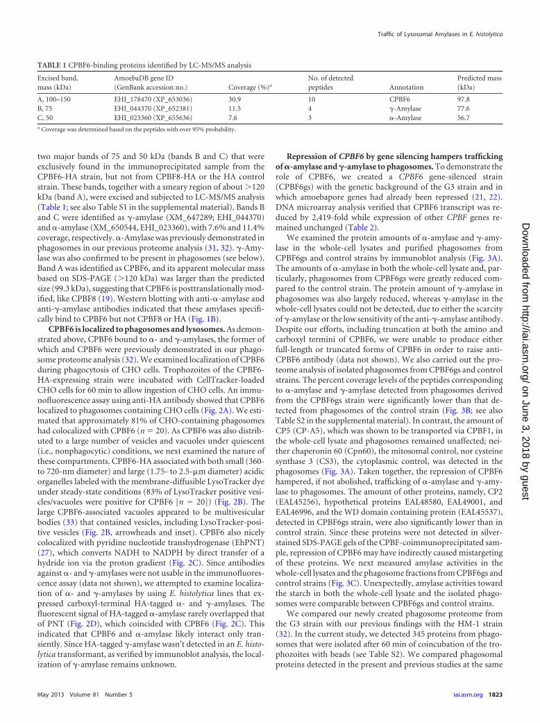
CPBF6 is localized to phagosomes and lysosomes. As demon-strated above, CPBF6 bound to �- and �-amylases, the former ofwhich and CPBF6 were previously demonstrated in our phago-some proteome analysis (32). We examined localization of CPBF6during phagocytosis of CHO cells. Trophozoites of the CPBF6-HA-expressing strain were incubated with CellTracker-loadedCHO cells for 60 min to allow ingestion of CHO cells. An immu-nofluorescence assay using anti-HA antibody showed that CPBF6localized to phagosomes containing CHO cells (Fig. 2A). We esti-mated that approximately 81% of CHO-containing phagosomeshad colocalized with CPBF6 (n � 20). As CPBF6 was also distrib-uted to a large number of vesicles and vacuoles under quiescent(i.e., nonphagocytic) conditions, we next examined the nature ofthese compartments. CPBF6-HA associated with both small (360-to 720-nm diameter) and large (1.75- to 2.5-�m diameter) acidicorganelles labeled with the membrane-diffusible LysoTracker dyeunder steady-state conditions (83% of LysoTracker positive vesi-cles/vacuoles were positive for CPBF6 [n � 20]) (Fig. 2B). Thelarge CPBF6-associated vacuoles appeared to be multivesicularbodies (33) that contained vesicles, including LysoTracker-posi-tive vesicles (Fig. 2B, arrowheads and inset). CPBF6 also nicelycolocalized with pyridine nucleotide transhydrogenase (EhPNT)(27), which converts NADH to NADPH by direct transfer of ahydride ion via the proton gradient (Fig. 2C). Since antibodiesagainst �- and �-amylases were not usable in the immunofluores-cence assay (data not shown), we attempted to examine localiza-tion of �- and �-amylases by using E. histolytica lines that ex-pressed carboxyl-terminal HA-tagged �- and �-amylases. Thefluorescent signal of HA-tagged �-amylase rarely overlapped thatof PNT (Fig. 2D), which coincided with CPBF6 (Fig. 2C). Thisindicated that CPBF6 and �-amylase likely interact only tran-siently. Since HA-tagged �-amylase wasn’t detected in an E. histo-lytica transformant, as verified by immunoblot analysis, the local-ization of �-amylase remains unknown.
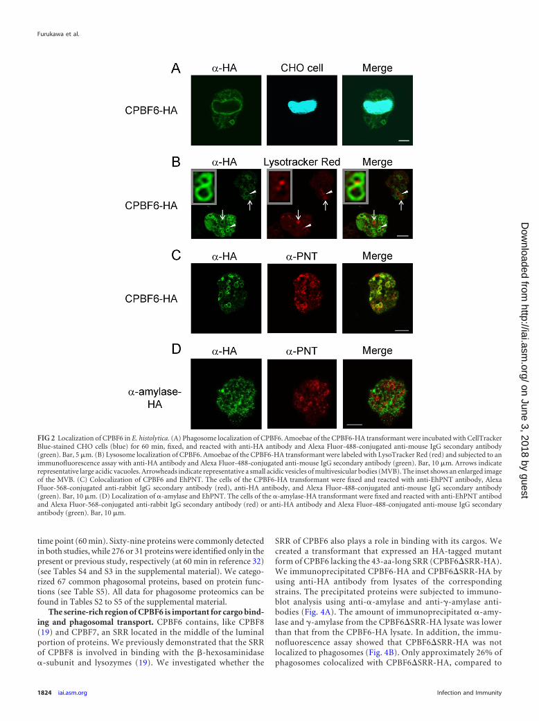
Repression of CPBF6 by gene silencing hampers traffickingof �-amylase and �-amylase to phagosomes. To demonstrate therole of CPBF6, we created a CPBF6 gene-silenced strain(CPBF6gs) with the genetic background of the G3 strain and inwhich amoebapore genes had already been repressed (21, 22).DNA microarray analysis verified that CPBF6 transcript was re-duced by 2,419-fold while expression of other CPBF genes re-mained unchanged (Table 2).
We examined the protein amounts of �-amylase and �-amy-lase in the whole-cell lysates and purified phagosomes fromCPBF6gs and control strains by immunoblot analysis (Fig. 3A).The amounts of �-amylase in both the whole-cell lysate and, par-ticularly, phagosomes from CPBF6gs were greatly reduced com-pared to the control strain. The protein amount of �-amylase inphagosomes was also largely reduced, whereas �-amylase in thewhole-cell lysates could not be detected, due to either the scarcityof �-amylase or the low sensitivity of the anti-�-amylase antibody.Despite our efforts, including truncation at both the amino andcarboxyl termini of CPBF6, we were unable to produce eitherfull-length or truncated forms of CPBF6 in order to raise anti-CPBF6 antibody (data not shown). We also carried out the pro-teome analysis of isolated phagosomes from CPBF6gs and controlstrains. The percent coverage levels of the peptides correspondingto �-amylase and �-amylase detected from phagosomes derivedfrom the CPBF6gs strain were significantly lower than that de-tected from phagosomes of the control strain (Fig. 3B; see alsoTable S2 in the supplemental material). In contrast, the amount ofCP5 (CP-A5), which was shown to be transported via CPBF1, inthe whole-cell lysate and phagosomes remained unaffected; nei-ther chaperonin 60 (Cpn60), the mitosomal control, nor cysteinesynthase 3 (CS3), the cytoplasmic control, was detected in thephagosomes (Fig. 3A). Taken together, the repression of CPBF6hampered, if not abolished, trafficking of �-amylase and �-amy-lase to phagosomes. The amount of other proteins, namely, CP2(EAL45256), hypothetical proteins EAL48580, EAL49001, andEAL46996, and the WD domain containing protein (EAL45537),detected in CPBF6gs strain, were also significantly lower than incontrol strain. Since these proteins were not detected in silver-stained SDS-PAGE gels of the CPBF-coimmunoprecipitated sam-ple, repression of CPBF6 may have indirectly caused mistargetingof these proteins. We next measured amylase activities in thewhole-cell lysates and the phagosome fractions from CPBF6gs andcontrol strains (Fig. 3C). Unexpectedly, amylase activities towardthe starch in both the whole-cell lysate and the isolated phago-somes were comparable between CPBF6gs and control strains.
We compared our newly created phagosome proteome fromthe G3 strain with our previous findings with the HM-1 strain(32). In the current study, we detected 345 proteins from phago-somes that were isolated after 60 min of coincubation of the tro-phozoites with beads (see Table S2). We compared phagosomalproteins detected in the present and previous studies at the same
TABLE 1 CPBF6-binding proteins identified by LC-MS/MS analysis
Excised band,mass (kDa)
AmoebaDB gene ID(GenBank accession no.) Coverage (%)a
No. of detectedpeptides Annotation
Predicted mass(kDa)
A, 100–150 EHI_178470 (XP_653036) 30.9 10 CPBF6 97.8B, 75 EHI_044370 (XP_652381) 11.5 4 �-Amylase 77.6C, 50 EHI_023360 (XP_655636) 7.6 3 �-Amylase 56.7a Coverage was determined based on the peptides with over 95% probability.
Traffic of Lysosomal Amylases in E. histolytica
May 2013 Volume 81 Number 5 iai.asm.org 1823
on June 3, 2018 by guesthttp://iai.asm
.org/D
ownloaded from
time point (60 min). Sixty-nine proteins were commonly detectedin both studies, while 276 or 31 proteins were identified only in thepresent or previous study, respectively (at 60 min in reference 32)(see Tables S4 and S3 in the supplemental material). We catego-rized 67 common phagosomal proteins, based on protein func-tions (see Table S5). All data for phagosome proteomics can befound in Tables S2 to S5 of the supplemental material.
The serine-rich region of CPBF6 is important for cargo bind-ing and phagosomal transport. CPBF6 contains, like CPBF8(19) and CPBF7, an SRR located in the middle of the luminalportion of proteins. We previously demonstrated that the SRRof CPBF8 is involved in binding with the �-hexosaminidase�-subunit and lysozymes (19). We investigated whether the
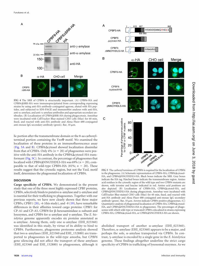
SRR of CPBF6 also plays a role in binding with its cargos. Wecreated a transformant that expressed an HA-tagged mutantform of CPBF6 lacking the 43-aa-long SRR (CPBF6�SRR-HA).We immunoprecipitated CPBF6-HA and CPBF6�SRR-HA byusing anti-HA antibody from lysates of the correspondingstrains. The precipitated proteins were subjected to immuno-blot analysis using anti-�-amylase and anti-�-amylase anti-bodies (Fig. 4A). The amount of immunoprecipitated �-amy-lase and �-amylase from the CPBF6�SRR-HA lysate was lowerthan that from the CPBF6-HA lysate. In addition, the immu-nofluorescence assay showed that CPBF6�SRR-HA was notlocalized to phagosomes (Fig. 4B). Only approximately 26% ofphagosomes colocalized with CPBF6�SRR-HA, compared to
FIG 2 Localization of CPBF6 in E. histolytica. (A) Phagosome localization of CPBF6. Amoebae of the CPBF6-HA transformant were incubated with CellTrackerBlue-stained CHO cells (blue) for 60 min, fixed, and reacted with anti-HA antibody and Alexa Fluor-488-conjugated anti-mouse IgG secondary antibody(green). Bar, 5 �m. (B) Lysosome localization of CPBF6. Amoebae of the CPBF6-HA transformant were labeled with LysoTracker Red (red) and subjected to animmunofluorescence assay with anti-HA antibody and Alexa Fluor-488-conjugated anti-mouse IgG secondary antibody (green). Bar, 10 �m. Arrows indicaterepresentative large acidic vacuoles. Arrowheads indicate representative a small acidic vesicles of multivesicular bodies (MVB). The inset shows an enlarged imageof the MVB. (C) Colocalization of CPBF6 and EhPNT. The cells of the CPBF6-HA transformant were fixed and reacted with anti-EhPNT antibody, AlexaFluor-568-conjugated anti-rabbit IgG secondary antibody (red), anti-HA antibody, and Alexa Fluor-488-conjugated anti-mouse IgG secondary antibody(green). Bar, 10 �m. (D) Localization of �-amylase and EhPNT. The cells of the �-amylase-HA transformant were fixed and reacted with anti-EhPNT antibodand Alexa Fluor-568-conjugated anti-rabbit IgG secondary antibody (red) or anti-HA antibody and Alexa Fluor-488-conjugated anti-mouse IgG secondaryantibody (green). Bar, 10 �m.
Furukawa et al.
1824 iai.asm.org Infection and Immunity
on June 3, 2018 by guesthttp://iai.asm
.org/D
ownloaded from
80% with full-length CPBF6-HA (n � 20). These data areconsistent with the notion that the SRR in CPBF6 is importantfor both the binding to �-amylase and �-amylase and for theirphagosomal transport. Immunoblot analysis showed thatCPBF6-HA was detected as a broad smeary band of 110 to 150kDa (Fig. 1B and 4A), whereas CPBF6�SRR-HA was detectedas an apparently single 100-kDa band. These data are consis-tent with the premise that the SRR contains posttranslationalmodifications.
The cytosolic domain is involved in the phagosomal traffick-ing of CPBF6. We speculated that the cytosolic domain con-taining the Yxx� motif of CPBF proteins may also be involvedin binding with accessory molecules and, therefore, plays animportant role in the determination of localization. However,our previously study showed that the localization of CPBF1 wasnot altered by mutation of the Yxx� motif, suggesting that themotif does not primarily determine its localization of CPBF1(20). We asked if the cytoplasmic region containing the Yxx�motif is involved in the phagosomal trafficking of CPBF6. Wecreated two deletion mutants of CPBF6, CPBF6�cytosol andCPBF6�NNGYHDLS, which lacked the entire 15-aa-long cytoso-
TABLE 2 Specific repression of CPBF6 by gene silencinga
Gene CPBF6gs strain Control strain Fold change
CPBF1 3,110 (�1,010) 2,780 (�268) 1.12CPBF2 135 (�15.4) 141 (�16.7) 0.95CPBF3 638 (�45.0) 482 (�18.4) 1.32CPBF4 1,120 (�347) 1,040 (�269) 1.08CPBF5 127 (�44.0) 136 (�38.9) 0.93CPBF6 1.58 (�0.75) 3,820 (�87.8) 4.13 � 10 4
CPBF7 1,730 (�282) 1,540 (�190) 1.13CPBF8 5,620 (�556) 5,630 (�454) 1.00CPBF9 311 (�24.9) 248 (�30.7) 1.26CPBF10 123 (�90.2) 94.5 (�26.8) 1.30CPBF11 96.3 (�14.6) 77.0 (�10.7) 1.25a Total RNA was extracted from the CPBF6gs and control strains, and expressionanalysis was performed using a custom-made E. histolytica DNA microarray. Average(� standard deviation) normalized signal intensities from DNA microarrays weredetermined in triplicate and indicate relative levels of the transcripts, in arbitrary units,of CPBF genes (CPBF1 to CPBF11) in the CPBF6gs and control strains. The fold changeindicates the relative change in signal intensity in the CPBF6gs strain relative to that inthe control strain.
FIG 3 Phenotypic changes of CPBF6 gene silencing. (A) Immunoblot analysis of the whole-cell lysate and the phagosome fraction. Approximately 20 �g of thewhole-cell lysate and 2 �g of the phagosome fraction were electrophoresed by SDS-PAGE and subjected to immunoblot analysis using anti-�-amylase,anti-�-amylase, CPBF1, CP5, Cpn60, and CS3 antibodies. CPBF1 and CP5 are phagosomal proteins, while CPN60 is a mitosomal marker and CS3 is a cytosolicmarker. Faint smeary bands in the whole-cell lysate of the control and CPBF6gs strains, detected with anti-�-amylase antibody, were considered backgroundbecause of their low intensities compared to those in the phagosome fraction. (B) Proteomic analysis of isolated phagosomes from CPBF6gs and control strains.Each dot represents a protein identified from phagosomes. The x axis indicates the percentage of coverage from CPBF6gs minus the percentage of coverage fromthe control strain), and it is theoretically close to 0 when the repression of CPBF6 does not affect the trafficking of a protein. Similarly, y axis indicates thepercentage of coverage from CPBF6gs divided by the percentage of coverage from the control strain, and it is close to 1 when the repression of CPBF6 does notaffect the trafficking of a protein. Individual data are shown in Table S2 in the supplemental material. (C) Enzymatic activities of amylase in whole-cell lysates andphagosomes from CPBF6gs and control strains. The amoeba lysate, culture supernatant, or phagosomal fraction was mixed with 200 �g/ml of substrate solutioncontaining BODIPY FL-conjugated DQ starch in 100 �l of 0.1 M MOPS (pH 6.9). The fluorescence was measured at excitation and emission wavelengths of 485and 530 nm, respectively, at 25°C for 60 min. Data shown are the means � standard deviations of three independent experiments.
Traffic of Lysosomal Amylases in E. histolytica
May 2013 Volume 81 Number 5 iai.asm.org 1825
on June 3, 2018 by guesthttp://iai.asm
.org/D
ownloaded from
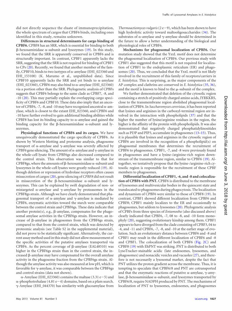
lic portion after the transmembrane domain or the 8-aa carboxyl-terminal portion containing the Yxx� motif. We examined thelocalization of these proteins in an immunofluorescence assay(Fig. 5A and B). CPBF6�cytosol showed localization dissimilarfrom that of CPBF6. Only 4% (n � 20) of phagosomes were pos-itive with the anti-HA antibody in the CPBF6�cytosol-HA trans-formant (Fig. 5C). In contrast, the percentage of phagosomes thatlocalized with CPBF6�NNGYHDLS-HA was 69% (n � 20), com-parable to that of wild-type CPBF6-HA (81%; n � 20). Theseresults suggest that the cytosolic region, but not the YxxL motifitself, determines the phagosomal localization of CPBF6.
DISCUSSIONCargo specificity of CPBF6. We demonstrated in the presentstudy that one of the three most highly expressed CPBF proteins,CPBF6, selectively binds to putative �-amylase and �-amylase andis essential for their trafficking to phagosomes. Together with ourprevious reports, we have now clearly shown that three majorCPBFs, CPBF1 (20), -6 (this study), and -8 (19), have remarkabledifferences in their affinities toward cargo proteins: CPBF1 forCP-A1 and CP-A5, CPBF8 for �-hexosaminidase �-subunit andlysozymes, and CPBF6 for �-amylase and �-amylase. The E. his-tolytica genome apparently encodes six proteins annotated as�-amylase. Among them, only one �-amylase (EHI_023360)was identified in this study, by virtue of its ability to bind toCPBF6. Furthermore, phagosome proteome analysis showedthat two �-amylases (EHI_023360 and EHI_152880) are trans-ported to phagosomes in the wild-type amoeba, but CPBF6gene silencing did not affect the transport of these amylases(EHI_023360 and EHI_152880) to phagosomes, although it
abolished transport of another �-amylase (EHI_023360).Therefore, �-amylase (EHI_023360) appears to be a major, andperhaps the sole, �-amylase transported via CPBF6. In con-trast, �-amylase is encoded by a single gene in the E. histolyticagenome. These findings altogether underline the strict cargospecificity of CPBF6 in trafficking of lysosomal enzymes. As we
FIG 4 The SRR of CPBF6 is structurally important. (A) CPBF6-HA andCPBF6�SRR-HA were immunoprecipitated from corresponding expressingstrains by using anti-HA-antibody-conjugated agarose, eluted with HA pep-tides, and subjected to SDS-PAGE and immunoblot analyses with anti-HA,anti-�-amylase, and anti-�-amylase antibodies and appropriate secondary an-tibodies. (B) Localization of CPBF6�SRR-HA during phagocytosis. Amoebaewere incubated with CellTracker Blue-stained CHO cells (blue) for 60 min,fixed, and reacted with anti-HA antibody and Alexa Fluor-488-conjugatedanti-mouse IgG secondary antibody (green). Bar, 10 �m.
FIG 5 The carboxyl terminus of CPBF6 is required for the localization of CPBF6to the phagosome. (A) Schematic representation of CPBF6-HA, CPBF6�cytosol-HA, and CPBF6�NNGYHDLS-HA. Black boxes indicate the SRR. Gray boxesindicate the HA tag. Hatched boxes indicate the transmembrane region. Aminoacid residues in the cytosolic region of the wild type and two CPBF6 mutants areshown, with tyrosine and leucine indicated in red. Amino acid positions arealso depicted. (B) Localization of CPBF6-HA, CPBF6�cytosol-HA, andCPBF6�NNGYHDLS-HA during phagocytosis. Amoebae were incubated withCellTracker Blue-stained CHO cells (blue) for 60 min, fixed, and reacted withanti-HA antibody and Alexa Fluor-488-conjugated anti-mouse IgG secondaryantibody (green). Bar, 10 �m. Arrows indicate CPBF6-positive phagosomes. (C)Quantitative analysis of phagosomal localization of CPBF6-HA, CPBF6�cytosol-HA, and CPBF6�NNGYHDLS-HA to phagosomes. The percentages of phago-somes with which wild-type or truncated CPBF6 colocalized in strains expressingCPBF6-HA, CPBF6�cytosol-HA, or CPBF6�NNGYHDLS-HA are shown.
Furukawa et al.
1826 iai.asm.org Infection and Immunity
on June 3, 2018 by guesthttp://iai.asm
.org/D
ownloaded from
did not directly sequence the eluate of immunoprecipitation,the whole spectrum of cargos that CPBF6 binds, including onesidentified in this study, remains unknown.
Differences in structural determinants for cargo binding ofCPBF6. CPBF8 has an SRR, which is essential for binding to both�-hexosaminidase �-subunit and lysozymes (19). In this study,we found that the SRR is apparently conserved in CPBF6 and isstructurally important. In contrast, CPBF1 apparently lacks theSRR, suggesting that the SRR is not required for binding of CPBF1to CPs (20). Recently, we found that another member of the fam-ily, CPBF10, binds to two isotypes of �-amylase (EHI_023360 andEHI_153100) (K. Marumo et al., unpublished data). SinceCPBF10 apparently lacks the SRR and yet binds to �-amylase(EHI_023360), CPBF6 may also bind to �-amylase (EHI_023360)via a portion other than the SRR. Phylogenetic analysis of CPBFssuggests that CPBF6 belongs to the same clade as CPBF7, -8, and-10 (20). This may partially explain the overlapping cargo speci-ficity of CPBF6 and CPBF10. These data also imply that an ances-tor of CPBF6, -7, -8, and -10 may have recognized ancestral �-am-ylase, which is closest to the extant EHI_023360, and CPBF6 and-10 have further evolved to gain additional binding abilities whileCPBF8 has lost its binding capacity to �-amylase and gained thebinding capacity for the �-hexosaminidase �-subunit and ly-sozymes.
Physiological functions of CPBF6 and its cargos. We haveunequivocally demonstrated the cargo specificity of CPBF6. Asshown by Western blotting and proteome analysis, phagosometransport of �-amylase and �-amylase was severely affected byCPBF6 gene silencing. The amount of �-amylase and �-amylase inthe whole-cell lysate from CPBF6gs also decreased compared tothe control strain. This observation was similar to that forCPBF8gs, where the amounts of �-hexosaminidase �-subunit andlysozymes in the whole-cell lysates were greatly reduced (19). Al-though deletion or repression of hydrolase receptors often causesmissecretion of cargos (20), gene silencing of CPBF8 did not resultin missecretion of the �-hexosaminidase �-subunit and ly-sozymes. This can be explained by swift degradation of non- ormistargeted �-amylase and �-amylase by proteasomes in theCPBF8gs strain. Although we have clearly demonstrated that pha-gosomal transport of �-amylase and �-amylase is mediated byCPBF6, enzymatic activities toward the starch were comparablebetween the control strain and CPBF6gs. These data indicate thatanother protein(s), e.g., �-amylase, compensates for the phago-somal amylase activities in the CPBF6gs strain. However, an in-crease of �-amylase in phagosomes from the CPBF6gs strain,compared to that from the control strain, which was detected byproteomic analysis (see Table S2 in the supplemental material),did not prove to be statistically significant. Alternatively, the cur-rent assay method used in this study did not allow measurement ofthe specific activities of the putative amylases transported viaCPBF6. As the percent coverage of �-amylase (EAL48510) washigher in the CPBF6gs strain than in the control strain, the in-creased �-amylase may have compensated for the overall amylaseactivity in the phagosome fraction from the CPBF6gs strain. Al-though the amylase activity was also measured at low pH, which isfavorable for �-amylase, it was comparable between the CPBF6gsand control strains (data not shown).
�-Amylase (EHI_023360) contains the maltase (3.31 e 5) and�-phosphotrehalase (4.81 e 4) domains, based on a pfam search.�-Amylase (EHI_044370) has similarity with glucoamylase from
Thermoactiomyces vulgaris (2 e 9), which has been shown to havehigh hydrolytic activity toward maltooligosaccharides (34). Thesubstrates of �-amylase and �-amylase should be determined inthe future to allow a better understanding of the biological andphysiological roles of CPBF6.
Mechanisms for phagosomal localization of CPBF6. Ourmutation study showed that the YxxL motif does not determinethe phagosomal localization of CPBF6. Our previous study withCPBF1 also suggested that this motif is not required for localiza-tion of CPBF1 to the endoplasmic reticulum (ER) and phago-somes (20). Thus, we concluded that the YxxL motif is not likelyinvolved in the recruitment of this family of receptors/carriers inE. histolytica. This is surprising, as the major components of theAP complex and clathrins are conserved in E. histolytica (35, 36),and the motif is known to bind to the �-subunit of the complex.
We further demonstrated that deletion of the cytosolic regioncontaining a stretch of positively charged amino acids, IYRKKRS,close to the transmembrane region abolished phagosomal local-ization of CPBF6. In Saccharomyces cerevisiae, it has been reportedthat lysines and arginines in the carboxyl-terminal region are in-volved in the interaction with phospholipids (37) and that thehigher the number of lysine/arginine residues in the region, thehigher is the affinity of the protein with phospholipids. It was alsodemonstrated that negatively charged phosphatidylinositidessuch as PI3P and PIP3, accumulate in phagosomes (13–15). Thus,it is plausible that lysines and arginines in the cytosolic region ofCPBF6 are involved in the recognition of a phospholipid(s) onphagosomal membranes that determines the recruitment ofCPBF6 to phagosomes. CPBF1, -7, and -8 were previously foundin phagosomes and have a lysine-/arginine-rich region down-stream of the transmembrane region, similar to CPBF6 (19). Al-together, we tentatively propose that the lysine-/arginine-rich cy-tosolic region is responsible for the recruitment of these CPBFmembers to phagosomes.
Differential localization of CPBF1, -6, and -8 and colocaliza-tion of CPBF6 with PNT. CPBF6 is distributed to the membraneof lysosomes and multivesicular bodies in the quiescent state andtranslocated to phagosomes during phagocytosis. The localizationand dynamism of CPBF6 were similar to those of CPBF8 (19). Incontrast, CPBF1 showed different localization from CPBF6 andCPBF8; CPBF1 mainly localizes to the ER and occasionally tophagosomes, but seldom to lysosomes (20). Phylogenetic analysisof CPBFs from three species of Entamoeba (also discussed above)clearly indicated that CPBF6, -7, 08 to -8, and -10 form mono-phyly (20), suggesting evolutionary kinship among them. CPBF1appears to have diverged from the clade consisting of CPBF3, 04 to-4, and -11 and CPBF6, -7, -8, and -10 at the earlier stage of evo-lution. Such an evolutionary distance between CPBF6 and -8 andCPBF1 may result in the different localization of CPBF6 and -8and CPBF1. The colocalization of both CPBF6 (Fig. 2C) andCPBF8 (19) with EhPNT was striking. PNT is distributed to bothLysoTracker-stainable acidic (late endosomes, lysosomes, andphagosomes) and nonacidic vesicles and vacuoles (27), and there-fore is not necessarily a lysosomal marker, despite the fact thatPNT utilizes the proton gradient across the membrane. Thus, it istempting to speculate that CPBF6/8 and PNT are cotransportedand that the enzymatic reactions of putative �-amylase, �-amy-lase, �-hexosaminidase �-subunit, and lysozymes transported byCPBF6/8, require NADPH produced by PNT. The mechanisms oflocalization of PNT to lysosomes, endosomes, and phagosomes
Traffic of Lysosomal Amylases in E. histolytica
May 2013 Volume 81 Number 5 iai.asm.org 1827
on June 3, 2018 by guesthttp://iai.asm
.org/D
ownloaded from
are still unknown. We previously demonstrated that all domains,such as the amino-terminal extension, 11–13 transmembrane,and linker regions are essential for the vesicular/vacuolar distribu-tion of PNT (27). However, in contrast to CPBF6, no apparentlysine-/arginine-rich region is present in PNT.
ACKNOWLEDGMENTS
We are grateful to Nicholas Sherman, W.M. Keck Biomedical Mass Spec-trometry Laboratory at the University of Virginia, for technical supporton MS analyses. We are also grateful to Kazuo Ebine and Kisaburo Naga-mune for helpful discussions.
This work was supported by a Grant-in-Aid for Scientific Researchfrom the Ministry of Education, Culture, Sports, Science and Technology(MEXT) of Japan (23117001, 23117005, and 23390099), a Grant-in-Aidon Bilateral Programs of Joint Research Projects and Seminars from theJapan Society for the Promotion of Science, a Grant-in-Aid on StrategicInternational Research Cooperative Program (SICP) from the Japan Sci-ence and Technology Agency, a grant for research on emerging and re-emerging infectious diseases from the Ministry of Health, Labor and Wel-fare (MHLW) of Japan (H23-Shinkosaiko-ippan-014), a grant forresearch to promote the development of anti-AIDS pharmaceuticals fromthe Japan Health Sciences Foundation (KHA1101), and by the GlobalCOE Program (Global COE for Human Metabolomic Systems Biology)from MEXT, Japan.
REFERENCES1. Bracha R, Kobiler D, Mirelman D. 1982. Attachment and ingestion of
bacteria by trophozoites of Entamoeba histolytica. Infect. Immun. 36:396 –406.
2. Tsutsumi V, Ramírez-Rosales A, Lanz-Mendoza H, Shibayama M,Chávez B, Rangel-López E, Martínez-Palomo A. 1992. Entamoeba his-tolytica: erythrophagocytosis, collagenolysis, and liver abscess productionas virulence markers. Trans. R. Soc. Trop. Med. Hyg. 86:170 –172.
3. Guerrant RL, Brush J, Ravdin JI, Sullivan JA, Mandell GL. 1981.Interaction between Entamoeba histolytica and human polymorphonu-clear neutrophils. J. Infect. Dis. 143:83–93.
4. Bracha R, Mirelman D. 1984. Virulence of Entamoeba histolytica tropho-zoites. Effects of bacteria, microaerobic conditions, and metronidazole. J.Exp. Med. 160:353–368.
5. Hirata KK, Que X, Melendez-Lopez SG, Debnath A, Myers S, HerdmanDS, Orozco E, Bhattacharya A, McKerrow JH, Reed SL. 2007. A phago-cytosis mutant of Entamoeba histolytica is less virulent due to deficientproteinase expression and release. Exp. Parasitol. 115:192–199.
6. Katz U, Ankri S, Stolarsky T, Nuchamowitz Y, Mirelman D. 2002.Entamoeba histolytica expressing a dominant negative N-truncated lightsubunit of its Gal-lectin are less virulent. Mol. Biol. Cell 13:4256 – 4265.
7. Orozco E, Guarneros G, Martinez-Palomo A, Sánchez T. 1983. Ent-amoeba histolytica. Phagocytosis as a virulence factor. J. Exp. Med. 158:1511–1521.
8. Que X, Brinen LS, Perkins P, Herdman S, Hirata K, Torian BE, RubinH, McKerrow JH, Reed SL. 2002. Cysteine proteinases from distinctcellular compartments are recruited to phagocytic vesicles by Entamoebahistolytica. Mol. Biochem. Parasitol. 119:23–32.
9. Andrä J, Herbst R, Leippe M. 2003. Amoebapores, archaic effectorpeptides of protozoan origin, are discharged into phagosomes and killbacteria by permeabilizing their membranes. Dev. Comp. Immunol. 27:291–304.
10. Mann BJ. 2002. Structure and function of the Entamoeba histolytica Gal/GalNAc lectin. Int. Rev. Cytol. 216:59 – 80.
11. Petri WA, Jr, Haque R, Mann BJ. 2002. The bittersweet interface ofparasite and host: lectin-carbohydrate interactions during human inva-sion by the parasite Entamoeba histolytica. Annu. Rev. Microbiol. 56:39 –64.
12. Voigt H, Olivo JC, Sansonetti P, Guillén N. 1999. Myosin IB fromEntamoeba histolytica is involved in phagocytosis of human erythrocytes.J. Cell Sci. 112:1191–1201.
13. Byekova YA, Powell RR, Welter BH, Temesvari LA. 2010. Localizationof phosphatidylinositol (3,4,5)-trisphosphate to phagosomes in Ent-amoeba histolytica achieved using glutathione S-transferase- and greenfluorescent protein-tagged lipid biosensors. Infect. Immun. 78:125–137.
14. Ghosh SK, Samuelson J. 1997. Involvement of p21racA, phosphoinosi-tide 3-kinase, and vacuolar ATPase in phagocytosis of bacteria and eryth-rocytes by Entamoeba histolytica: suggestive evidence for coincidental evo-lution of amebic invasiveness. Infect. Immun. 65:4243– 4249.
15. Nakada-Tsukui K, Okada H, Mitra BN, Nozaki T. 2009. Phosphatidyl-inositol-phosphates mediate cytoskeletal reorganization during phagocy-tosis via a unique modular protein consisting of RhoGEF/DH and FYVEdomains in the parasitic protozoon Entamoeba histolytica. Cell. Microbiol.11:1471–1491.
16. Somlata Bhattacharya S, Bhattacharya A. 2011. A C2 domain proteinkinase initiates phagocytosis in the protozoan parasite Entamoeba histo-lytica. Nat. Commun. 2:230.
17. Boettner DR, Huston CD, Linford AS, Buss SN, Houpt E, Sherman NE,Petri WA, Jr. 2008. Entamoeba histolytica phagocytosis of human eryth-rocytes involves PATMK, a member of the transmembrane kinase family.PLoS Pathog. 4:e8. doi:10.1371/journal.ppat.0040008.
18. Labruyère E, Zimmer C, Galy V, Olivo-Marin Guillén J-CN. 2003.EhPAK, a member of the p21-activated kinase family, is involved in thecontrol of Entamoeba histolytica migration and phagocytosis. J. Cell Sci.116:61–71.
19. Furukawa A, Nakada-Tsukui K, Nozaki T. 2012. Novel transmembranereceptor involved in phagosome transport of lysozymes and �-hexo-saminidase in the enteric protozoan Entamoeba histolytica. PLoS Pathog.8:e1002539. doi:10.1371/journal.ppat.1002539.
20. Nakada-Tsukui K, Tsuboi Furukawa K, Yamada A, Nozaki Y, T. 2012.A novel class of cysteine protease receptors that mediate lysosomal trans-port. Cell. Microbiol. 14:1299 –1317.
21. Bracha R, Nuchamowitz Y, Mirelman D. 2003. Transcriptional silencingof an amoebapore gene in Entamoeba histolytica: molecular analysis andeffect on pathogenicity. Eukaryot. Cell 2:295–305.
22. Zhang H, Alramini H, Tran V, Singh U. 2011. Nucleus-localized anti-sense small RNAs with 5=-polyphosphate termini regulate long term tran-scriptional gene silencing in Entamoeba histolytica G3 strain. J. Biol.Chem. 286:44467– 44479.
23. Diamond LS, Mattern CF, Bartgis IL. 1972. Viruses of Entamoeba histo-lytica. I. Identification of transmissible virus-like agents. J. Virol. 9:326 –341.
24. Diamond LS, Harlow DR, Cunnick CC. 1978. A new medium for theaxenic cultivation of Entamoeba histolytica and other Entamoeba. Trans.R. Soc. Trop. Med. Hyg. 72:431– 432.
25. Saito-Nakano Y, Mitra BN, Nakada-Tsukui K, Sato D, Nozaki T. 2007.Two Rab7 isotypes, EhRab7A and EhRab7B, play distinct roles in biogen-esis of lysosomes and phagosomes in the enteric protozoan parasite Ent-amoeba histolytica. Cell. Microbiol. 9:1796 –1808.
26. Nozaki T, Asai T, Sanchez LB, Kobayashi S, Nakazawa M, Takeuchi T.1999. Characterization of the gene encoding serine acetyltransferase, aregulated enzyme of cysteine biosynthesis from the protist parasites Ent-amoeba histolytica and Entamoeba dispar. Regulation and possible func-tion of the cysteine biosynthetic pathway in Entamoeba. J. Biol. Chem.274:32445–32452.
27. Yousuf MA, Mi-Ichi F, Nakada-Tsukui K, Nozaki T. 2010. Localizationand Targeting of an unusual pyridine nucleotide transhydrogenase in Ent-amoeba histolytica. Eukaryot. Cell 9:926 –933.
28. Gilchrist CA, Houpt E, Trapaidze N, Fei Z, Crasta O, Asgharpour A,Evans C, Martino-Catt S, Baba DJ, Stroup S, Hamano S, EhrenkauferG, Okada M, Singh U, Nozaki T, Mann BJ, Petri WA, Jr. 2006. Impactof intestinal colonization and invasion on the Entamoeba histolytica tran-scriptome. Mol. Biochem. Parasitol. 147:163–176.
29. Husain A, Jeelani G, Sato D, Nozaki T. 2011. Global analysis of geneexpression in response to L-cysteine deprivation in the anaerobic proto-zoan parasite Entamoeba histolytica. BMC Genomics 12:275. doi:10.1186/1471-2164-12-275.
30. Penuliar GM, Furukawa A, Nakada-Tsukui K, Husain A, Sato D,Nozaki T. 2012. Transcriptional and functional analysis of trifluorome-thionine resistance in Entamoeba histolytica. J. Antimicrob. Chemother.67:375–386.
31. Okada M, Huston CD, Mann BJ, Petri WA, Kita K, Nozaki T. 2005.Proteomic analysis of phagocytosis in the enteric protozoan parasiteEntamoeba histolytica. Eukaryot. Cell 4:827– 831.
32. Okada M, Huston CD, Oue M, Mann BJ, Petri WA, Jr, Kita K, NozakiT. 2006. Kinetics and strain variation of phagosome proteins of Ent-amoeba histolytica by proteomic analysis. Mol. Biochem. Parasitol. 145:171–183.
Furukawa et al.
1828 iai.asm.org Infection and Immunity
on June 3, 2018 by guesthttp://iai.asm
.org/D
ownloaded from
33. Saito-Nakano Y, Yasuda T, Nakada-Tsukui K, Leippe M, Nozaki T.2004. Rab5-associated vacuoles play a unique role in phagocytosis of theenteric protozoan parasite Entamoeba histolytica. J. Biol. Chem. 279:49497– 49507.
34. Uotsu-Tomita R, Tonozuka T, Sakai H, Sakano Y. 2001. Novel glu-coamylase-type enzymes from Thermoactinomyces vulgaris and Methano-coccus jannaschii whose genes are found in the flanking region of thea-amylase genes. Appl. Microbiol. Biotechnol. 56:465– 473.
35. Clark CG, Alsmark UCM, Tazreiter M, Saito-Nakano Y, Ali V, Marion S,Weber C, Mukherjee C, Bruchhaus I, Tannich E, Leippe M, Sicheritz-Ponten T, Foster PG, Samuelson J, Noël CJ, Hirt RP, Embley TM, GilchristCA, Mann BJ, Singh U, Ackers JP, Bhattacharya S, Bhattacharya A, LohiaA, Guillén N, Duchêne M, Nozaki T, Hall N. 2007. Structure and content ofthe Entamoeba histolytica genome. Adv. Parasitol. 65:51–190.
36. Loftus B, Anderson I, Davies R, Alsmark UCM, Samuelson J, AmedeoP, Roncaglia P, Berriman M, Hirt RP, Mann BJ, Nozaki T, Suh B, PopM, Duchene M, Ackers J, Tannich E, Leippe M, Hofer M, Bruchhaus I,Willhoeft U, Bhattacharya A, Chillingworth T, Churcher C, Hance Z,Harris B, Harris D, Jagels K, Moule S, Mungall K, Ormond D, SquaresR, Whitehead S, Quail MA, Rabbinowitsch E, Norbertczak H, Price C,Wang Z, Guillén N, Gilchrist C, Stroup SE, Bhattacharya S, Lohia A,Foster PG, Sicheritz-Ponten T, Weber C, Singh U, Mukherjee C, El-Sayed NMWAP, Jr, Clark CG, Embley TM, Barrell B, Fraser CM, HallN. 2005. The genome of the protist parasite Entamoeba histolytica. Nature433:865– 868.
37. Scheglmann D, Werner K, Eiselt G, Klinger R. 2002. Role of paired basicresidues of protein C-termini in phospholipid binding. Protein Eng. 15:521–527.
Traffic of Lysosomal Amylases in E. histolytica
May 2013 Volume 81 Number 5 iai.asm.org 1829
on June 3, 2018 by guesthttp://iai.asm
.org/D
ownloaded from

![Entamoeba histolytica / E. dispar - qu.edu.iqqu.edu.iq/vmjou/wp-content/uploads/2015/01/Vol.-111-8-14.pdf · [Entamoeba histolytica / E. dispar] ... Entamoeba histolytica trophozoite](https://img.pdfslide.net/doc/110x75/5aa7ee767f8b9ab8228ce260/entamoeba-histolytica-e-dispar-queduiqqueduiqvmjouwp-contentuploads201501vol-111-8-14pdfentamoeba.jpg)