Embed Size (px)
Citation preview

Cysteine-Rich Protein 61 and Connective TissueGrowth Factor Induce Deadhesion and Anoikis ofRetinal Pericytes
Haibo Liu, Ru Yang, Babben Tinner, Annam Choudhry, Norbert Schutze, and Brahim Chaqour
Department of Anatomy and Cell Biology (H.L., R.Y., A.C., B.C.), State University of New York Downstate Medical Center,Brooklyn, New York 11203; Health Science Department (B.T.), QBM Cell Science, Ottawa, Canada K1A 0R6; andOrthopedic Center for Musculoskeletal Research (N.S.), University of Wurzburg, 97070 Wurzburg, Germany
Loss of retinal pericytes is one of the distinctive features ofdiabetic retinopathy (DR), which is characterized by retinalcapillary obliteration. The matricellular proteins, cysteine-rich protein 61 (Cyr61) and connective tissue growth factor(CTGF), are aberrantly expressed in the retinal vasculaturefrom the early stages of DR, but their effects on retinal peri-cytes are unknown. We show herein that rat retinal pericytes(RRPs) exposed to advanced glycosylation-end products, animportant injurious stimulus of diabetes, express increasedlevels of both Cyr61 and CTGF, and concomitantly undergoanoikis, a form of apoptosis by loss of cell-matrix interactions.Adenovirus-mediated expression of Cyr61 and/or CTGF con-ferred an anoikis-prone phenotype to rat retinal pericytes,including decreased phosphotyrosine protein levels at focaladhesion points and formation of cortical actin rings. Whenused as substrates for pericyte attachment and compared
with other matrix proteins (e.g. type IV collagen), recombi-nant Cyr61 and CTGF proteins exhibited antiadhesive andapoptogenic activities. Phosphatase inhibitors reversed theseeffects, suggesting that Cyr61 and CTGF promote dephos-phorylation events. Furthermore, Cyr61- and CTGF-inducedapoptosis was mediated through the intrinsic pathway andinvolved the expression of genes that have been functionallygrouped as p53 target genes. Expression of the matrix metal-loproteinase-2 gene, a known target of p53, was increased inpericytes overexpressing either Cyr61 or CTGF. Inhibition ofmatrix metalloproteinase-2 had, at least in part, a protectiveeffect against Cyr61- and CTGF-induced apoptosis. Taken to-gether, these findings support the involvement of Cyr61 andCTGF in pericyte detachment and anoikis, implicating theseproteins in the pathogenesis of DR. (Endocrinology 149:1666–1677, 2008)
THE CYSTEINE-RICH PROTEIN 61 (Cyr61) and connec-tive tissue growth factor (CTGF), also known as CCN1
and CCN2, respectively, belong to a subset of extracellularmatrix (ECM) molecules termed matricellular proteins,which exhibit highly regulated expression during develop-ment and in pathological events (1, 2). These immediate earlygene-encoded proteins are induced by and modulate, at leastin part, the activity of growth factors, hormones, and me-chanical forces (3, 4). Structurally, the Cyr61 and CTGF pro-teins share 40–50% amino acid homology with one anotherand contain, each, four conserved modular domains withsequence similarities to IGF binding proteins, the von Wil-lebrand factor type C repeat, the thrombospondin type I
repeat, and a carboxy-terminal domain containing a cystine-knot motif (5). These modules contain binding sites for in-tegrins, low-density lipoprotein receptors, transmembraneproteoglycans, growth factor receptors, and/or ECM pro-teins (6–12). Functionally, the Cyr61 and CTGF proteins actas adaptors and modulators of cell-matrix interactions, al-though their biological activities are contextual and cell typedependent. Cell exposure to these proteins modulate celladhesion, migration, proliferation, differentiation, survival,and ECM protein synthesis (13). Through these activities,Cyr61 and CTGF regulate diverse biological processes includ-ing angiogenesis, development, fibrosis, and hypertrophy.
Numerous studies have described increased Cyr61 andCTGF protein levels in the microvascular complications ofdiabetes including diabetic retinopathy (14, 15). CTGF wasshown to be expressed in microglia, but its localization shiftsto retinal microvascular pericytes in a large subset of diabeticpersons (16). Similarly, Cyr61 protein levels markedly in-crease in retinal blood vessels at the early stages of diabetesand in late stages of vitreoproliferative disorders (17). How-ever, what particular function(s) these proteins manifest inretinal blood vessels is unknown. Existing hypotheses asso-ciate the activity of these proteins with ECM remodeling (18,19). In active ophthalmopathy, Cyr61 was suggested to havea role in both orbital inflammation and adipogenesis andserve as an early marker of ocular diseases (20). Twigg et al.(19) and Zhou et al., (21) reported that advanced glycosyla-tion end (AGE) products, a major injurious stimulus asso-
First Published Online January 10, 2008Abbreviations: Ad-CTGF, Adenovirus of CTGF; Ad-Cyr61, adeno-
virus of CYR61; Ad-GFP, adenovirus encoding green fluorescent pro-tein; AGE, advanced glycosylation end; Bax, Bcl-2-associated X protein;Bcl2, B-cell LL/lymphoma 2; CT, cycle threshold; CTGF, connectivetissue growth factor; Cyr61, cysteine-rich protein 61; DAPI, 4�,6-dia-midino-2-phenylindole; ECM, extracellular matrix; EthD-1, ethidiumhomodimer; FA, focal adhesion; FITC, fluorescein isothiocyanate;GAPDH, glyceraldehyde 3-phosphate dehydrogenase; MMP, matrixmetalloproteinase; PTEN, phosphatase and tensin homolog; RRP, ratretinal pericyte; SDS, sodium dodecyl sulfate; TRITC, tetramethyl rho-damine isothiocyanate; TUNEL, terminal deoxynucleotidyl transferase-mediated deoxyuridine triphosphate nick end labeling; VEGF, vascularendothelial growth factor.Endocrinology is published monthly by The Endocrine Society (http://www.endo-society.org), the foremost professional society serving theendocrine community.
0013-7227/08/$15.00/0 Endocrinology 149(4):1666–1677Printed in U.S.A. Copyright © 2008 by The Endocrine Society
doi: 10.1210/en.2007-1415
1666
ciated with diabetes, act, in part, through cell-derived CTGFto induce fibronectin and type IV collagen expression infibroblasts and mesangial cells, respectively. However, theactual biological activities of these proteins in the retinalcapillaries are unknown.
One of the distinctive features of the process that leads todiabetic retinopathy is the initial loss of retinal capillarypericytes, which outnumber endothelial cells in the retinamore than in any other vascular bed (22). Pericytes aresmooth muscle-like cells defined by their perivascular local-ization within the basement membrane that they producetogether with endothelial cells. Their putative functions in-clude maintenance of capillary structure and integrity, reg-ulation of endothelium homeostasis, and capillary diameterand angiogenesis (23). Because of their tissue localizationoutside the blood retinal barrier, retinal pericytes rely en-tirely on survival signals derived from their surroundingECM (24). However, diabetes-induced hyperglycemia andassociated toxicities affect both the ECM composition andfunction leading to accelerated death of retinal pericytes andendothelial cells and the loss of the innate vascular autoreg-ulation characteristic of the early stages of diabetic retinop-athy (25, 26). Although both Cyr61 and CTGF have beenshown to be up-regulated in the retinal capillaries, theireffects on pericytes are still unknown (17, 27). This studyidentifies Cyr61 and CTGF as antiadhesive molecules initi-ating retinal pericyte anoikis, a form of apoptosis resultingfrom loss of cell adhesion and/or cell adhesion-dependentsignaling.
Materials and MethodsReagents
All chemicals were of reagent grade. Caspase inhibitors were pur-chased from Millipore/Chemicon (San Diego, CA). Pharmacologicalinhibitors were from Sigma-Aldrich. AGE-BSA and Bcl-2-associated Xprotein (Bax) and B-cell LL/lymphoma 2 (Bcl2) antibodies were fromBiovison (Mountain View, CA). Recombinant CYR61 was produced inbaculovirus Sf21 cells and purified as previously described (28). Re-combinant CTGF was from Novus Biologicals (Littleton, CO). EHS lami-nin and type IV collagen were from Invitrogen (Carlsbad, CA). Type Icollagen was prepared from rat tail tendon. Anti-Cyr61 and anti-CTGFantibodies were described previously (4). Antipaxillin and antiphos-photyrosine antibodies were from Cell Signaling Inc. (Danvers, MA) The4�,6-diamidino-2-phenylindole (DAPI), fluorescein isothiocyanate(FITC)-conjugated goat antimouse IgG and tetramethyl rhodamine iso-thiocyanate (TRITC)-conjugated goat antirabbit IgG antibodies werefrom Vector Laboratories (Burlingame, CA). TRITC-conjugated phal-loidin was from Cytoskeleton Inc. (Denver, CO).
Cell culture and treatment
Rat retinal pericytes (RRPs) were isolated from a pool of SpragueDawley rat retinas by selective sieving at QBM Cell Science Laboratories(Ottawa, Canada). The cells were plated in 35-mm dishes in predefinedpericyte medium containing 2% fetal bovine serum obtained from CellBiolabs, Inc. (San Diego, CA). RRPs were characterized by immuno-staining using NG2 (chondroitin sulfate proteoglycan) and desmin an-tibodies (Chemicon). These cells grow at a very slow rate and can bepassaged only twice.
Cyr61 and CTGF adenoviral vector preparationand utilization
Mouse Cyr61 and CTGF cDNAs were isolated by PCR amplificationusing DNA templates obtained from American Type Culture Collection
(Manassas, VA) and cloned into a shuttle vector. The recombinant ad-enoviruses, Ad-Cyr61 and Ad-CTGF, were produced by cotransfectingan adenoviral shuttle vector with a viral backbone in which the recom-binant cDNA is driven by the cytomegalovirus promoter. Recombinantadenovirus amplification was carried out by Vector Biolabs (Philadel-phia, PA). The empty adenovirus, Ad-V, and the adenovirus encodinggreen fluorescent protein (Ad-GFP) were used as controls for infection.All adenoviruses were replication deficient and used at 20 multiplicityof infection. Cells at 80% confluence were incubated with adenoviralvectors first, in serum-free medium for up to 3 h and then in serum-containing medium. Cells were then incubated in serum-free mediumfor defined periods of time and then processed for various analyses.
Western immunoblotting and fluorescence microscopy
For Western blot analysis, protein samples (24 �g) were fractioned ina 10% sodium dodecyl sulfate (SDS)-polyacrylamide gel and transferredto nitrocellulose membrane, and Western blot analysis was performedwith anti-Cyr61, anti-CTGF, or anti-glyceraldehyde 3-phosphate dehy-drogenase (GAPDH) antibodies. Immunodetection was performed us-ing enhanced chemiluminescence (Amersham Biosciences, Piscataway,NJ) according to the manufacturer’s recommendations.
For immunocytochemical analyses, cells plated on glass coverslipswere fixed in 0.4% formaldehyde-PBS for 20 min and permeabilized in0.1% Triton X-100 at room temperature for 5 min. For visualization ofactin stress fibers, cells were stained with TRITC-phalloidin. For im-munodetection of specific proteins, cells were incubated with the indi-cated primary antibodies overnight at 4 C and then treated with TRITC-or FITC-conjugated secondary antibodies. Coverslips were washed sev-eral times in PBS between incubations. Images were acquired using the1024 MDC laser scanning fluorescence imaging system (Bio-Rad Lab-oratories, Hercules, CA).
Anoikis and terminal deoxynucleotidyl transferase-mediateddeoxyuridine triphosphate nick end labeling (TUNEL) assays
Cells undergoing death by anoikis were identified and quantifiedusing the CytoSelect anoikis assay kit according to the manufacturer’sinstructions (Cell Bioloab). Briefly, cells to be assayed for anoikis werecollected, pelleted by centrifugation, and resuspended in culture me-dium. Cells (400,000) in suspension were added into either an anchor-age-resistant poly-Hema-coated or a control uncoated plate. After a 24-hincubation time at 37 C, ethidium homodimer (EthD-1) was added intoeach well to detect apoptotic cells. Free Eth-D1 is virtually undetectablebefore interacting with cell membrane. Fluorescence was measured at515 and 590 nm using a Fluorolite-1000 plate reader (Dynex Technol-ogies, Chantilly, VA).
TUNEL staining was performed in 35-mm dishes using an in situdeath detection kit according to the manufacturer’s instructions (RocheMolecular Biochemicals, Indianapolis, IN). The percentage of TUNEL-positive cells (relative to total counterstained with DAPI) was deter-mined by counting approximately 200 cells in 10 randomly chosen fieldsper coverslip for each experiment.
Cell adhesion assay
Microtiter 96-well plates were coated overnight at 4 C with 50 �l/wellof substrate, e.g. Cyr61, CTGF, laminin, type I or IV collagen, or BSA. Thewells were then blocked with 0.5% BSA in PBS (100 �l/well) for 2 h at37 C. Subconfluent cells were trypsinized, washed, and resuspended inadhesion buffer (Hank’s balanced salt solution supplemented with 1 mmMgCl2). An aliquot of the cell suspension (40,000 cells) was first prein-cubated with a vehicle solution or chemical inhibitors for 30 min andthen added to the coated plates in a final volume of 50 �l and allowedto attach at 37 C in a humidified incubator for 30 min. Nonattached cellswere removed by two washes with PBS. Attached cells were fixed with4% paraformaldehyde at room temperature for 10 min and stained for10 min with crystal violet (5 mg/ml in 2% ethanol). After several washeswith water, the wells were dried, and the dye was extracted with 2% SDSfor 30 min at room temperature. Adhesion was quantified by measure-ment of absorbance at 550 nm. Nonspecific cell attachment (attachmentto wells coated with BSA) was always less than 5%.
Liu et al. • Cyr61- and CTGF-Induced Anoikis Endocrinology, April 2008, 149(4):1666–1677 1667

RNA isolation and quantitative analysis of mRNA
Total RNA was extracted from cells using RNAEasy column pu-rification protocol (QIAGEN, Valencia, CA). Quantitative real-timeRT-PCR assay was performed to quantify the mRNA levels of nu-merous genes using TaqMan technology on an Applied Biosystems7000 sequence detection system (Foster City, CA). Highly specificprimers were designed using the Web-based primer design program,Primer3 (Whitehead Institute for Biomedical Research, Cambridge,MA). The forward and reverse primers were: 5�-TGAGCTCCAG-CACCATCAAG-3� and 5�-AGTGCCAGCCTGGTCAAGTG-3� forCyr61; 5�-CGCCAACCGCAAGATTG-3� and 5�-ACGGACCCAC-CGAAGACA-3� for CTGF, 5�-TGACGATGAGCTGTGGACTC-3� and5�-ACAAGAAGGGGAACTTGCAG-3� for matrix metalloproteinase(MMP)-2; 5�-ACCCCACTCACATTCTCCAG-3� and 5�-GCCCAT-CAAAAGGGATAAAG-3� for MMP-3; 5�-TACGGGCTCGAAGCA-GAC-3� and 5�-AAAGGCGTGTGCCAGTAGAC-3� for MMP-9; 5�-ACAGAAGGGGAGCAGAAAGC-3� and 5�-AGATGTCCACCAGGGTCTCA-3� for vascular endothelial growth factor (VEGF); 5�-CAGACGGCAACTTCAACTGG-3� and 5�-CAGCCCATGATGGT-TCTGAT-3� for Bax; 5�-CAGGCAGCCAATAACAGTCA-3� and 5�-CTCCTCCTCCATCCCTTCAT-3� for Bcl-2 antagonist of cell death(Bad); 5�-GATTGGGGAATGGGTTGGTAG-3� and 5�-TGCAGAGT-GGAGGAAATGGG-3� for p53; 5�-GGGACTCGCACTTGCAATA-3�and 5�-AGTTATGGTGCGCTGACTCC-3� for GADD45a; 5�-TGTTGT-GATGGTGGCTTGAG-3� and 5�-CTCCTGACCCTTCATCCGTA-3�for apoptosis inhibitor 2; 5�-GTTGCCTCCTCTCCTTTTCG-3� and 5�-AGCGTGGAAGATTGTTCAGC-3� for activating transcription factor5; and 5�-GAGGCAGACCAGCCTAACAG-3� and 5�-GGCACT-TCAGGGCTTTCTCT-3� for p21WAF1. These primers were designed tospan exon-exon junctions so that genomic DNA would not be de-tected. Primers for �1(IV) collagen and �1-laminin chain were ob-tained commercially (SuperArray Bioscience Corp., Frederick, MD).The cycling parameters for PCR amplification reactions were: Am-pliTaq activation 95 C for 10 min, denaturation 95 C for 15 sec, andannealing/extension 60 C for 1 min (40 cycles). Triplicate CT valueswere analyzed with Microsoft Excel using the comparative cyclethreshold (CT) (hhCT) method as described by the manufacturer (Ap-plied Biosystems). Transcript levels (2�hhCT) were obtained by nor-malizing to an endogenous reference (18S rRNA).
In-gel zymography
Ten micrograms of proteins, as measured using the Bradford assay(Bio-Rad) from conditioned media were mixed with loading buffer andassayed for MMP activity by gel zymography using gelatin-containingprecast gels from Invitrogen according to the manufacturer’s instruc-tions. Clear digested regions representing MMPs activity were quanti-fied using a Kodak documentation system (Rochester, NY), and molec-ular weights were estimated using prestained molecular weightmarkers.
Pathway-specific microarray gene expression profiling
The Oligo GEArray rat apoptosis system from SuperArray Biosciencewas used. The array contains 113 oligonucleotide probes representinggenes involved in apoptosis. Side-by-side hybridization experimentsdetermine differential gene expression between two samples. Total RNAfrom Ad-V and Ad-Cyr61-infected cells subjected to DNase digestion toeliminate genomic DNA contamination was used as a template forRT-PCR. The TrueLabeling-AMP 2.0 linear amplification and labelingkit (Superarray, Frederick, MD) was used to convert total RNA to am-plified and biotinylated cRNA, which was then hybridized overnight at60 C with nylon membrane arrays in a hybridization oven. After wash-ing, arrays were incubated with streptavidin and immunodetection wasperformed using the chemiluminescence detection kit from AmershamBiotech (Pisataway, NJ). The membranes were dried and exposed tox-ray films. Images were quantified using a data analysis software.
Statistical analysis
Data were expressed as means � se. To test differences among severalmeans for significance, a one-way ANOVA with the Newman-Keuls
multiple comparison test was used. Where appropriate, post hoc un-paired t test was used to compare two means/groups, and P � 0.05 or �0.01 were considered significant. Statistical analyses were performedusing the Prism software for Windows version 4 (GraphPad Inc., SanDiego, CA).
ResultsEffects of AGE-BSA on Cyr61 and CTGF gene expressionin RRPs
As a basis for establishing the potential involvement ofCyr61 and CTGF in retinal pericyte pathology, we first ex-amined the effects of AGE-BSA on the expression of theCyr61 and CTGF genes. Cultured RRPs were treated withAGE-BSA (100 �g/ml) for various periods of time and theexpression of the Cyr61 and CTGF genes was analyzed byreal-time PCR and Western immunoblotting. As shown in(Fig. 1A), AGE-BSA induced nearly 2- and 4-fold increases ofeither Cyr61 or CTGF mRNA levels after incubation with
FIG. 1. AGE-BSA induces Cyr61 and CTGF gene expression and ap-optosis of RRPs. Cultured pericytes were exposed to AGE-BSA (100�g/ml) for up to 120 h. A, The Cyr61 and CTGF transcript levels weredetermined by real-time PCR using specifically designed primersoverlapping two adjacent exons and normalized to 18S rRNA levels.Data are given as means � SE of three separate experiments in whicheach time point was tested in quadruplicate. To facilitate comparisonsamong different experiments, the levels of either Cyr61 or CTGFmRNAs in the control were set to 100%. *, P � 0.05; and **, P � 0.01vs. control cells. B, Cyr61 and CTGF proteins in RRPs exposed toAGE-BSA were detected in cellular lysates by Western immunblot-ting. To control for total protein loading, the same blots were probedwith an anti-GAPDH antibody. Immunodetection was performed byenhanced chemiluminescence. C, Apoptotic cells were identified usingthe TUNEL assay. Representative histograms are shown for 24, 28,and 96 h after AGE-BSA treatment. Experiments have been repeatedat least five times. Data are mean � SE. **, P � 0.01 vs. the exper-imental points 24 and 48 h of incubation.
1668 Endocrinology, April 2008, 149(4):1666–1677 Liu et al. • Cyr61- and CTGF-Induced Anoikis

AGE-BSA for 4 and 96 h, respectively. These changes cor-related well with those seen at the protein level (Fig. 1B).Cells treated with BSA alone did not result in Cyr61 andCTGF gene induction, suggesting specificity of AGE-BSAeffects on retinal pericytes (data not shown).
AGE-BSA-induced pericyte death was determined usingthe TUNEL assay. Ninety-six-hour exposure to AGE-BSAsignificantly increased the number of TUNEL-positive cells(Fig. 1C). There was no significant increase in the number ofapoptotic cells at earlier time points (e.g. 24 and 48 h). BSAtreatment of the cells for 4 d resulted in less than 5% of celldeath (data not shown). AGE-BSA-induced cell death wasconcomitant with the stronger elevation of Cyr61 and CTGFlevels, suggesting a potential role of these proteins in thepro-apoptotic effects of AGE products.
Adenovirus-mediated expression of Cyr61 and/or CTGFinduces apoptosis of retinal pericytes
We examined the specific effects of Cyr61 and CTGF pro-teins on retinal pericyte death by transducing the cells withreplication-deficient adenoviruses overexpressing either theAd-Cyr61 or Ad-CTGF genes. Cells infected with an emptyadenoviral vector (Ad-V) were used as controls. A progres-sive increase of Cyr61 and CTGF proteins was detected byWestern immunoblotting after the adenoviruses were added(Fig. 2A). Upon incubation in serum-free medium, cells over-expressing Cyr61 or CTGF underwent a dramatic change incell morphology and adherence (data not shown). Cell deathwas, first, assessed using the TUNEL assay. As shown in Fig.2B, TUNEL-positive nuclei were readily detected in cellsinfected with either Ad-Cyr61 or A-CTGF but not the controlAd-V. Statistical analyses of data indicate that 50 and 31% ofcells underwent apoptosis upon expression of Cyr61 and
CTGF, respectively (Fig. 2C, **, P � 0.01 vs. Ad-V). Overex-pression of both Cyr61 and CTGF in RRPs had an additiveeffect suggesting that their coexpression amplified the deathsignal.
Apoptosis of RRPs is mediated by anoikis
For many anchorage-dependent cells, the adherence to thematrix is a necessary condition for survival. Disruption ofcell-matrix interactions results in loss of prosurvival signalsculminating in cell death referred to as anoikis. We used theCytoSelect anoikis assay to determine whether RRPs un-dergo anoikis either when they were exposed to AGE-BSA orupon overexpression of Cyr61 or CTGF. First, cells weretreated with either BSA alone or AGE-BSA and collected(both floating and attached cells) after 4 d. The cells were thenplated in anchorage-resistant Hema-coated and controlplates. EthD-1 was added after 6 h and the fluorescence wasmeasured. As shown in Fig. 3A, AGE-BSA significantly in-creased the amount of apoptotic cells plated in anchorage-resistant and control plates. BSA treatment alone resulted in22% increase (P � 0.05) of cell death when cells were platedin anchorage-resistant plates vs. control plates. AGE-BSA-induced cell death was increased by 63% if the cells wereincubated in anchorage-resistant plates vs. control plates(P � 0.01). These data clearly support the idea that AGE-BSA-induced cell death was, at least in part, mediated byaltered cell-matrix interactions, i.e. anoikis.
In parallel experiments, cells were incubated with Ad-Cyr61, Ad-CTGF, or Ad-V. Twenty four hours later, cellswere collected and tested for anoikis. Figure 3B shows thatthe amount of Ad-V-transduced cells able to survive andreadhere to the tissue culture flasks significantly decreasedwhen the cells were maintained in suspension in the anchor-age-resistant vs. control plates, indicating that RRP adher-ence is critical for their survival. Overexpression of Cyr61resulted in 74% increase of cell death in the anchorage-re-sistant plate and 59% increase in the control plate (P � 0.05).Differences between the control and Poly-Hema-coatedplates may be due, at least in part, to the ability of the cellsto produce their own matrix and readhere to it in the controlplates. Similarly, overexpression of CTGF increased celldeath in the anchorage-resistant (46%) vs. control plates(39%). Thus, both Cyr61 and CTGF induce anoikis-like celldeath.
Effects of Cyr61 and CTGF on retinal pericyte adhesion
Cyr61 and CTGF have been shown to support adhesion ofskin fibroblasts and endothelial cells but they induce dead-hesion of mesangial cells (29, 30). Interestingly, one of thestriking changes observed in retinal pericytes as they expressincreasing amounts of Cyr61 or CTGF proteins upon trans-duction with recombinant adenoviruses is their adoption ofa rounded morphology and their detachment from the ECM.A large amount of cells appeared to either be completelydetached or have less than five focal adhesion sites/cell (datanot shown). Because anoikis is linked primarily to the loss ofcell adhesion or the absence of cell adhesion-dependent sig-naling, we analyzed the effects of Cyr61 and CTGF as sub-strates on pericyte adhesion. As shown in Fig. 4A, a poor cell
FIG. 2. Adenovirus-mediated expression of Cyr61 and/or CTGF in-duces apoptosis of RRPs. Cells were plated for 24 h in serum-con-taining medium with Ad-V, Ad-Cyr61, or Ad-CTGF. Cells were in-cubated in serum-free medium and processed for further analyses. A,Cell lysates were prepared and their protein content fractioned in a10% SDS-polyacrylamide gel and analyzed for the expression of Cyr61and CTGF proteins by Western blot/immunodetection assay. B, RRPswere fixed, permeabilized, and subjected to the TUNEL assay. Cellswere counterstained with DAPI for nuclear localization. TUNEL-positive cells were detected with an FITC-conjugated antibody. C,Quantification of TUNEL-positive RRPs upon overexpression of theCyr61 and/or CTGF genes. The percentage of cells scored for apoptosiswas determined and represent mean � SE (n � 4). **, P � 0.01 vs. Ad-V.
Liu et al. • Cyr61- and CTGF-Induced Anoikis Endocrinology, April 2008, 149(4):1666–1677 1669

adhesion occurs on BSA, whereas laminin, type IV collagen,or type I collagen dose-dependently increased cell adhesion.Maximal adhesion was supported on type IV collagen-coatedwells, which is consistent with the notion that basementmembrane proteins are the natural support for pericytes inretinal vessels. In contrast, coating concentrations of Cyr61 orCTGF ranging from 0.5 to 20 �g/ml resulted in a markedlydecreased pericyte adhesion. Cyr61 and CTGF proteins sup-
ported well endothelial cell adhesion within the range of theconcentrations used (data not shown), suggesting that directcytotoxicity of these proteins did not cause deadhesion ofpericytes. Similarly, Cyr61 and CTGF used as substrate de-creased, in a dose-dependent manner, pericyte adhesion totype IV collagen-coated wells, which is consistent with theantiadhesive properties of Cyr61 and CTGF vis-a-vis peri-cytes (Fig. 4B).
Cyr61 and CTGF alter anchorage-dependent signaling inretinal pericytes
The anchorage of cells to ECM proteins is mainly mediatedby integrins, which undergo phosphorylation of their ty-rosine and serine residues, glycosylation of their extracellu-lar amino acid residues, and oligomerization/clustering.Clusters of integrins induce autophosphorylation and re-cruitment of cytoplasmic tyrosine kinase proteins to focaladhesion (FA) complexes composed essentially of focal ad-hesion kinase, paxillin, talin, and actin. As the cells releasetheir ECM attachments, they reorganize their FA complexes,which disassemble and reconcentrate ventrally underneaththe cells (31). In this process, FA structural proteins are de-phosphorylated and their cell content diminishes. The earlytyrosine dephosphorylation of FA kinase and the decreasedamount of paxillin are sufficient to initiate cell death byanoikis (32, 33). Therefore, we examined whether pericytetreatment with AGE-BSA affects the tyrosine phosphoryla-tion state of the proteins at focal adhesion points. As shownin Fig. 5A, phosphotyrosine staining revealed an intensepunctate at the cell periphery in BSA-treated cells charac-teristic of phosphotyrosine proteins at focal adhesion points.However, phosphotyrosine staining was markedly reducedin AGE-BSA-treated cells, which appeared to have lost theirelongated shape as well, a typical feature of cells undergoingapoptosis. A similar pattern for paxillin staining can be ob-served, suggesting a marked decrease of paxillin in AGE-BSA-treated cells (Fig. 5B).
We also examined whether these anoikis-associated eventsoccur upon overexpression of either Cyr61 or CTGF in peri-cytes. Immunohistochemical analyses with antiphosphoty-rosine and antipaxillin antibodies revealed typical arrow-head-shaped dots localized at the periphery of control cellsat focal adhesion sites (Fig. 5, C and D, respectively). In
FIG. 4. Adhesion of RRPs to Cyr61 and CTGF. A, RRPs were platedin microtiter plates precoated with increasing concentrations of BSA(0.75, 1.25, 2.5, 5, 10 �g/ml); Cyr61 (1.5, 3, 6, 12, 24 �g/ml); CTGF (1.5,3, 6, 12, 24 �g/ml); EHS laminin (1.5, 2.5, 5, 10, 20 �g/ml), type IVcollagen (0.75, 1.25, 2.5, 5, 10 �g/ml); or type I collagen (0.75, 1.25, 2.5,5, 10 �g/ml). After incubation at 37 C for 30 min, attached cells werefixed with paraformaldehyde and stained with crystal violet. The dyewas solubilized with 2% SDS and quantified by absorbance at 550 nm.The data shown are the means � SE of three determinations. Theexperiments were repeated twice with similar results. B, RRPs wereplated in microtiter plates precoated with type IV collagen (1.50�g/ml) and increasing concentrations of either Cyr61 (1, 10, 25, 50�g/ml) or CTGF (1, 10, 25, 50 �g/ml). The data shown are the means �SE of three determinations.
FIG. 3. Apoptosis of RRPs is mediated by anoikis. A, RRPswere incubated with either BSA or AGE-BSA (100 �g/ml)for up to 4 d. The cells were then collected and plated inanchorage-resistant ploy-Hema-coated and control non-coated plates. EthD-1 was added after 6 h, and the fluo-rescence due to the interaction of EthD-1 with damaged cellmembranes was measured at 590 nm. B, Cells were incu-bated with Ad-V, Ad-Cyr61, or Ad-CTGF. Twenty-fourhours after incubation with the adenoviruses, cells werecollected and subjected to the anoikis assay as described inA. Values are the means � SE (n � 3). **, P � 0.01.
1670 Endocrinology, April 2008, 149(4):1666–1677 Liu et al. • Cyr61- and CTGF-Induced Anoikis

contrast, phosphotyrosine protein and paxillin staining dis-tinctly decreased in Cyr61-treated cells, which adopted arounded morphology after an incubation time of 6 h, as thecells begin expressing increasing amounts of Cyr61. CTGF-treated cells also showed a reduced staining, although lessintense than that of Cyr61-treated cells. Western blot analysisshowed decreased phosphotyrosine protein and paxillinband intensities in Cyr61- and CTGF-treated cells after anincubation time of 6 h (Fig. 5E). The changes of protein bandintensity were not as dramatic as those seen by immuno-staining, suggesting that the immunohistochemical ap-proach was more sensitive than Western blotting analysis ofproteins. However, a longer incubation time period (24 h)resulted in cell detachment and drastic decreases of phos-photyrosine protein and paxillin band intensities (Fig. 5F).
We further examined actin cytoskeletal alterations using
phalloidin staining. As shown in Fig. 6, control cells wereelongated and contained an extensive network of stress fi-bers. In contrast, Cyr61- and CTGF-expressing cells exhibitedmostly a peripheral membrane-associated ring of actin char-acteristic of apoptotic cells. However, these changes devel-oped rapidly in Cyr6- expressing cells and relatively slowlyin CTGF-expressing cells, which may account for the higherproapoptotic potential of Cyr61, compared with that ofCTGF. Taken together, these observations suggest that Cyr61and CTGF induce morphological and cytoskeletal alterationsconsistent with anoikis.
Cyr61 and CTGF induce dephosphorylation events in RRPs
Because the loss of matrix adhesion is a known inducer ofcell death, the antiadhesive activity of Cyr61 and CTGF maybe viewed as the mechanism for their proapoptotic activity.By decreasing phosphotyrosine protein levels at the cell sur-face, Cyr61 and CTGF clearly affect phosphorylation eventsin the cells. To further substantiate this hypothesis, cells wereplated in microtiter plates coated with Cyr61 and/or type IVcollagen in the presence and in the absence of either phos-phatase or kinase inhibitors. The extent of cell adhesion wasdetermined. As shown in Fig. 7A, both sodium orthovana-date (20 �m), a tyrosine phosphatase inhibitor, and okadaicacid (50 nm), a serine threonine phosphatase inhibitor, sig-nificantly increased cell adhesion in type IV collagen/Cyr61-coated wells. In contrast, genistein, a tyrosine kinase inhib-itor, increased cell detachment on type IV collagen.Consistent with these data, incubation of the cells with eitherorthovanadate or okadaic acid decreased Cyr61-inducedanoikis, whereas genistein further increased the levels of cellundergoing apoptosis as determined by the anoikis assay(Fig. 7B). Thus, by inhibiting dephosphorylation events,phosphatase inhibitors maintain the target of tyrosine andserine/threonine kinases in a phosphorylation/active stateand prevent cell detachment and apoptosis.
Cyr61 and CTGF activate the intrinsic mitochondrialpathway of apoptosis in RRPs
To examine the apoptotic pathways associated with theproanoikis effects of Cyr61 and CTGF, we determined thelevels Bax and Bcl2 as well as the effects of specific inhibitorsof caspases on RRPs. Western blot and densitometric anal-yses showed that adenovirus-mediated expression of eitherCyr61 or CTGF induced a 2-fold increase (Fig. 8A; **, P � 0.05vs. Ad-V) of Bax protein levels, a proapoptotic molecule
FIG. 5. Effects of AGE-BSA, Cyr61, and CTGF on anchorage-depen-dent signaling. A and B, RRPs were incubated with either BSA orAGE-BSA (100 �g/ml) for up to 4 d. Cells were fixed in paraformal-dehyde solution and immunostained with either antiphosphotyrosine(P-Tyr; A) or antipaxillin antibody (B). The immunostaining wasdetected with FITC- and TRITC-conjugated IgG, respectively, andvisualized by fluorescence microscopy. C and D, RRPs were incubatedwith Ad-V, Ad-Cyr61, or Ad-CTGF for 6 h; fixed in paraformaldehydesolution; and immunostained with either anti-P-Tyr (C) or antipax-illin (D) antibodies. E and F, Equal amounts of proteins (20 �g) fromcells treated with Ad-V, Ad-Cyr61, or Ad-CTGF for 6 h (E) or 24 h (F)were fractioned by electrophoresis; transferred to a nitrocellulosemembrane; and immunoblotted sequentially with anti-P-Tyr, anti-paxillin, and anti-GAPDH antibodies. Signals were detected bychemiluminescence. The experiments have been repeated at leasttwice with nearly similar results.
FIG. 6. Cyr61 and CTGF induce reorganization of cytoskeletal actin.RRPs transduced with Ad-V, Ad-Cyr61, or Ad-CTGF were fixed inparaformaldehyde solution, permeabilized with a detergent, andstained with TRITC-conjugated phalloidin.
Liu et al. • Cyr61- and CTGF-Induced Anoikis Endocrinology, April 2008, 149(4):1666–1677 1671
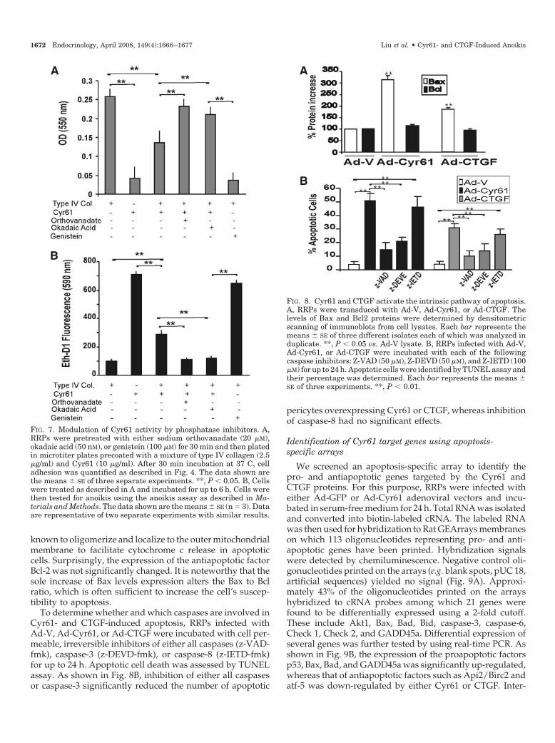
known to oligomerize and localize to the outer mitochondrialmembrane to facilitate cytochrome c release in apoptoticcells. Surprisingly, the expression of the antiapoptotic factorBcl-2 was not significantly changed. It is noteworthy that thesole increase of Bax levels expression alters the Bax to Bclratio, which is often sufficient to increase the cell’s suscep-tibility to apoptosis.
To determine whether and which caspases are involved inCyr61- and CTGF-induced apoptosis, RRPs infected withAd-V, Ad-Cyr61, or Ad-CTGF were incubated with cell per-meable, irreversible inhibitors of either all caspases (z-VAD-fmk), caspase-3 (z-DEVD-fmk), or caspase-8 (z-IETD-fmk)for up to 24 h. Apoptotic cell death was assessed by TUNELassay. As shown in Fig. 8B, inhibition of either all caspasesor caspase-3 significantly reduced the number of apoptotic
pericytes overexpressing Cyr61 or CTGF, whereas inhibitionof caspase-8 had no significant effects.
Identification of Cyr61 target genes using apoptosis-specific arrays
We screened an apoptosis-specific array to identify thepro- and antiapoptotic genes targeted by the Cyr61 andCTGF proteins. For this purpose, RRPs were infected witheither Ad-GFP or Ad-Cyr61 adenoviral vectors and incu-bated in serum-free medium for 24 h. Total RNA was isolatedand converted into biotin-labeled cRNA. The labeled RNAwas then used for hybridization to Rat GEArrays membraneson which 113 oligonucleotides representing pro- and anti-apoptotic genes have been printed. Hybridization signalswere detected by chemiluminescence. Negative control oli-gonucleotides printed on the arrays (e.g. blank spots, pUC 18,artificial sequences) yielded no signal (Fig. 9A). Approxi-mately 43% of the oligonucleotides printed on the arrayshybridized to cRNA probes among which 21 genes werefound to be differentially expressed using a 2-fold cutoff.These include Akt1, Bax, Bad, Bid, caspase-3, caspase-6,Check 1, Check 2, and GADD45a. Differential expression ofseveral genes was further tested by using real-time PCR. Asshown in Fig. 9B, the expression of the proapoptotic factorsp53, Bax, Bad, and GADD45a was significantly up-regulated,whereas that of antiapoptotic factors such as Api2/Birc2 andatf-5 was down-regulated by either Cyr61 or CTGF. Inter-
FIG. 7. Modulation of Cyr61 activity by phosphatase inhibitors. A,RRPs were pretreated with either sodium orthovanadate (20 �M),okadaic acid (50 nM), or genistein (100 �M) for 30 min and then platedin microtiter plates precoated with a mixture of type IV collagen (2.5�g/ml) and Cyr61 (10 �g/ml). After 30 min incubation at 37 C, celladhesion was quantified as described in Fig. 4. The data shown arethe means � SE of three separate experiments. **, P � 0.05. B, Cellswere treated as described in A and incubated for up to 6 h. Cells werethen tested for anoikis using the anoikis assay as described in Ma-terials and Methods. The data shown are the means � SE (n � 3). Dataare representative of two separate experiments with similar results.
FIG. 8. Cyr61 and CTGF activate the intrinsic pathway of apoptosis.A, RRPs were transduced with Ad-V, Ad-Cyr61, or Ad-CTGF. Thelevels of Bax and Bcl2 proteins were determined by densitometricscanning of immunoblots from cell lysates. Each bar represents themeans � SE of three different isolates each of which was analyzed induplicate. **, P � 0.05 vs. Ad-V lysate. B, RRPs infected with Ad-V,Ad-Cyr61, or Ad-CTGF were incubated with each of the followingcaspase inhibitors: Z-VAD (50 �M), Z-DEVD (50 �M), and Z-IETD (100�M) for up to 24 h. Apoptotic cells were identified by TUNEL assay andtheir percentage was determined. Each bar represents the means �SE of three experiments. **, P � 0.01.
1672 Endocrinology, April 2008, 149(4):1666–1677 Liu et al. • Cyr61- and CTGF-Induced Anoikis

estingly, several of the Cyr61 target genes such as akt1, Bad,Bax, Bid, caspase-3, caspase-6, and GADD45a have beenfunctionally grouped as p53- and DNA damage-inducedapoptosis genes (34). Indeed, several members of the Bclfamily such as Bax, Bad, and Bid are known to be under thetranscriptional control of p53. These proteins constitute acritical intracellular checkpoint of apoptosis. Meanwhile, inaddition to the genes imprinted on the GEArray, we foundthat the overexpression of Cyr61 or CTGF reduced the tran-script levels of the small GTPase, p21WAF1, which is also a p53target gene. On another hand, the differential expression ofother p53 gene targets such as AIP-1, Fas, Prdx2, Bnip3, andRnf7 could not be confirmed by real-time PCR (data notshown), which underscores limitations of the GEArray ap-proach. Taken together, these data suggest that the p53-mediated anoikis pathway plays an important role in Cyr61-and CTGF-induced retinal pericyte apoptosis.
Cyr61- and CTGF-induced MMP-2 gene expressionmediates, at least in part, RRP apoptosis
Expression of either Cyr61 or CTGF has been previouslyassociated with changes of gene programs for cell prolifer-
ation, ECM synthesis, and angiogenesis (35, 36). Therefore,we determined the mRNA levels of several putative Cyr61and CTGF target genes including VEGF-A, type IV collagen,laminins, and MMP-2, -3, and -9 in cells expressing Cyr61and CTGF. We found that either Cyr61 or CTGF induced a2- to 3-fold increase in the mRNA levels for MMP-2, com-pared with control (Fig. 10). The mRNA levels of VEGF-Awas significantly increased in Cyr61- but not CTGF-overex-pressing cells. Conversely, neither Cyr61 nor CTGF affectedthe expression profile of �1(IV) collagen, �1-laminin,MMP-3, and MMP-9. In-gel gelatin zymography revealed asingle MMP-2 band corresponding to the activated form ofMMP-2 in conditioned medium from Cyr61- and CTGF-overexpressing cells (Fig. 11A). Neither the active nor latentforms of MMP-2 were detected in GFP-overexpressing cells.Thus, the MMP-2 gene, which has been shown to be underthe control of p53 (37), is a Cyr61 and CTGF target gene aswell.
Increased perivascular localization of MMP-2 has previ-ously been observed in the retinal capillaries at the earlystages of diabetes and has appeared to occur concomitantlywith increased Cyr61 and CTGF gene expression, suggestinga cause-and-effect relationship (17, 38). To determinewhether MMP-2 expression and activation mediate the pro-apoptotic effects of Cyr61 and/or CTGF on pericytes, RRPsinfected with Ad-V, Ad-Cyr61, or Ad-CTGF were incubatedwith SB3CT, a specific inhibitor of MMP-2, and cell death wasassessed using the TUNEL assay. As shown in Fig. 11B, thenumber of apoptotic cells was significantly reduced but notcompletely abolished in the presence of the MMP-2 inhibitor,SB3CT in cells overexpressing either Cyr61 or CTGF. Con-sistent with this result, incubation of RRPs with purifiedactive MMP-2 significantly increased the number of apopto-tic cells (�19%) (Fig. 11C). Taken together, these results sug-gest that the apoptogenic activity of the Cyr61 and CTGFproteins is partly mediated by MMP-2.
Discussion
Anoikis, a form of cell death triggered by inappropriate orinadequate contacts between the cells and the ECM, regu-
FIG. 9. Identification of Cyr61 target genes using pathway-specificGEArray profiling. A, Total RNA (2 �g) from Ad-GFP- and Ad-Cyr61-infected RRPs was converted to biotin-labeled cRNA using theTrueLabeling-AMP amplification system as described in Materialsand Methods. The cRNAs were hybridized to separate rat apoptosisOligo GEArray membranes on which 113 pro- and antiapoptotic geneshave been spotted. The hybridization signals were detected by chemi-luminescence. B, The mRNA levels of selected Cyr61 target genes,p53, Bax, BAD, GAD/GADD45a, Api2, atf-5, and p21WAF1 were quan-tified by real-time PCR and normalized to the 18S rRNA levels. Dataare given as means � SE, n � 3. ** P � 0.01; *, P � 0.05 vs. the controlAd-GFP.
FIG. 10. Expression profile of putative Cyr61 and CTGF target genesin RRPs. RRPs were transduced with Ad-GFP, Ad-Cyr61, or Ad-CTGFand incubated in serum-free medium for 24 h. Genomic DNA-freeRNA was prepared and reverse transcribed. The mRNA levels ofMMP-2, -3, and -9, VEGF-A, �1(IV) collagen and �1-laminin chainwere determined using real-time PCR and normalized to the 18SrRNA levels. Data are given as means � SE, n � 4. **, P � 0.05 vs.Ad-GFP control.
Liu et al. • Cyr61- and CTGF-Induced Anoikis Endocrinology, April 2008, 149(4):1666–1677 1673

lates both physiological (e.g. tissue homeostasis and turn-over) and pathological processes (e.g. tissue degenerationand tumorigenesis). Anoikis was initially observed for en-dothelial and epithelial cells for which attachment to theECM, even in the presence of serum, is required to preventcell death. However, initiation of anoikis is still a debatableissue as the initiating factors that bring about cell detachmentfrom their substrate and propel the cells in the apoptoticpathway are not well understood. Our data showed that theCyr61 and/or CTGF proteins promote detachment andanoikis of retinal pericytes. When used as substrates for cellattachment and compared with the constitutively expressedECM proteins such as type IV collagen and laminin, bothCyr61 and CTGF exhibited antiadhesive and apoptogenicactivities vis-a-vis retinal pericytes. Clearly, dynamicchanges of the ECM composition, and thus cell-matrix in-teractions, are conducive to compromising pericyte survival.
As matricellular proteins, Cyr61 and CTGF do not assumea direct structural role in the matrix but modulate cell func-tion and cell-matrix interactions. Other matricellular proteinssuch as thrombospondin, tenascin-C, and secreted proteinacidic and rich in cysteine have been found to negativelyregulate cell adhesion as well and reduce it to, at least, a stateof intermediate cell adhesion (39). The type I repeat of throm-bospondin 1, which is found in thrombospondin, Cyr61, andCTGF, has been shown to specifically induce endothelial celldeadhesion and apoptosis/anoikis, although adhesive prop-erties have been assigned to this domain in hepatic stellatecells (40, 41). The antiadhesion properties of thrombospondinentail disassembly of focal adhesion characterized by un-bundling of actin stress fibers and depletion of several focaladhesion proteins. Cyr61 and CTGF act, seemingly, along asimilar pathway and their antiadhesive activity provides amechanism for pericyte detachment and death. In Cyr61-treated cells, cytoskeletal actin rearranges into a peripheralring in preparation of blebbing, which is characteristic of cellsduring the execution phase of apoptosis. The cytoskeletalchanges develop rapidly in Cyr61-expressing cells and rel-atively slowly in CTGF-expressing cells, which may accountfor the higher proapoptotic potential of Cyr61. During thisprocess, the link between the actin cytoskeleton and theplasma membrane may be broken focally allowing blebs toprotrude at sites in which the plasma membrane is no longer
attached to the cytoskeleton (42). However, membrane bleb-bing is cell type and stimulus dependent (42, 43). Whetherboth Cyr61 and CTGF induce membrane blebbing is un-known and will be investigated in future studies.
Interestingly, our data are at variance with a previousreport showing that Cyr61 and CTGF trigger death of rat-1fibroblasts through their cell adhesive function mediated by�6�1 integrin (44). Cyr61 and CTGF have been shown torecognize various integrin receptors, although such interac-tions are complex and vary as a function of the type ofintegrins, the domain location in these proteins, and the celltype (45). Paradoxically, Cyr61-�6�1 integrin interactionspromote spreading/survival of skin fibroblasts and activatea gene program for angiogenesis (29, 46). Complicating fur-ther the matter is the observation that Cyr61 and CTGFpromote cell migration/motility as well, which implies thatthey also exhibit anti-adhesive effects to allow efficient cellmigration (47, 48). Whereas these observations are intriguingin their own right because cell adhesion presumably advo-cates cell survival instead of cell migration or death, theyunderscore the complex mechanisms of matricellular activ-ities. Namely, the matricellular concept of these proteins mayaccount for their puzzling features and the apparently con-tradictory properties reported in different studies. The ac-tivities of matricellular proteins are, indeed, cell type depen-dent and contextual in that they depend on the availabilityof receptors/binding partners and the ECM composition,which varies for each cell type. Anchorage-dependent cellssuch as cultured pericytes produce their own ECM to whichthey adhere (49). A potential explanation for the antiadhesiveactivities of Cyr61 and CTGF vis-a-vis pericytes is that assubstrates, these proteins compete with the constitutivelyexpressed ECM proteins for integrin binding. The excessiveaccumulation of these protein in the ECM may initiate dead-hesion between pericytes and their surrounding ECM pro-tein substrate, by converting strong cell-ECM interactionsinto weak ones. Under these conditions, the intracellularsignaling pathways (e.g. tyrosine kinase phosphorylationcascades) that are normally triggered by contact betweenintegrins and the constitutively expressed ECM proteins mayeither be turned off or replaced by others.
Two types of data support these observations. First, thechanges of cell shape and morphology of pericytes indicate
FIG. 11. Cyr61- and CTGF-induced MMP-2 gene expression mediates RRP apoptosis. A, In-gel zymography of conditioned medium fromAd-GFP-, Ad-Cyr61-, and Ad-CTGF-transduced cells. B, Inhibition of MMP-2 reduced Cyr61- and CTGF-induced apoptosis. RRPs expressingAd-GFP, Ad-Cyr61, or Ad-CTGF were incubated with SB3CT (1 nM) for 24 h in serum-free medium. The percentage of apoptotic cells wasdetermined using TUNEL assay. **, P � 0.05 vs. Ad-GFP; *, P � 0.05 vs. Ad-Cyr61 or Ad-CTGF, (n � 4). C, Cultured RRPs were treated withactive MMP-2 (1 nM) for up to 16 h. Apoptotic cells were identified by TUNEL assay and counted. **, P � 0.05 vs. Ad-GFP (n � 3).
1674 Endocrinology, April 2008, 149(4):1666–1677 Liu et al. • Cyr61- and CTGF-Induced Anoikis

that Cyr61 and CTGF provoke cell detachment possibly byinducing integrin disengagement. It is now well appreciatedthat integrin engagement or disengagement regulates cellproliferation and survival (50). In osteoblasts, unoccupied�v�3-integrin induces apoptosis by transmitting a positivedeath signal, whereas in epithelial cells, the integrin �4 sub-unit can be cleaved by caspases, which disrupts hemides-mosome assembly and induces cell death (51, 52). There is,however, also clear evidence that integrin signaling alone isnot sufficient to prevent anoikis (53). Second, the antiadhe-sive activity of Cyr61 and CTGF was associated with de-phosphorylation events. Antagonizing the activity of phos-phatases by sodium orthovanadate or okadaic acidmaintained the targets of tyrosine kinases in a phosphory-lation state and reduced pericyte deadhesion and death.However, these Cyr61-associated dephosphorylation eventsare cell type specific because in human primary fibroblasts,immobilized Cyr61 activates focal adhesion kinase and pax-illin (54). Therefore, conflicting cell type-specific signals arisefrom Cyr61-cell interactions, and further work is needed todefine the integration and regulation of the subsequentresponses.
Meanwhile, our data showed that both Cyr61 and CTGFup-regulated the expression of the MMP-2 gene and in-creased MMP-2 activity, which mediates, at least in part,pericyte apoptosis. Morphological manifestations of cells un-dergoing anoikis can be seen in recombinant MMP-2-treatedcells (data not shown). Thus, the process of Cyr61- andCTGF-induced anoikis embraces, perhaps, the dissolution byMMP-2 of interactions through pericellular proteolysis ofmolecules involved in cell-matrix and cell-cell interactions.In fact, there are several potential mechanisms for the ap-optogenic activity of MMP-2. First, the collagenolytic activityof MMP-2 may induce pericyte death via degradation andremoval of survival signals emanating from the ECM pro-teins. Type IV collagen, the major structural element of thebasement membranes in which pericytes are embedded inretinal capillaries, is the prime target of MMP-2 activity.Second, MMP-2 may, in part, induce ECM degradation andrelease of modular breakdown products with a potent pro-apoptotic activity. In particular, proteolytic fragments of typeIV collagen (e.g. tumstatin) and type XVII collagen (e.g. en-dostatin) have been shown to induce apoptosis of endothelialcells (55). Third, the proper proapoptotic activity of MMP-2may compromise survival of anchorage-dependent cells likeretinal pericytes. In ventricular myocytes, MMP-2 mediates�-adrenergic receptor-stimulated apoptosis via both directinteraction with �1-integrins and poly-ADP-ribose-polymer-ase cleavage (56). Further studies are needed to elucidatethe mechanism of MMP-2-mediated apoptosis of retinalpericytes.
Another interesting finding of this study is the identifica-tion of the proapoptotic genes activated by Cyr61. Theseinclude Bad, Bax, Casp3, Casp6, Check1, Check2 Gadd45a,p53, and Trp53inp1. Most of these genes have been func-tionally grouped as p53 target genes. The MMP-2 gene hasalso been shown to be transcriptionally activated by p53,suggesting an important role of p53 as a mediator of Cyr61and CTGF activity in pericytes (37). The p53 protein is knownas both a downstream target of Cyr61 in cancer cells and a
critical regulatory factor of program cell death during anoikis(34, 57). In particular, studies have shown that anoikis can besuppressed by transfection of fibroblasts with a dominant-negative form of p53 (43). Survival signals mediated by thea6b4 integrin can be effective only in p53-defective cells (58).Mechanistically, p53 was shown to target the protein calledphosphatase and tensin homolog or phosphatase and tensinhomolog (PTEN), which dephosphorylates focal adhesionkinase and phosphatidyl inositol 3,4,5 triphosphate, therebyantagonizing their function in cell survival (59). The p53-PTEN interregulation provides a plausible link between p53activation and suppression of integrin-mediated survivalsignaling because PTEN was shown to restore anchoragedependency in anoikis-resistant cells (43).
Functionally, accumulation of Cyr61 and/or CTGF in nor-mal retinal capillaries is likely to compromise retinal capil-lary integrity and precipitate their closure and degeneration,an important vascular feature of background diabetic reti-nopathy. In vivo studies have shown that both Cyr61 andCTGF are downstream effectors of AGE products in thediabetic retina and that mice treated with AGE productssignificantly up-regulated the expression of the Cyr61 andCTGF genes (17). Deleterious effects of AGE in diabetic micecan be prevented by injection of aminoguanidine, whichdown-regulates the expression of the Cyr61 and CTGF genes.In line with these observations, AGE-BSA-induced Cyr61and CTGF gene expression in pericytes was markedly de-creased in the presence of phosphatase inhibitors, whichreversed, at least in part, the proapoptotic effects of Cyr61and CTGF (data not shown). Thus, the accumulation of thesematricellular proteins in the retinal capillaries at the onset ofdiabetes may alter the retinal capillary structure and orga-nization. Studies have shown that impaired angiogenesisand reduced capillarization of skeletal muscles was associ-ated with increased Cyr61 and CTGF levels in diabetic skel-etal muscle (60). In addition, these matricellular proteins aresusceptible to proteolytic degradation, which yields peptidefragments with, perhaps, biological activities of their own. Arecent study showed that an N-terminal peptide derivedfrom the CTGF protein accumulates in the vitreous of pa-tients with active proliferative diabetic retinopathy (61).Therefore, Cyr61, CTGF, and/or their individual domainsprovide potential targets in therapeutic and biotechnologicalcontexts.
Acknowledgments
The authors thank Dr. Julie Rushbrook for her critical discussion andreview of this work. Our thanks go to S. Jatzke for technical help withthe preparation of recombinant Cyr61.
Received October 16, 2007. Accepted December 31, 2007.Address all correspondence and requests for reprints to: Dr. Brahim
Chaqour, Department of Anatomy and Cell Biology, State University ofNew York Downstate Medical Center, Brooklyn, New York 11203. E-mail: [email protected].
This work was supported by grants from the Juvenile Diabetes Re-search Foundation (to B.C.) and the Deutsche Forschungsgemeinschaft(to N.S.).
Disclosure statement: H.L., R.Y., A.C., N.S., and B.C. have nothing todeclare. B.T. is employed by QBM Cell Science.
Liu et al. • Cyr61- and CTGF-Induced Anoikis Endocrinology, April 2008, 149(4):1666–1677 1675
References
1. Chen Y, Du XY 2007 Functional properties and intracellular signaling ofCCN1/Cyr61. J Cell Biochem 100:1337–1345
2. Perbal B 2004 CCN proteins: multifunctional signalling regulators. Lancet363:62–64
3. Chaqour B, Goppelt-Struebe M 2006 Mechanical regulation of the Cyr61/CCN1 and CTGF/CCN2 proteins. FEBS J 273:3639–3649
4. Chaqour B, Yang R, Sha Q 2006 Mechanical stretch modulates the promoteractivity of the profibrotic factor CCN2 through increased actin polymerizationand NF-�B activation. J Biol Chem 281:20608–20622
5. O’Leary JM, Hamilton JM, Deane CM, Valeyev NV, Sandell LJ, DowningAK 2004 Solution structure and dynamics of a prototypical chordin-like cys-teine-rich repeat (von Willebrand factor type C module) from collagen IIA.J Biol Chem 279:53857–53866
6. Chen CC, Young JL, Monzon RI, Chen N, Todorovic V, Lau LF 2007 Cyto-toxicity of TNF� is regulated by integrin-mediated matrix signaling. EMBO J26:1257–1267
7. Gao R, Brigstock DR 2003 Low density lipoprotein receptor-related protein(LRP) is a heparin-dependent adhesion receptor for connective tissue growthfactor (CTGF) in rat activated hepatic stellate cells. Hepatol Res 27:214–220
8. Leask A, Abraham DJ 2006 All in the CCN family: essential matricellularsignaling modulators emerge from the bunker. J Cell Sci 119:4803–4810
9. Leu SJ, Lam SC, Lau LF 2002 Pro-angiogenic activities of CYR61 (CCN1)mediated through integrins �v�3 and �6�1 in human umbilical vein endo-thelial cells. J Biol Chem 277:46248–46255
10. Leu SJ, Liu Y, Chen N, Chen CC, Lam SC, Lau LF 2003 Identification of a novelintegrin �6�1 binding site in the angiogenic inducer CCN1 (CYR61). J BiolChem 278:33801–33808
11. Schober JM, Chen N, Grzeszkiewicz TM, Jovanovic I, Emeson EE, UgarovaTP, Ye RD, Lau LF, Lam SC 2002 Identification of integrin �(M)�(2) as anadhesion receptor on peripheral blood monocytes for Cyr61 (CCN1) andconnective tissue growth factor (CCN2): immediate-early gene products ex-pressed in atherosclerotic lesions. Blood 99:4457–4465
12. Segarini PR, Nesbitt JE, Li D, Hays LG, Yates III JR, Carmichael DF 2001 Thelow density lipoprotein receptor-related protein/�2-macroglobulin receptor isa receptor for connective tissue growth factor. J Biol Chem 276:40659–40667
13. Rachfal AW, Brigstock DR 2005 Structural and functional properties of CCNproteins. Vitam Horm 70:69–103
14. Brigstock DR 2003 The CCN family: a new stimulus package. J Endocrinol178:169–175
15. Wilkinson-Berka JL, Fletcher EL 2004 Angiotensin and bradykinin: targets forthe treatment of vascular and neuro-glial pathology in diabetic retinopathy.Curr Pharm Des 10:3313–3330
16. Kuiper EJ, Witmer AN, Klaassen I, Oliver N, Goldschmeding R, Schlinge-mann RO 2004 Differential expression of connective tissue growth factor inmicroglia and pericytes in the human diabetic retina. Br J Ophthalmol 88:1082–1087
17. Hughes JM, Kuiper EJ, Klaassen I, Canning P, Stitt AW, Van Bezu J, Schalk-wijk CG, Van Noorden CJ, Schlingemann RO 2007 Advanced glycation endproducts cause increased CCN family and extracellular matrix gene expressionin the diabetic rodent retina. Diabetologia 50:1089–1098
18. He Z, Way KJ, Arikawa E, Chou E, Opland DM, Clermont A, Isshiki K, MaRC, Scott JA, Schoen FJ, Feener EP, King GL 2005 Differential regulation ofangiotensin II-induced expression of connective tissue growth factor by pro-tein kinase C isoforms in the myocardium. J Biol Chem 280:15719–15726
19. Twigg SM, Chen MM, Joly AH, Chakrapani SD, Tsubaki J, Kim HS, Oh Y,Rosenfeld RG 2001 Advanced glycosylation end products up-regulate con-nective tissue growth factor (insulin-like growth factor-binding protein-relatedprotein 2) in human fibroblasts: a potential mechanism for expansion of ex-tracellular matrix in diabetes mellitus. Endocrinology 142:1760–1769
20. Lantz M, Vondrichova T, Parikh H, Frenander C, Ridderstrale M, Asman P,Aberg M, Groop L, Hallengren B 2005 Overexpression of immediate earlygenes in active Graves’ ophthalmopathy. J Clin Endocrinol Metab 90:4784–4791
21. Zhou G, Li C, Cai L 2004 Advanced glycation end-products induce connectivetissue growth factor-mediated renal fibrosis predominantly through trans-forming growth factor �-independent pathway. Am J Pathol 165:2033–2043
22. Podesta F, Romeo G, Liu WH, Krajewski S, Reed JC, Gerhardinger C,Lorenzi M 2000 Bax is increased in the retina of diabetic subjects and isassociated with pericyte apoptosis in vivo and in vitro. Am J Pathol 156:1025–1032
23. Sakagami K, Wu DM, Puro DG 1999 Physiology of rat retinal pericytes:modulation of ion channel activity by serum-derived molecules. J Physiol521(Pt 3):637–650
24. Stitt AW, Hughes SJ, Canning P, Lynch O, Cox O, Frizzell N, Thorpe SR,Cotter TG, Curtis TM, Gardiner TA 2004 Substrates modified by advancedglycation end-products cause dysfunction and death in retinal pericytes byreducing survival signals mediated by platelet-derived growth factor. Diabe-tologia 47:1735–1746
25. Beltramo E, Buttiglieri S, Pomero F, Allione A, D’Alu F, Ponte E, Porta M 2003
A study of capillary pericyte viability on extracellular matrix produced byendothelial cells in high glucose. Diabetologia 46:409–415
26. Brownlee M 1992 Glycation products and the pathogenesis of diabetic com-plications. Diabetes Care 15:1835–1843
27. Suzuma K, Naruse K, Suzuma I, Takahara N, Ueki K, Aiello LP, King GL2000 Vascular endothelial growth factor induces expression of connectivetissue growth factor via KDR, Flt1, and phosphatidylinositol 3-kinase-akt-dependent pathways in retinal vascular cells. J Biol Chem 275:40725–40731
28. Schutze N, Kunzi-Rapp K, Wagemanns R, Noth U, Jatzke S, Jakob F 2005Expression, purification, and functional testing of recombinant CYR61/CCN1.Protein Expr Purif 42:219–225
29. Chen N, Chen CC, Lau LF 2000 Adhesion of human skin fibroblasts to Cyr61is mediated through integrin �6�1 and cell surface heparan sulfate proteo-glycans. J Biol Chem 275:24953–24961
30. Crean JK, Furlong F, Mitchell D, McArdle E, Godson C, Martin F 2006Connective tissue growth factor/CCN2 stimulates actin disassembly throughAkt/protein kinase B-mediated phosphorylation and cytoplasmic transloca-tion of p27(Kip-1). FASEB J 20:1712–1714
31. Bannerman DD, Sathyamoorthy M, Goldblum SE 1998 Bacterial lipopoly-saccharide disrupts endothelial monolayer integrity and survival signalingevents through caspase cleavage of adherens junction proteins. J Biol Chem273:35371–35380
32. Gilmore AP 2005 Anoikis. Cell Death Differ 12(Suppl 2):1473–147733. van de WB, Nagelkerke JF, Stevens JL 1999 Dephosphorylation of focal
adhesion kinase (FAK) and loss of focal contacts precede caspase-mediatedcleavage of FAK during apoptosis in renal epithelial cells. J Biol Chem 274:13328–13337
34. Michalak E, Villunger A, Erlacher M, Strasser A 2005 Death squads enlistedby the tumour suppressor p53. Biochem Biophys Res Commun 331:786–798
35. Chen CC, Mo FE, Lau LF 2001 The angiogenic factor Cyr61 activates a geneticprogram for wound healing in human skin fibroblasts. J Biol Chem 276:47329–47337
36. Zhou D, Herrick DJ, Rosenbloom J, Chaqour B 2005 Cyr61 mediates theexpression of VEGF, �v-integrin, and �-actin genes through cytoskeletallybased mechanotransduction mechanisms in bladder smooth muscle cells.J Appl Physiol 98:2344–2354
37. Bian J, Sun Y 1997 Transcriptional activation by p53 of the human type IVcollagenase (gelatinase A or matrix metalloproteinase 2) promoter. Mol CellBiol 17:6330–6338
38. Yang R, Liu H, Williams I, Chaqour B 2007 Matrix metalloproteinase-2 ex-pression and apoptogenic activity in retinal pericytes: implications in diabeticretinopathy. Ann NY Acad Sci 1103:196–201
39. Murphy-Ullrich JE 2001 The de-adhesive activity of matricellular proteins: isintermediate cell adhesion an adaptive state? J Clin Invest 107:785–790
40. Guo N, Krutzsch HC, Inman JK, Roberts DD 1997 Thrombospondin 1 andtype I repeat peptides of thrombospondin 1 specifically induce apoptosis ofendothelial cells. Cancer Res 57:1735–1742
41. Tong ZY, Brigstock DR 2006 Intrinsic biological activity of the throm-bospondin structural homology repeat in connective tissue growth factor. JEndocrinol 188:R1–R8
42. Frisch SM, Screaton RA 2001 Anoikis mechanisms. Curr Opin Cell Biol 13:555–562
43. Grossmann J 2002 Molecular mechanisms of “detachment-induced apopto-sis—anoikis.” Apoptosis 7:247–260
44. Todorovicc V, Chen CC, Hay N, Lau LF 2005 The matrix protein CCN1(CYR61) induces apoptosis in fibroblasts. J Cell Biol 171:559–568
45. Lau LF, Lam SC 1999 The CCN family of angiogenic regulators: the integrinconnection. Exp Cell Res 248:44–57
46. Chen Y, Abraham DJ, Shi-Wen X, Pearson JD, Black CM, Lyons KM, LeaskA 2004 CCN2 (connective tissue growth factor) promotes fibroblast adhesionto fibronectin. Mol Biol Cell 15:5635–5646
47. Gao R, Brigstock DR 2006 A novel integrin �5�1 binding domain in module4 of connective tissue growth factor (CCN2/CTGF) promotes adhesion andmigration of activated pancreatic stellate cells. Gut 55:856–862
48. Grzeszkiewicz TM, Kirschling DJ, Chen N, Lau LF 2001 CYR61 stimulateshuman skin fibroblast migration through Integrin �v�5 and enhances mito-genesis through integrin �v�3, independent of its carboxyl-terminal domain.J Biol Chem 276:21943–21950
49. Canfield AE, Allen TD, Grant ME, Schor SL, Schor AM 1990 Modulation ofextracellular matrix biosynthesis by bovine retinal pericytes in vitro: effects ofthe substratum and cell density. J Cell Sci 96(Pt 1):159–169
50. Stupack DG, Cheresh DA 2003 Apoptotic cues from the extracellular matrix:regulators of angiogenesis. Oncogene 22:9022–9029
51. Werner ME, Chen F, Moyano JV, Yehiely F, Jones JC, Cryns VL 2007 Caspaseproteolysis of the integrin �4 subunit disrupts hemidesmosome assembly,promotes apoptosis, and inhibits cell migration. J Biol Chem 282:5560–5569
52. Zhao H, Ross FP, Teitelbaum SL 2005 Unoccupied �(v)�3 integrin regulatesosteoclast apoptosis by transmitting a positive death signal. Mol Endocrinol19:771–780
53. Chen CS, Mrksich M, Huang S, Whitesides GM, Ingber DE 1997 Geometriccontrol of cell life and death. Science 276:1425–1428
54. Chen CC, Chen N, Lau LF 2001 The angiogenic factors Cyr61 and connective
1676 Endocrinology, April 2008, 149(4):1666–1677 Liu et al. • Cyr61- and CTGF-Induced Anoikis

tissue growth factor induce adhesive signaling in primary human skin fibro-blasts. J Biol Chem 276:10443–10452
55. Sudhakar A, Sugimoto H, Yang C, Lively J, Zeisberg M, Kalluri R 2003Human tumstatin and human endostatin exhibit distinct antiangiogenic ac-tivities mediated by �v�3 and �5�1 integrins. Proc Natl Acad Sci USA 100:4766–4771
56. Menon B, Singh M, Ross RS, Johnson JN, Singh K 2006 �-Adrenergic re-ceptor-stimulated apoptosis in adult cardiac myocytes involves MMP-2-me-diated disruption of �1 integrin signaling and mitochondrial pathway. Am JPhysiol Cell Physiol 290:C254–C261
57. Tong X, Xie D, O’Kelly J, Miller CW, Muller-Tidow C, Koeffler HP 2001Cyr61, a member of CCN family, is a tumor suppressor in non-small cell lungcancer. J Biol Chem 276:47709–47714
58. Bachelder RE, Ribick MJ, Marchetti A, Falcioni R, Soddu S, Davis KR,Mercurio AM 1999 p53 inhibits �6�4 integrin survival signaling by promotingthe caspase 3-dependent cleavage of AKT/PKB. J Cell Biol 147:1063–1072
59. Stambolic V, MacPherson D, Sas D, Lin Y, Snow B, Jang Y, Benchimol S, MakTW 2001 Regulation of PTEN transcription by p53. Mol Cell 8:317–325
60. Kivela R, Silvennoinen M, Touvra AM, Lehti TM, Kainulainen H, Vihko V2006 Effects of experimental type 1 diabetes and exercise training on angio-genic gene expression and capillarization in skeletal muscle. FASEB J 20:1570–1572
61. Hinton DR, Spee C, He S, Weitz S, Usinger W, LaBree L, Oliver N, Lim JI2004 Accumulation of NH2-terminal fragment of connective tissue growthfactor in the vitreous of patients with proliferative diabetic retinopathy. Di-abetes Care 27:758–764
Endocrinology is published monthly by The Endocrine Society (http://www.endo-society.org), the foremost professional society serving theendocrine community.
Liu et al. • Cyr61- and CTGF-Induced Anoikis Endocrinology, April 2008, 149(4):1666–1677 1677