Embed Size (px)
Citation preview
Brit. J. Ophthal. (1962) 46, 45.
CYSTIC LYMPHANGIOMA OF THE ORB1T*BY
ALY MORTADADepartment of Ophthalmology, Faculty ofMedicine, Cairo University, Egypt
CYSTIC lymphangiomata are occasionally found in the neck, axilla, groin,sacral region, floor of the mouth, and retro-peritoneal space. A few caseshave been described in the orbit by Gradle (1920), Weldige-Cremer (1920),Niosi (1921), and Meisner (1926). Among 120 primary intra-orbital tumoursdiagnosed histologically, I have seen three cystic lymphangiomata. Therarity of these lesions and the varying presenting clinical features makethese three cases of general interest.
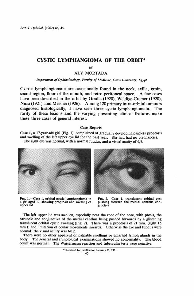
Case ReportsCase 1, a 17-year-old girl (Fig. 1), complained of gradually developing painless proptosisand swelling of the left upper eye lid for the past year. She had had no pregnancies.The right eye was normal, with a normal fundus, and a visual acuity of 6/9.
FiG. 1.-Case 1, orbital cystic lymphangioma in FIG. 2.-Case 1, translucent orbital cysta girl aged 17, showing proptosis and swelling of pushing forward the medial canthus con-upper lid. junctiva.
The left upper lid was swollen, especially near the root of the nose, with ptosis, thecaruncle and conjunctiva of the medial canthus being pushed forwards by a glisteningtranslucent orbital cystic swelling (Fig. 2). There was a proptosis of 21 mm. (right 15mm.); and limitation of ocular movements inwards. Otherwise the eye and fundus werenormal; the visual acuity was 6/12.There were no other apparent or palpable swellings or enlarged lymph glands in the
body. The general and rhinological examinations showed no abnormality. The bloodcount was normal. The Wassermann reaction and tuberculin tests were negative.
* Received for publication January 13, 1961.45

An x ray of the orbits showed nothing abnormal, but orbital exploration revealed alarge cyst containing clear fluid in the medial side of the orbit, outside the muscle cone.A non-encapsulated translucent tumour, measuring 0-5 x 0 5 cm., was excised from the
conjunctival surface of the upper lid.
Histological Examination.-The large orbital cyst was'lined by flat endothelial cells, andwas in fact a cystic lymphangioma, with numerous Meibomian glands, accessory lacrimalglands, and cavernous lymphangiomatous spaces (Fig. 3).
FIG. 3.-Case 1, tumour removed frompalpebral conjunctiva, showing Meibomianglands in tarsus, accessory lacrimal glands,and cavernous lymphangiomatous spaces.x 120.
Case 2, a male infant aged 18 months, had right proptosis and a cystic orbital swellingwhich had rapidly pushed the right upper lid forwards in 2 days (Fig. 4).
*.:.~~~~~~~~~~~~~~~~~~~~~~~~~~~~~- i:..:..
FIG. 4.-Case 2, orbital cystic lymphangioma in a male infant aged18 months, showing the orbital cyst pushing the upper eyelidforwards.
There was no history of trauma. The left eye was normal, with a normal fundus.The right lower lid was normal, but the upper lid showed ecchymosis, ptosis, and a
large non-pulsating cystic swelling, partially compressible and not attached to the skin,which prevented the examination of the right eye.The child's general condition was good. There were no other apparent or palpable
swellings in the body, and no allergic manifestations. The Wassermann reaction andtuberculin tests were negative. The blood count and faeces and urine examinations werenormal. The clotting time was 2-5 min. and the bleeding time 2 min. A postero-anterior x ray of the skull showed a wider right orbit (Fig. 5, opposite).
After the aspiration of dark blood, the cyst diminished in size, and the eye could beexamined. There was a right proptosis of 20 mm. (left 15 mm.) and limitation of ocularmovements upwards. Otherwise the right eye was normal with a normal fundus.
46 ALY MORTADA

CYSTIC LYMPHANGIOMA OF THE ORBIT
FIG. 5.-Case 2, Postero-anterior skull x ray,showing wider right orbit.
Orbital exploration revealed a large cyst, 1 5 x 2 cm. in size, which was full of blood andoccupied the upper part of the orbit, above the globe. The medial part of the upper lidshowed a small yellowish non-encapsulated tumour, measuring 05 x 1 cm., under thepalpebral and fornix conjunctivae.
Histological Examination.-The conjunctival tumour showed clear spaces lined byendothelial cells separated by connective tissue trabeculae, surrounding Krause's glands(Fig. 6).
4.~ g4 FIG. 6.-Case 2, tumour removed from
palpebral conjunctiva, showing cavernous% Krause's glands. x 120.
Some spaces were full of lymphocytes. The fibrous trabeculae between the spacesshowed areas of chronic inflammation rich in eosinophils.The large orbital cyst was lined by flat endothelial cells and contained blood.The picture is consistent with that of a cavernous lymphangioma of the lid and con-
junctiva, with haemorrhage into a cystic lymphangioma of the orbit.
Case 3, a 22-year-old woman (Fig. 7, overleaf), complained of gradual painless proptosisand diminution of vision in the right eye of 7 months' duration. She had one healthychild and no other pregnancies. The left eye had been shrunken since childhood.The right eyelids were externally normal and the visual acuity was 1/60. A small
translucent mass measuring 0 5 x0*5 cm. was present in the lower conjunctival fornix.
47

The fundus showed post-papilloedemic optic atrophy. There was a proptosis of 21 mm.and a slight limitation of ocular movements downwards. A deep orbital mass was feltbetween the globe and the lower orbital margin.
FIG. 7.-Case 3, orbital cystic lymphangioma in a woman aged 22,showing proptos
The patient's general condition was good. There were no other apparent or palpableswellings in the body. The blood count was normal. The Wassermann and tuberculintests were negative. X rays of both orbits showed no abnormality.The lower conjunctival fomix tumour was removed. The orbital mass proved to be
a translucent cyst in the muscle cone space which contained clear fluid and measured1 x l-5 cm.
Histological Examination.-The fornix conjunctival mass was a cavernous lymphan-gioma (Fig. 8).
FIG.7.as3,oritatFIG. 8.-Case 3, tumour removed fromshoglower conjunctival fonix, showing caver-
p ~~ 7: ~ nous lymphangiomatous spaces. x 120.
The orbital cyst was a cystic lymphangioma lined by flat endothelial cells.
DiscussionFew orbital cystic lymphangiomata have been recorded in the literature.
Even orbital cavernous lymphangiomata are rare-only some forty caseshave been reported since the original observations of Forster (1886) andWiesner (1886). As the orbit is free of lymphatic vessels, orbital lymphan-giomata originate either from displaced foetal cells of the lymphatic channels(Watson and McCarthy, 1940) or from lymphangiomata extending from the
ALY MORTADA48
CYSTIC LYMPHANGIOMA OF THE ORBIT
lid and conjunctiva (Werncke, 1904). The tumour has usually a congenitalbasis and proptosis may be evident at birth (Cabannes, 1903), but it usuallygrows so very slowly that advice is not sought until the patient has reachedadult life (Ayres, 1895). Orbital lymphangiomata may be associated withlymphangioma of cheek (Israel, 1895), or lips and palate (Waldstein, 1910),or with hypertrophy of the face (Jess, 1936).
Wintersteiner (1898), Franklin and Cordes (1924), and Wolff (1932)observed that recurrent attacks of inflammation or haemorrhage in cases oforbital lymphangiomata manifest clinically as intermittent proptosis. Visualdamage due to pressure on the optic nerve by an orbital cavernous lym-phangioma was noted by Kahn (1906) and Smith (1925).
Orbital cystic lymphangiomata have to be differentiated from otherorbital cysts, especially the serous cysts classically described as hygromata,which are usually instances of serous tendonitis or dilatation of fascialelements associated with the muscle tendons (Duke-Elder, 1952).The safest treatment for cystic orbital lymphangioma is by surgical removal.
Any cavernous lymphangiomata can be treated by electrolysis, 75 per centalcohol injection (Wray, 1915), or radiation (Mackay, 1915), but the besttreatment is by excision.
SummaryThree cases of cystic orbital lymphangiomata were found among 120
histopathologically diagnosed primary orbital tumours. All three casesshowed small cavernous lymphangiomata on the conjunctival surface of theeye lids. As the orbit is free from lymphatic vessels, the most probableexplanation of the presence of an orbital cystic lymphangioma is that itcomprises an extension of dilated lymphangiomatous spaces from the lidconjunctival tumour.The presenting clinical feature differed in the three cases:
(a) Gradual proptosis and swelling of the upper lid.
(b) Rapid proptosis and cystic swelling of the upper lid after haemorrhage in thetumour. The condition simulated an orbital haemorrhage after trauma.Histologically the tumour trabeculae showed chronic inflammatory cellsrich in eosinophils.
(c) Gradual slight proptosis and marked diminution of vision due to the pres-sure of the tumour on the optic nerve causing post-papilloedemic opticatrophy.
The three cases were all treated successfully by excision of both the cysticorbital lymphangioma and the cavernous lid conjunctival lymphangioma.These are the first three orbital cystic lymphangiomata to be reported fromEgypt.4
49
50 AL Y MORTADA
REFERENCES
AYREs, S. C. (1895). Amer. J. Ophthal., 12, 321.CABANNES (1903). These, Bordeaux.DUKE-ELDER, S. (1952). "Text-book of Ophthalmology", vol. 5, p. 5225. Kimpton, London.FORSTER, J. (1886). Munch. med. Wschr., 33, 617.FRANKLIN, W. S., and CoRDES, F. C. (1924). J. Amer. med. Ass., 83, 1741.GRADLE, H. S. (1920). Arch. Ophthal. (N. Y.), 49, 520.ISRAEL, J. (1895). Berl. klin. Wschr., 32, 616.JESS, A. (1936). Ber. ophthal. Ges. (Heidelberg), 51, 390.KAHN, H. (1906). Beitr. Augenheilk., 7, 290, No. 65, p. 16.MACKAY, G. (1915). Trans. ophthal. Soc. U.K., 35, 180.MEISNER (1926). Klin. Mbl. Augenheilk., 76, 876.Niosi, F. (1921). Arch. Ottal., 28, 219.SMrrH, E. T. (1925). Trans. Amer. ophthal. Soc., 23, 240.WALDSTEiN (1910). Dtsch. med. Wschr., 36, 2030.WATSON, W. L., and MCCARTHY, W. D. (1940). Surg. Gynec. Obstet., 71, 569.WELDIGE-CREMER, DE (1920). Z. Augenheilk., 65, 44.WERNCKE, T. (1904). Mitt. Augenki. Jurjew, 2, 81.WIESNER, B. (1886). v. Graefes Arch. Ophthal., 32, py. 2, 205.WINTERSTEINER, H. (1898). Ibid., 45, 613.WOLFF, E. (1932). Trans. ophthal. Soc. U.K., 52, 298.WRAY, C. (1915). Ibid., 35, 189.


![Unilocular Cystic Lymphangioma of the Small Omentum in a Girl … · 2017-06-15 · [14,16,21]. Laparoscopic management has the advantages of lower cost and decreased morbidity compared](https://img.pdfslide.net/doc/110x75/5f0ee6187e708231d4417ba4/unilocular-cystic-lymphangioma-of-the-small-omentum-in-a-girl-2017-06-15-141621.jpg)