Embed Size (px)
Citation preview
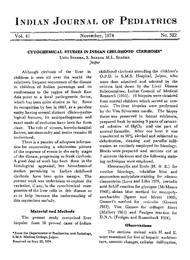
INDIAN JOURNAL oF PEDIATRICS Vol. 41 November, 1974 No. 322
CYTOCHEMICAL STUDIES IN INDIAN CHILDHOOD CIRRHOSIS* USI-IA SFIARMA, S. SAXENA M.L. SHARMA
yaipur
Although cirrhosis of the liver in
children is seen all over the world the relatively frequent occurrence of the disease in children of Indian parentage and its confinement to the region of South East Asia point to a local pathogenetic factor, which has been quite elusive so far. Since its recognition by Sen in 1887, as a peculiar entity having several distinct clinico-patho- logical features, its aetiopathogenesis and exact mode of evolution have been far from clear. The role of viruses, heredo-familial factors, autoimmunity and toxins remains ill understood.
There is a paucity of adequate informa- tion for constructing a wholesome picture of the sequence of events in the early stages of the disease, progressing to frank cirrhosis. A good deal of work has been done in the histological appraisal, but histochemical studies pertaining to Indian childhood cirrhosis have been quite meagre. The present work was undertaken to explore the variation, if any, in the cytochemical com- ponents of the liver cells in this disease so as to help increase the understanding of this mysterious malady.
Material and Methods
The present study comprised liver biopsies from 50 proved cases of Indian
*From the Departments of Paediatrics, and Pathology, S.M.S. Medical College, Jaipur. Received on May 29, 1974.
childhood cirrhosis attending the children's O.P.D. in S.M.S. Hospital, Jaipur, who were then admitted and selected by the criteria laid down by the Liver Disease Subcommittee, Indian Council of Medical Research (1955). 10 biopsies were obtained from normal children which served as con- trols. The liver biopsies were performed by the Vim Silverman needle. The biopsy tissue was preserved in formal sublimate, prepared fresh by mixing 9 parts of saturat- ed solution of HgC12 and one part of neutral formalin. After one hour it was transferred to 50% alcohol and subjected to dehydration, clearing and paraffin infil- tration as routinely employed for histology. Blocks were prepared and sections cut at 7 microns thickness and the following stain- ing techniques were employed.
Hematoxylin and Eosin (H. & E.) for routine histology, toluidine blue and ammonium molybdatestaining for ribonu- cleoproteins (Love and Liles 1959, periodic acid Schiff reaction for glycogen (McManus 1948), alcian blue method for mueopoly- saccharides (Spieer and Meyer 1960), Gomori's method for reticulin (Gomori 1937), Von Giesson for collagen fibers (Mallory 1961) and Feulgen reaction for D.N.A. (Feutgen and Rossenback 1924).
Observat ions
The sections stained with H. and E. were examined for loss of hepatic architec- ture, necrotic changes, cellular infiltration,
346 INDIAN JOURNAL OF PEDIATRICS VoL. 41, No. 322
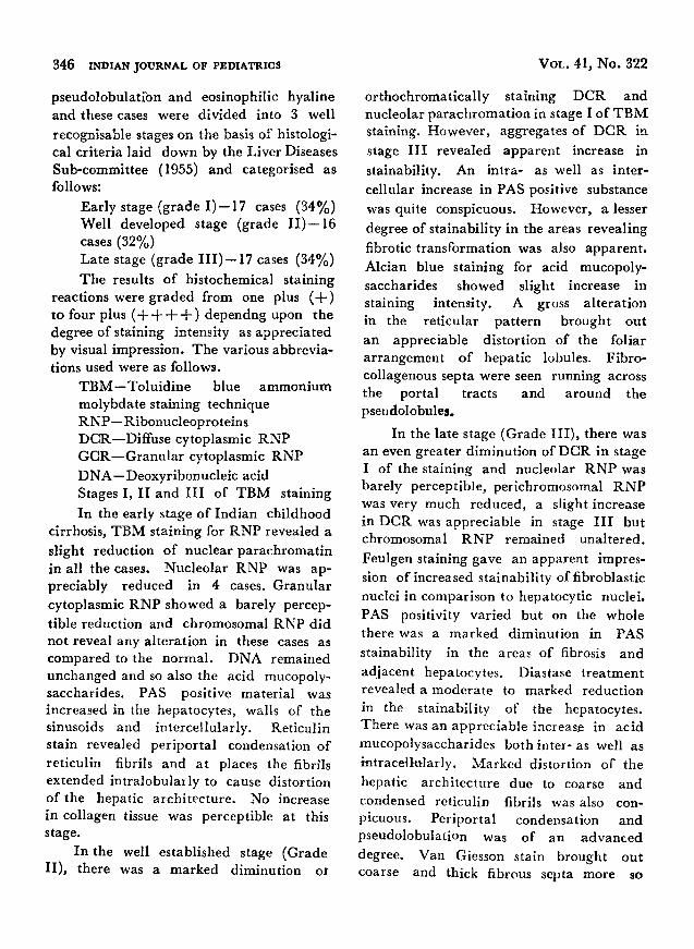
pseudolobulatibn and eosinophilic hyaline and these cases were divided into 3 well
recognisable stages on the basis of histologi- cal criteria laid down by the Liver Diseases Sub-committee (1955) and categorised as follows:
Early stage (grade I ) - -17 cases (34%) Well developed stage (grade I I ) - -16 eases (32%) Late stage (grade I I I ) - - 1 7 cases (34%)
The results of histochemical staining reactions were graded from one plus ( + ) to four plus ( + + + + ) dependug upon the degree of staining intensity as apprecia ted by visual impression. The various abbrevia- tions used were as follows.
T B M - - T o l u i d i n e blue ammonium molybdate staining technique R N P - - Ribonucleoproteins DCR--Diffuse cytoplasmic RNP G C R - - G r a n u l a r cytoplasmic RNP
DNA--Deoxyribonucle ic acid Stages I, I I and I I I of TBM staining
In the early stage of Indian childhood cirrhosis, T B M staining for RNP revealed a
slight reduction of nuclear parachromat in in all the cases. Nucleolar RNP was ap- preciably reduced in 4 cases. Granular cytoplasmic RNP showed a barely percep-
tible reduction and chromosomal RNP did not reveal any al terat ion in these eases as compared to the normal . DNA remained unchanged and so also the acid mucopoly- saccharides. PAS positive material was increased in the hepatocytes, walls of the sinusoids and intercellularly. Reticulin stain revealed per ipor ta l condensation of reticulin fibrils and at places the fibrils extended intralobula, ly to cause distortion of the hepatic archi tecture. No increase in collagen tissue was perceptible at this stage.
In the well established stage (Grade II) , there was a marked diminution ot
or thochromat ical ly staining DCR and nucleolar parachromat ion in stage [ of TB1V[ staining. However , aggregates of DCR in
stage I I I revealed apparent increase in
stainability. An intra- as well as inter-
cellular increase in PAS positive substance was quite conspicuous. However, a lesser
degree of stainability in the areas revealing
fibrotic transformation was also apparent . Alcian blue staining for acid mucopoly- saccharides showed slight increase in staining intensity. A gross alteration in the reticular pat tern brought out
an appreciable distort ion of the foliar arrangement of hepatic lobules. Fibro- collagenous septa were seen running across the portal tracts and around the pseudolobules.
In the late stage (Grade I I I ) , there was an even greater diminution of DCR in stage I of the staining and nucleolar RNP was barely perceptible, perichromosomal R N P was very much reduced, a slight increase in DCR was appreciable in stage I I I but chromosomal RN P remained unaltered.
Fenlgen staining gave an apparent impres- sion of increased stainability of fibroblastic nuclei in comparison to hepatocytic nuclei. PAS positivity varied but on the whole there was a marked diminution in PAS
stainability in the area~ of fibrosis and
adjacent hepatocytes. Diastase t reatment revealed a moderate to marked reduct ion in the stainability of the hepatoeytes. There was an appreciable increase in acid mucopolysaccharides both inter- as well as intrace~lularly. Marked distortion of the
hepatic archi tecture due to coarse and condensed reticulin fibrils was also con- picuous. Periportal condensation and pseudolobulation was of an advanced
degree. Van Giesson stain brought out coarse and thick fibrous septa more so
SHARMA E T A L . - - C Y T O C H E M I C A L STUDIES IN I N D I A N C H I L D H O O D CIRRHOSIS 347
around the portal t ract and the pseudo- lobules. Eosinophilic hyaline bodies were
seen in a few cases in the advanced stage.
These were intensely PAS positive and gave a purple reaction with the T B M method,
thereby indicating the presence of RNA containing substance in it. There was no positive staining for acid mucopolysaccha- rides but a magenta red colour was appre- ciated by PAS techniques which persisted in spite o f diastase digestion, suggesting the presence of diastase resistant PAS positive substance in their chemical composition.
D i s c u s s i o n
A lot of work has been done to unravel the aetiopathogenetic mechanisms involved in Indian childhood cirrhosis but the histochemical field has barely been probed. O f course, in any histochemical work the stainability is likely to be modified by several factors i.e. pH, temperature, the physicochemical state and quantum of
reacting substances, and individual apprai- sal. From the findings in the present study it is quite apparent that there is a progres- sive decrease in various types of RNPs like nuclear parachromat in and or thochromatic DCR in stage I of the staining, with a fur ther diminution proport ional to the advanced "stage of this disease.
As these two types of RNPs are closely related to the protein synthetic mechanism of the cell, their reduction would indicate
of a gross disturbance of intracellular bio- logy cells. This may lend some support to the hypothesis of protein deficiency as a significant factor in the evolution of Indian chi ldhood cirrhosis.
A striking feature was the progressive diminution of RNPs parallel with progres- sion of the disease process. Such a reduction in cytoplasmic RNPs has been also observed by Achar and Chacko (1954) and Prasad
and Prasad (1961). The re was no appre- ciable GCR or other types of RNPs. T h e same holds good for DNP which also did not have any appreciable alteration but for difference in staining intensity of Fuelgen positive material in hepatocytes and adjacent fibroblastic nuclei which should not be taken to imply that there is an actual increase in the DNP content of these cells. Ra the r such an al terat ion can be easily brought about by the condensation of the nuclear apparatus due to compression and spindalord appearance in contrast to the relatively loose and dispersed state of chromat in in the nuclei of the hepatocytes.
The liver is intr icately involved in carbohydra te metabolism. Hence it is not suprising to find a variable alteration in PAS positive substance in various l iver l diseases including Indian childhood cirrho- sis. There was a conspicuous increase in PAS positive substance both intra- as well as intercellularly. This has also been ob- served by earlier workers (Mangal and Bhardwaj 1962, Agarwal 1969). Another interesting aspect was the light PAS positive staining of collagen and the adjacent hepa-
tocytes suggesting a role for the metabolic turnover of carbohydrate , protein-carbo- hydra te complexes, or any other material giving PAS positive reaction in this enigmatic malady. Acidmucopholysacc- harides as brought out by Alcina Blue staining also revealed a progresssve increase parallel to the advancement of the disease process suggesting its involvement in fibrogenesis. Some changes in the ground substance of the hepatic architec-
ture may well be related to the evolution of this disease.
It is worth mentioning here that the eosinophilic hyaline material which has been considered to be a pathognomonic feature of the disease (Nayak et al. 1972)
f f ~ INDIAN JOURNAL O1~ PItDIATI~I~S Vw.. 41, No.
revealed a densely stained PAS positive material which was diastase resistant, and faintly staining RNPs which is possible made up of a potysaccharide-protein com- plex having cytoplasmic tibonucleoproteins. The genesis of hyaline can be explained b~t a peculiar type of degenerative change.
There was an appreciable difference in the intensity of Feulgen positive materiaI in the hepatocytes.
Suntnmry
The histochemical study of liver bropsy m Indian ghi|dhood cirrhosis showed a progressive diminution of ~rthocbxomatic, DCR, nuclear parachromatin, perichromo- s ~ a l RNP and metachromatic DCR (Stage IIl~ as the disease advanced from early to late stages. Nucleotar paTachro- matin and GCK were not demonstrable while chromosomal RNP was unaltered. Feulgen stain did not reveal any significant abnormality. PAS positive substance was deposited both intra- as wail as extracellu- larly. Cytoplasmic hyaline had localised densely stained PAS positive material. Sulphated mucopolysaccharides were de- monstrable in progressively increasing intensity in different stages of the disease.
References
Achar, S.T. and C, hacko, P. (1954). Pathological changes in the liver in hepatic cirrhosis of childhood commonly known as infantile biliary cirrhosis, lnd/a~ 3. Mad. Sd. 8, 442.
Agar~a]~ V.P. (1969). Personal communication. Feulgen, R. and Rossenback, H. (1924).
Mikroskopich-Chemischer Nachweis einer, Nuclein- sanse Von Typus der Thymonucleinsanre ur~l auf die darauf beruhende, elective forlmng, Von- Zeykernen in mLikroskepichen praparaten. 5. PAyr~ C,/u~. 135, 203.
Gomori, G. (1937). Silver impregnation of
reticulum in paraffin sections. Ames. ~. Path, 13, 993. Jelliffe, D.B., Bias, G. and Mukherjee, K.L.
(1957). Venoocclusive disease of the liver.and Indian childhood cirrhosis. Arch. Dis. Child. 32, 369.
Liver Disease Subcommittee. (1955). Infantile cirrhosis of liver. Indian 3. Med. Ras. 43, 723.
Love, R. and Liles, K.H. (1959). Differentiation of nucleoproteins by inactivation of protein bound amino groups and staining with toluidine blue and ammonium mobydate. 37. Histod~rn. Cytodwm. 7, 164.
Mallory, F.B. (1961L Pathological staining technique. New York, Hafts P~lishirq Co. p. 152.
Mangal, H.N. and Bhardwaj, T. (1962). Abstracts. Annual meetings, Indian Society of Psdiatrics, Jaipur.
McManus,J.F.A. (1946). The histologic demon- stration of mucin after periodic acid. aVature. 158, 202.
Prasad, L.S. and Prasad. P.R. (1961). In Asian Pediatrics, p. 145.
Sen, B.C. (1887). Enlargement of the Liver in Children. Indian ~Ied. Gaz. 22, 338.
Spicer, S.S. and Meyer, D.B. (1960). Tech. Bull. P~gist. Mad Tech. 30, 53. Wakers J.H. and Waterlow, J.C. (1954) Span',
Rap. Set Mad. Res. Coun. London. No. 285.
INDIAN JOURNAL OF PEDIATRIC~ P L A T E I
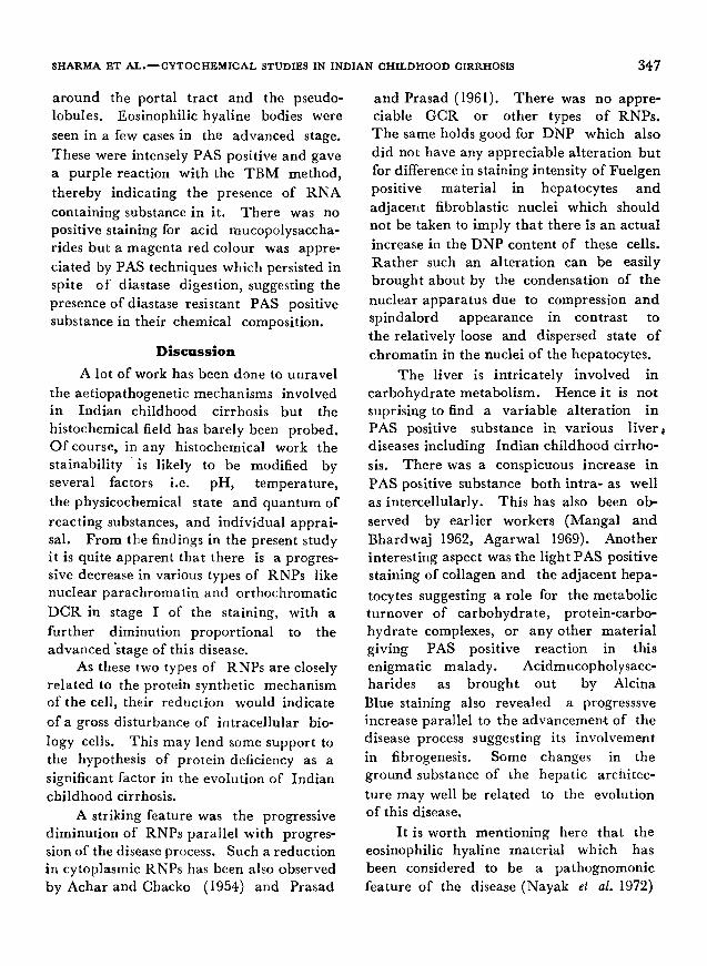
Fig. 1. Vacuolar degeneration. (H. & E. X 450).
Fig. 2. Eosinophilic hyaline in grade lI . (H. & E. •
Fig. 3. Distribution of DCR and perJchro- mosomal RNP. (TBM stain X 1000).
SHARMA ET AL.--CYTOCHEMICAL STUDIES IN INDIAN CHILDHOOD CIRRHOSIS.
P L A T E I I iNDIAN JOURNAL OF PEDIATRIC~
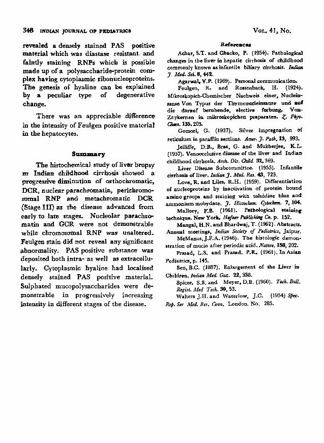
Fig. 1, Intraventricular and irltracerebellar haemorrhages (H & E X 100)"
Fix. 2. Embolus of cerebeltar tissue in the pulmonary arteriole. (H & g • 100).
MADFIAVAN ET. AL. ~EMBOLISM OF CEREBELLAR TISSUE IN 3"HE LUNO.
Fig. 3. Absence of nails in both the thumbs. Finger nails are hypoplastic (Case III-2).
Fig, 4. Iliac horn and webbing of the elbows (Case II-2).
AGARWAL ET AL.--AN INDIAN FAMILY WITH THE NAIL PATELLA SYNDROME.