Embed Size (px)
Citation preview
Cytological preparations for molecular analysis.
Gilda da Cunha Santos MD, PhD,FRCPC, FIACAssociate Professor
UNIVERSITY of TORONTO
University Health Network - Laboratory Medicine Program
Department of Laboratory Medicine and Pathobiology
University of Toronto, Ontario, Canada
Pre-analytical issues for EBUS TBNA specimens.
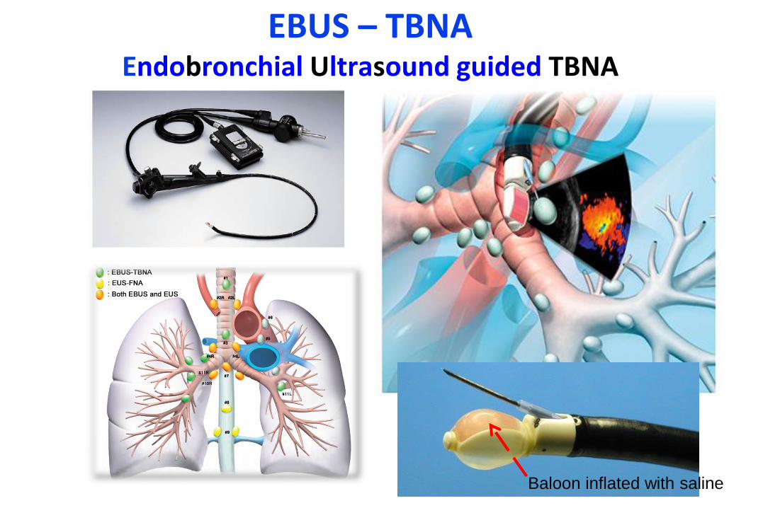
EBUS – TBNAEndobronchial Ultrasound guided TBNA
Baloon inflated with saline
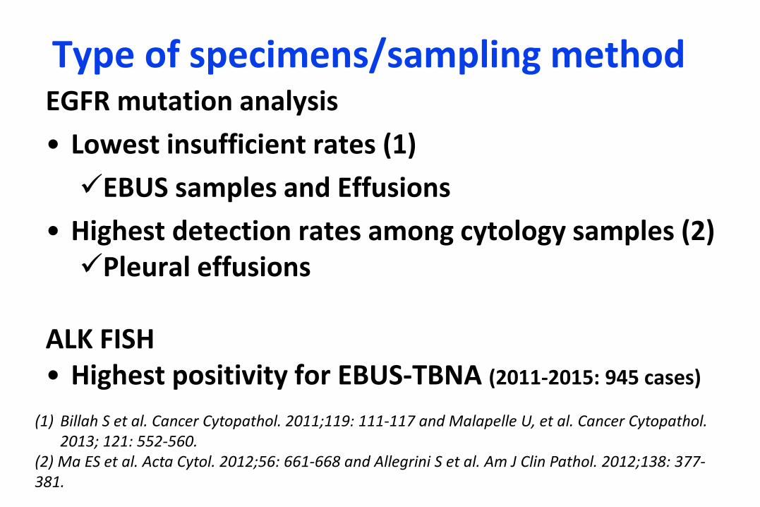
Type of specimens/sampling methodEGFR mutation analysis
• Lowest insufficient rates (1)
EBUS samples and Effusions
• Highest detection rates among cytology samples (2)Pleural effusions
ALK FISH• Highest positivity for EBUS-TBNA (2011-2015: 945 cases)
(1) Billah S et al. Cancer Cytopathol. 2011;119: 111-117 and Malapelle U, et al. Cancer Cytopathol. 2013; 121: 552-560.
(2) Ma ES et al. Acta Cytol. 2012;56: 661-668 and Allegrini S et al. Am J Clin Pathol. 2012;138: 377-381.
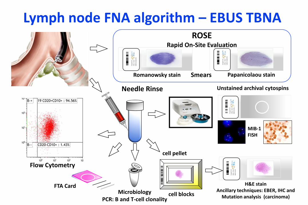
Needle Rinse
Lymph node FNA algorithm – EBUS TBNA
Flow Cytometry
Unstained archival cytospins
cell blocks
Sam
ple
#12345/2
011
Zid
ler,
Har
old
MR
N:1
23456/2
01
1
H&E stainAncillary techniques: EBER, IHC and
Mutation analysis (carcinoma)
cell pellet
MIB-1FISH
FTA Card
Sam
ple
#12345/2
011
Zid
ler,
Har
old
MR
N:1
23456/2
01
1
Papanicolaou stain Smears Romanowsky stain
Sam
ple
#12345/2
011
Zid
ler,
Har
old
MR
N:1
23456/2
01
1
Sam
ple
#12345/2
011
Zid
ler,
Har
old
MR
N:1
23456/2
01
1
ROSERapid On-Site Evaluation
MicrobiologyPCR: B and T-cell clonality
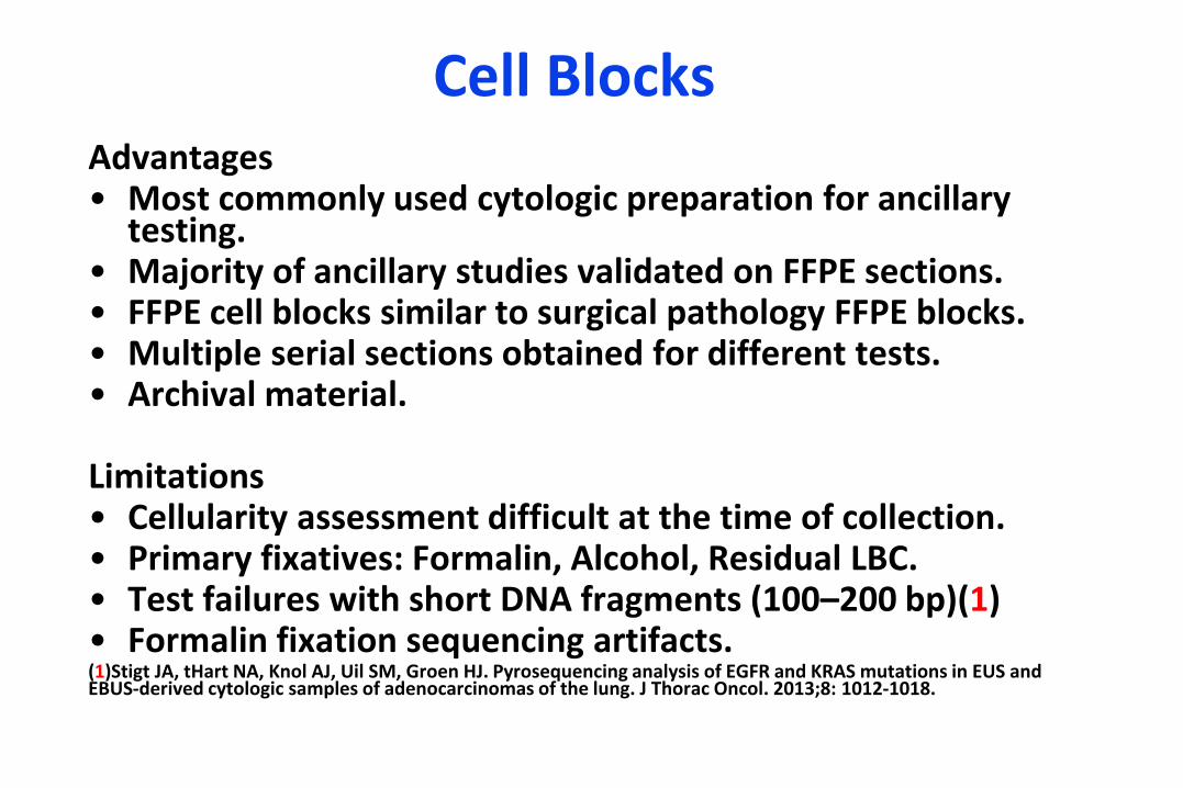
Cell BlocksAdvantages• Most commonly used cytologic preparation for ancillary
testing. • Majority of ancillary studies validated on FFPE sections.• FFPE cell blocks similar to surgical pathology FFPE blocks.• Multiple serial sections obtained for different tests.• Archival material.
Limitations• Cellularity assessment difficult at the time of collection.• Primary fixatives: Formalin, Alcohol, Residual LBC.• Test failures with short DNA fragments (100–200 bp)(1) • Formalin fixation sequencing artifacts.(1)Stigt JA, tHart NA, Knol AJ, Uil SM, Groen HJ. Pyrosequencing analysis of EGFR and KRAS mutations in EUS and EBUS-derived cytologic samples of adenocarcinomas of the lung. J Thorac Oncol. 2013;8: 1012-1018.
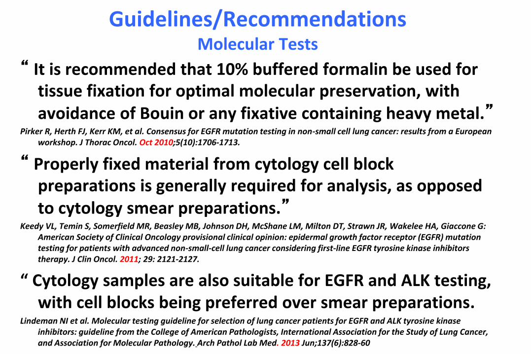
Guidelines/RecommendationsMolecular Tests
“ It is recommended that 10% buffered formalin be used for tissue fixation for optimal molecular preservation, with avoidance of Bouin or any fixative containing heavy metal.”
Pirker R, Herth FJ, Kerr KM, et al. Consensus for EGFR mutation testing in non-small cell lung cancer: results from a European workshop. J Thorac Oncol. Oct 2010;5(10):1706-1713.
“ Properly fixed material from cytology cell block preparations is generally required for analysis, as opposed to cytology smear preparations.”
Keedy VL, Temin S, Somerfield MR, Beasley MB, Johnson DH, McShane LM, Milton DT, Strawn JR, Wakelee HA, Giaccone G: American Society of Clinical Oncology provisional clinical opinion: epidermal growth factor receptor (EGFR) mutation testing for patients with advanced non-small-cell lung cancer considering first-line EGFR tyrosine kinase inhibitors therapy. J Clin Oncol. 2011; 29: 2121-2127.
“ Cytology samples are also suitable for EGFR and ALK testing, with cell blocks being preferred over smear preparations.
Lindeman NI et al. Molecular testing guideline for selection of lung cancer patients for EGFR and ALK tyrosine kinase inhibitors: guideline from the College of American Pathologists, International Association for the Study of Lung Cancer, and Association for Molecular Pathology. Arch Pathol Lab Med. 2013 Jun;137(6):828-60
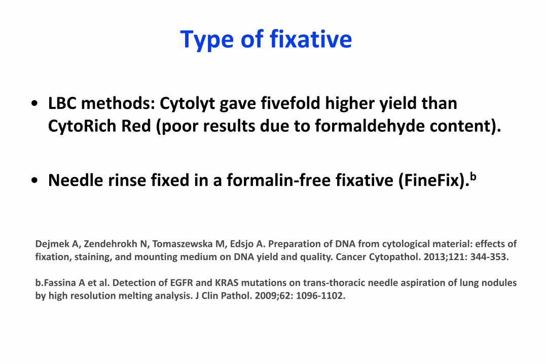
Type of fixative
• LBC methods: Cytolyt gave fivefold higher yield than CytoRich Red (poor results due to formaldehyde content).
• Needle rinse fixed in a formalin-free fixative (FineFix).b
Dejmek A, Zendehrokh N, Tomaszewska M, Edsjo A. Preparation of DNA from cytological material: effects of fixation, staining, and mounting medium on DNA yield and quality. Cancer Cytopathol. 2013;121: 344-353.
b.Fassina A et al. Detection of EGFR and KRAS mutations on trans-thoracic needle aspiration of lung nodules by high resolution melting analysis. J Clin Pathol. 2009;62: 1096-1102.
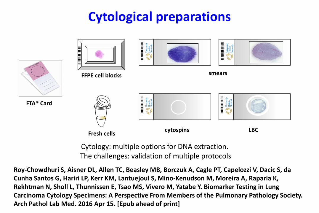
Cytological preparations
FTA® Card
Fresh cells
FFPE cell blocks smears
cytospins LBC
Cytology: multiple options for DNA extraction. The challenges: validation of multiple protocols
Roy-Chowdhuri S, Aisner DL, Allen TC, Beasley MB, Borczuk A, Cagle PT, Capelozzi V, Dacic S, da Cunha Santos G, Hariri LP, Kerr KM, Lantuejoul S, Mino-Kenudson M, Moreira A, Raparia K, Rekhtman N, Sholl L, Thunnissen E, Tsao MS, Vivero M, Yatabe Y. Biomarker Testing in Lung Carcinoma Cytology Specimens: A Perspective From Members of the Pulmonary Pathology Society. Arch Pathol Lab Med. 2016 Apr 15. [Epub ahead of print]
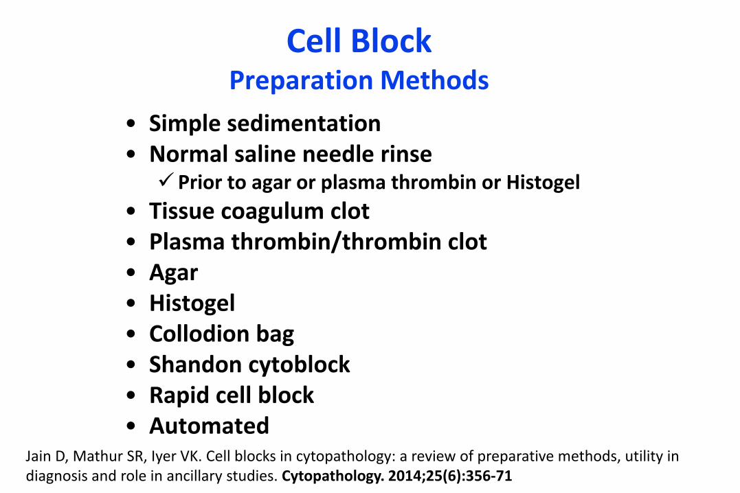
Cell Block Preparation Methods
• Simple sedimentation• Normal saline needle rinse
Prior to agar or plasma thrombin or Histogel
• Tissue coagulum clot• Plasma thrombin/thrombin clot• Agar• Histogel• Collodion bag• Shandon cytoblock• Rapid cell block• Automated
Jain D, Mathur SR, Iyer VK. Cell blocks in cytopathology: a review of preparative methods, utility in diagnosis and role in ancillary studies. Cytopathology. 2014;25(6):356-71
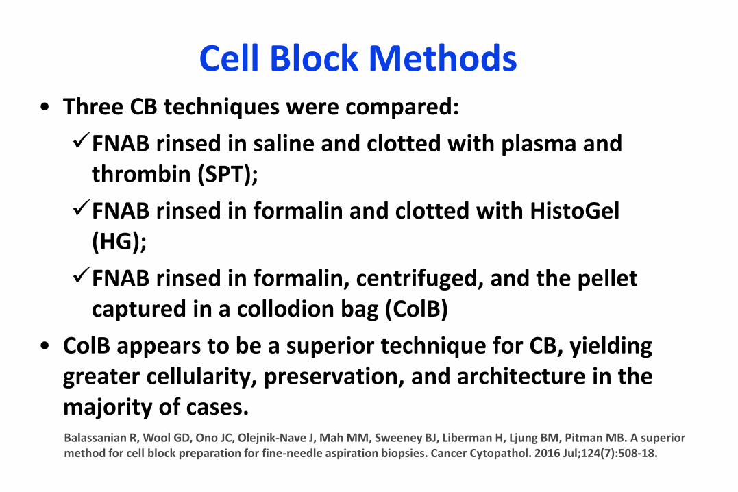
Cell Block Methods• Three CB techniques were compared:
FNAB rinsed in saline and clotted with plasma and thrombin (SPT);
FNAB rinsed in formalin and clotted with HistoGel(HG);
FNAB rinsed in formalin, centrifuged, and the pellet captured in a collodion bag (ColB)
• ColB appears to be a superior technique for CB, yielding greater cellularity, preservation, and architecture in the majority of cases.Balassanian R, Wool GD, Ono JC, Olejnik-Nave J, Mah MM, Sweeney BJ, Liberman H, Ljung BM, Pitman MB. A superior method for cell block preparation for fine-needle aspiration biopsies. Cancer Cytopathol. 2016 Jul;124(7):508-18.
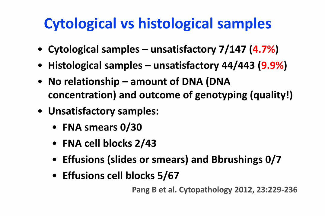
Cytological vs histological samples
• Cytological samples – unsatisfactory 7/147 (4.7%)
• Histological samples – unsatisfactory 44/443 (9.9%)
• No relationship – amount of DNA (DNA concentration) and outcome of genotyping (quality!)
• Unsatisfactory samples:
• FNA smears 0/30
• FNA cell blocks 2/43
• Effusions (slides or smears) and Bbrushings 0/7
• Effusions cell blocks 5/67Pang B et al. Cytopathology 2012, 23:229-236
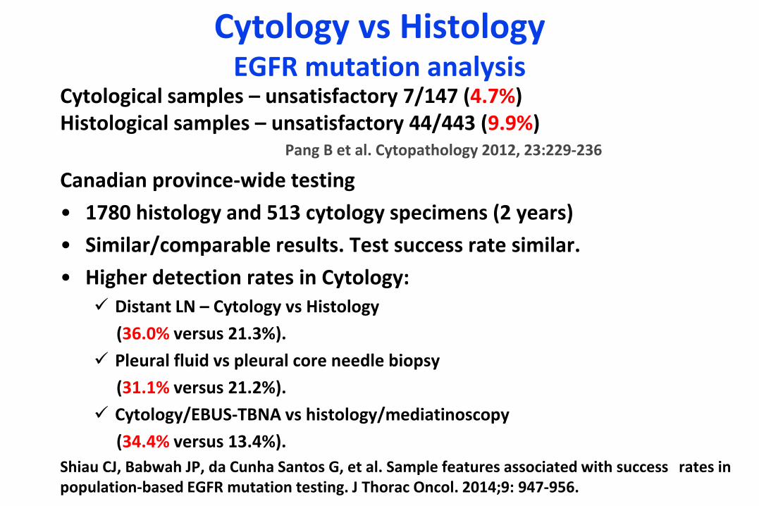
Cytology vs HistologyEGFR mutation analysis
Cytological samples – unsatisfactory 7/147 (4.7%)Histological samples – unsatisfactory 44/443 (9.9%)
Pang B et al. Cytopathology 2012, 23:229-236
Canadian province-wide testing
• 1780 histology and 513 cytology specimens (2 years)
• Similar/comparable results. Test success rate similar.
• Higher detection rates in Cytology: Distant LN – Cytology vs Histology
(36.0% versus 21.3%).
Pleural fluid vs pleural core needle biopsy
(31.1% versus 21.2%).
Cytology/EBUS-TBNA vs histology/mediatinoscopy
(34.4% versus 13.4%).
Shiau CJ, Babwah JP, da Cunha Santos G, et al. Sample features associated with success rates in population-based EGFR mutation testing. J Thorac Oncol. 2014;9: 947-956.
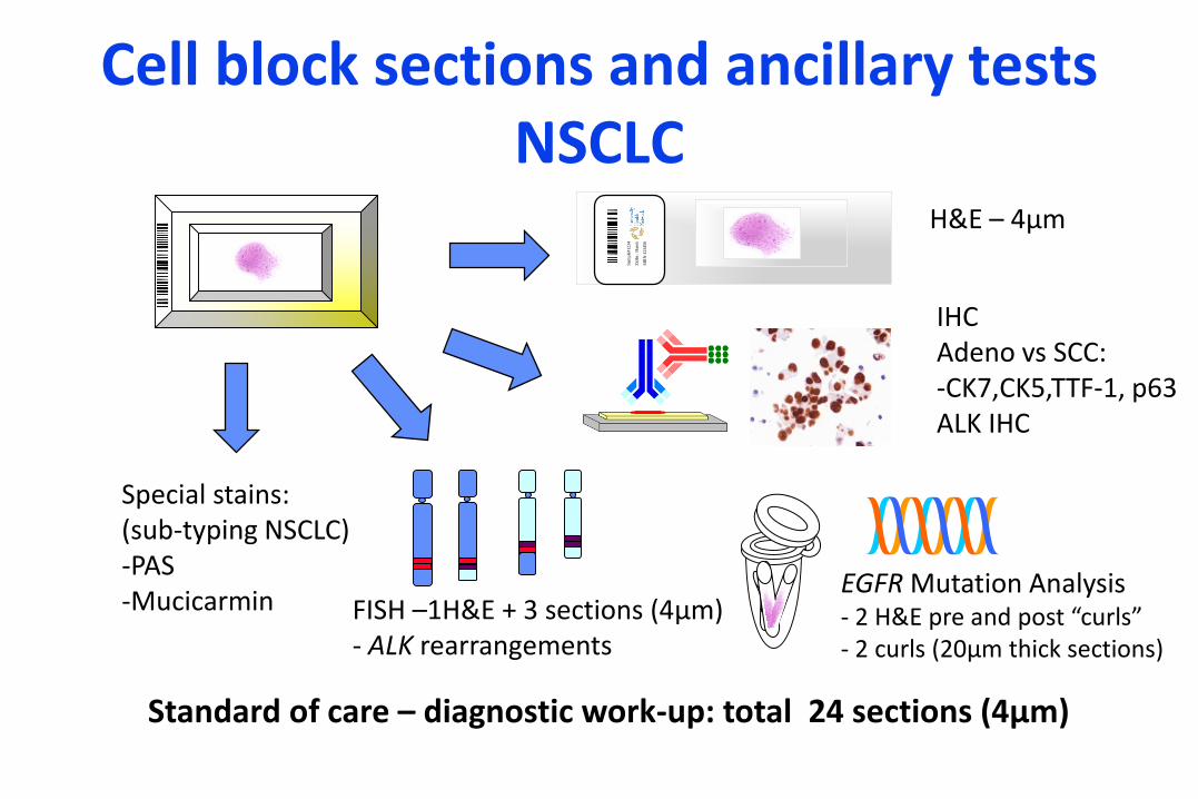
Cell block sections and ancillary testsNSCLC
IHCAdeno vs SCC:-CK7,CK5,TTF-1, p63ALK IHC
EGFR Mutation Analysis- 2 H&E pre and post “curls”- 2 curls (20μm thick sections)
Special stains:(sub-typing NSCLC)-PAS-Mucicarmin FISH –1H&E + 3 sections (4μm)
- ALK rearrangements
H&E – 4μm
Sam
ple
#12345/2
011
Zid
ler,
Har
old
MR
N:1
23456/2
01
1
Standard of care – diagnostic work-up: total 24 sections (4μm)
TTF-1CK7
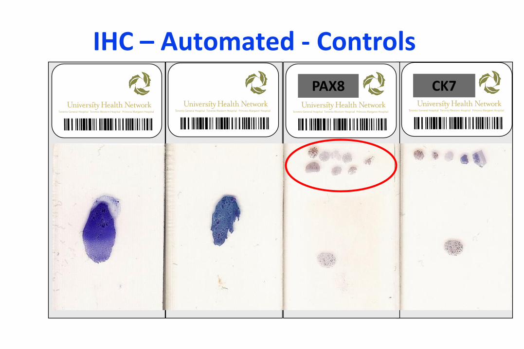
IHC – Automated - Controls
CK7PAX8
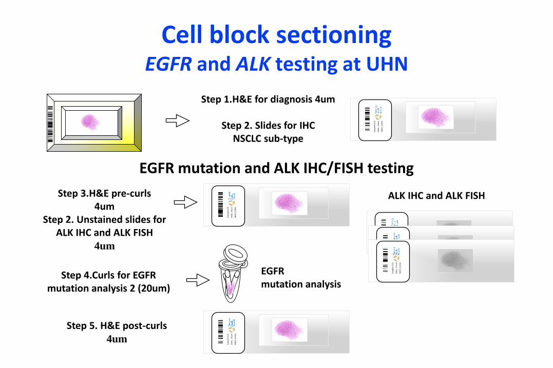
Cell block sectioningEGFR and ALK testing at UHN
Sam
ple
#12345/2
011
Zid
ler,
Har
old
MR
N:1
23456/2
01
1
EGFR mutation and ALK IHC/FISH testing
Step 1.H&E for diagnosis 4um
Step 2. Slides for IHC NSCLC sub-type
Sam
ple
#12345/2
011
Zid
ler,
Har
old
MR
N:1
23456/2
01
1
Sam
ple
#12345/2
011
Zid
ler,
Har
old
MR
N:1
23456/2
01
1
Step 3.H&E pre-curls4um
Step 2. Unstained slides for ALK IHC and ALK FISH
4um Sam
ple
#12345/2
011
Zid
ler,
Har
old
MR
N:1
23456/2
01
1
Sam
ple
#12345/2
011
Zid
ler,
Har
old
MR
N:1
23456/2
01
1
Sam
ple
#12345/2
011
Zid
ler,
Har
old
MR
N:1
23456/2
01
1
Step 4.Curls for EGFR mutation analysis 2 (20um)
ALK IHC and ALK FISH
EGFR mutation analysis
Step 5. H&E post-curls4um
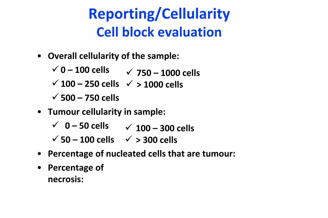
Reporting/CellularityCell block evaluation
• Overall cellularity of the sample:
0 – 100 cells
100 – 250 cells
500 – 750 cells
• Tumour cellularity in sample:
0 – 50 cells
50 – 100 cells
• Percentage of nucleated cells that are tumour:
• Percentage of necrosis:
750 – 1000 cells > 1000 cells
100 – 300 cells > 300 cells
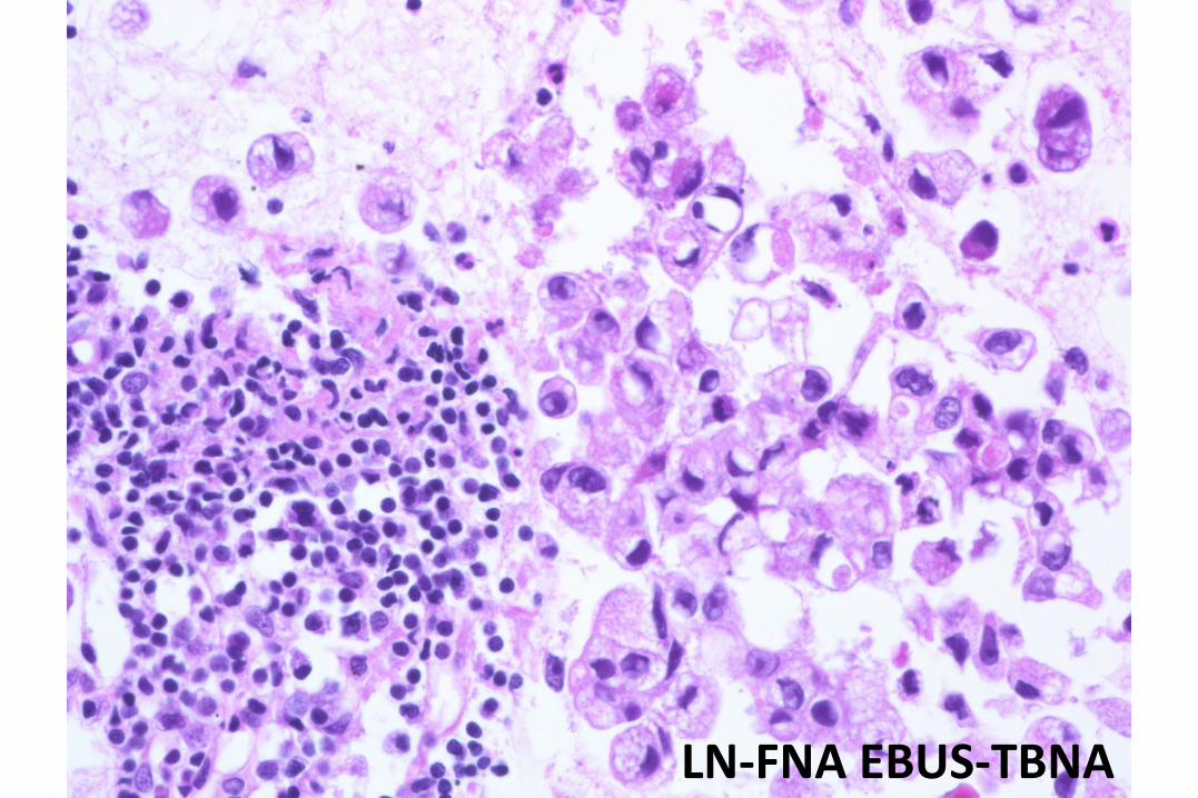
LN-FNA EBUS-TBNA

Evaluation of tumor cellularity
H&E post-curls
H&E pre-curlsCurls
Cellularity Assessment
da Cunha Santos G, Wyeth T, Reid A et al. A proposal for cellularity assessment for EGFR mutational analysis with a correlation with DNA yield and evaluation of the number of sections obtained from cell blocks for immunohistochemistry in non-small cell lung carcinoma. J Clin Pathol. 2016 Jul;69(7):607-11.
“Evaluation of a postcurl slide is an unnecessary practice.”
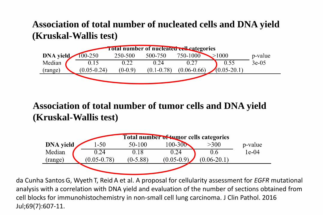
Association of total number of nucleated cells and DNA yield
(Kruskal-Wallis test)
Association of total number of tumor cells and DNA yield
(Kruskal-Wallis test)
da Cunha Santos G, Wyeth T, Reid A et al. A proposal for cellularity assessment for EGFR mutational analysis with a correlation with DNA yield and evaluation of the number of sections obtained from cell blocks for immunohistochemistry in non-small cell lung carcinoma. J Clin Pathol. 2016 Jul;69(7):607-11.

ALK rearrangement - FISH
• Most common ALK rearrangement: Paracentric inversion on the short arm of chromosome 2 jusxtaposing the 5’ end of EML4 gene with the 3’ end of the ALK gene.
q pALKEML4
2p23
3’ 5’
2p21Chromosome 2
ALK probe
ALK EML4
Inversion
ALK EML4
InversionDeletion
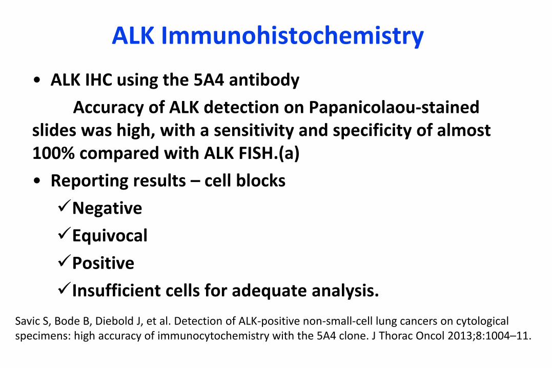
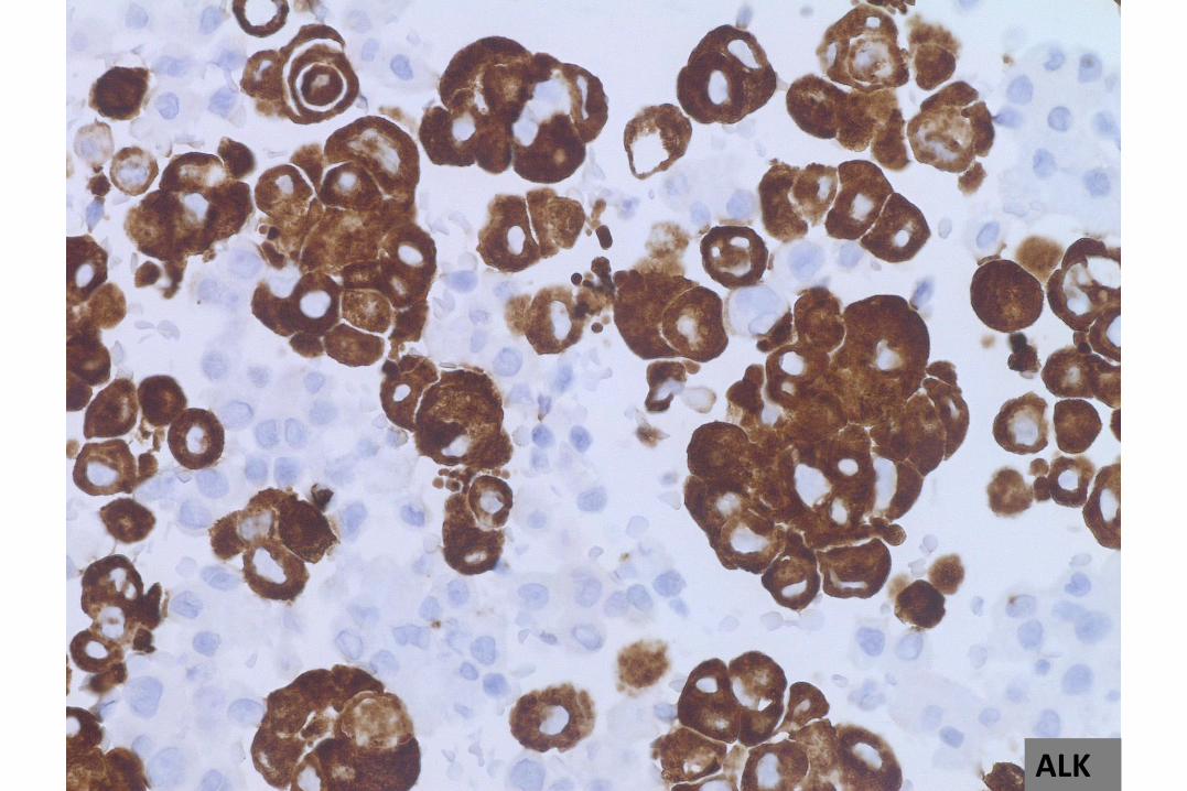
ALK Immunohistochemistry
• ALK IHC using the 5A4 antibody
Accuracy of ALK detection on Papanicolaou-stained slides was high, with a sensitivity and specificity of almost 100% compared with ALK FISH.(a)
• Reporting results – cell blocks
Negative
Equivocal
Positive
Insufficient cells for adequate analysis.
Savic S, Bode B, Diebold J, et al. Detection of ALK-positive non-small-cell lung cancers on cytological specimens: high accuracy of immunocytochemistry with the 5A4 clone. J Thorac Oncol 2013;8:1004–11.
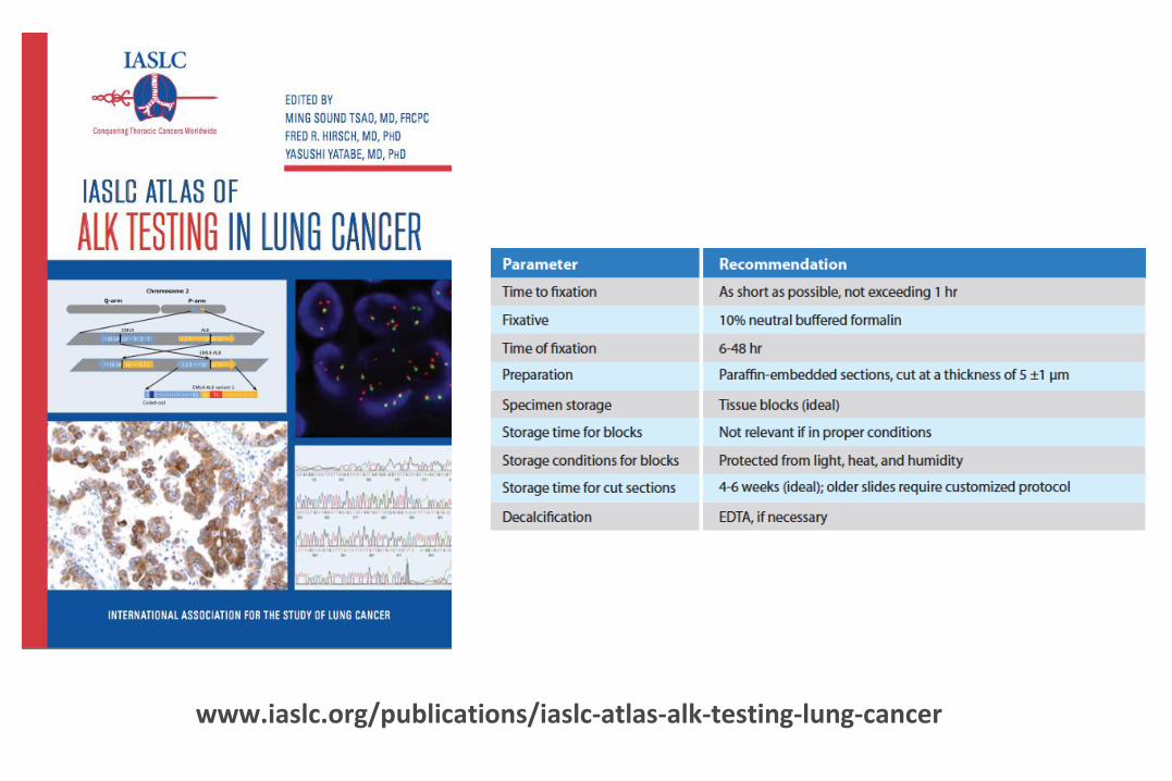
www.iaslc.org/publications/iaslc-atlas-alk-testing-lung-cancer
ALK
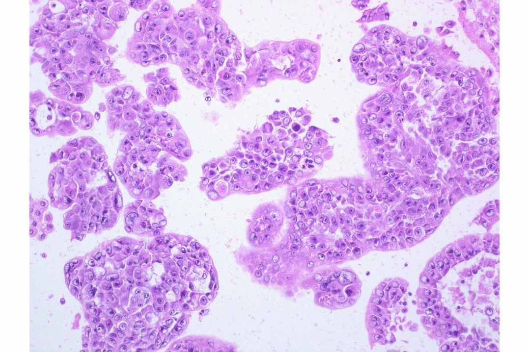

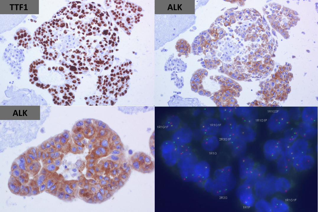
TTF1 ALK
ALK
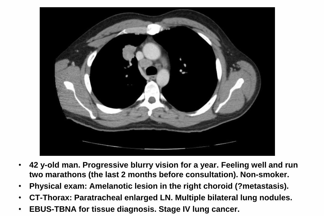
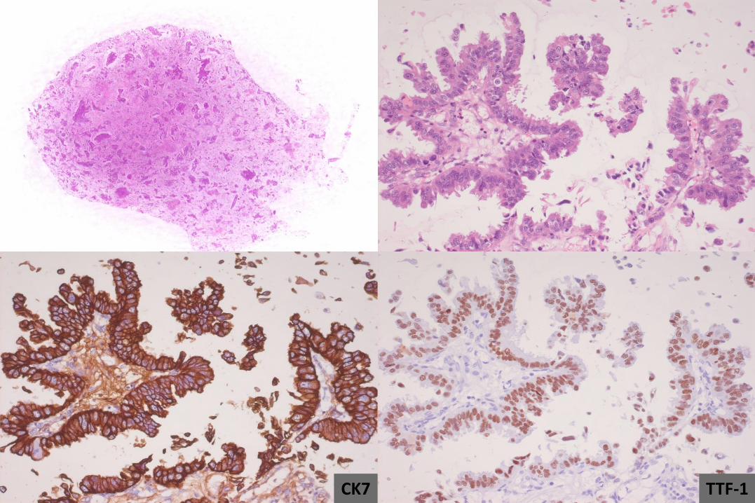
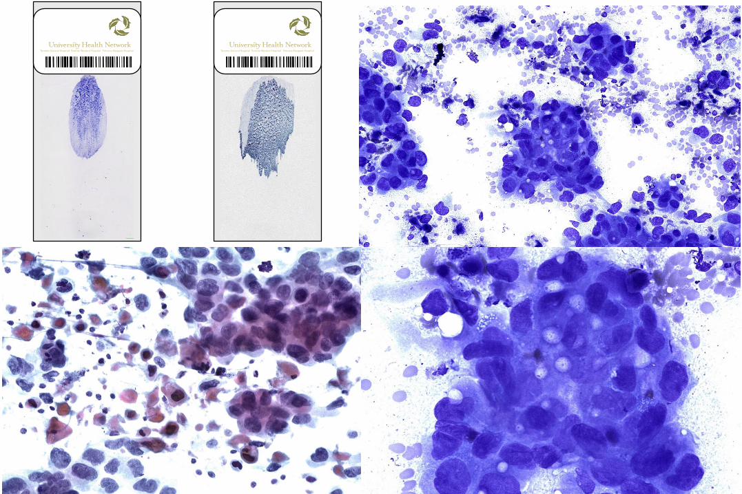
• 42 y-old man. Progressive blurry vision for a year. Feeling well and run
two marathons (the last 2 months before consultation). Non-smoker.
• Physical exam: Amelanotic lesion in the right choroid (?metastasis).
• CT-Thorax: Paratracheal enlarged LN. Multiple bilateral lung nodules.
• EBUS-TBNA for tissue diagnosis. Stage IV lung cancer.
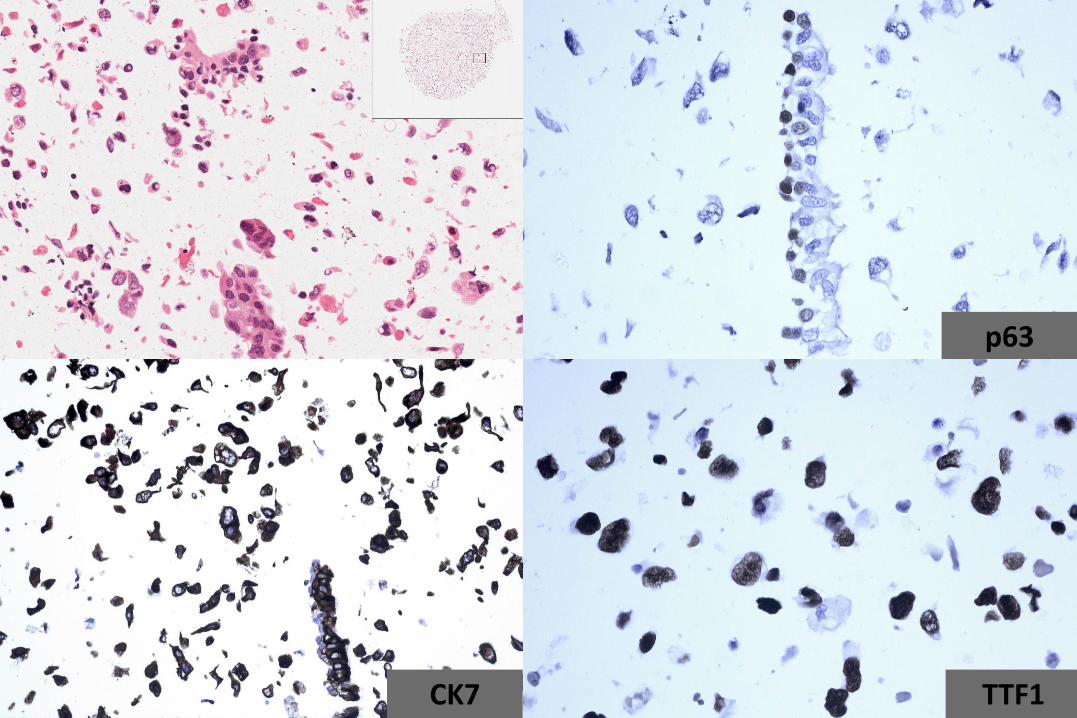
p63
CK7 TTF1
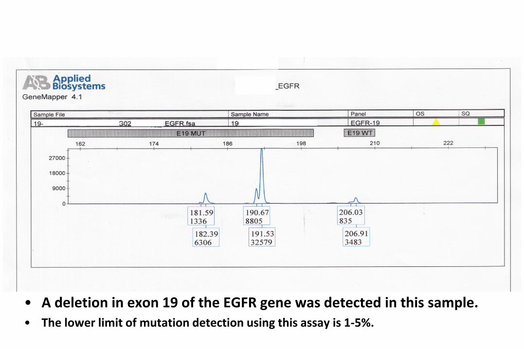
• A deletion in exon 19 of the EGFR gene was detected in this sample.• The lower limit of mutation detection using this assay is 1-5%.

• 54 y-old man. Visit in June. Cough since Jan/Feb. Antibiotics (two
cycles) with no relief. Chest X-ray: Masses in the LUL.
• EBUS-TBNA for diagnosis.
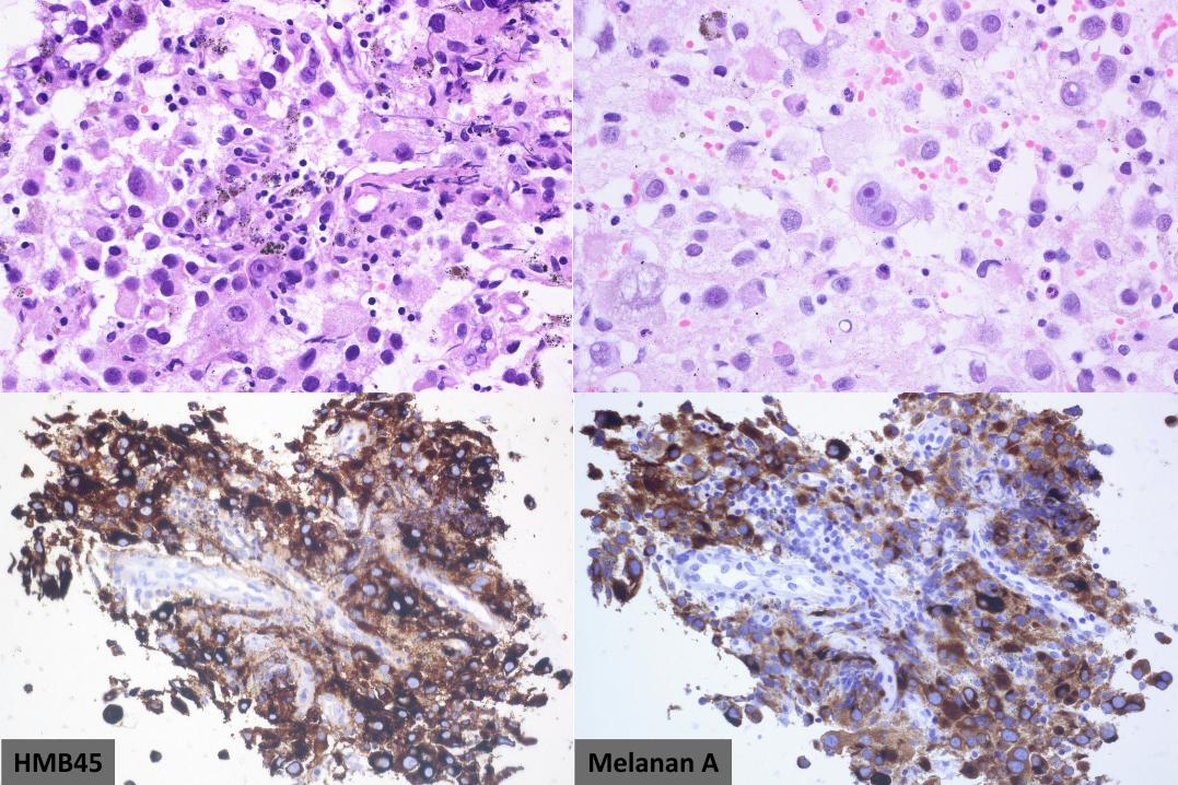
HMB45 Melanan A
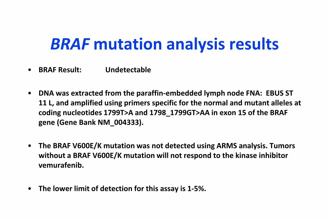
BRAF mutation analysis results
• BRAF Result: Undetectable
• DNA was extracted from the paraffin-embedded lymph node FNA: EBUS ST 11 L, and amplified using primers specific for the normal and mutant alleles at coding nucleotides 1799T>A and 1798_1799GT>AA in exon 15 of the BRAF gene (Gene Bank NM_004333).
• The BRAF V600E/K mutation was not detected using ARMS analysis. Tumors without a BRAF V600E/K mutation will not respond to the kinase inhibitor vemurafenib.
• The lower limit of detection for this assay is 1-5%.
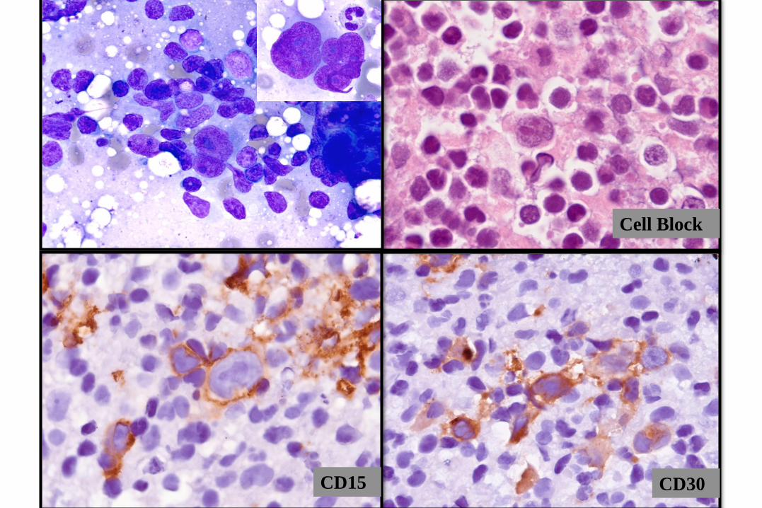
CD30CD15
Cell Block
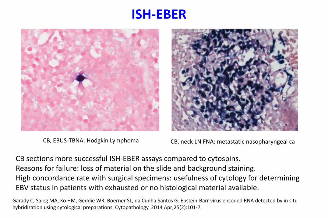
CB, neck LN FNA: metastatic nasopharyngeal ca
ISH-EBER
CB sections more successful ISH-EBER assays compared to cytospins. Reasons for failure: loss of material on the slide and background staining. High concordance rate with surgical specimens: usefulness of cytology for determining EBV status in patients with exhausted or no histological material available.
Garady C, Saieg MA, Ko HM, Geddie WR, Boerner SL, da Cunha Santos G. Epstein-Barr virus encoded RNA detected by in situ hybridization using cytological preparations. Cytopathology. 2014 Apr;25(2):101-7.
CB, EBUS-TBNA: Hodgkin Lymphoma
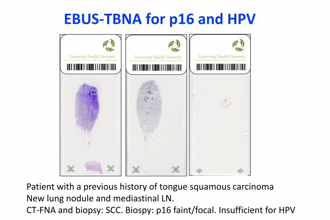
EBUS-TBNA for p16 and HPV
Patient with a previous history of tongue squamous carcinomaNew lung nodule and mediastinal LN. CT-FNA and biopsy: SCC. Biospy: p16 faint/focal. Insufficient for HPV
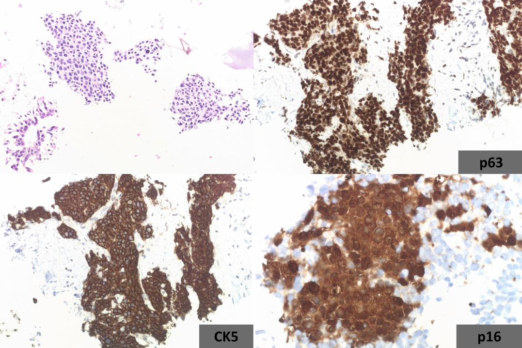
CK5
p63
p16
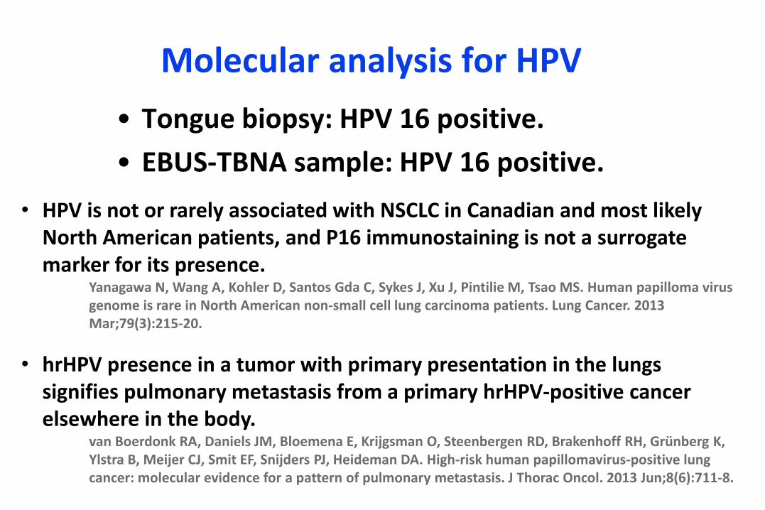
Molecular analysis for HPV
• Tongue biopsy: HPV 16 positive.
• EBUS-TBNA sample: HPV 16 positive.
• HPV is not or rarely associated with NSCLC in Canadian and most likely North American patients, and P16 immunostaining is not a surrogate marker for its presence.
Yanagawa N, Wang A, Kohler D, Santos Gda C, Sykes J, Xu J, Pintilie M, Tsao MS. Human papilloma virus genome is rare in North American non-small cell lung carcinoma patients. Lung Cancer. 2013 Mar;79(3):215-20.
• hrHPV presence in a tumor with primary presentation in the lungs signifies pulmonary metastasis from a primary hrHPV-positive cancer elsewhere in the body.
van Boerdonk RA, Daniels JM, Bloemena E, Krijgsman O, Steenbergen RD, Brakenhoff RH, Grünberg K, Ylstra B, Meijer CJ, Smit EF, Snijders PJ, Heideman DA. High-risk human papillomavirus-positive lung cancer: molecular evidence for a pattern of pulmonary metastasis. J Thorac Oncol. 2013 Jun;8(6):711-8.

CK7
GATA3 ER
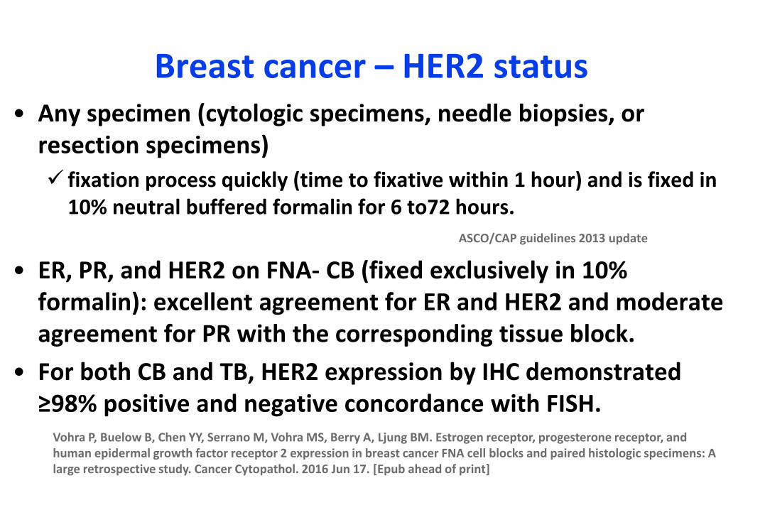
Breast cancer – HER2 status• Any specimen (cytologic specimens, needle biopsies, or
resection specimens)
fixation process quickly (time to fixative within 1 hour) and is fixed in 10% neutral buffered formalin for 6 to72 hours.
ASCO/CAP guidelines 2013 update
• ER, PR, and HER2 on FNA- CB (fixed exclusively in 10% formalin): excellent agreement for ER and HER2 and moderate agreement for PR with the corresponding tissue block.
• For both CB and TB, HER2 expression by IHC demonstrated ≥98% positive and negative concordance with FISH.
Vohra P, Buelow B, Chen YY, Serrano M, Vohra MS, Berry A, Ljung BM. Estrogen receptor, progesterone receptor, and human epidermal growth factor receptor 2 expression in breast cancer FNA cell blocks and paired histologic specimens: A large retrospective study. Cancer Cytopathol. 2016 Jun 17. [Epub ahead of print]
Opportunities and Perspectives
• Cytopathologists should provide detailed
information in molecular reports about type of
specimens, fixation and sample preparation
(standardization still required)
• Cytopathologists are essential for alliquoting
material for multiple studies for prognostic and
predictive markers.
• Techniques for cell enrichment.
THANK YOU!


![Cytological and molecular characterization of three ... · embryos of Clausena excavata [20] and homozygous short-lived plantlets of Rhode Red Valencia sweet orange [21] have also](https://img.pdfslide.net/doc/110x75/6075143509321d163c34d893/cytological-and-molecular-characterization-of-three-embryos-of-clausena-excavata.jpg)