Embed Size (px)
Citation preview

1
To be published in The Plant Cytoskeleton (Bo Liu, Ed.), Advances in Plant Biology vol. 2
3.7. Cytoskeleton and root hair growth
Eunsook Park and Andreas Nebenführ
University of Tennessee, Department of Biochemistry and Cellular and Molecular Biology,
Knoxville, TN 37996-0840, USA
Abstract
Root hairs are long tubular outgrowths of root epidermis cell that form to increase the root
surface in order to assist in the uptake of water and nutrients from soil. Root hair development
consists of two distinct processes: root hair initiation and tip growth. During both events, the
dynamic organization of the cytoskeleton translates local signaling events into a focused growth
response that is critical during root hair growth. Microtubules are primarily important for
maintenance of the direction of tip growth. The actin cytoskeleton, on the other hand, is crucial
for the selection of the root hair initiation site and maintenance of tip growth. The unique
cytoskeletal organization found in growing root hairs controls the polar delivery of membranes
to the apex in order to enlarge the cell unidirectionally. Signaling factors, such as calcium and
reactive oxygen species, are instrumental in maintaining polarity of the cytoskeleton, vesicle
trafficking, and ultimately root hair growth. Interestingly, these regulatory factors are
interdependent upon each other, so that an elaborate set of feedback loops forms that results in a
stable self-organized cell polarity.
2
3.7.1. Introduction Root hairs are highly polarized outgrowths of a subset of root epidermis cells, the so-
called trichoblasts. The biological function of root hairs is to increase the surface area of roots in
order to facilitate the absorption of water and nutrients from soil. Root hairs are also the site of
initial interaction with microorganisms (Geitmann and Emons, 2000). The patterning of root
trichoblasts varies between plant species and can also be regulated by environmental factors
(Dolan and Costa, 2001). These genetic and environmental regulatory mechanisms have been
studied intensively and were reviewed elsewhere (Ishida et al., 2008). Trichoblasts are unique
plant cells that first elongate by diffuse growth over their entire surface with the other root cells
and subsequently form an outgrowth, the root hair, which elongates only at its very tip. This kind
of tip growth also occurs in pollen tubes, which are discussed elsewhere in this volume.
The mechanism of root hair growth can be conceptually divided into two distinct events:
root hair initiation, which breaks the symmetry of the root epidermis and results in the formation
of a bulge, and unidirectional root hair elongation by tip growth in which all secretion of new
cell wall material occurs in a small area at the tip of the hair. Root hairs are not critical for plant
growth, so that plants can grow normally on growth media in the lab even with defective root
hairs. This permitted studies about the molecular mechanisms underlying root hair development
based on mutant screens for defects in root hair growth (Schiefelbein and Somerville, 1990).
These mutants showed diverse phenotypes from abnormal length or shape of root hairs to
additional root hair initiation. In independent studies, pharmacological analyses using chemicals
to disrupt cytoskeletal organization revealed the importance of the cytoskeleton, especially actin
filaments, for tip growth (Bibikova et al., 1999; Baluska et al., 2000). More recently, studies with
GFP-fused proteins related to root hair growth provided further support for the emerging
regulatory network and opened up the additional dimension of temporal dynamics (Hepler et al.,
2001; Carol and Dolan, 2002; Cole and Fowler, 2006).
Collectively, these studies have established that three factors are very important for this
special type of cell morphogenesis. First, signaling from the trichoblast determination pathway
leads to a rearrangement of the cytoskeleton in the root epidermis for bulge formation and
ultimately for support of tip growth in the emerging hair (Bibikova et al., 1999; Baluska et al.,
2000). Second, polar membrane trafficking is required to provide new plasma membrane and cell

3
wall components to the growing tip (Ovecka et al., 2005). Finally, there is a unique distribution
of regulatory factors, primarily calcium (Felle and Hepler, 1997), reactive oxygen species
(Monshausen et al., 2007), and pH (Bibikova et al., 1998) which regulate each other to modulate
tip growth (reviewed in Cole and Fowler, 2006). Pollen tubes, the other classical model for tip
growth in plants, share a similar growth mechanism that involves similar key regulators (Cole
and Fowler, 2006). However, there are also some distinct differences between pollen tubes and
root hairs, such as vacuole position and growth rate. It remains to be seen whether a common
model can simultaneously explain the specific behavior of root hairs and pollen tubes. However,
in this chapter, we will forgo this comparison and focus on the mechanisms of root hair growth.
3.7.2. Root hair Initiation
The first visible step during root hair initiation is that a part of the outer epidermis cell
starts swelling in response to local signaling. The position of the initial swelling depends on the
plant species. For example, Arabidopsis root hairs are initiated on the basal part of the
trichoblast, i.e. closest to the root tip (Carol and Dolan 2002), while maize roots form the root
hair approximately in the middle of an epidermal cell (Baluska et al., 2000; Fischer et al., 2007).
The positioning of root hairs on trichoblasts depends at least partially on hormonal signaling as
revealed by mutant analysis. As the first step of root hair initiation, a membrane trafficking
effector, ROP-GTPase, and a cell wall loosening enzyme, expansin, accumulate at the initiation
site (Molendijk et al., 2001). Both actin and microtubule cytoskeleton then rearrange (Van
Bruaene et al., 2004) and local alkalization of the cytosol occurs accompanied by acidification of
the cell wall (Bibikova et al., 1998). In addition, the cytosolic calcium concentration increases
locally due to a massive uptake of calcium from the environment (Very and Davies, 2000).
Finally, actin filaments accumulate and the cell bulges outward and eventually begins tip growth
(Ishida et al., 2008).
3.7.2.1. Selection of the bulge site Auxin and ethylene signaling appears to be a key regulator in determining the position of
root hairs, since many root hair defective mutants are directly or indirectly related to auxin or
ethylene responses (Guimil and Dunand, 2007). In particular, ethylene signaling mutants showed
altered position of root hairs on a trichoblast, suggesting that bulge site selection occurs
4
downstream of a hormone signaling pathway (Masucci and Schiefelbein, 1994). The auxin
transport mutant, aux1, also displayed root hairs in an apically shifted position and often carried
two root hairs on a cell (Grebe et al., 2002). The apical and basal membrane localization of
AUX1 on root epidermis cells appeared to be required for maintaining planar root hair polarity
by facilitating the uptake of auxin from more distal cells to maintain a local proximal auxin
maximum in the trichoblast (Swarup et al., 2001). Moreover, pharmacological interruption of
auxin transport also disrupted proper root hair positioning, supporting the critical role of auxin in
determining the root hair position (Grebe, 2004).
How auxin and ethylene signaling affect cytoskeletal reorganization is still not clear.
However, ROP-GTPase (Rho-like GTPase of plants) may be part of the signaling cascade. In
Arabidopsis, immunolocalization of ROP proteins revealed their accumulation under the plasma
membrane of the root hair initiation site even before swelling started, and this accumulation was
maintained during root hair growth (Fu et al., 2002). This localization, together with evidence
that overexpression of ROP-GTPase resulted in multiple root hair formation on a single
trichoblast (Yang et al., 2007), further supports the notion that ROP-GTPases are key regulators
for root hair initiation. ROP accumulation was not sensitive to cytoskeleton-disrupting drugs
suggesting that ROP accumulation is an upstream event of actin and microtubule rearrangement
in trichoblasts (Baluska et al., 2000; Molendijk et al., 2001). How localization and activity of
ROP-GTPase is functionally regulated by polar auxin transport still remains an open question to
be investigated. Recently, a mathematical simulation model hypothesized that the auxin gradient
might function to balance the activity of ROPs, so that the root hair can be formed at the proper
position of root epidermis cells (Payne and Grierson, 2009). Although there is as of yet little
experimental support for this hypothesis, this model is a good starting point to identify the
mechanisms that ultimately lead to selection of the bulge site.
3.7.2.2. The cytoskeleton in bulge formation
Root epidermis cells have highly organized transverse cortical microtubule arrays. Once a
trichoblast starts root hair formation, this microtubule array becomes irregular and randomized at
the site of the future root hair bulge (Baluska et al., 2000). This is correlated with local wall
acidification which leads to a change in cell wall formation and ultimately results in wall
thinning so that the cell can swell in this region (Bibikova et al., 1998). Remarkably, the apex of
5
the bulge is devoid of microtubules when the root hair swelling is ready to initiate tip growth
(Baluska et al., 2000). At the same time, actin filaments at the future bulge site become arranged
parallel to the long axis of the cell (Baluska et al., 2000). G-actin and profilin also accumulate at
the tip of the root hair bulge, as observed by immunofluorescence in maize (Braun et al., 1999),
and a fine actin meshwork later forms within the bulge (Baluska et al., 2000).
Actin mutants, act2 and act8, showed root hair bulges but no proper tip growth
suggesting that the actin cytoskeleton does not have a critical role in the early steps of root hair
initiation (Ringli et al., 2002; Kandasamy et al., 2009). However, actin mutants often formed
multiple sites of root hair bulges implying that actin might be involved in the process of
determining the root hair initiation site on the trichoblast (Kandasamy et al., 2009). Lettuce
seedlings that were germinated on media containing 10µM cytochalasin B, an inhibitor of actin
polymerization, did not produce any root hairs (Takahashi et al., 2003). This result suggests that
longitudinal redistribution of actin filaments on the site of root hair emergence is necessary for
root hair initiation. However, cytochalasin D treatment of vetch roots did not show any effects
on root hair bulge formation (Miller et al., 1999). Note, however, that cytochalasin variants have
different side effects beside actin disruption. Thus, it is conceivable that long-term exposure of
lettuce roots to cytochalasin B may have induced additional secondary effects that inhibited root
hair development. The actin cytoskeleton is clearly more important in later steps of root hair
initiation, namely, during the transition from bulge formation to tip growth since actin mutants as
well as pharmacological interventions showed that a defect of actin organization prevented the
initiation of root hair tip growth (Miller et al., 1999; Baluska et al., 2000; Ringli et al., 2002;
Kandasamy et al., 2009).
3.7.3. Root hair tip growth: general considerations and organelle distribution Once a bulge is established, root hairs continue to increase their surface area only at their
tip, away from the root epidermis. Arabidopsis root hairs have been reported to be able to grow
to a length of about 700µm with the growth rate of 1-2µm/min. However, these parameters can
differ significantly depending on growth conditions (Ojangu et al., 2007; Monshausen et al.,
2008). Conventionally, root hairs are divided into three zones from the tip toward the root
epidermis, apex, subapex, and shank. This subcellular organization of growing root hairs shares
some similarity with pollen tubes with some notable differences (compare chapter 3.2 for a

6
discussion of pollen tube growth). In growing root hairs, the dome-shaped apex, where active
growth occurs, is predominantly filled with secretory vesicles. Large organelles are prevented
from entering this region, presumably by mesh-like short, randomly distributed actin fragments
in the subapex (Baluska et al., 2003). Unlike pollen tubes, apex and subapex of root hairs are
often not clearly distinguished; the vesicle-filled apical zone is narrower than in pollen tubes and
the actin mesh spreads more broadly. A major difference between root hairs and pollen tubes is
the position of the large central vacuole. While the vacuole in pollen tubes is located far behind
the tip in the shank, the vacuole in growing root hairs can reach into subapex (Cole and Fowler,
2006).
When root hairs stop growing, the vesicle accumulation in the apex disappears and the
vacuole can fill the entire root hair. These characteristic changes in organelle distribution and
cell morphology depend strongly on the cytoskeleton which in turn is regulated by a feedback
loop involving membrane transport and several signaling molecules. [Figure 1 near here]
3.7.3.1. Actin cytoskeleton in root hair tip growth Actin filaments are fundamental for root hair growth, similar to what was found in other
cell types (reviewed in Smith and Oppenheimer, 2005; Hussey et al. 2006). Visualization of the
actin cytoskeleton has been performed with several different probes in fixed or living root hairs.
While longitudinal actin bundles along the shank of growing root hairs are relatively consistent
in images with different probes, the existence of actin at the subapex and apex is somewhat
controversial. Confocal microscopy images of actin visualized with a freeze-shattering technique
using actin antibodies showed high actin accumulation at the maize root hair apex (Baluska et
al., 2000). Labeling of globular actin (G-actin) by fluorescein isothiocyanate (FITC)-DNase I
also showed that G-actin accumulated extensively in the apex in growing root hairs of wheat.
This accumulation disappeared and was replaced by thick filamentous actin (F-actin) extending
into the tip in fully grown root hairs (He et al., 2006). Co-visualization of G-actin and F-actin by
labeling with FITC-DNase I and Tetramethyl Rhodamine Isothiocyanate (TRITC)-Phalloidin,
respectively, showed distinct localization of G-actin and F-actin in growing root hairs. While G-
actin accumulated at the apex, F-actin was presented in the shank of the growing root hairs and
could not penetrate into the apex (He et al., 2006).

7
GFP-conjugated actin binding proteins also have been used to visualize actin filaments.
This technique offers the advantage of revealing F-actin dynamics during root hair growth.
However, this has to be balanced with potential artifacts resulting from over-expression of the
labels. Initial experiments with GFP-talin transformed Arabidopsis displayed the high
accumulation of actin at the apex of growing root hairs (Baluska et al., 2000). In subsequent
years, it was revealed that over-expression of GFP-talin could cause severe developmental
defects in transgenic plants (Ketelaar et al., 2004). Many studies now employ a fimbrin-based
marker to visualize actin dynamics in all cells of several species (Figure 3.7.1.A). The second
actin binding domain of fimbrin fused to the C-terminus of GFP (GFP-FABD2) displayed more
fine actin filaments than GFP-talin and did not show any obvious developmental defects (Voigt
et al., 2005). In contrast to GFP-talin, GFP-FABD2 (or GFP-FABD2-GFP for brighter signals)
did not accumulate in the apex of growing root hairs of transgenic Arabidopsis seedlings
(Sheahan et al., 2004; Wang et al., 2008). However, FABD2-GFP transformed Medicago
truncatula displayed accumulation of actin at the apex (Miller et al., 1999; Voigt et al., 2005;
Timmers et al., 2007), suggesting that actin organization might differ between the species.
Recently, a new actin marker, Lifeact has been introduced which is based on the actin-binding
domain of yeast ABP1 (Era et al., 2009). Lifeact-Venus revealed a similar actin cytoskeleton as
GFP-FABD2, however, it had a better resolution at the root hair tip, so that an irregular actin
mesh in the subapex and highly dynamic fine filaments reaching into the tip of the apex could be
observed (Era et al., 2009).
Based on results from a variety of cell types, it is generally assumed that a central
function of actin filaments is to deliver membrane compartments to the apex of root hairs in
order to provide cell wall components and membrane lipids necessary for growth, as well as
several regulatory factors (Baluska and Volkmann, 2002). The contribution of actin dynamics to
root hair tip growth has been exposed by treatment with actin disrupting drugs, Latrunculin B
(LatB) (Bibikova et al., 1997; Baluska et al., 2000) and cytochalasin D (Miller et al., 1999). The
effect of LatB is dosage-dependent. A one hour treatment of as little as 50 nM LatB reduced root
hair growth by 25% while concentrations higher than 500 nM led to complete cessation of root
hair growth in Arabidopsis (Bibikova et al., 1999). Concentrations of more than 1 µM
cytochalasin D also caused cytoplasmic streaming to stop within 30 min and 10 µM cytochalasin
D could kill root hairs within 15 min in Medicago (Miller et al., 1999). Overall, disruption of

8
actin filaments causes root hairs to stop growing, suggesting that actin organization is critical for
tip growth. Genetic studies have corroborated this conclusion. Over decades, numerous root
hair mutants of Arabidopsis have been isolated in several studies (reviewed in Guimill and
Dunand, 2007) and some of them are mutants of either components of actin filaments or
regulators of actin dynamics. For example, der1, a mutant of ACTIN2 (ACT2), displayed a
cessation of root hair growth after bulging (Ringli et al., 2002) with additional pleiotropic
phenotypes in different tissues (Gilliland et al., 2002). ACT8, the isoform most similar to ACT2,
also contributes to root hair tip growth. act8 mutants showed around 50% shorter root hairs than
wild type and overexpression of ACT8 could complement the act2 mutant phenotype
(Kandasamy et al., 2009). Mutation of several regulators of actin dynamics also displayed an
arrest of tip growth in root hairs; induced overexpression of AIP1, a F-actin capping protein,
resulted in short root hairs (Ketelaar et al., 2007), and overexpression of PFN1, an Arabidopsis
profilin isoform, stimulated root hair tip growth and resulted in root hairs that were twice as long
as wild type (Ramachandran et al., 2000). Finally, mutation of AtFH8, an Arabidopsis group Ie
formin known to regulate actin dynamics, caused an arrest of root hair tip growth after bulge
formation (Deeks et al., 2005).
3.7.3.2. Myosin in root hair tip growth
While the actin cytoskeleton has been recognized as an important factor for tip growth,
the involvement of myosins, motor proteins that utilize F-actin as a track, was not clear until
recently. Based on evidence from pollen tubes and circumstantial evidence from the importance
of the actin cytoskeleton (Tang et al., 1989; Yokota et al., 2000), a contribution of myosins on
root hair tip growth has been proposed (Tominaga et al., 2000). Recent studies with truncated
class XI myosins fused to GFP variants at their N-terminus showed their localization on various
membrane compartments suggesting a function in intracellular vesicle trafficking (Li and
Nebenführ, 2007; Avisar et al., 2009). Direct evidence supporting the involvement of myosin in
root hair tip growth was provided by the identification of a mutant of Arabidopsis thaliana, xi-k.
xi-k plants showed short root hairs and abnormal trichome branching patterns (Ojangu et al.,
2007; Peremyslov et al., 2008). MYA2, another one of the 13 class XI myosin genes in
Arabidopsis, also seems to be involved in root hair tip growth based on the fact that mya2
mutants also displayed short root hairs (Peremyslov et al., 2008). Interestingly, in these mutants,

9
movements of three organelles, Golgi, peroxisomes, and mitochondria, were dramatically slower
than in wild type (Peremyslov et al., 2008). It is still unknown how a single myosin can
contribute to the movement of three distinct organelles, particularly, since none of the tested
YFP-myosin tail fusions localized to these organelles (Li and Nebenführ, 2007; Reisen and
Hanson, 2007; Sparkes et al., 2008). A possible hypothesis is that one of the cargoes of XI-K
might be a regulator of cytoskeleton dynamics, thus resulting in defects of movements of many
organelles. In support of this contention, recent observation using variable-angle evanescent
wave microscopy and spinning disc confocal microscopy showed that actin turnover might be
required for myosin-based mitochondrial trafficking (Zheng et al., 2009). Thus, while it is now
firmly established that myosins are involved in root hair tip growth, more research is needed to
decipher the mechanism by which myosins operate in this process.
3.7.3.3. Microtubules and root hair tip growth
The organization of microtubules has been initially visualized in root hairs using electron
microscopy and immunofluorescence with anti-tubulin antibodies (Lloyd and Wells, 1985; Traas
et al., 1985). Later, transgenic plants expressing fluorescent proteins fused to α-tubulin 6 (GFP-
TUA6) or to the microtubule binding domain of microtubule associated protein 4 (GFP-MBD),
were used to reveal dynamic changes of microtubule organization during root hair growth (Bao
et al., 2001; Timmers et al., 2007). After the reorganization event during root hair bulging, dense
cortical microtubules (CMTs) are arranged longitudinally along the shank of the root hairs.
Similar to the organization of F-actin, CMTs have not been detected in the apex of growing root
hairs (Figure 3.7.1.B). Once root hairs are fully grown and the central vacuole approaches the
root hair tip, longer and less dense CMTs are arranged longitudinally or spirally along the root
hairs and can reach to the very tip of the root hairs (Van Bruaene et al., 2004; Timmers et al.,
2007). Endoplasmic microtubules (EMTs) initially have been observed in CLSM images in the
interior of root hairs expressing GFP-MBD in Medicago truncatula (Sieberer et al., 2002). EMTs
displayed a more irregular arrangement than CMTs and predominantly accumulated around the
nucleus as well as in the subapex. This distribution later has also been shown to exist in
Arabidopsis root hairs (Van Bruaene et al., 2004). EMTs in the subapex of growing root hairs are
highly dynamic. While the majority of CMTs array parallel to the shank of root hairs, EMTs at
the subapex continuously change their directions and lengths (Van Bruaene et al., 2004).

10
In contrast to actin filaments, which play a major role in tip growth, microtubules are
generally thought of being primarily important for maintaining the direction of root hair growth.
Treatment with the microtubule depolymerizing drug, oryzalin, and the microtubule stabilizing
drug, taxol, showed a loss of directionality of Arabidopsis root hair growth (Bibikova et al.,
1999). Both taxol and oryzalin-treated root hairs displayed wavy root hairs as well as branched
root hairs. Both drugs showed a similar effect on the angular deviation from straight root hairs,
however, taxol was more effective in triggering branching than oryzalin (Bibikova et al., 1999),
suggesting that the two drugs can distinguish between two distinct roles of microtubules during
root hair growth. Analysis of mutants in α-tubulin (tua6) (Bao et al., 2001) and in a microtubule-
associated protein (mor1) (Whittington et al., 2001) also revealed branched or wavy root hair
growth, consistent with the pharmacological analysis. Thus, microtubules appear to be required
for maintaining a stable polarity for straight growth.
The growth rates of Arabidopsis root hairs did not change during treatment with the
microtubule disrupting drugs treatments (Bibikova et al., 1999) although Medicago truncatula
root hairs had only 60% of root hair growth rate of untreated root hairs (Sieberer et al., 2002).
Thus, it is likely that microtubules do not play a direct the role in elongating root hair cell
surface. Recent studies of mutants in armadillo repeat-containing kinesins (ARKs or MRH2) of
Arabidopsis showed abnormal root hair growth but no reduced root hair growth rate, further
supporting a microtubule function in restricting the elongation zone of root hair tip growth (Yang
et al., 2007; Sakai et al., 2008; Yoo et al., 2008).
Given their co-alignment patterns (Geitmann and Emons, 2000) and evidence from other
cell types (Collings et al., 2006), an interaction between microtubules and the actin cytoskeleton
has been proposed. Using propyzamide to depolymerize microtubules and cytochalasin B to
destabilize actin filaments, the interaction between microtubules and actin cytoskeleton has been
studied in detail in the root hairs of Hydrocharis dubia (Tominaga et al., 1997). Simultaneous
treatment with both inhibitors was sufficient to stop cytoplasmic streaming and growth. Washout
of Cytochalasin B in the presence of propyzamide did not allow full recovery of the longitudinal
actin cytoskeleton indicating that longitudinal microtubules are required to establish the actin
cables normally found in the root hair shank (Tominaga et al., 1997). At the same time, actin
filaments appeared to be required for normal MT dynamics since longer treatment of root hairs
with LatB permitted the formation of MT bundles in the apical or subapical regions (Timmers et

11
al., 2007). A possible candidate for this interaction between actin filaments and microtubules is
ARK/MRH2 kinesin, since it could be shown that ARK1/MRH2 can bind to actin filaments in
vitro (Yang et al., 2007). Further work will be necessary to confirm this hypothesis.
3.7.3.4. Membrane trafficking and tip growth
To increase the cell size, massive secretion at the tip is required to provide membrane and
cell wall components. Additionally, active endocytosis occurs at the apex of root hairs to recycle
regulators and remove excess membrane (Ovecka et al., 2005). These membrane trafficking
activities are reflected in the accumulation of secretory and endocytic vesicles in the apex of
growing root hairs. The small GTPase, RabA4b, has been successfully used as a vesicle marker
in root hairs (Figure 3.7.1.C). In growing root hairs, RabA4b-YFP labeled vesicles accumulated
at the apex of root hairs and LatB treatment released this accumulation coincident with a
cessation of growth (Preuss et al., 2004).
It is not surprising that many root hair mutants are defective in genes encoding proteins
involved in membrane trafficking (Gilliland et al., 2002). Members of the Ras-like small GTPase
superfamily have roles on various endomembrane compartments and several mutants of these
proteins showed impaired tip growth. A major example is ROP, a Rho-like GTPase that
coordinates actin organization and membrane trafficking by stimulating multiple signaling
pathways (Yalovsky et al., 2008). Beside its contribution to root hair initiation (see section
3.7.2.2.), ROP-GTPase functions as a critical player in root hair tip growth (Molendijk et al.,
2001). ROP-GTPase is normally localized to almost the entire plasma membrane of plant cells
but is restricted to the apical plasma membrane of growing root hairs. ROP-GTPase activates
phosphatidyl-inositol (PtdIns)-monophosphate kinase (PIPK), a key regulator to maintain tip
focused membrane trafficking and actin organization (Yalovsky et al., 2008). It also stimulates
NADPH oxidase activity, which leads to the production of reactive oxygen species (Sheahan et
al., 2004), a critical factor involved in regulating calcium gradients at the apex of root hairs
(Baxter-Burrell et al., 2002). Many regulators of ROP-GTPase activity were also found to be
involved in root hair tip growth. An Arabidopsis root hair defective mutant, supercentipede 1
(scn1), has a similar phenotype to ROP-GTPase mutants, such that mutants have multiple root
hair initiation sites in a cell and root hair tip growth is aborted (Carol et al., 2005). SCN1 encodes
a Rho-GTPase GDP dissociation inhibitor (RhoGDI) that restricts ROP activity to the apex in

12
order to maintain a polarity for root hair growth. Similarly, ROP localization on the plasma
membrane is a prerequisite for its function and S-acylation of ROP-GTPase is necessary for its
membrane localization (Yalovsky et al., 2008). TIP1 encodes a S-acyl transferase (Hemsley et
al., 2005) and Arabidopsis tip1 mutants were initially isolated based on their extremely short,
sometimes branched root hair phenotype (Schiefelbein et al., 1993). Since S-acyl transferases
tend to modify only specific target proteins, it will be interesting to test whether ROPs are targets
for TIP1.
Other regulators of root hair tip growth are specific phosphoinositides that can serve as a
recognition landmark for certain cytosolic proteins, thus recruiting them to membrane patches
where they then perform their functions. For example, PI(4,5)P2 accumulates in the apical
plasma membrane of root hairs and many modifying proteins of this lipids are important for the
root hair tip growth mechanism (Cole and Fowler, 2006; Xue et al., 2009). RabA4b interacts
with PtdIns-4OH Kinase, PI-4Kβ1 that synthesizes a precursor of PI(4,5)P2 and both proteins
colocalize to small vesicles at the apex of growing root hairs (Preuss et al., 2006). A pi4kβ1/2
double mutant displayed shorter root hairs than wild type, which were often branched or jagged.
PI-4Kβ1 also binds to CBL1, a calcium sensor, implicating that activity of PI-4Kβ1 is depending
on the calcium gradient present at the root hair apex. Another root hair defective mutant, rhd4,
showed short and wavy root hairs and RabA4b-YFP accumulation at the apex was altered in the
mutants (Thole et al., 2008). RHD4 encodes PI4P-phophatase and might function to balance
PI4P levels to maintain polar tip growth at the root hair apex. RHD4 might also have a function
in actin organization since rhd4 mutants showed more patchy actin organization and thinner
filaments in root epidermis cells compared with wild type, however, the detailed mechanism of
this interaction is not yet clear.
3.7.3.5. Signaling factors in tip growth
It has long been known that intracellular calcium gradients in the root hair cytoplasm
have an important role in root hair growth (Hepler et al., 2001). Besides a role of calcium as a
second messenger in signaling cascades, it has been suggested that calcium might restrict the site
of membrane trafficking and also modify the cytoskeleton at the root hair tip so that the root hair
can grow straight (Wymer et al., 1997). For example, calcium can affect F-actin polymerization
by controlling actin-binding protein activity, such as profilin, actin-depolymerizing factor

13
(ADF), or villin (reviewed in Hussey et al., 2006). In support of this, profilin also accumulates in
the root hair tip (Baluska et al., 2000) and can lead to sequestration of G-actin in a calcium
dependent manner (Kovar et al., 2000). Calcium may also control tip growth by several other
mechanisms in addition to a modulation of F-actin dynamics. For example, G-actin accumulation
at the apex of wheat root hairs as visualized with FITC-DNase I could be disrupted by treatment
with dibromo-l,2-bis(o-aminophenoxy)ethane-N,N,N’,N’-tetraacetic acid (BAPTA) (He et al.,
2006). BAPTA is well known to rapidly dissipate calcium gradients at the growing root hair tip
and consequently stops root hair tip growth (Felle and Hepler, 1997). It was also shown that
membrane trafficking is regulated by the calcium gradient since treatment with the calcium
ionophore A23187 caused the release of the accumulation of RabA4b-YFP from the apex and a
stop of root hair growth (Preuss et al., 2006). Despite these insights, the precise mechanism by
which calcium regulates tip growth is still unknown. Given the role of calcium as a key
regulatory factor for many cellular events, it is likely that the calcium gradient coordinates tip
growth by acting through multiple regulatory factors at the same time.
Similar to pollen tubes, the tip-focused calcium gradient in root hairs has been shown to
oscillate within a range from 0.2 µM to more than 1.5 µM (Bibikova et al., 1997; Wymer et al.,
1997). These calcium oscillations are tightly correlated with the cell growth rate (Monshausen et
al., 2008). Recent studies have investigated this connection in more detail by using the
ratiometric calcium marker, yellow Cameleon 3.6 (Monshausen et al., 2008). This study
demonstrated that root hair growth rate oscillations are typically followed by cytosolic calcium
oscillation at the root hair apex with about 5 sec lag time. This observation suggests that a high
concentration of cytosolic calcium at the root hair apex restricts root hair growth. This
hypothesis was supported by observations that treatment with 200 µM La3+, a blocker of Ca2+
channels, led to an acceleration of root hair growth. Similarly, treatment with 10 µM A23187, a
calcium ionophore, blocked root hair growth (Monshausen et al., 2008). Cytosolic calcium
gradients are accompanied by pH changes and a ROS gradient in the cytosol as well as in the cell
wall space. ROS are also considered a critical factor to sustain polar growth, since rhd2, a mutant
of Arabidopsis NADPH oxidase, displayed short root hairs and the typical calcium gradient in
root hair tip was absent (Foreman et al., 2003). Given that addition of external ROS can recover
the root hair elongation and ROS can activate calcium channels, it is likely that accumulation of
ROS at the tip is required to open calcium channels at the plasma membrane of the root hair apex
14
in order to increase the cytosolic calcium concentration at the tip (Foreman et al., 2003).
Interestingly, unlike animal NADPH oxidase, plant NADPH oxidases have two EF hand motifs
at their N-terminus suggesting their regulation by calcium (Sagi and Fluhr, 2001). Indeed, RHD2
can be activated by calcium in the growing root hair, resulting in a positive feedback loop (Sagi
and Fluhr, 2001; Takeda et al., 2008). This inter-dependence of calcium and ROS signaling leads
to alternating oscillations of ROS and Ca2+gradients in the growing root hair tip, which appears
to be necessary for maintaining polar root hair growth (Monshausen et al., 2007).
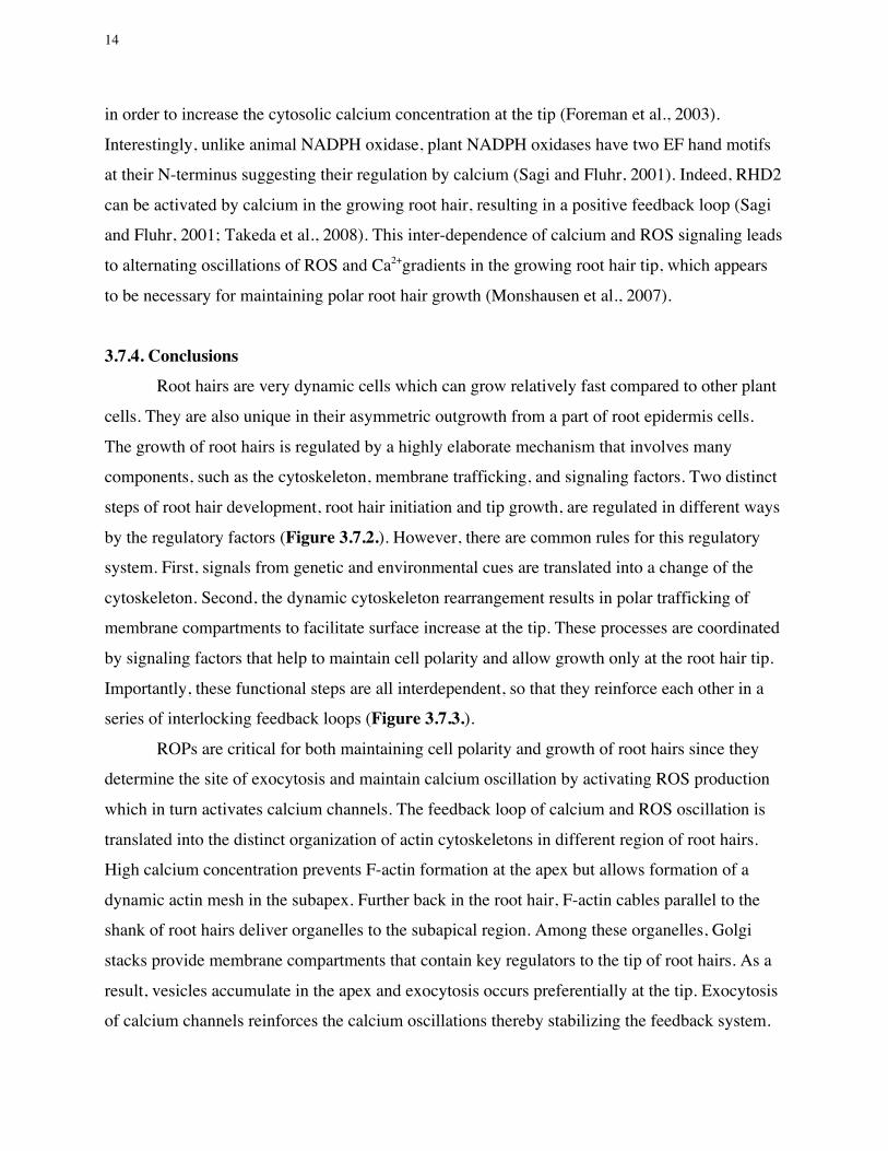
3.7.4. Conclusions Root hairs are very dynamic cells which can grow relatively fast compared to other plant
cells. They are also unique in their asymmetric outgrowth from a part of root epidermis cells.
The growth of root hairs is regulated by a highly elaborate mechanism that involves many
components, such as the cytoskeleton, membrane trafficking, and signaling factors. Two distinct
steps of root hair development, root hair initiation and tip growth, are regulated in different ways
by the regulatory factors (Figure 3.7.2.). However, there are common rules for this regulatory
system. First, signals from genetic and environmental cues are translated into a change of the
cytoskeleton. Second, the dynamic cytoskeleton rearrangement results in polar trafficking of
membrane compartments to facilitate surface increase at the tip. These processes are coordinated
by signaling factors that help to maintain cell polarity and allow growth only at the root hair tip.
Importantly, these functional steps are all interdependent, so that they reinforce each other in a
series of interlocking feedback loops (Figure 3.7.3.).
ROPs are critical for both maintaining cell polarity and growth of root hairs since they
determine the site of exocytosis and maintain calcium oscillation by activating ROS production
which in turn activates calcium channels. The feedback loop of calcium and ROS oscillation is
translated into the distinct organization of actin cytoskeletons in different region of root hairs.
High calcium concentration prevents F-actin formation at the apex but allows formation of a
dynamic actin mesh in the subapex. Further back in the root hair, F-actin cables parallel to the
shank of root hairs deliver organelles to the subapical region. Among these organelles, Golgi
stacks provide membrane compartments that contain key regulators to the tip of root hairs. As a
result, vesicles accumulate in the apex and exocytosis occurs preferentially at the tip. Exocytosis
of calcium channels reinforces the calcium oscillations thereby stabilizing the feedback system.

15
At the same time, PtdIns 4,5-P2 (PIP2), a derived membrane lipid that is deposited at the apex by
exocytotic vesicles, activates ROP proteins to stimulate ROS production, while ROP activity is
spatially limited by interaction with RhoGDI in the subapex. Remarkably, ROPs activate PIP-
kinase to produce PIP2 in the membrane, resulting in a positive feedback loop that stabilizes ROP
activity at the apex. The net result of these interdependent feedback loops is a series of stable
oscillation that ensures polar growth over long periods.
Every factor in this regulatory network plays a pivotal role, so that slight imbalances of
any of these factors affect the entire system, resulting in failure of normal root hair growth.
Recent studies have identified many of the genes responsible for root hair development and their
functions, and made big strides in revealing the underlying molecular mechanisms of root hair
development. However, it should be emphasized that many details still need to be clarified. For
example, the precise role of myosins on root hair development and how the contribution of
endocytosis to tip growth still remain in question. Further genetic, cell biological, and
biochemical studies to investigate the dynamic interplay of the various factors involved in root
hair growth combined with the collection of quantitative data and computational modeling will
be necessary to elucidate these and other questions.
3.7.5. References
Avisar, D., Abu-Abied, M., Belausov, E., Sadot, E., Hawes, C., and Sparkes, I.A. (2009). A comparative study of the involvement of 17 Arabidopsis myosin family members on the motility of Golgi and other organelles. Plant Physiol 150, 700-709.
Baluska, F., and Volkmann, D. (2002). Pictures in cell biology. Actin-driven polar growth of plant cells. Trends Cell Biol 12, 14.
Baluska, F., Wojtaszek, P., Volkmann, D., and Barlow, P. (2003). The architecture of polarized cell growth: the unique status of elongating plant cells. Bioessays 25, 569-576.
Baluska, F., Salaj, J., Mathur, J., Braun, M., Jasper, F., Samaj, J., Chua, N.H., Barlow, P.W., and Volkmann, D. (2000). Root hair formation: F-actin-dependent tip growth is initiated by local assembly of profilin-supported F-actin meshworks accumulated within expansin-enriched bulges. Dev Biol 227, 618-632.

16
Bao, Y., Kost, B., and Chua, N.H. (2001). Reduced expression of alpha-tubulin genes in Arabidopsis thaliana specifically affects root growth and morphology, root hair development and root gravitropism. Plant J 28, 145-157.
Baxter-Burrell, A., Yang, Z., Springer, P.S., and Bailey-Serres, J. (2002). RopGAP4-dependent Rop GTPase rheostat control of Arabidopsis oxygen deprivation tolerance. Science 296, 2026-2028.
Bibikova, T.N., Zhigilei, A., and Gilroy, S. (1997). Root hair growth in Arabidopsis thaliana is directed by calcium and an endogenous polarity. Planta 203, 495-505.
Bibikova, T.N., Blancaflor, E.B., and Gilroy, S. (1999). Microtubules regulate tip growth and orientation in root hairs of Arabidopsis thaliana. Plant J 17, 657-665.
Bibikova, T.N., Jacob, T., Dahse, I., and Gilroy, S. (1998). Localized changes in apoplastic and cytoplasmic pH are associated with root hair development in Arabidopsis thaliana. Development 125, 2925-2934.
Braun, M., Baluska, F., von Witsch, M., and Menzel, D. (1999). Redistribution of actin, profilin and phosphatidylinositol-4, 5-bisphosphate in growing and maturing root hairs. Planta 209, 435-443.
Carol, R.J., and Dolan, L. (2002). Building a hair: tip growth in Arabidopsis thaliana root hairs. Philos Trans R Soc Lond B Biol Sci 357, 815-821.
Carol, R.J., Takeda, S., Linstead, P., Durrant, M.C., Kakesova, H., Derbyshire, P., Drea, S., Zarsky, V., and Dolan, L. (2005). A RhoGDP dissociation inhibitor spatially regulates growth in root hair cells. Nature 438, 1013-1016.
Cole, R.A., and Fowler, J.E. (2006). Polarized growth: maintaining focus on the tip. Curr Opin Plant Biol 9, 579-588.
Collings, D.A., Lill, A.W., Himmelspach, R., and Wasteneys, G.O. (2006). Hypersensitivity to cytoskeletal antagonists demonstrates microtubule-microfilament cross-talk in the control of root elongation in Arabidopsis thaliana. New Phytol 170, 275-290.
Deeks, M.J., Cvrckova, F., Machesky, L.M., Mikitova, V., Ketelaar, T., Zarsky, V., Davies, B., and Hussey, P.J. (2005). Arabidopsis group Ie formins localize to specific cell membrane

17
domains, interact with actin-binding proteins and cause defects in cell expansion upon aberrant expression. New Phytol 168, 529-540.
Dolan, L., and Costa, S. (2001). Evolution and genetics of root hair stripes in the root epidermis. J Exp Bot 52, 413-417.
Era, A., Tominaga, M., Ebine, K., Awai, C., Saito, C., Ishizaki, K., Yamato, K.T., Kohchi, T., Nakano, A., and Ueda, T. (2009). Application of Lifeact reveals F-actin dynamics in Arabidopsis thaliana and the liverwort, Marchantia polymorpha. Plant Cell Physiol 50, 1041-1048.
Felle, H.H., and Hepler, P.K. (1997). The Cytosolic Ca2+ Concentration Gradient of Sinapis alba Root Hairs as Revealed by Ca2+-Selective Microelectrode Tests and Fura-Dextran Ratio Imaging. Plant Physiol 114, 39-45.
Fischer, U., Ikeda, Y., and Grebe, M. (2007). Planar polarity of root hair positioning in Arabidopsis. Biochem Soc Trans 35, 149-151.
Foreman, J., Demidchik, V., Bothwell, J.H., Mylona, P., Miedema, H., Torres, M.A., Linstead, P., Costa, S., Brownlee, C., Jones, J.D., Davies, J.M., and Dolan, L. (2003). Reactive oxygen species produced by NADPH oxidase regulate plant cell growth. Nature 422, 442-446.
Forer, A., and Fabian, L. (2005). Does 2,3-butanedione monoxime inhibit nonmuscle myosin? Protoplasma 225, 1-4.
Fu, Y., Li, H., and Yang, Z. (2002). The ROP2 GTPase controls the formation of cortical fine F-actin and the early phase of directional cell expansion during Arabidopsis organogenesis. Plant Cell 14, 777-794.
Geitmann, A., and Emons, A.M. (2000). The cytoskeleton in plant and fungal cell tip growth. J Microsc 198, 218-245.
Gilliland, L.U., Kandasamy, M.K., Pawloski, L.C., and Meagher, R.B. (2002). Both vegetative and reproductive actin isovariants complement the stunted root hair phenotype of the Arabidopsis act2-1 mutation. Plant Physiol 130, 2199-2209.
Grebe, M. (2004). Ups and downs of tissue and planar polarity in plants. Bioessays 26, 719-729.

18
Grebe, M., Friml, J., Swarup, R., Ljung, K., Sandberg, G., Terlou, M., Palme, K., Bennett, M.J., and Scheres, B. (2002). Cell polarity signaling in Arabidopsis involves a BFA-sensitive auxin influx pathway. Curr Biol 12, 329-334.
Guimil, S., and Dunand, C. (2007). Cell growth and differentiation in Arabidopsis epidermal cells. J Exp Bot 58, 3829-3840.
He, X., Liu, Y.M., Wang, W., and Li, Y. (2006). Distribution of G-actin is related to root hair growth of wheat. Ann Bot 98, 49-55.
Hemsley, P.A., Kemp, A.C., and Grierson, C.S. (2005). The TIP GROWTH DEFECTIVE1 S-acyl transferase regulates plant cell growth in Arabidopsis. Plant Cell 17, 2554-2563.
Hepler, P.K., Vidali, L., and Cheung, A.Y. (2001). Polarized cell growth in higher plants. Annu Rev Cell Dev Biol 17, 159-187.
Ishida, T., Kurata, T., Okada, K., and Wada, T. (2008). A genetic regulatory network in the development of trichomes and root hairs. Annu Rev Plant Biol 59, 365-386.
Kandasamy, M.K., McKinney, E.C., and Meagher, R.B. (2009). A single vegetative actin isovariant overexpressed under the control of multiple regulatory sequences is sufficient for normal Arabidopsis development. Plant Cell 21, 701-718.
Ketelaar, T., Anthony, R.G., and Hussey, P.J. (2004). Green fluorescent protein-mTalin causes defects in actin organization and cell expansion in Arabidopsis and inhibits actin depolymerizing factor's actin depolymerizing activity in vitro. Plant Physiol 136, 3990-3998.
Ketelaar, T., Allwood, E.G., and Hussey, P.J. (2007). Actin organization and root hair development are disrupted by ethanol-induced overexpression of Arabidopsis actin interacting protein 1 (AIP1). New Phytol 174, 57-62.
Kovar, D.R., Drobak, B.K., and Staiger, C.J. (2000). Maize profilin isoforms are functionally distinct. Plant Cell 12, 583-598.
Li, J.F., and Nebenführ, A. (2007). Organelle targeting of myosin XI is mediated by two globular tail subdomains with separate cargo binding sites. J Biol Chem 282, 20593-20602.

19
Lloyd, C.W., and Wells, B. (1985). Microtubules are at the tips of root hairs and form helical patterns corresponding to inner wall fibrils. J Cell Sci 75, 225-238.
Masucci, J.D., and Schiefelbein, J.W. (1994). The rhd6 Mutation of Arabidopsis thaliana Alters Root-Hair Initiation through an Auxin- and Ethylene-Associated Process. Plant Physiol 106, 1335-1346.
Miller, D., de Ruijter, N.C., Bisseling, T., and Emons, A.M. (1999). The role of actin in root hair morphogenesis: studies with lipochito-oligosaccharide as a growth stimulator and cytochalasin as an actin perturbing drug. Plant Journal 17, 141-154.
Molendijk, A.J., Bischoff, F., Rajendrakumar, C.S., Friml, J., Braun, M., Gilroy, S., and Palme, K. (2001). Arabidopsis thaliana Rop GTPases are localized to tips of root hairs and control polar growth. EMBO J 20, 2779-2788.
Monshausen, G.B., Messerli, M.A., and Gilroy, S. (2008). Imaging of the Yellow Cameleon 3.6 indicator reveals that elevations in cytosolic Ca2+ follow oscillating increases in growth in root hairs of Arabidopsis. Plant Physiol 147, 1690-1698.
Monshausen, G.B., Bibikova, T.N., Messerli, M.A., Shi, C., and Gilroy, S. (2007). Oscillations in extracellular pH and reactive oxygen species modulate tip growth of Arabidopsis root hairs. Proc Natl Acad Sci U S A 104, 20996-21001.
Ojangu, E.L., Jarve, K., Paves, H., and Truve, E. (2007). Arabidopsis thaliana myosin XIK is involved in root hair as well as trichome morphogenesis on stems and leaves. Protoplasma 230, 193-202.
Ovecka, M., Lang, I., Baluska, F., Ismail, A., Illes, P., and Lichtscheidl, I.K. (2005). Endocytosis and vesicle trafficking during tip growth of root hairs. Protoplasma 226, 39-54.
Payne, R.J., and Grierson, C.S. (2009). A theoretical model for ROP localisation by auxin in Arabidopsis root hair cells. PLoS One 4, e8337.
Peremyslov, V.V., Prokhnevsky, A.I., Avisar, D., and Dolja, V.V. (2008). Two class XI myosins function in organelle trafficking and root hair development in Arabidopsis. Plant Physiol 146, 1109-1116.
Preuss, M.L., Serna, J., Falbel, T.G., Bednarek, S.Y., and Nielsen, E. (2004). The Arabidopsis Rab GTPase RabA4b localizes to the tips of growing root hair cells. Plant Cell 16, 1589-1603.

20
Preuss, M.L., Schmitz, A.J., Thole, J.M., Bonner, H.K., Otegui, M.S., and Nielsen, E. (2006). A role for the RabA4b effector protein PI-4Kbeta1 in polarized expansion of root hair cells in Arabidopsis thaliana. J Cell Biol 172, 991-998.
Ramachandran, S., Christensen, H.E., Ishimaru, Y., Dong, C.H., Chao-Ming, W., Cleary, A.L., and Chua, N.H. (2000). Profilin plays a role in cell elongation, cell shape maintenance, and flowering in Arabidopsis. Plant Physiol 124, 1637-1647.
Reisen, D., and Hanson, M.R. (2007). Association of six YFP-myosin XI-tail fusions with mobile plant cell organelles. BMC Plant Biol 7, 6.
Ringli, C., Baumberger, N., Diet, A., Frey, B., and Keller, B. (2002). ACTIN2 is essential for bulge site selection and tip growth during root hair development of Arabidopsis. Plant Physiol 129, 1464-1472.
Sagi, M., and Fluhr, R. (2001). Superoxide production by plant homologues of the gp91(phox) NADPH oxidase. Modulation of activity by calcium and by tobacco mosaic virus infection. Plant Physiol 126, 1281-1290.
Sakai, T., Honing, H., Nishioka, M., Uehara, Y., Takahashi, M., Fujisawa, N., Saji, K., Seki, M., Shinozaki, K., Jones, M.A., Smirnoff, N., Okada, K., and Wasteneys, G.O. (2008). Armadillo repeat-containing kinesins and a NIMA-related kinase are required for epidermal-cell morphogenesis in Arabidopsis. Plant J 53, 157-171.
Schiefelbein, J., Galway, M., Masucci, J., and Ford, S. (1993). Pollen tube and root-hair tip growth is disrupted in a mutant of Arabidopsis thaliana. Plant Physiol 103, 979-985.
Schiefelbein, J.W., and Somerville, C. (1990). Genetic Control of Root Hair Development in Arabidopsis thaliana. Plant Cell 2, 235-243.
Sheahan, M.B., Staiger, C.J., Rose, R.J., and McCurdy, D.W. (2004). A green fluorescent protein fusion to actin-binding domain 2 of Arabidopsis fimbrin highlights new features of a dynamic actin cytoskeleton in live plant cells. Plant Physiol 136, 3968-3978.
Sieberer, B.J., Timmers, A.C., Lhuissier, F.G., and Emons, A.M. (2002). Endoplasmic microtubules configure the subapical cytoplasm and are required for fast growth of Medicago truncatula root hairs. Plant Physiol 130, 977-988.

21
Sparkes, I.A., Teanby, N.A., and Hawes, C. (2008). Truncated myosin XI tail fusions inhibit peroxisome, Golgi, and mitochondrial movement in tobacco leaf epidermal cells: a genetic tool for the next generation. J Exp Bot 59, 2499-2512.
Swarup, R., Friml, J., Marchant, A., Ljung, K., Sandberg, G., Palme, K., and Bennett, M. (2001). Localization of the auxin permease AUX1 suggests two functionally distinct hormone transport pathways operate in the Arabidopsis root apex. Genes Dev 15, 2648-2653.
Takahashi, H., Hirota, K., Kawahara, A., Hayakawa, E., and Inoue, Y. (2003). Randomization of cortical microtubules in root epidermal cells induces root hair initiation in lettuce (Lactuca sativa L.) seedlings. Plant Cell Physiol 44, 350-359.
Takeda, S., Gapper, C., Kaya, H., Bell, E., Kuchitsu, K., and Dolan, L. (2008). Local positive feedback regulation determines cell shape in root hair cells. Science 319, 1241-1244.
Tang, X.J., Hepler, P.K., and Scordilis, S.P. (1989). Immunochemical and immunocytochemical identification of a myosin heavy chain polypeptide in Nicotiana pollen tubes. J Cell Sci 92 ( Pt 4), 569-574.
Thole, J.M., Vermeer, J.E., Zhang, Y., Gadella, T.W., Jr., and Nielsen, E. (2008). Root hair defective4 encodes a phosphatidylinositol-4-phosphate phosphatase required for proper root hair development in Arabidopsis thaliana. Plant Cell 20, 381-395.
Timmers, A.C., Vallotton, P., Heym, C., and Menzel, D. (2007). Microtubule dynamics in root hairs of Medicago truncatula. Eur J Cell Biol 86, 69-83.
Tominaga, M., Yokota, E., Sonobe, S., and Shimmen, T. (2000). Mechanism of inhibition of cytoplasmic streaming by a myosin inhibitor, 2,3-butanedione monoxime. Protoplasma 213, 46-54.
Tominaga, M., Morita, K., Sonobe, S., Yokota, E., and Shimmen, T. (1997). Microtubules regulate the organization of actin filaments at the cortical region in root hair cells of hydrocharis. Protoplasma 199, 83-92.
Traas, J.A., Braat, P., Emons, A.M., Meekes, H., and Derksen, J. (1985). Microtubules in root hairs. J Cell Sci 76, 303-320.
Van Bruaene, N., Joss, G., and Van Oostveldt, P. (2004). Reorganization and in vivo dynamics of microtubules during Arabidopsis root hair development. Plant Physiol 136, 3905-3919.

22
Very, A.A., and Davies, J.M. (2000). Hyperpolarization-activated calcium channels at the tip of Arabidopsis root hairs. Proc Natl Acad Sci U S A 97, 9801-9806.
Voigt, B., Timmers, A.C., Samaj, J., Muller, J., Baluska, F., and Menzel, D. (2005). GFP-FABD2 fusion construct allows in vivo visualization of the dynamic actin cytoskeleton in all cells of Arabidopsis seedlings. Eur J Cell Biol 84, 595-608.
Wang, Y.S., Yoo, C.M., and Blancaflor, E.B. (2008). Improved imaging of actin filaments in transgenic Arabidopsis plants expressing a green fluorescent protein fusion to the C- and N-termini of the fimbrin actin-binding domain 2. New Phytol 177, 525-536.
Whittington, A.T., Vugrek, O., Wei, K.J., Hasenbein, N.G., Sugimoto, K., Rashbrooke, M.C., and Wasteneys, G.O. (2001). MOR1 is essential for organizing cortical microtubules in plants. Nature 411, 610-613.
Wymer, C.L., Bibikova, T.N., and Gilroy, S. (1997). Cytoplasmic free calcium distributions during the development of root hairs of Arabidopsis thaliana. Plant J 12, 427-439.
Xue, H.W., Chen, X., and Mei, Y. (2009). Function and regulation of phospholipid signalling in plants. Biochem J 421, 145-156.
Yalovsky, S., Bloch, D., Sorek, N., and Kost, B. (2008). Regulation of membrane trafficking, cytoskeleton dynamics, and cell polarity by ROP/RAC GTPases. Plant Physiol 147, 1527-1543.
Yang, G., Gao, P., Zhang, H., Huang, S., and Zheng, Z.L. (2007). A mutation in MRH2 kinesin enhances the root hair tip growth defect caused by constitutively activated ROP2 small GTPase in Arabidopsis. PLoS One 2, e1074.
Yokota, E., Muto, S., and Shimmen, T. (2000). Calcium-calmodulin suppresses the filamentous actin-binding activity of a 135-kilodalton actin-bundling protein isolated from lily pollen tubes. Plant Physiol 123, 645-654.
Yoo, C.M., Wen, J., Motes, C.M., Sparks, J.A., and Blancaflor, E.B. (2008). A class I ADP-ribosylation factor GTPase-activating protein is critical for maintaining directional root hair growth in Arabidopsis. Plant Physiol 147, 1659-1674.
Zheng, M., Beck, M., Muller, J., Chen, T., Wang, X., Wang, F., Wang, Q., Wang, Y., Baluska, F., Logan, D.C., Samaj, J., and Lin, J. (2009). Actin turnover is required for myosin-dependent mitochondrial movements in Arabidopsis root hairs. PLoS One 4, e5961.

23
Actin filaments
Microtubules
Small vesicles
Growing root hair Grown root hair
FIGURE 1
A
B
C
Figure 3.7.1. Distinct distribution of cytoskeletal elements and vesicles during root hair tip growth
(A) Actin cytoskeleton, visualized with FABD2-YFP. In growing root hairs, cortical F-actin
filaments array mostly longitudinally along the shank and are absent from subapex and
apex of root hairs. In contrast, thick bundles of actin cables reach into the apex in fully-
grown root hairs.
(B) Microtubules, visualized by GFP-MBD. Growing root hairs display longitudinal or
helical microtubules along the root hair shank. Note that microtubules do not reach the
extreme apex of the growing root hair while they do so in growing root hairs (modified
after Van Bruaene et al. 2004).
(C) RabA4b-YFP-labeled vesicles accumulate at the tip of growing root hairs. This
accumulation is tightly correlated with root hair growth; consequently, fully-grown root
hairs lack the accumulation of RabA4b-YFP vesicles at the tip.

24
A CB
FIGURE 2
D
Figure 3.7.2. Cellular architecture during root hair development
Schematic representation of root hairs during four stages of root hair development. Upper
panels show actin filaments (grey lines) and secretory vesicles (black dots). Lower panels
show microtubules (darker grey lines) and the tip-focused calcium gradient (darker color
represents higher calcium concentration).
(A) Early events after bulge site selection. Note the fragmentation of actin filaments and the
local loss of microtubule organization.
(B) Initial outgrowth and bulge formation. Actin filaments begin to form a dense mesh
behind the tip region where vesicles accumulate and the calcium gradient forms.
(C) Tip growth. Cytoskeletal elements are mostly longitudinal along the shank but do not
reach into the apex. Vesicle accumulation and calcium accumulation are maximal at the
apex.
(D) Fully grown hair. Vesicle accumulation and calcium gradient at the tip disappear and
longitudinal actin filaments and microtubules reach into the apex.
25
FIGURE 3
Ca2+
ROS
PIP2
ROP
Exocytosis
gro
wth
Vesicle
transport
organelle
movements
ROP
RhoGDI
Atr
bohC
Ca
-cha
nnel
Shank Subapex Apex
F-actin G-actinF-actin G-actinF-actin G-actin
Vesicle
accumulation
(RabA4b, PI4Kß1/ß2)
Endocytosis
Figure 3.7.3. Self-reinforcing feedback regulation of tip growth
Simplified model of regulatory mechanisms that affect the actin cytoskeleton in different
areas of the root hair during tip growth. For details see text.