Embed Size (px)
Citation preview
Cytoskeleton IV
Chapter 16
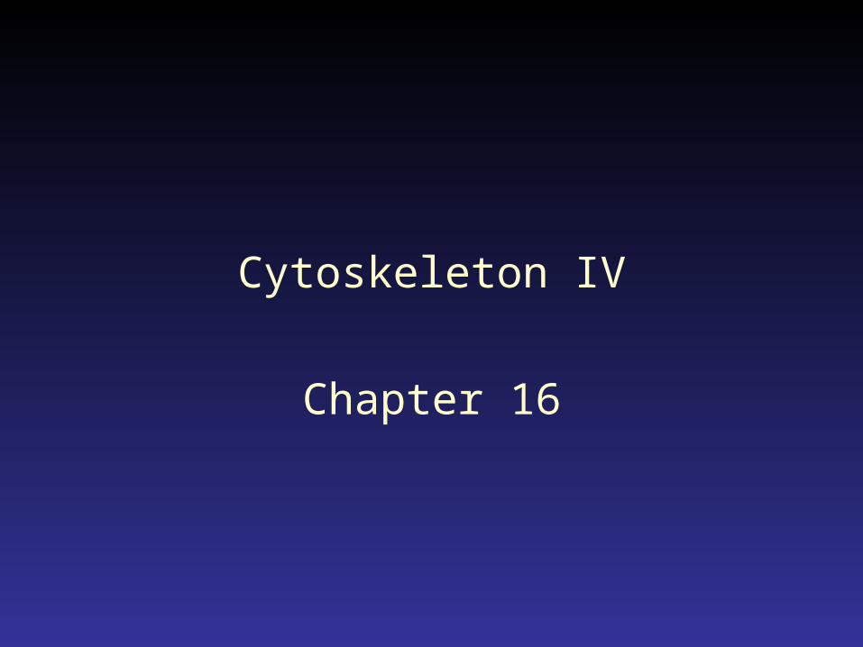
Mechanochemical cycle of myosin

DVD clip 74
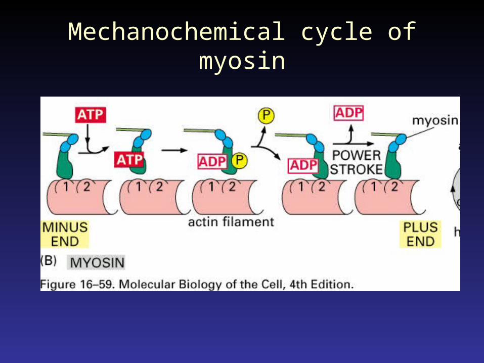
Striated vs. smooth muscle cells
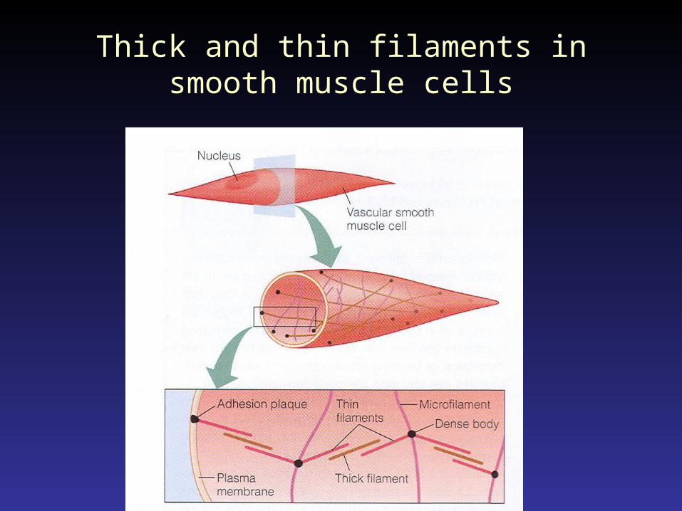
Thick and thin filaments in smooth muscle cells
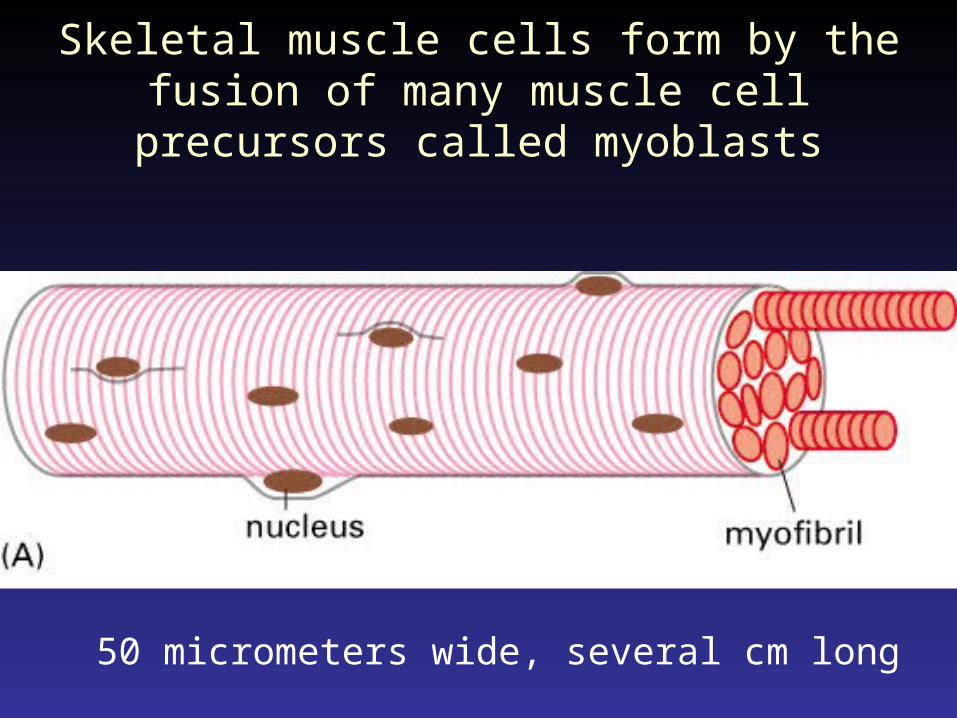
Skeletal muscle cells form by the fusion of many muscle cell precursors called myoblasts
50 micrometers wide, several cm long
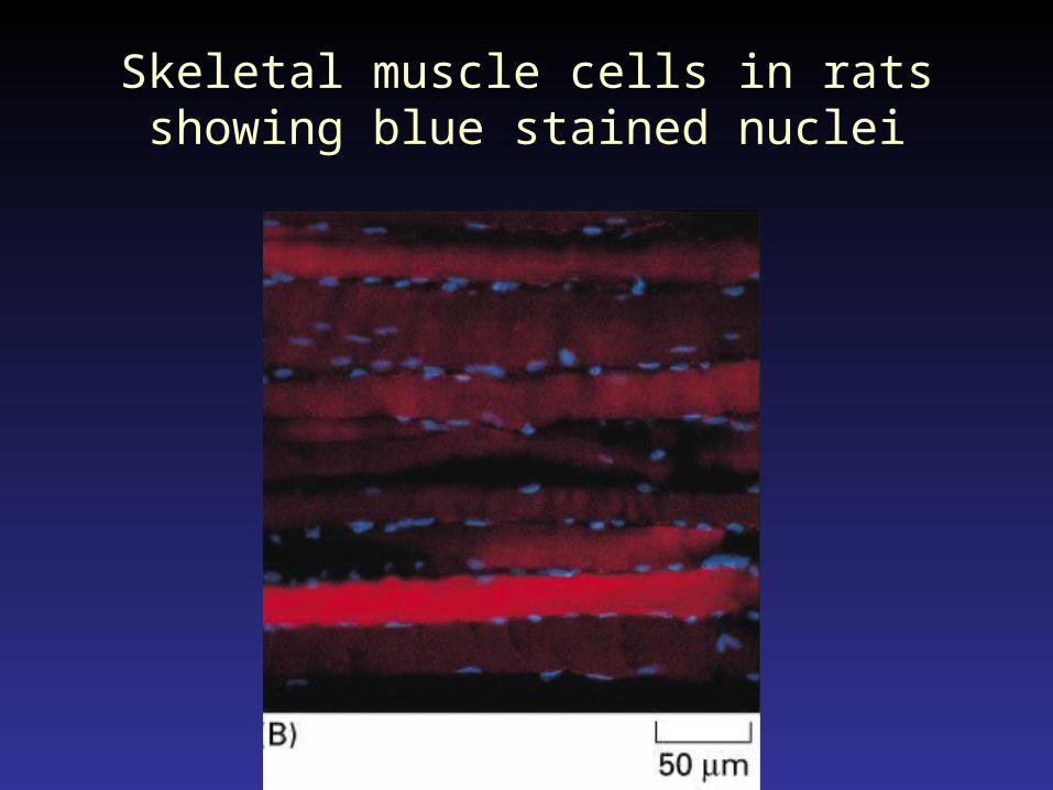
Skeletal muscle cells in rats showing blue stained nuclei
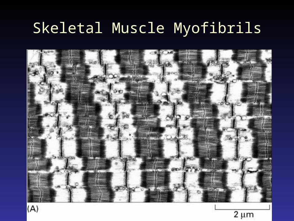
Skeletal Muscle Myofibrils
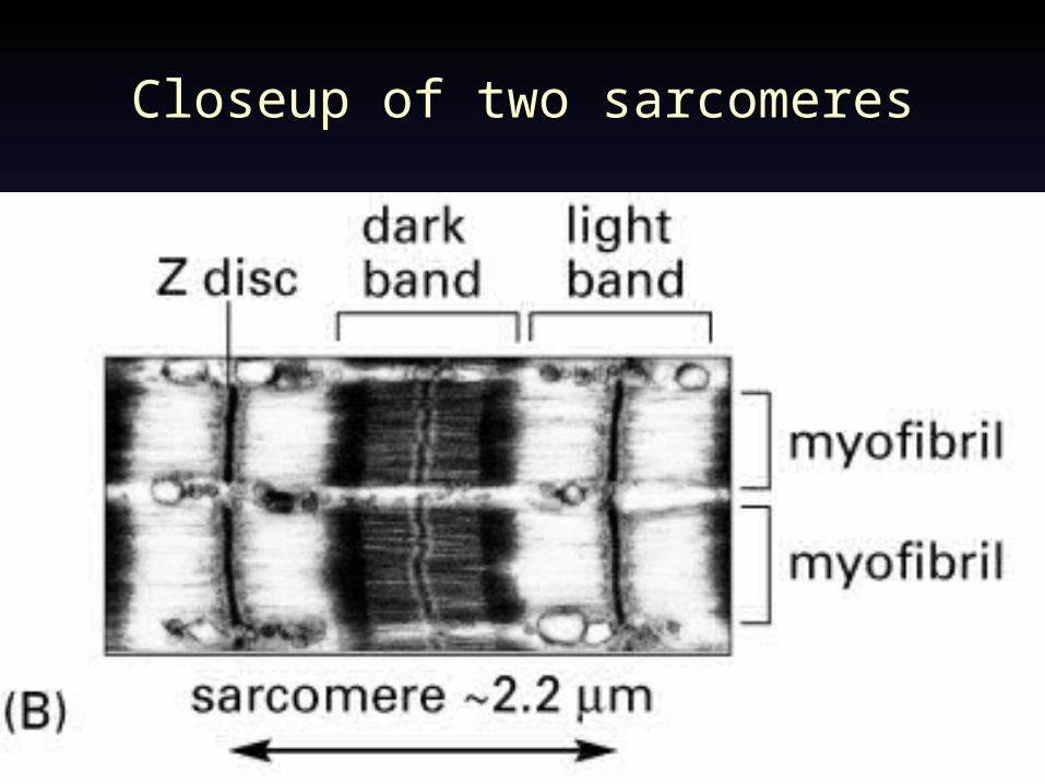
Closeup of two sarcomeres
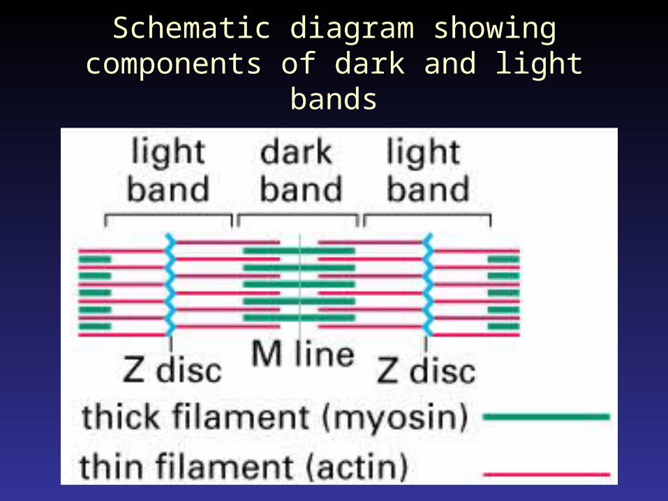
Schematic diagram showing components of dark and light bands
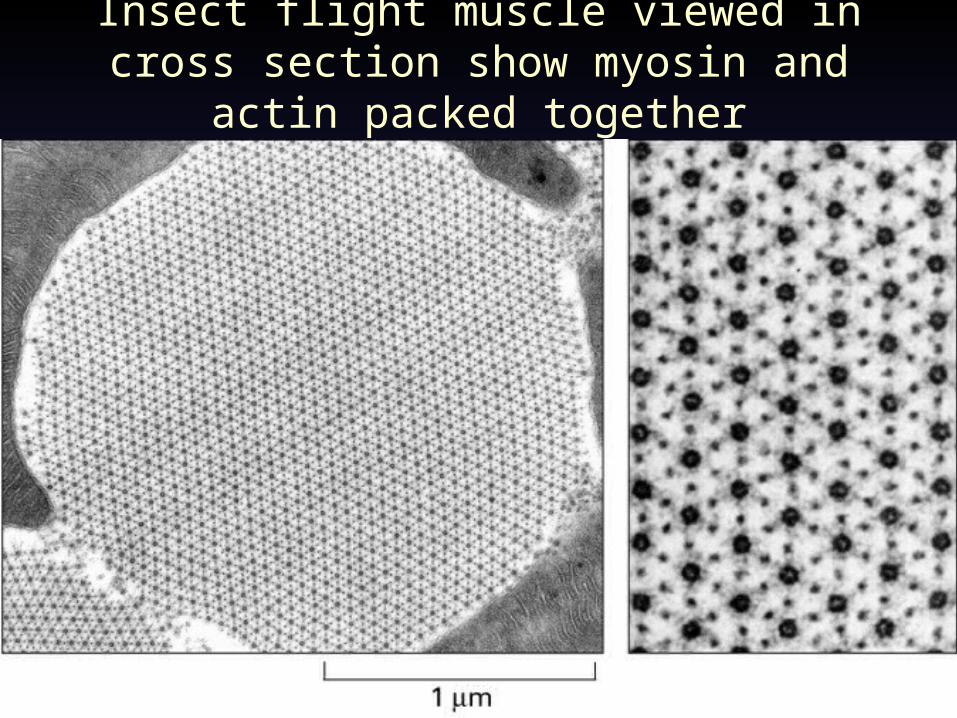
Insect flight muscle viewed in cross section show myosin and actin packed together
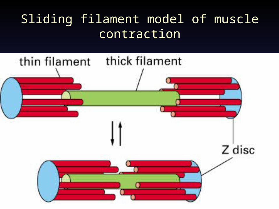
Sliding filament model of muscle contraction
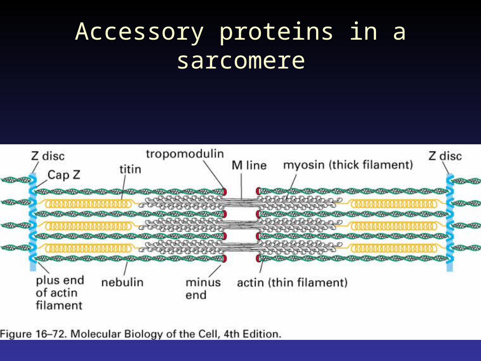
Accessory proteins in a sarcomere

Ion Channels at Neuromuscular Junction
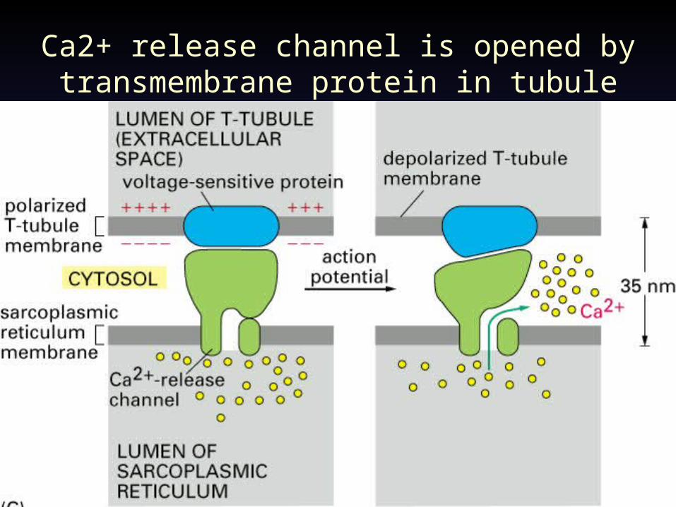
Ca2+ release channel is opened by transmembrane protein in tubule
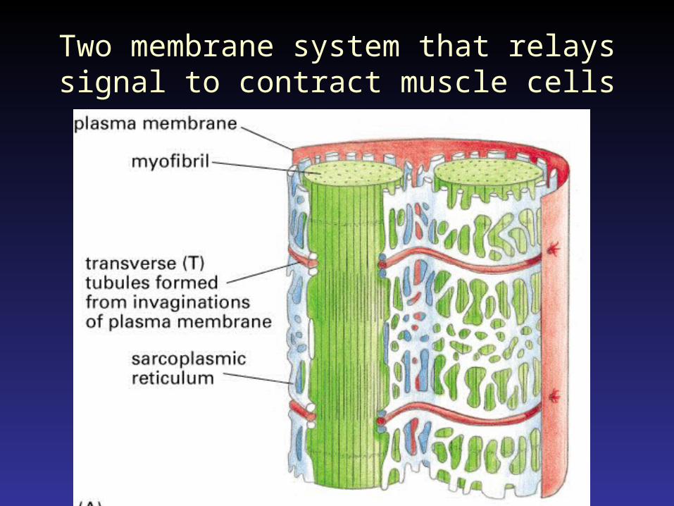
Two membrane system that relays signal to contract muscle cells
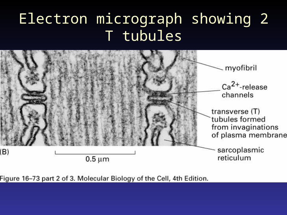
Electron micrograph showing 2 T tubules
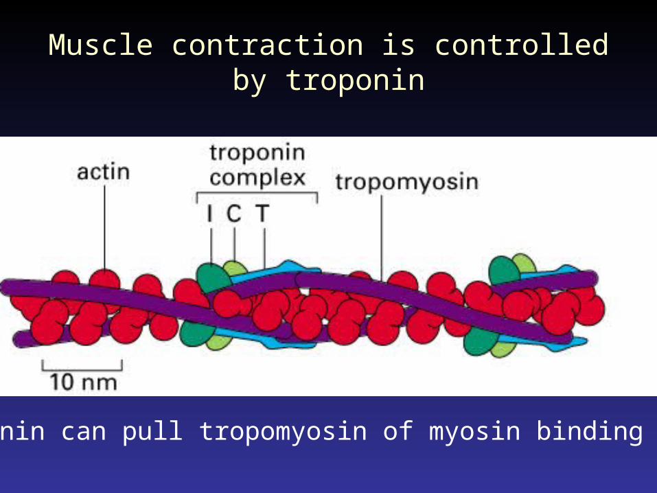
Muscle contraction is controlled by troponin
troponin can pull tropomyosin of myosin binding sites
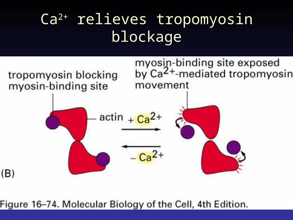
Ca2+ relieves tropomyosin blockage
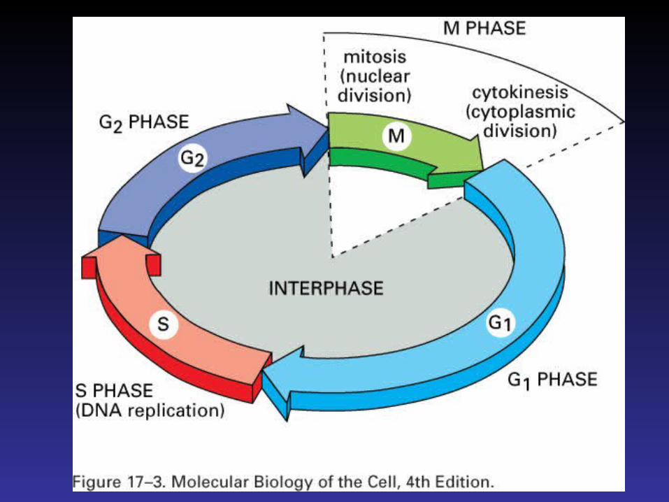
Cell Cycle and Programmed Cell Death
Chapter 17
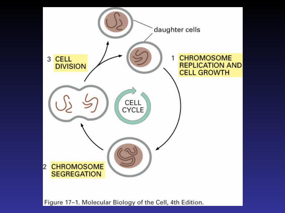


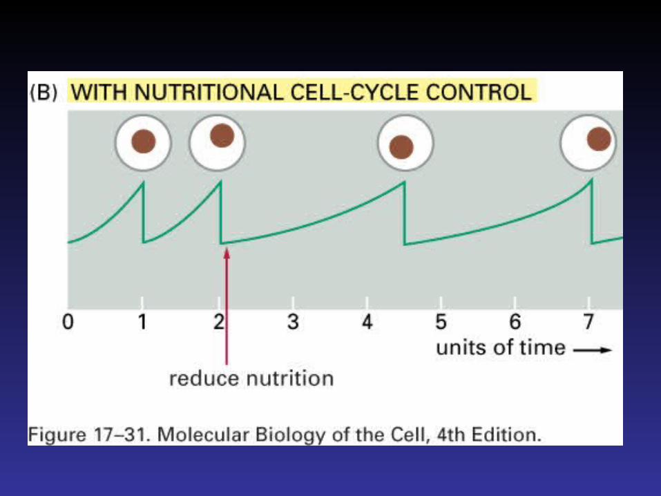
The cell cycle can be studied in a cell free system
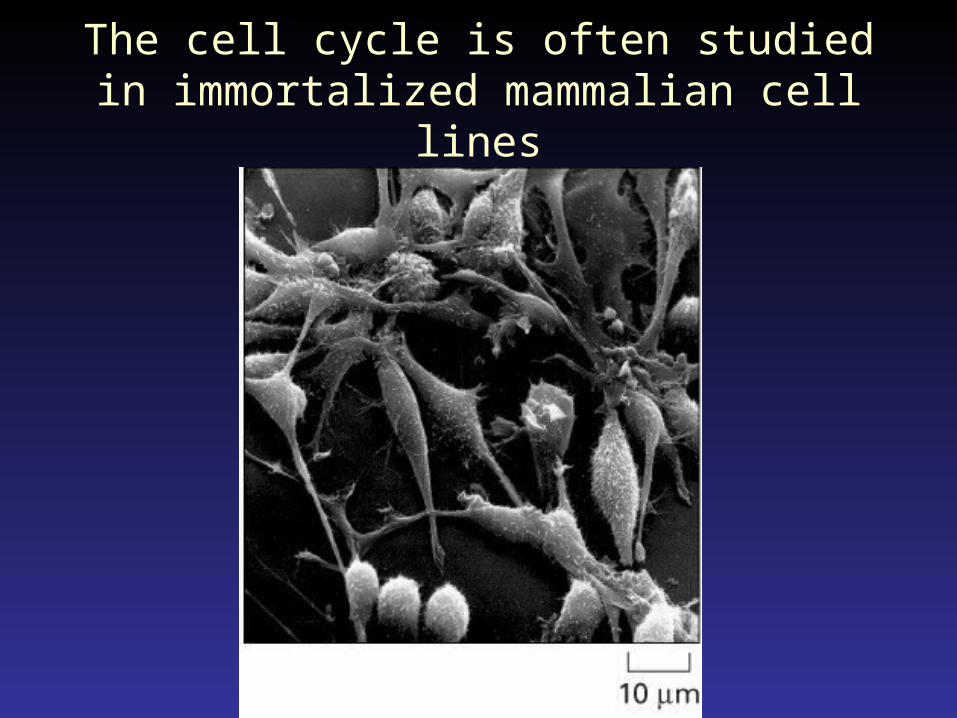
The cell cycle is often studied in immortalized mammalian cell lines
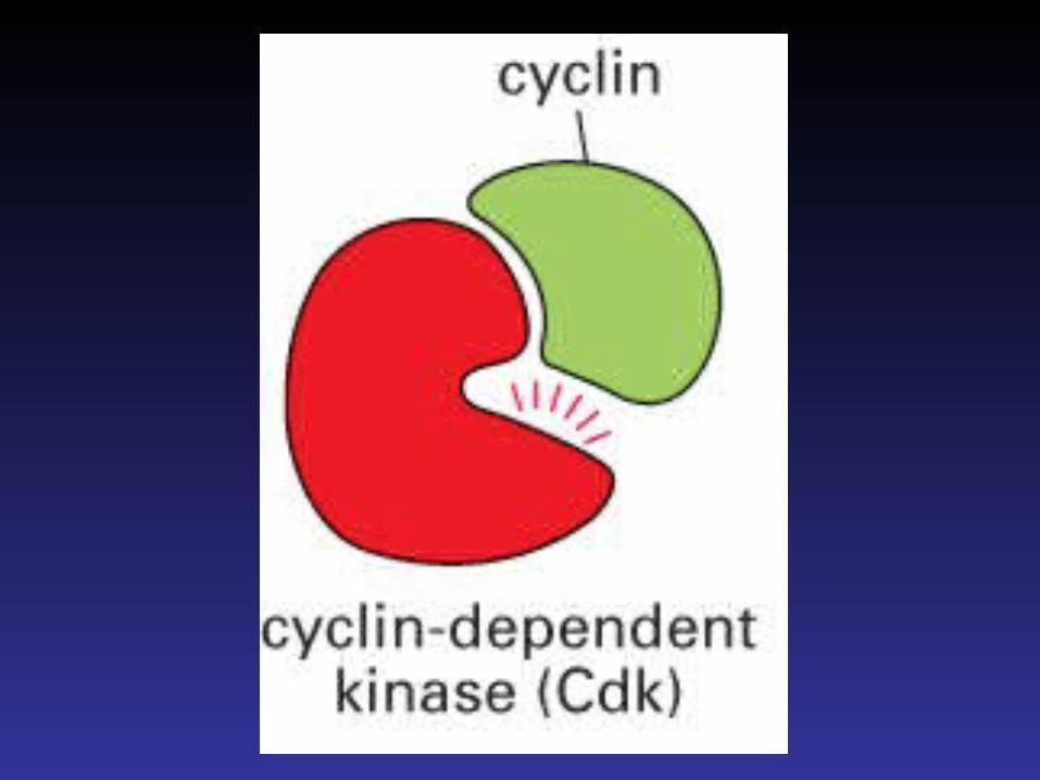
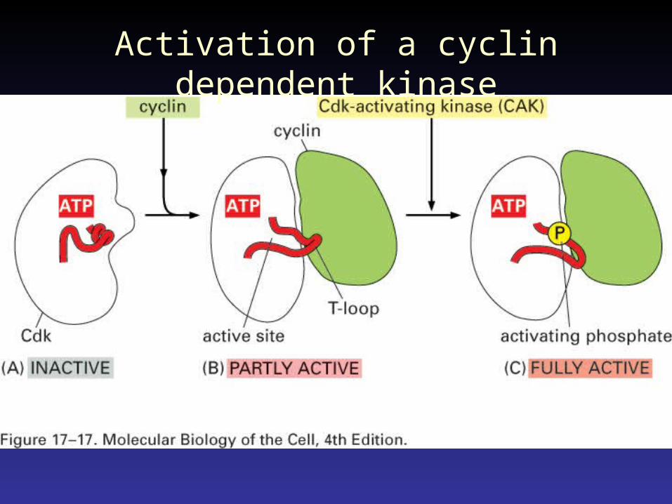
Activation of a cyclin dependent kinase
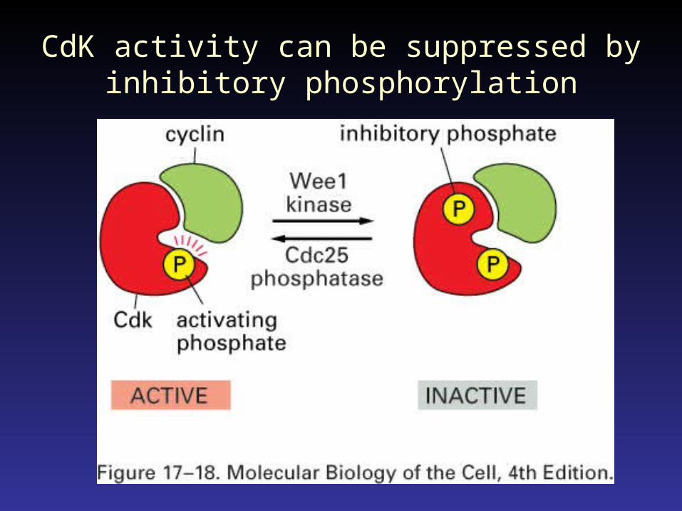
CdK activity can be suppressed by inhibitory phosphorylation
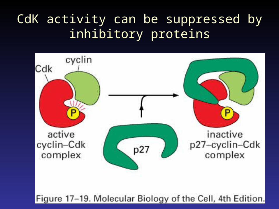
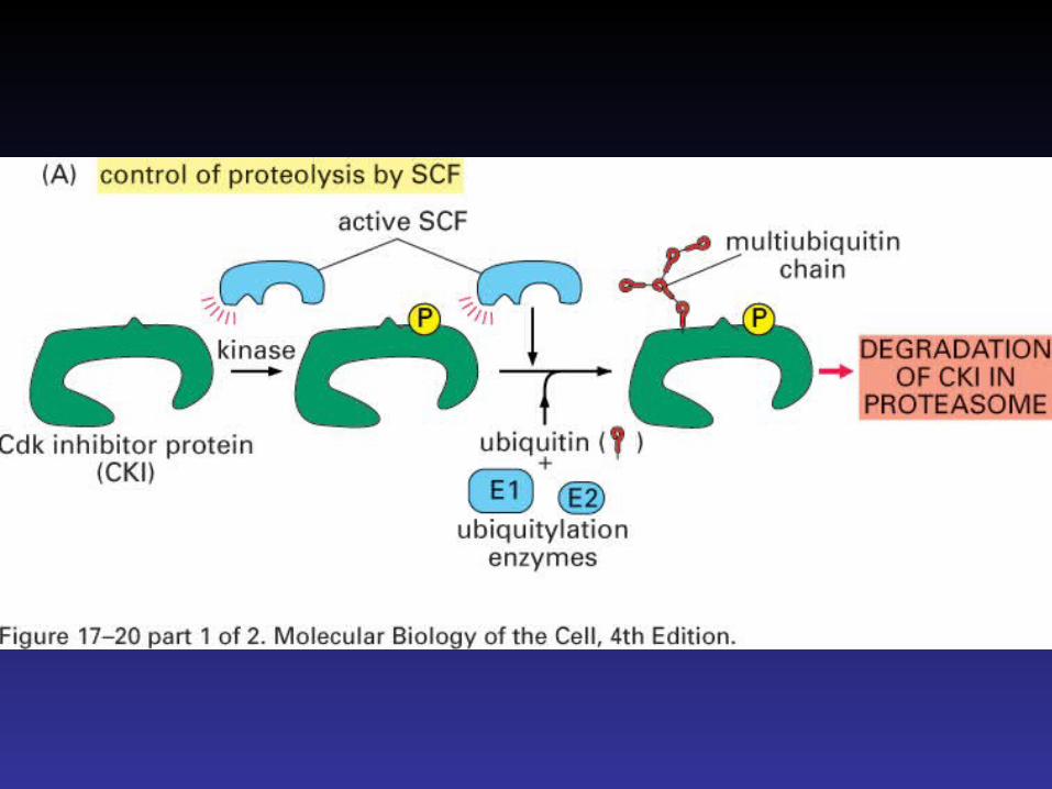
CdK activity can be suppressed by inhibitory proteins
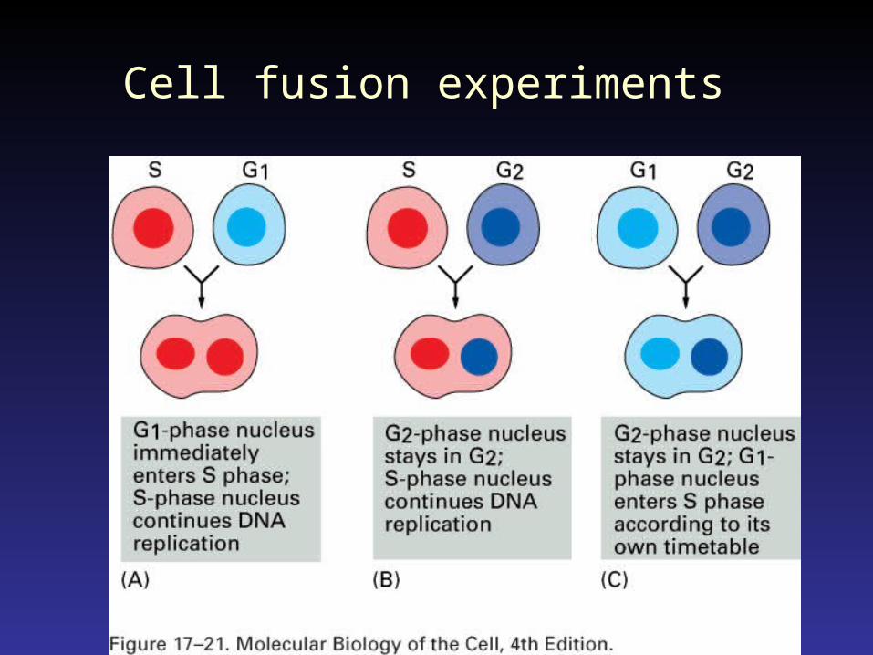
Cell fusion experiments
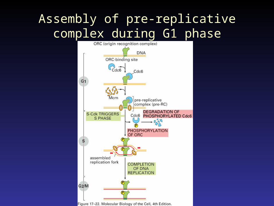
Assembly of pre-replicative complex during G1 phase
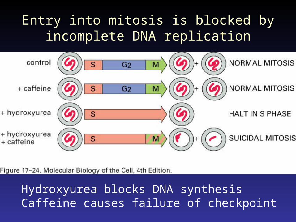
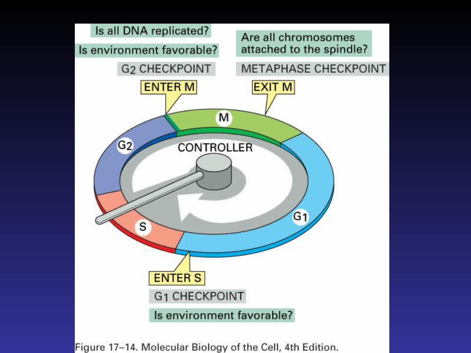
Entry into mitosis is blocked by incomplete DNA replication
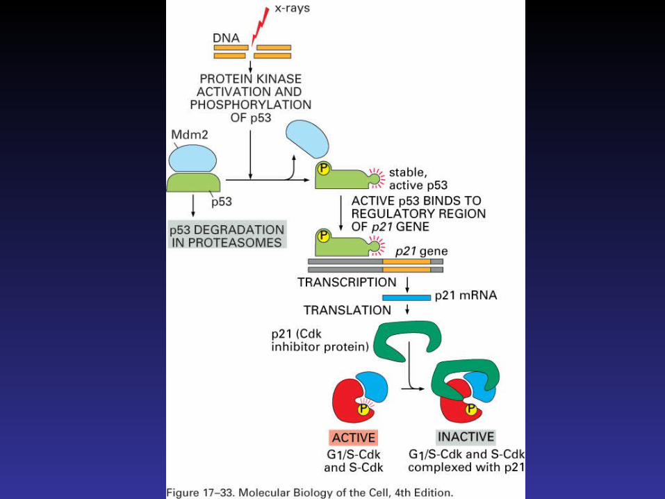
Hydroxyurea blocks DNA synthesisCaffeine causes failure of checkpoint
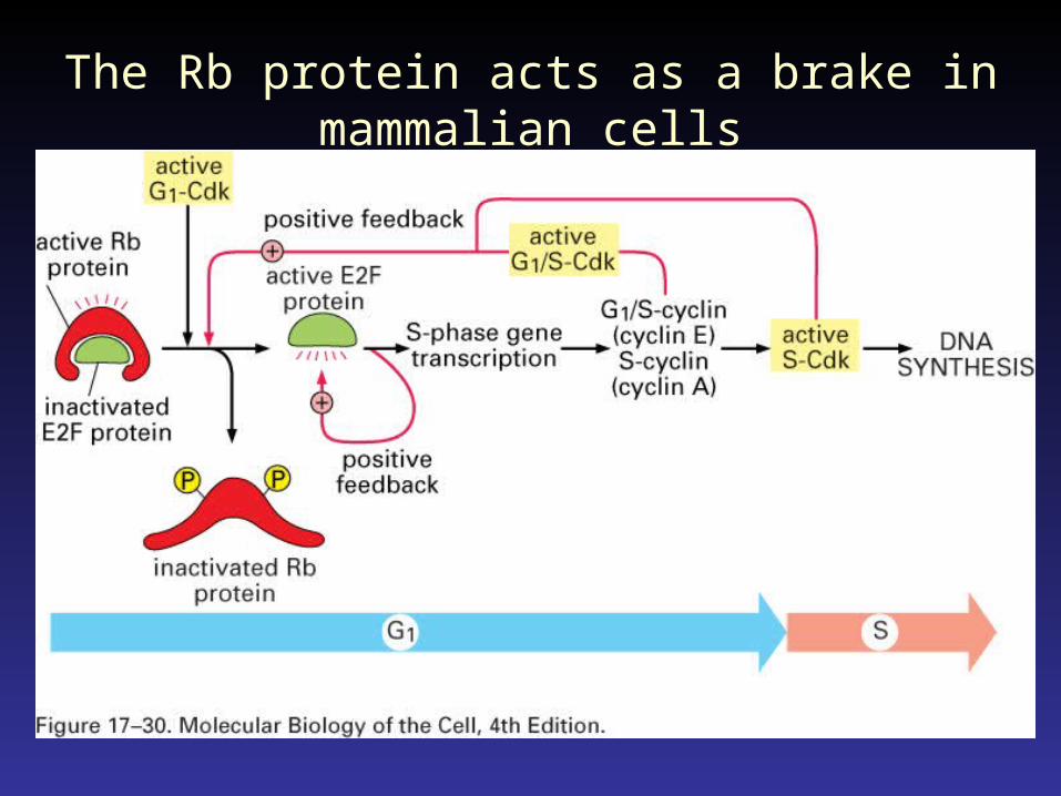
The Rb protein acts as a brake in mammalian cells
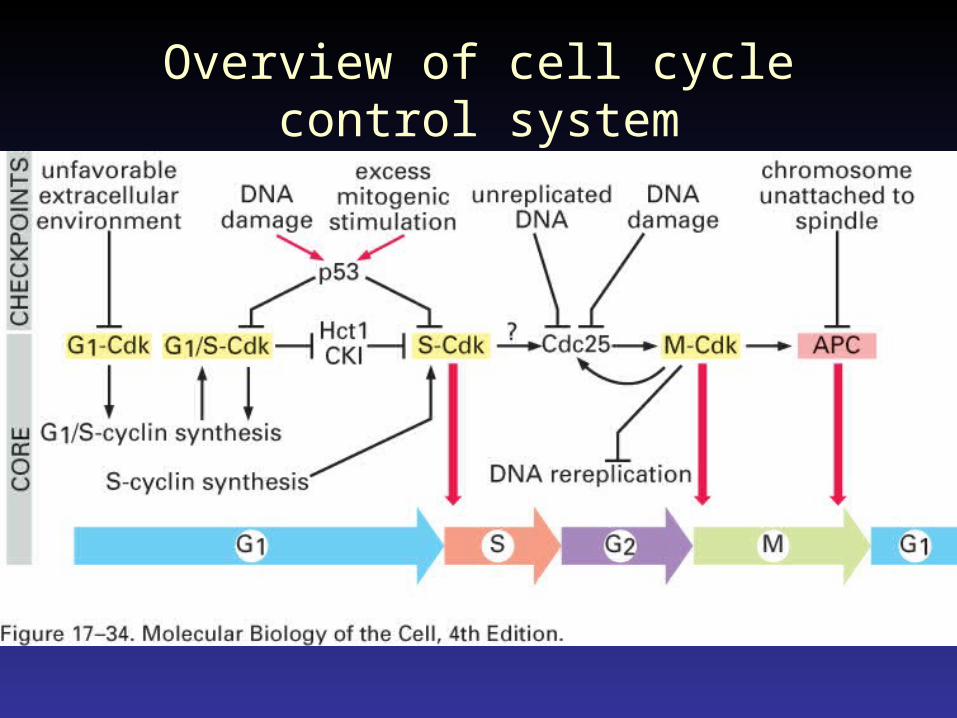
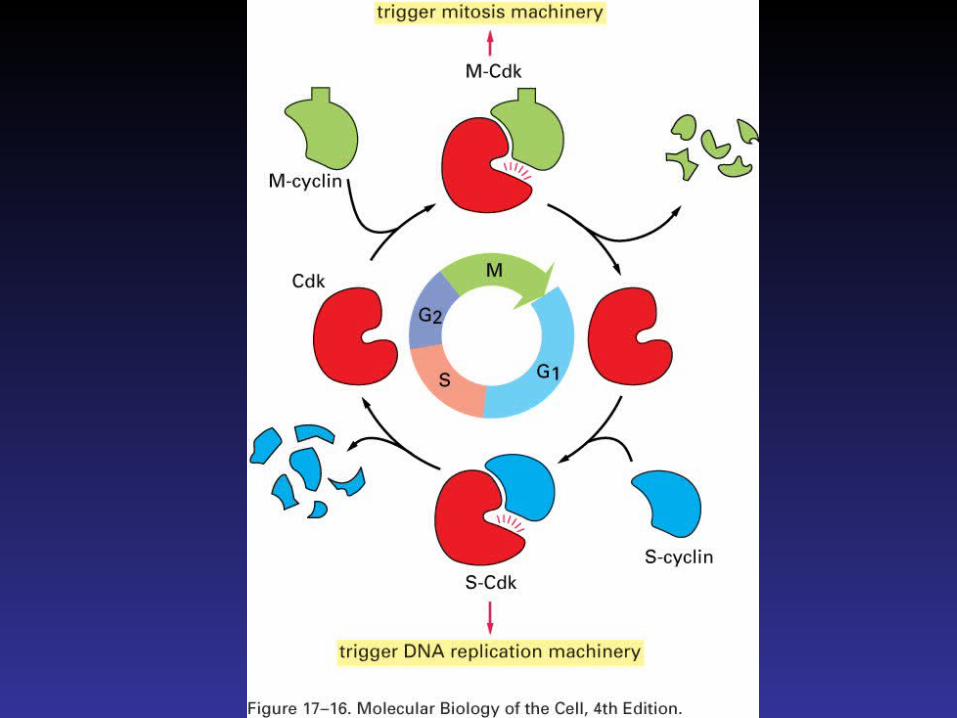
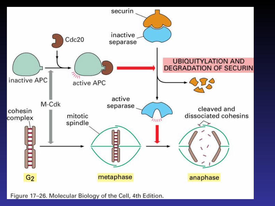
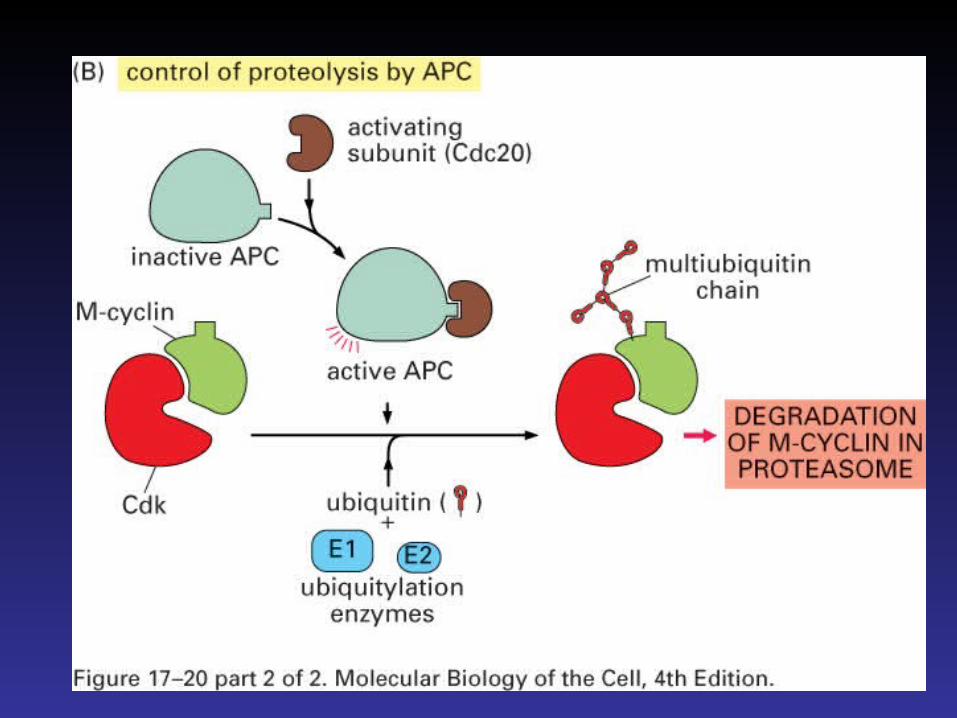
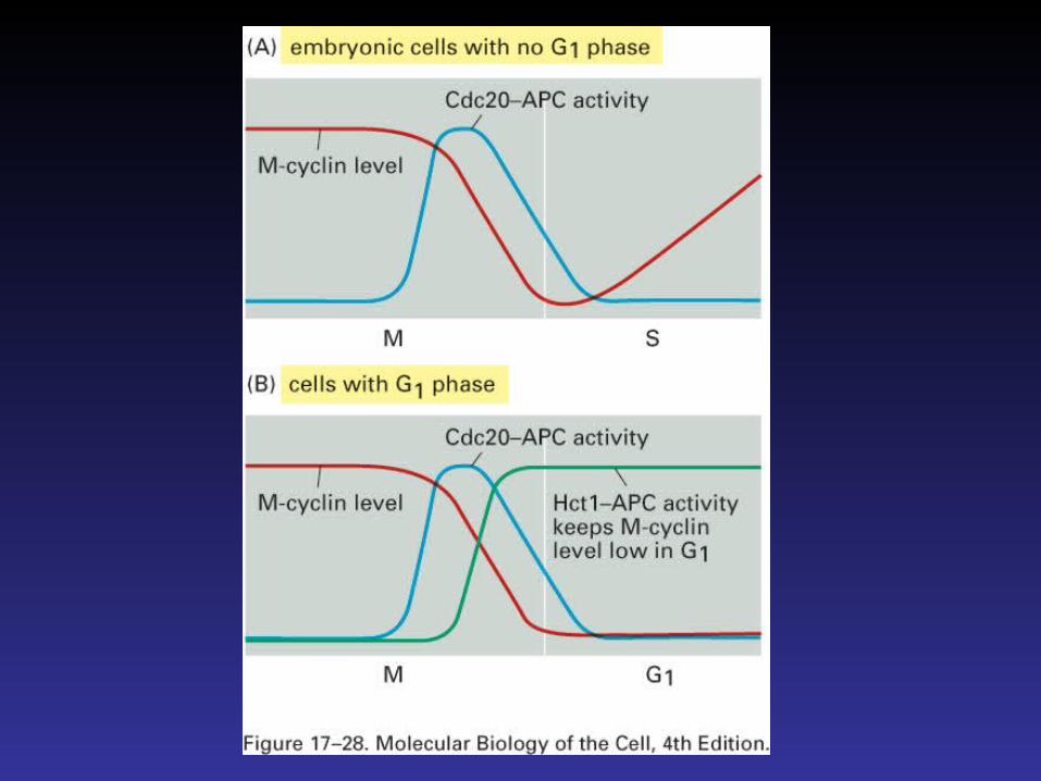
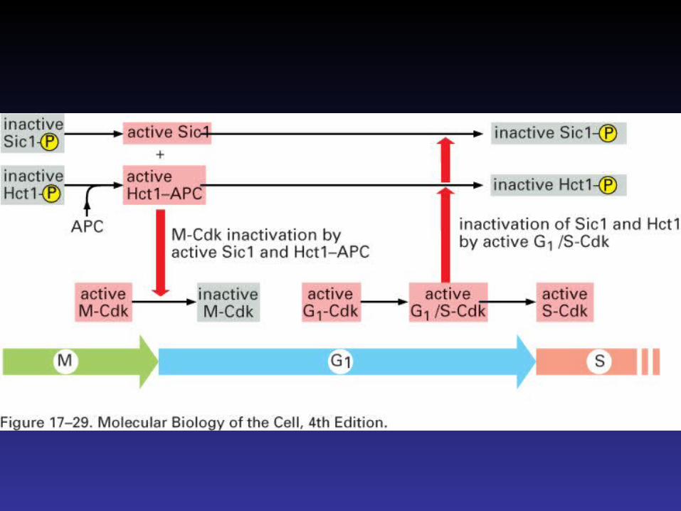
Overview of cell cycle control system