Embed Size (px)
Citation preview
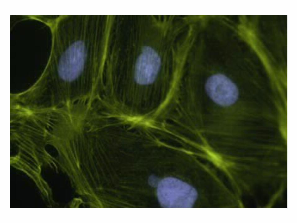
Cytoskeleton
Mobility of Cells
Crawling ameoba.mov
neutrophil chase.mov
Beating heart cell.mov
Adhesion between cells.mov
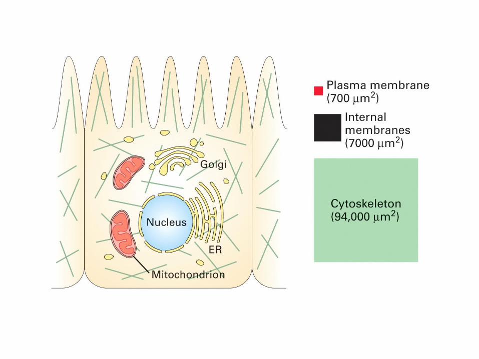
• The cytoskeleton is responsible for the organisation of the organelles inside a cell. It is the bulk of the cell and assists with cell motility, mitosis and meiosis, and maintaining the shape and stability of the cell. The cytoskeleton is made up of long fibres of polymers and subunits that form into one of the following:
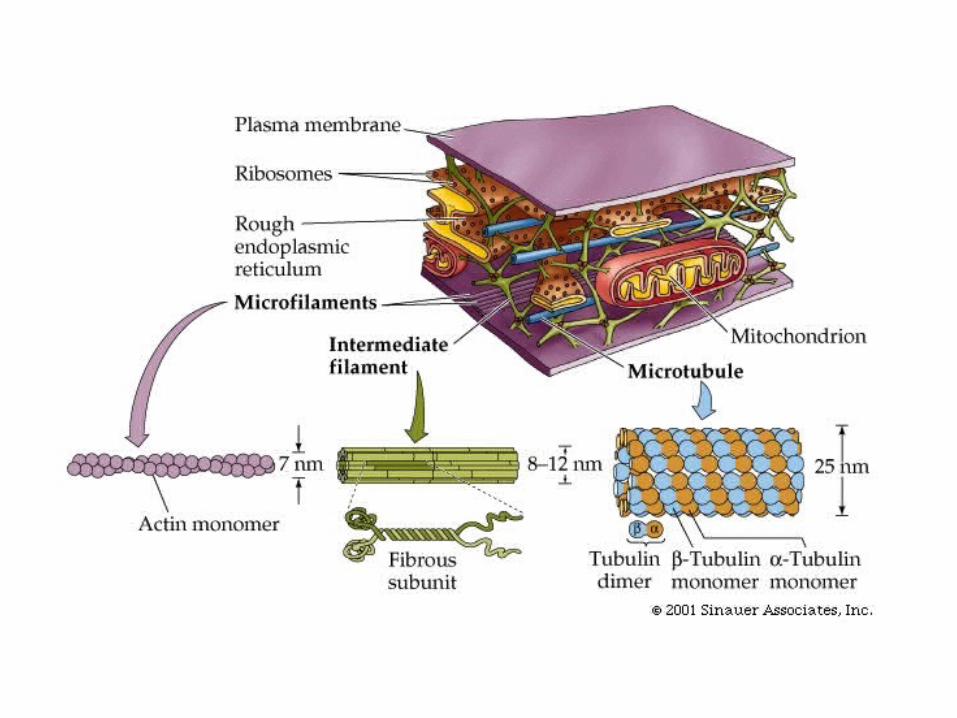
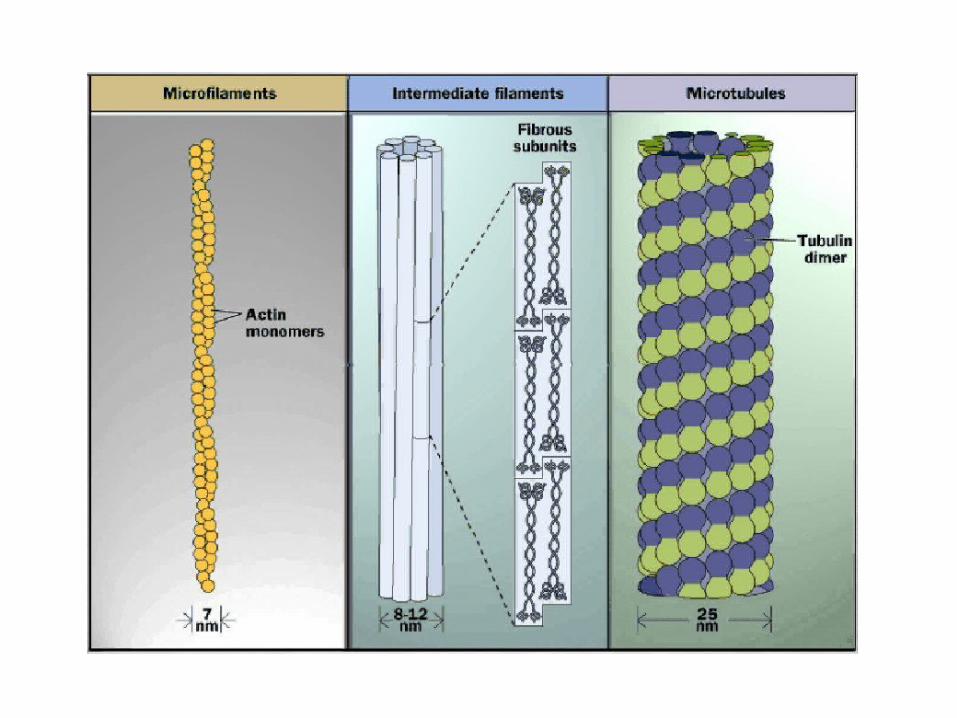
Microtubules Intermediate Filaments Microfilaments
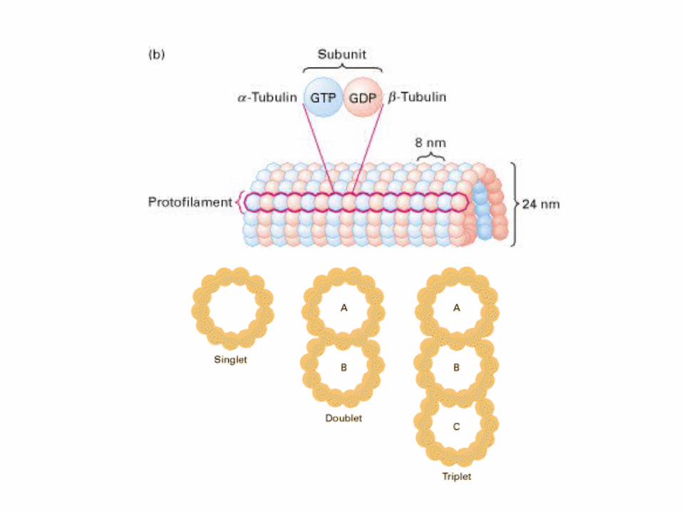
Microtubules• Microtubules are long, straight, hollow a
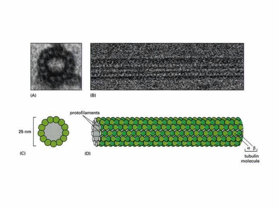
nd rigid cylinders of about 25nm in diameter.
• They are constructed of alternating identical subunits of alpha and beta protein tubulin, forming a protofilament.
• Its walls are made up of 13 protofilaments which correlate with one another laterally and the filament is able to increase or decrease in length by adding or removing proteins.
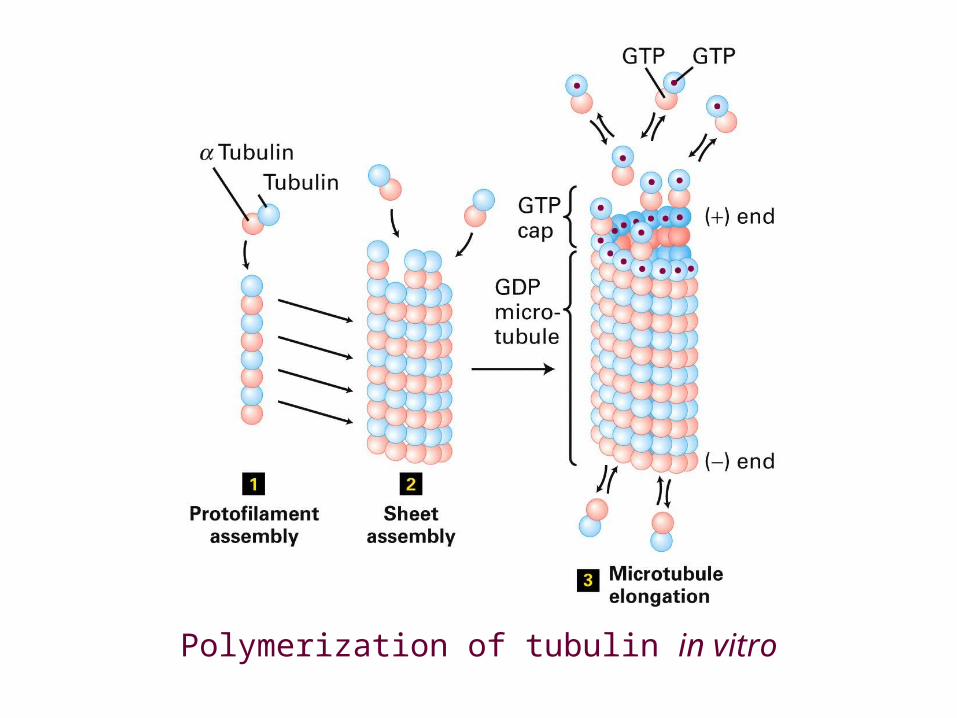
Polymerization of tubulin in vitro
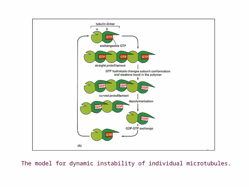
The model for dynamic instability of individual microtubules.
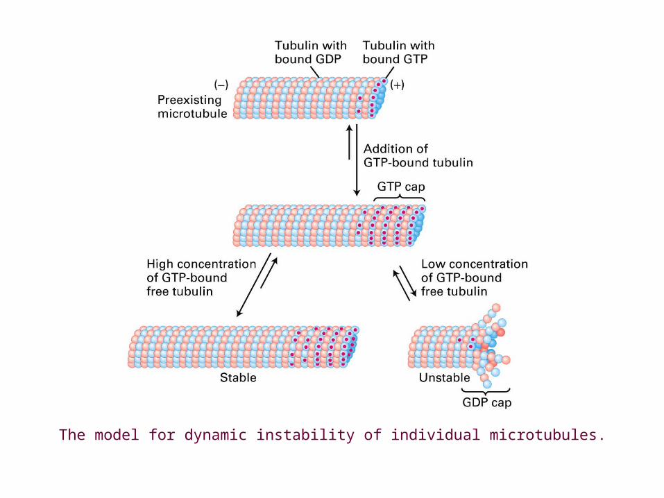
The model for dynamic instability of individual microtubules.
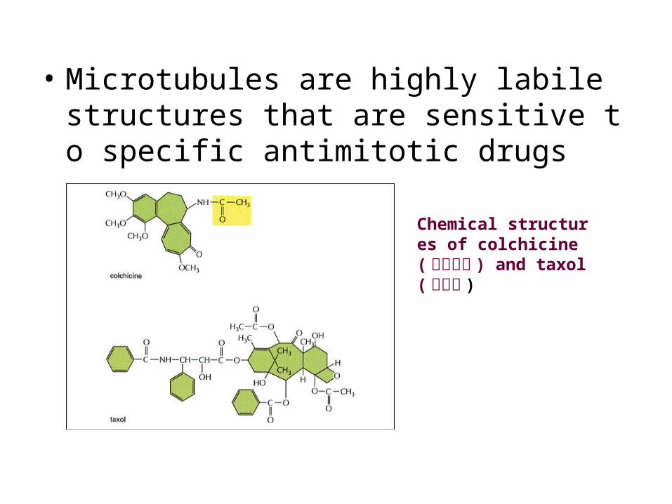
• Microtubules are highly labile structures that are sensitive to specific antimitotic drugs
Chemical structures of colchicine(秋水仙碱 ) and taxol(紫杉醇 )
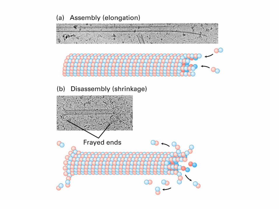
• Microtubules are dynamic structure which is seen in bundles and found in the cytoplasm of all eukaryotic cells. microtubule dynamic in vivo.mov
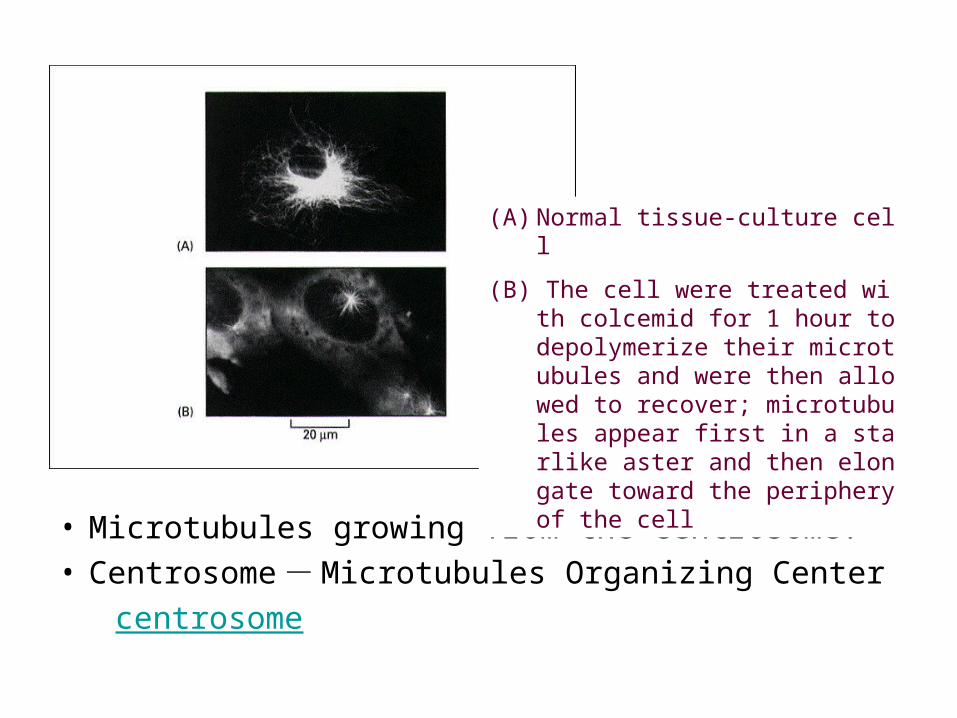
• Microtubules growing from the centrosome.• Centrosome - Microtubules Organizing Center centrosome
(A) Normal tissue-culture cell
(B) The cell were treated with colcemid for 1 hour to depolymerize their microtubules and were then allowed to recover; microtubules appear first in a starlike aster and then elongate toward the periphery of the cell
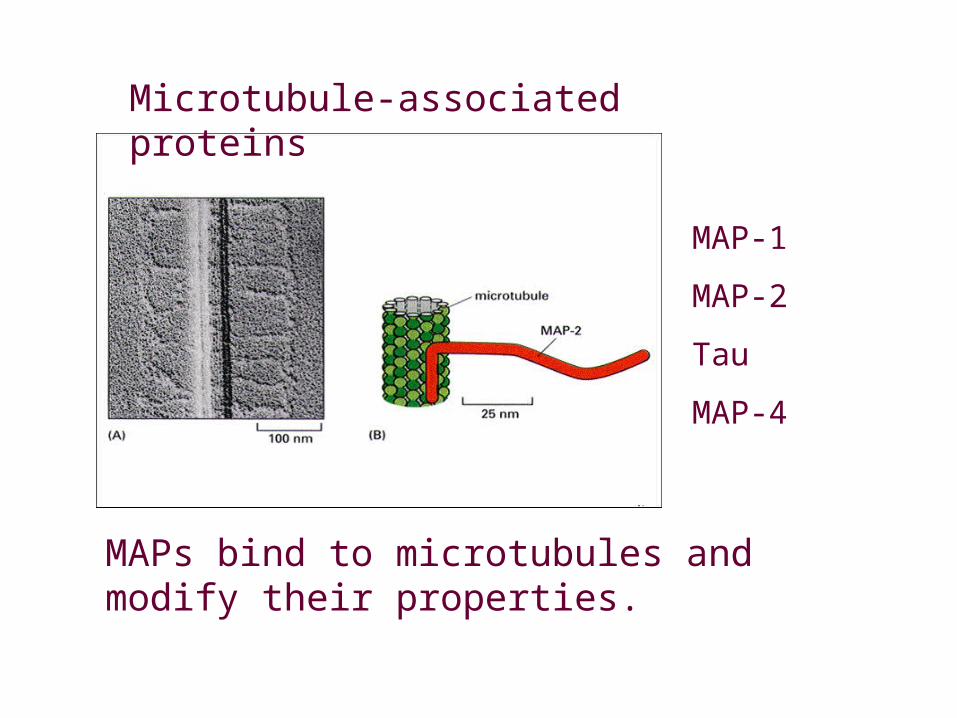
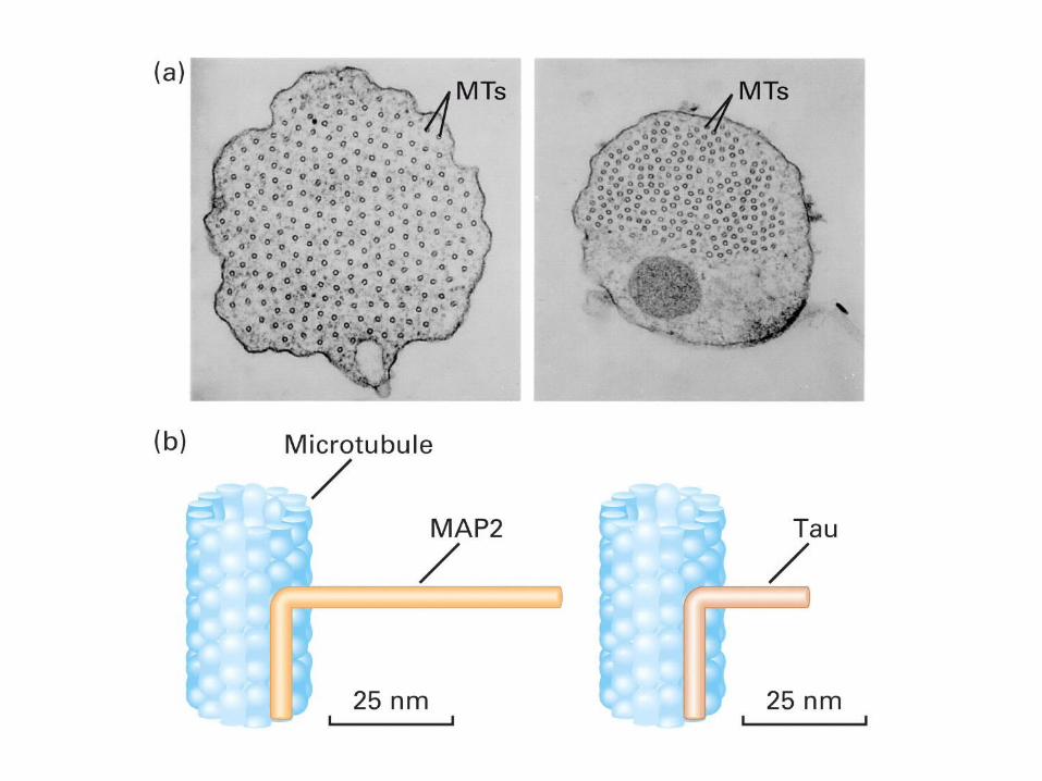
Microtubule-associated proteins
MAP-1
MAP-2
Tau
MAP-4
MAPs bind to microtubules and modify their properties.
Functions of Microtubules• Maintenance of the cell morphology• Cilia and flagella and centrioles are made fro
m microtubule• Maintenance organelles position and organe
lles movement• Membrane vesicle and protein transport• Chromosome movement and regulation of
mitosis• Cell signal transduction
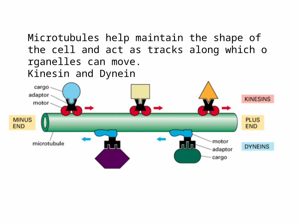
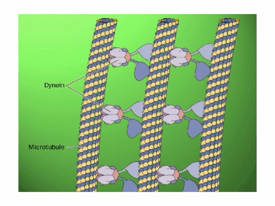
Microtubules help maintain the shape of the cell and act as tracks along which organelles can move.Kinesin and Dynein
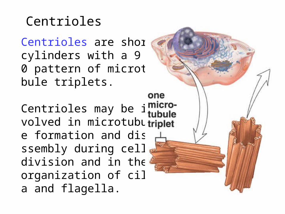
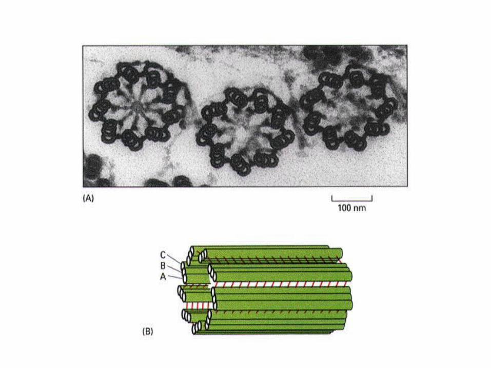
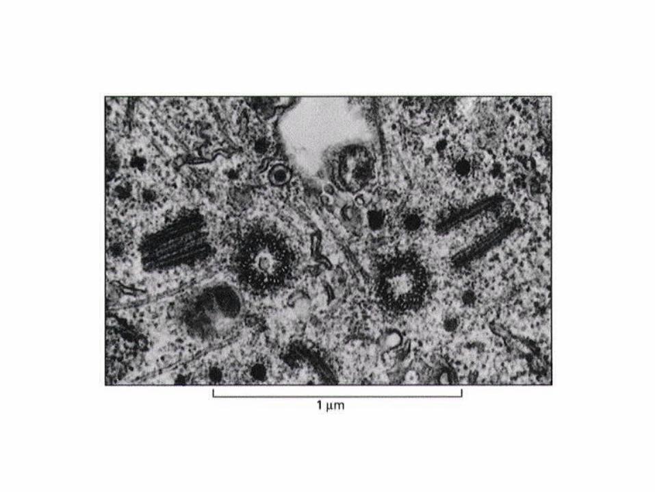
CentriolesCentrioles are short cylinders with a 9 + 0 pattern of microtubule triplets.
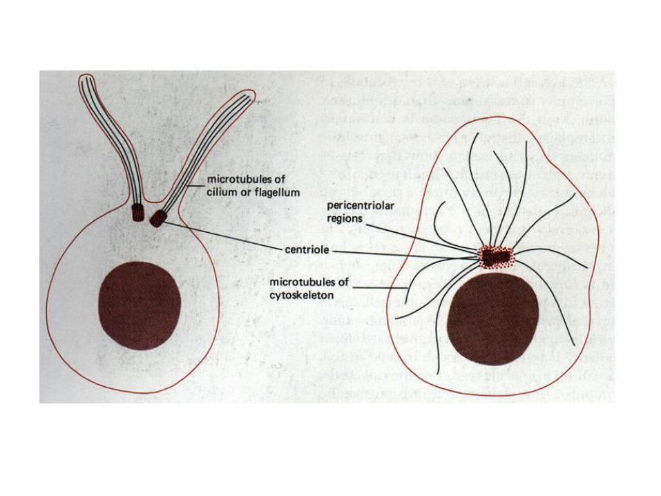
Centrioles may be involved in microtubule formation and disassembly during cell division and in the organization of cilia and flagella.
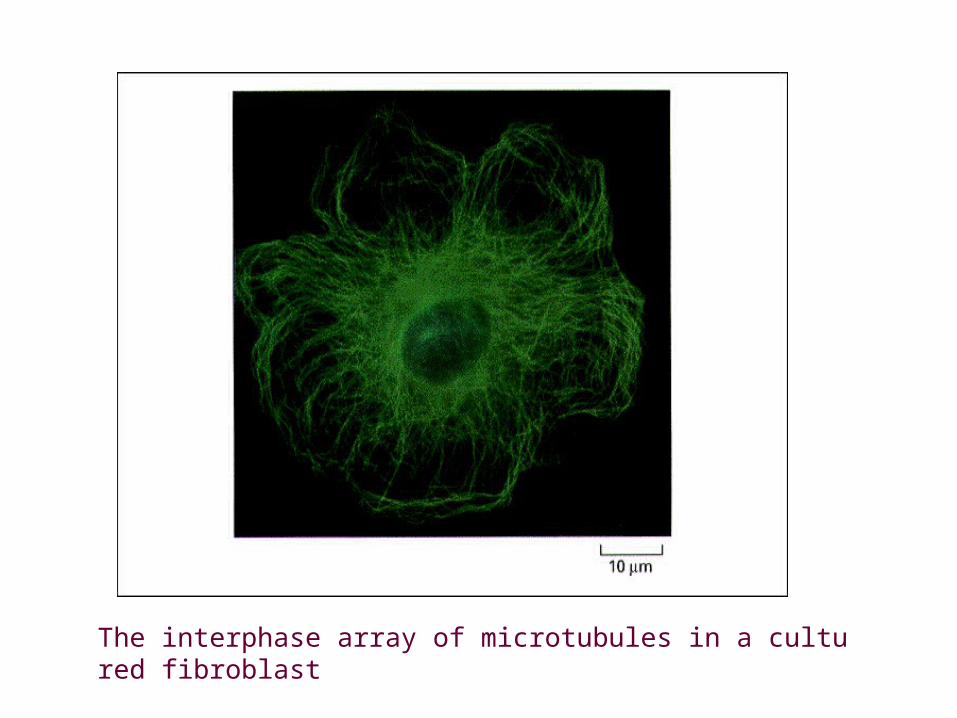
The interphase array of microtubules in a cultured fibroblast
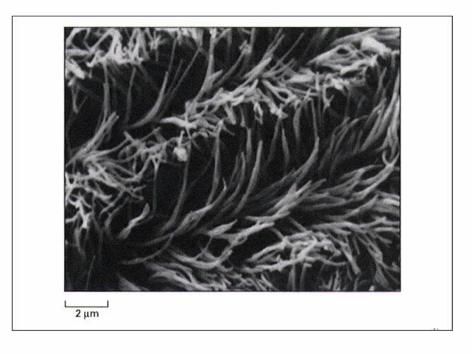
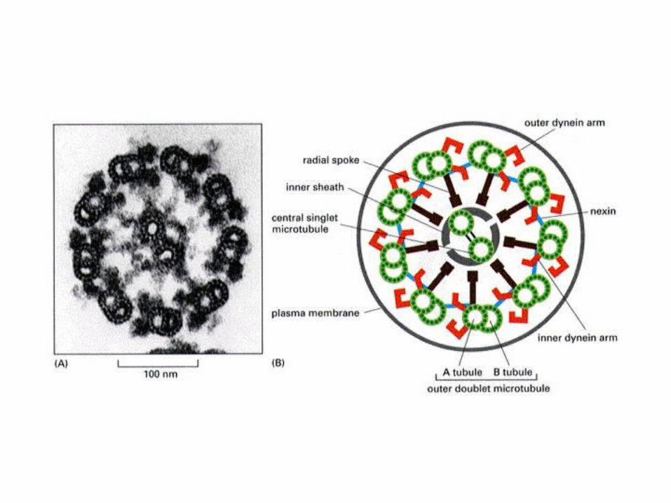
Cilia and flagella
Cilia (small and numerous) and flagella (large and single) have a 9 + 2 pattern of microtubules and are involved in cell movement.
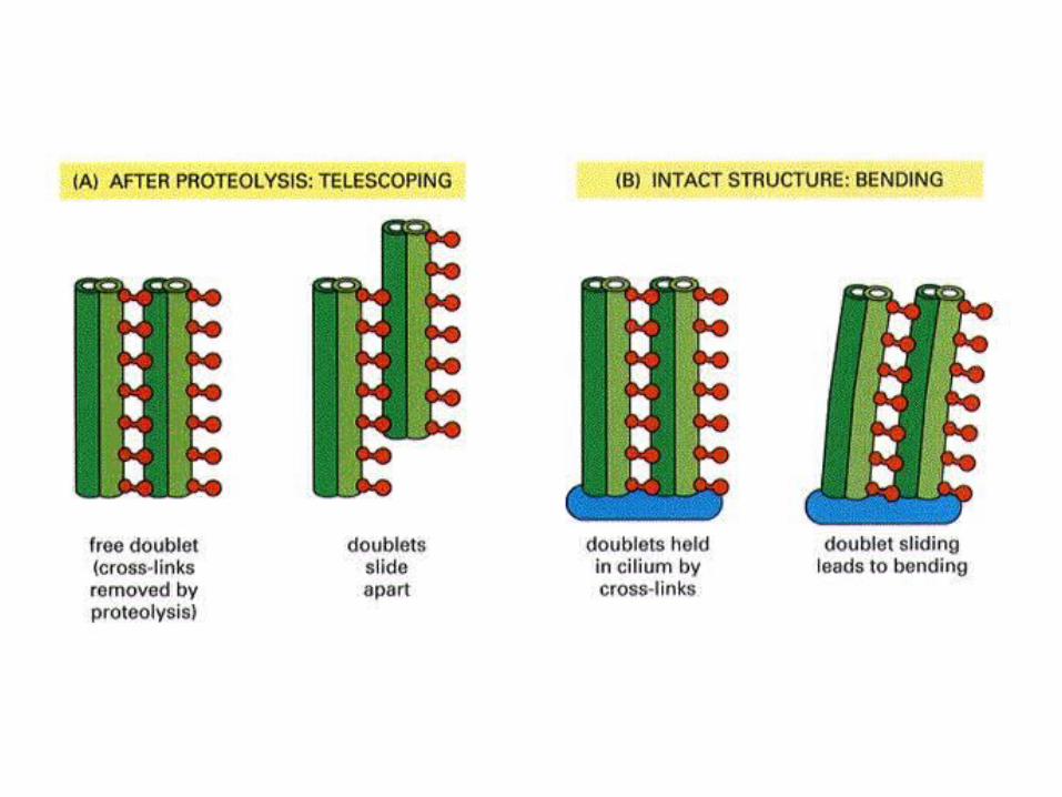
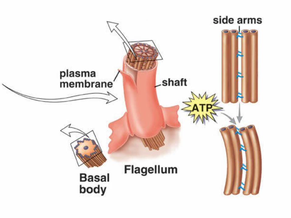
Cilia and flagella move when the microtubule doublets slide past one another.
Each cilium and flagellum has a basal body at its base.

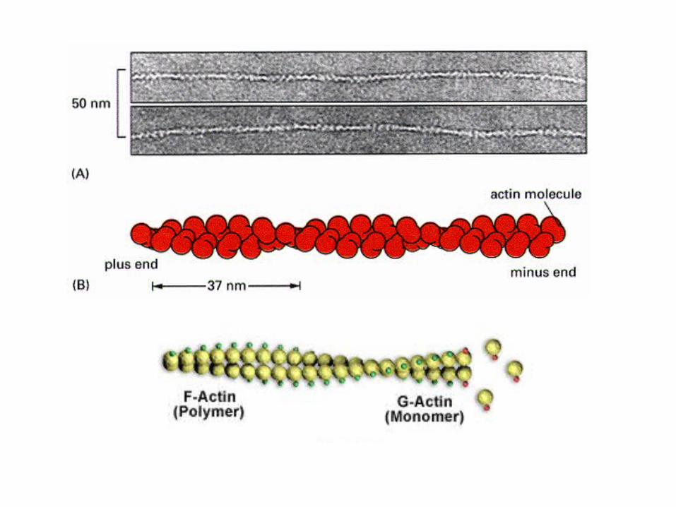
MicrofilamentsMicrofilaments• Microfilaments are left-handed helices f
ormed by two-strands of F-actin (fibrinous-actin) polymers.These are composed of rough spherical G-actin (globular-actin) monomers.
• They are the thinnest of the three cytoskeleton filaments and are about 8nm in diameter.
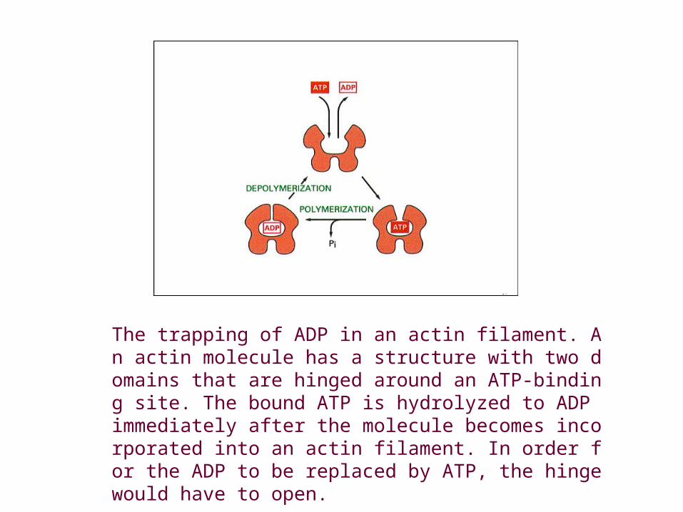
The trapping of ADP in an actin filament. An actin molecule has a structure with two domains that are hinged around an ATP-binding site. The bound ATP is hydrolyzed to ADP immediately after the molecule becomes incorporated into an actin filament. In order for the ADP to be replaced by ATP, the hinge would have to open.
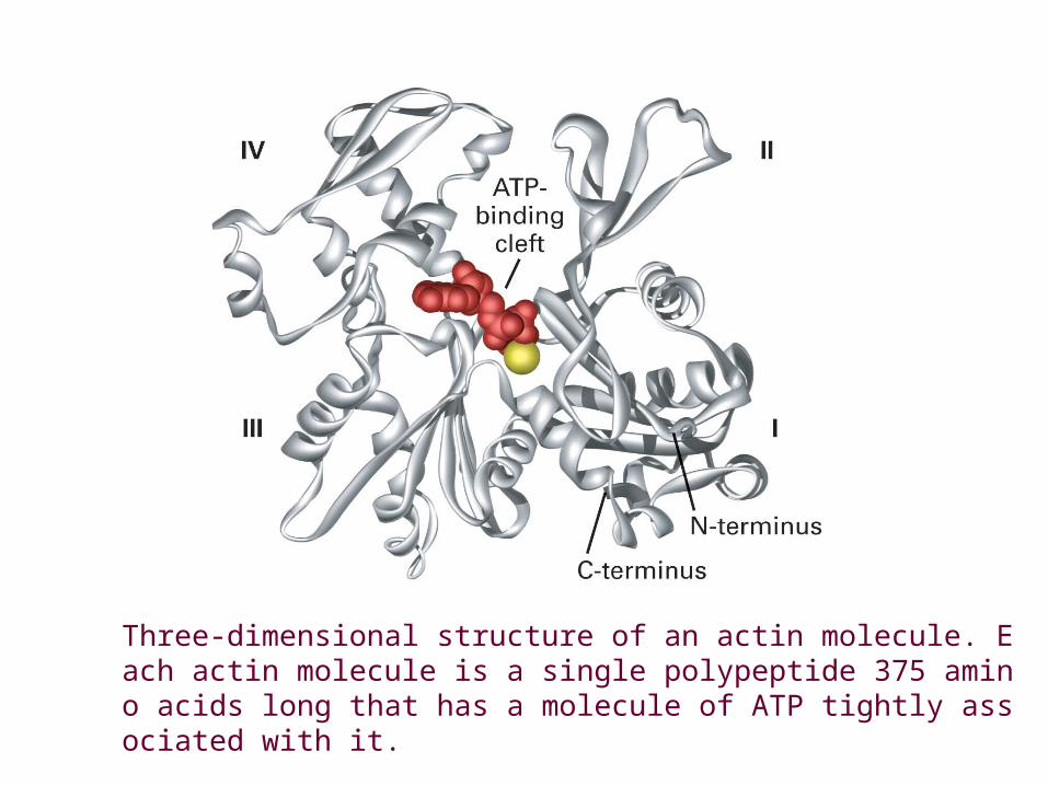
Three-dimensional structure of an actin molecule. Each actin molecule is a single polypeptide 375 amino acids long that has a molecule of ATP tightly associated with it.
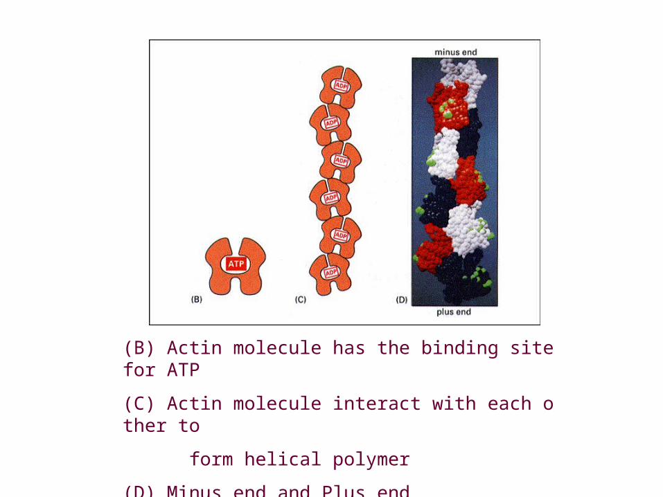
(B) Actin molecule has the binding site for ATP
(C) Actin molecule interact with each other to
form helical polymer
(D) Minus end and Plus end
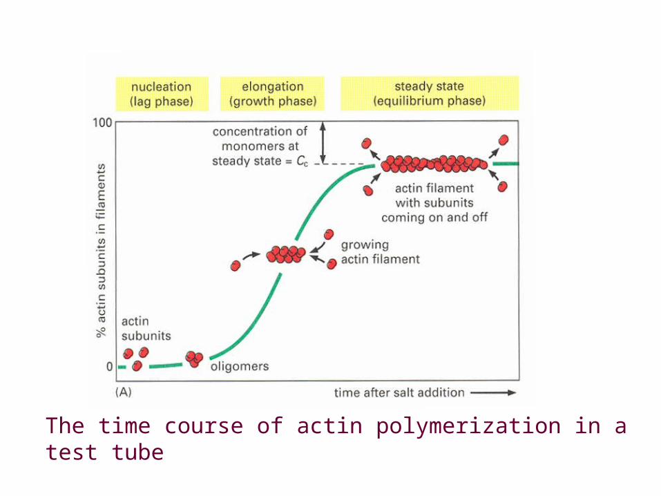
The time course of actin polymerization in a test tube
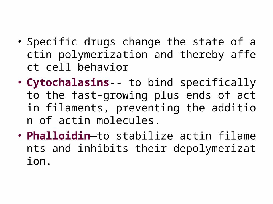
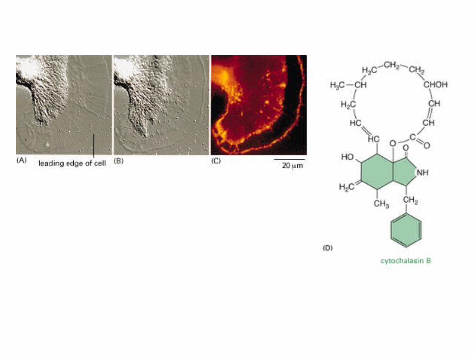
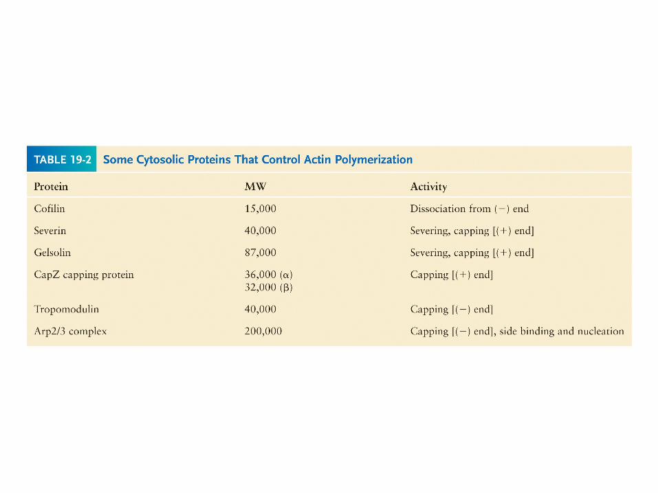
• Specific drugs change the state of actin polymerization and thereby affect cell behavior
• Cytochalasins-- to bind specifically to the fast-growing plus ends of actin filaments, preventing the addition of actin molecules.
• Phalloidin—to stabilize actin filaments and inhibits their depolymerization.
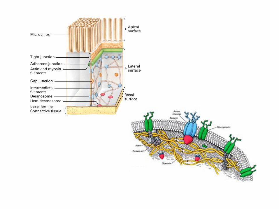
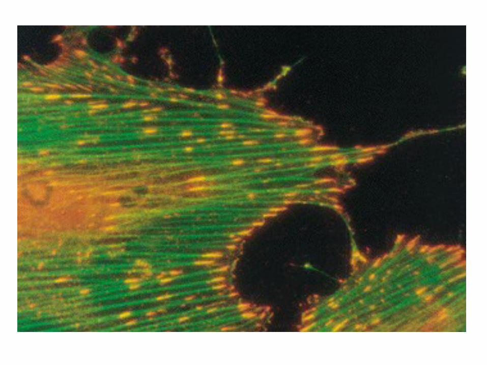
• The bulk of these filaments are anchored to the cytoplasmic side of cell plasma membranes and its nearby structures. The rest is evenly distributed throughout the cell.
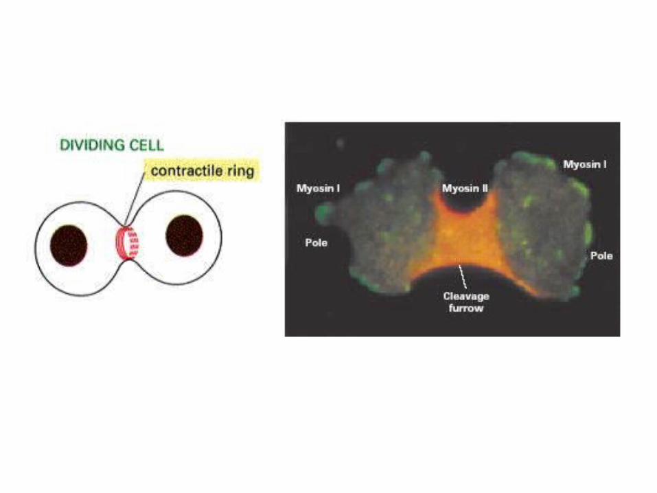
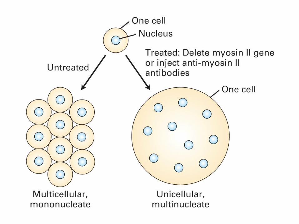
Functions of microfilaments• Maintenance of the cell shape• Muscle contraction • Cell division-contractile ring • Cell movements-cyclosis, ruffled membrane
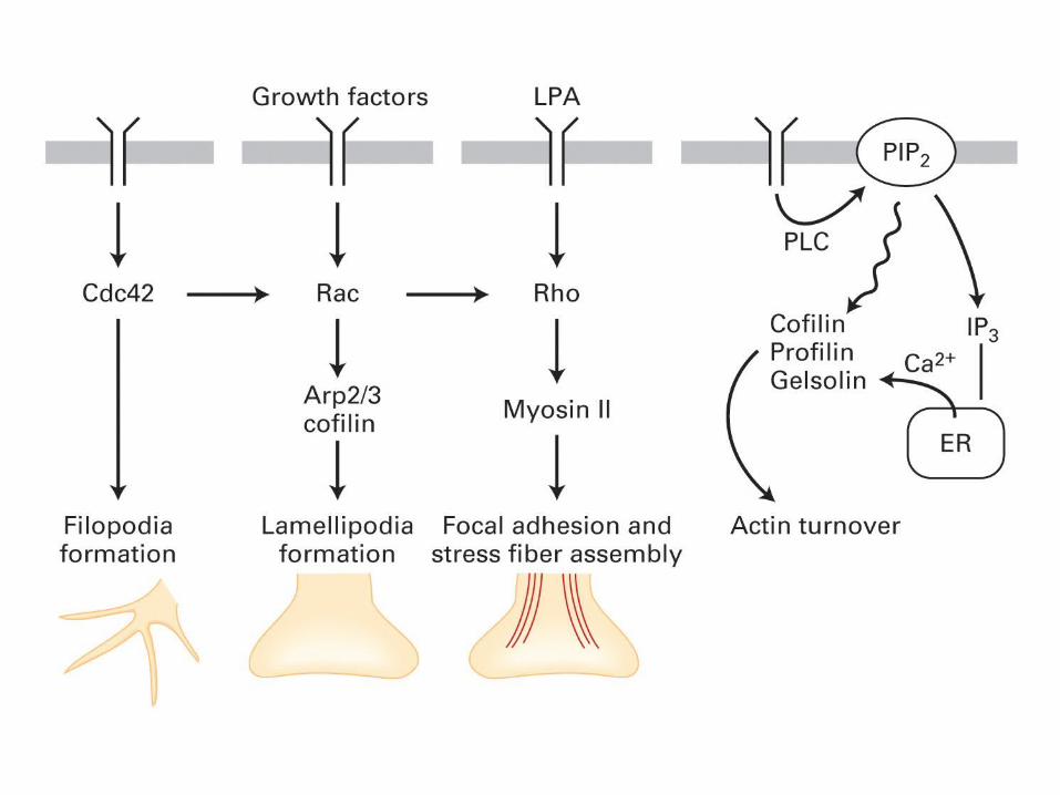
locomotion, phagocytosis• Cell signal transduction: Rho-GTPase• Membrane vesicle and protein transport
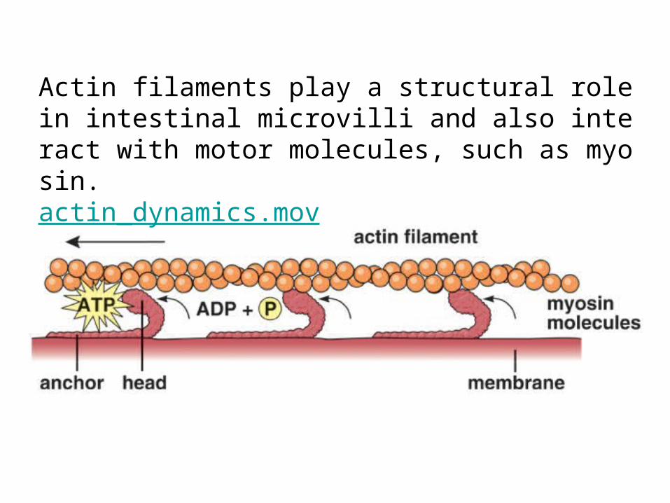
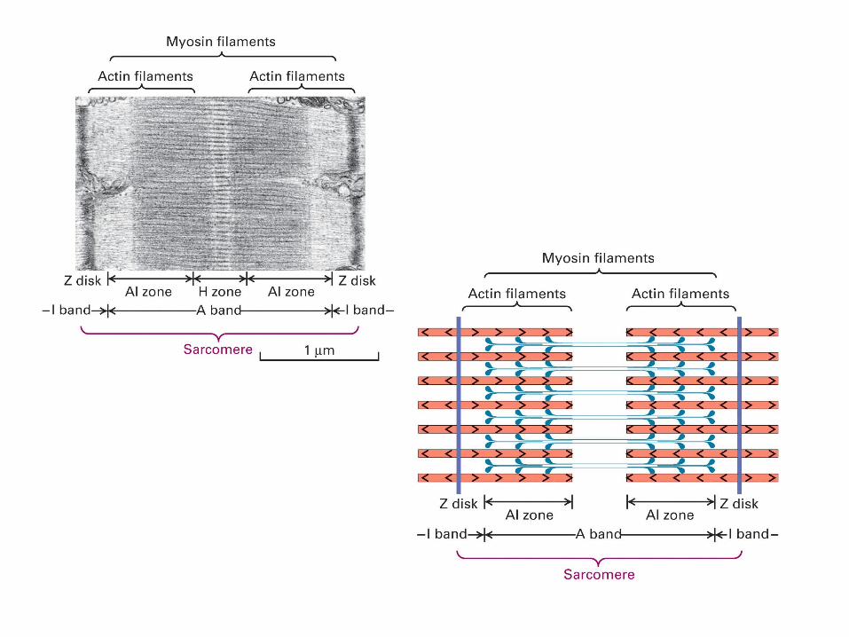
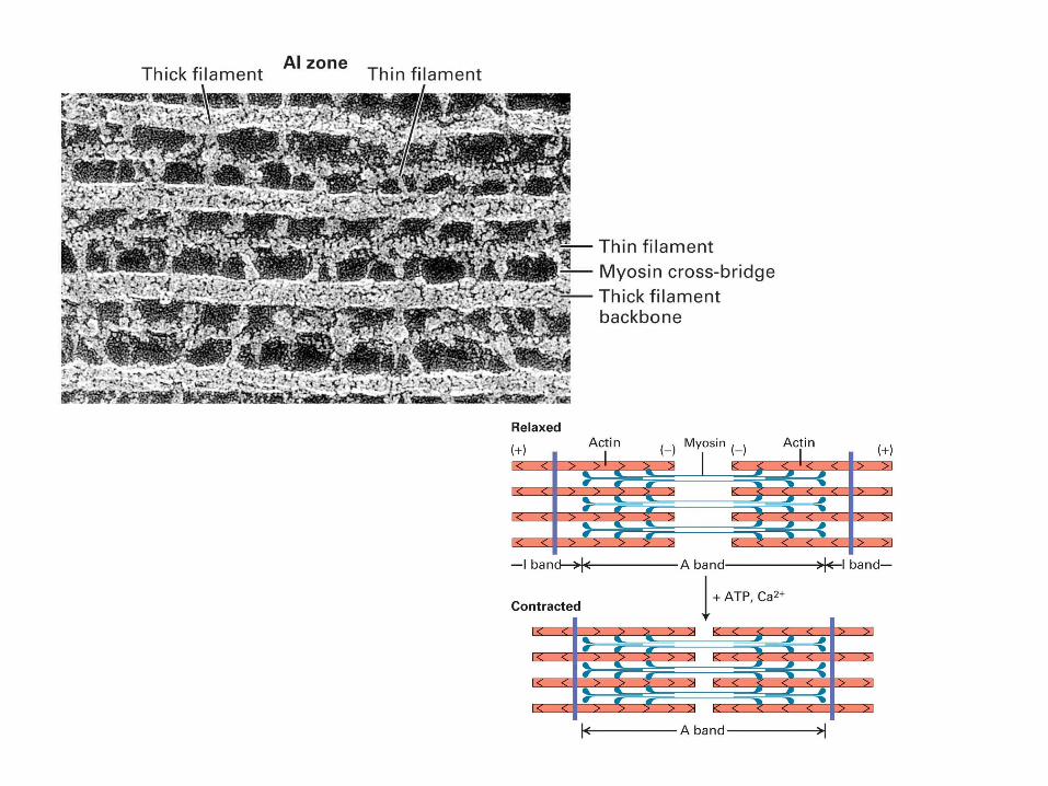
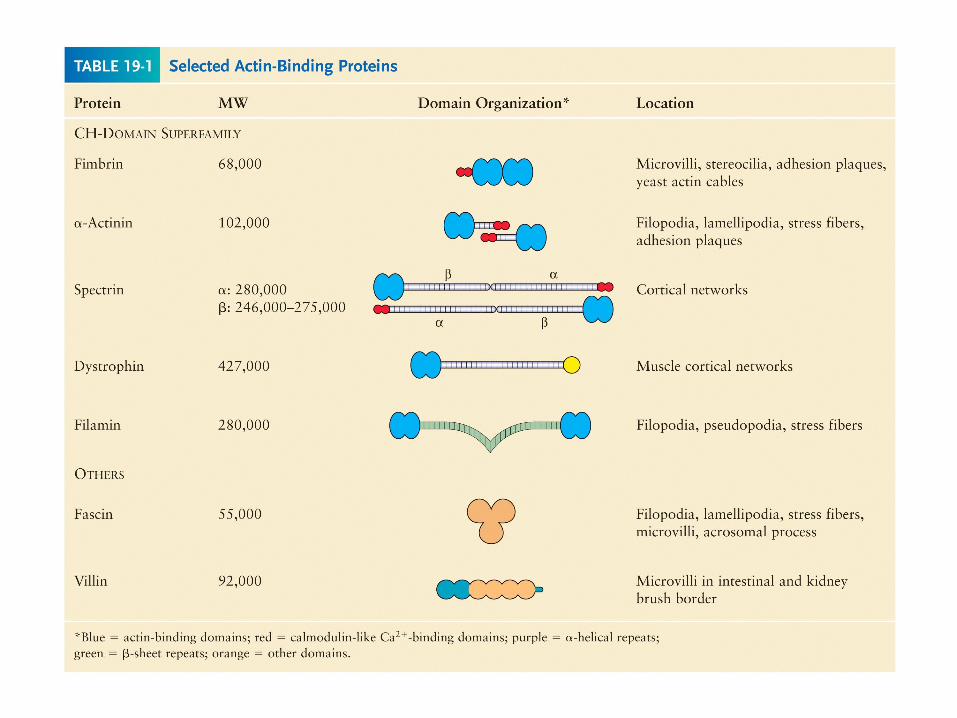
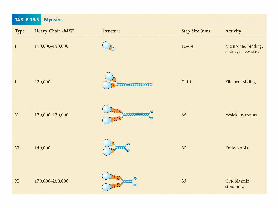
Actin filaments play a structural role in intestinal microvilli and also interact with motor molecules, such as myosin.actin_dynamics.mov
• Actin filaments can form both stable and labile structures in cells.
• Stable actin filaments form the core of microvilli ( 微绒毛 )and are a crucial component of the contractile apparatus of muscle cells. Many cell movements, however, depend on labile structures constructed from actin filaments.
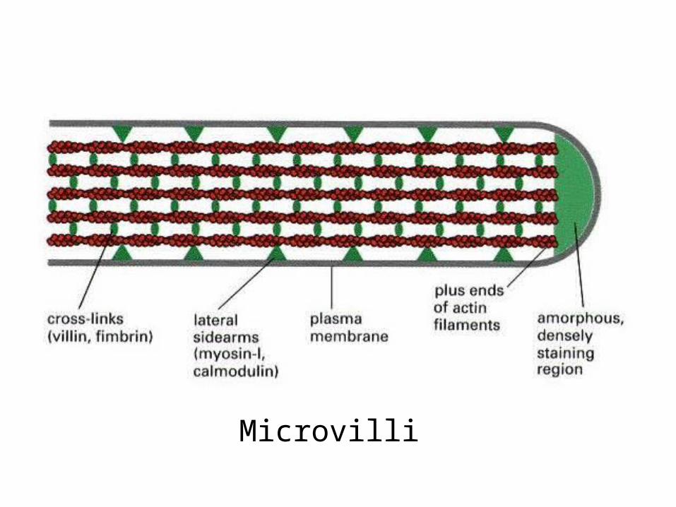
Microvilli

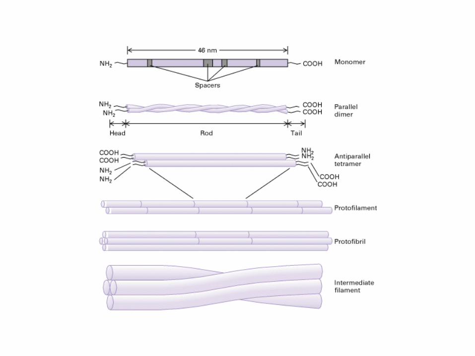
Intermediate FilamentsIntermediate Filaments• Intermediate filaments are rope like pol
ymers of fibrous polypeptides.• They are 10nm in diameter and are ther
efore intermediate in thickness between microfilaments and microtubules.
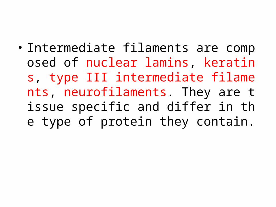
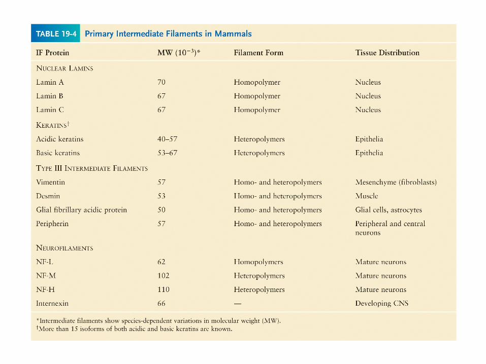
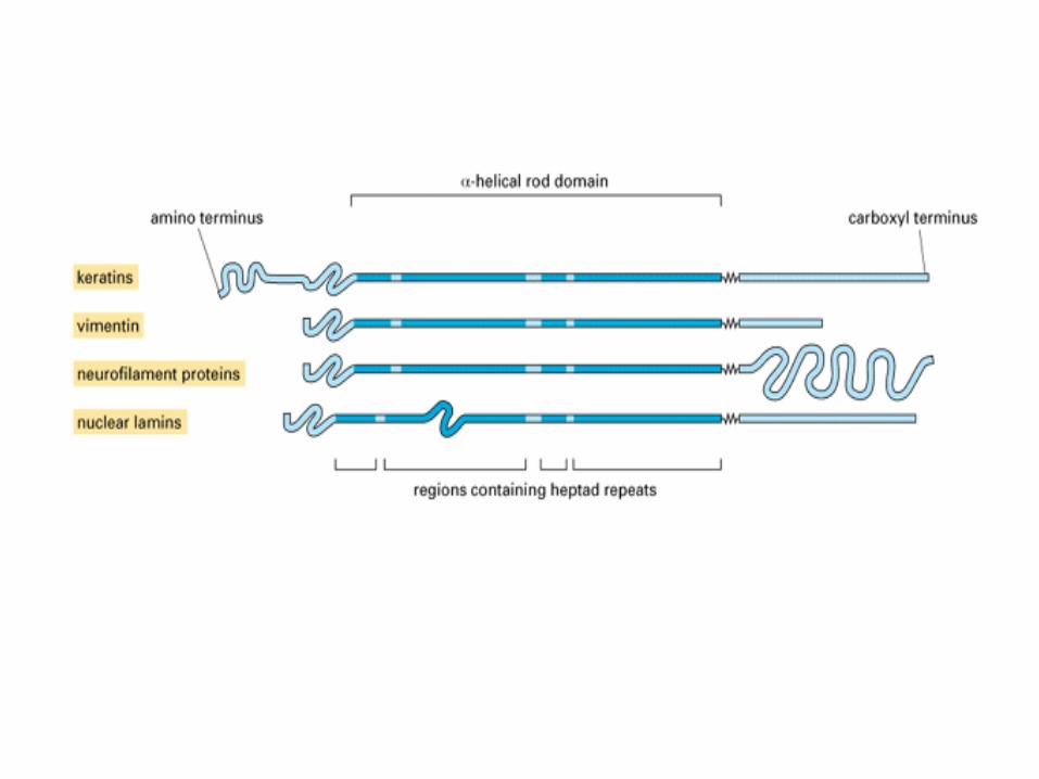
• Intermediate filaments are composed of nuclear lamins, keratins, type III intermediate filaments, neurofilaments. They are tissue specific and differ in the type of protein they contain.
Functions of IF• The major function of intermediate
filaments is to provide resistance to mechanical stress placed upon a cell. They are not directly involved in cell motility.
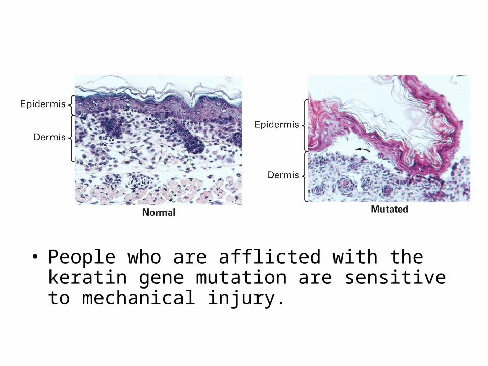
• People who are afflicted with the keratin gene mutation are sensitive to mechanical injury.