Embed Size (px)
Citation preview

DAH3.4 Protein interactions, sub –cellular locations and
arrays
Kathryn Lilley
Cambridge Centre for Proteomics
Department of Biochemistry
University of Cambridge
www.bio.cam.ac.uk/proteomics/
Part III Systems Biology
Outline
• Protein-protein interactions
• Protein sub-cellular location
• Protein Arrays
• Limitations of proteomics and systems biology
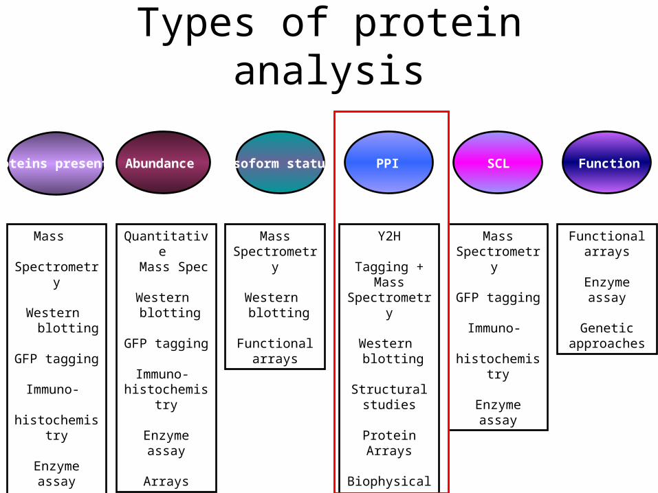
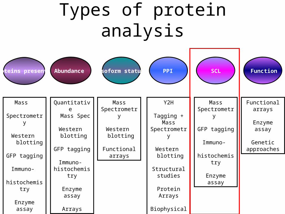
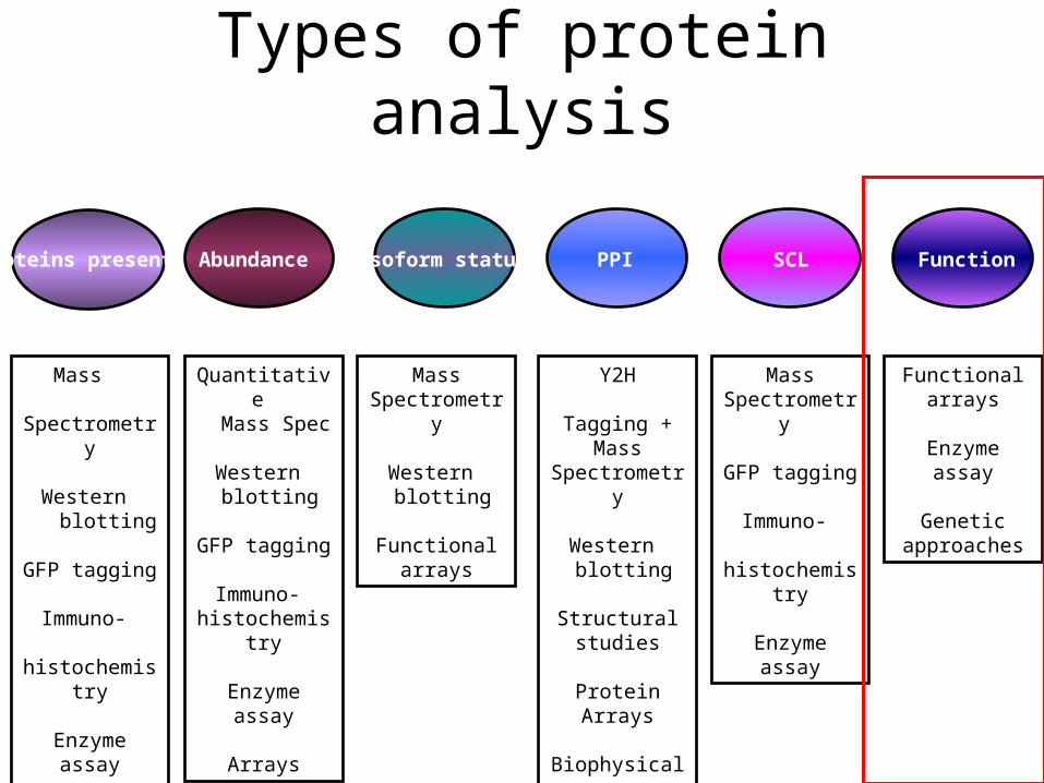
Types of protein analysis
Proteins present PPI SCL Function
Mass Spectrometry
Western blotting
GFP tagging
Immuno- histochemistry
Enzyme assay
Arrays
Abundance
Quantitative Mass Spec
Western blotting
GFP tagging
Immuno- histochemistry
Enzyme assay
Arrays
Isoform status
Mass Spectrometry
Western blotting
Functional arrays
Y2H
Tagging + Mass Spectrometry
Western blotting
Structural studies
Protein Arrays
Biophysical assays (e.g.ITC,
AUC)
Mass Spectrometry
GFP tagging
Immuno- histochemistry
Enzyme assay
Functional arrays
Enzyme assay
Genetic approaches
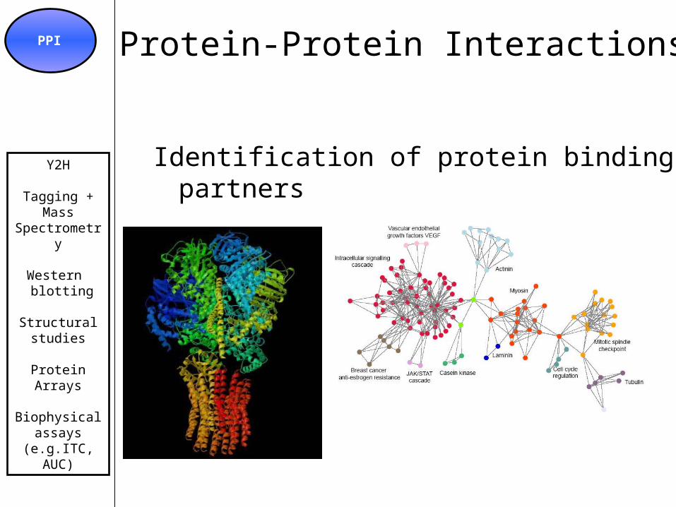
Protein-Protein InteractionsPPI
Y2H
Tagging + Mass Spectrometry
Western blotting
Structural studies
Protein Arrays
Biophysical assays (e.g.ITC,
AUC)
Identification of protein binding partners
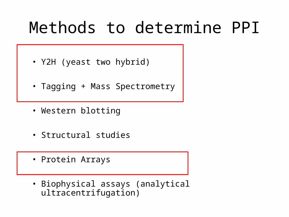
Methods to determine PPI
• Y2H (yeast two hybrid)
• Tagging + Mass Spectrometry
• Western blotting
• Structural studies
• Protein Arrays
• Biophysical assays (analytical ultracentrifugation)
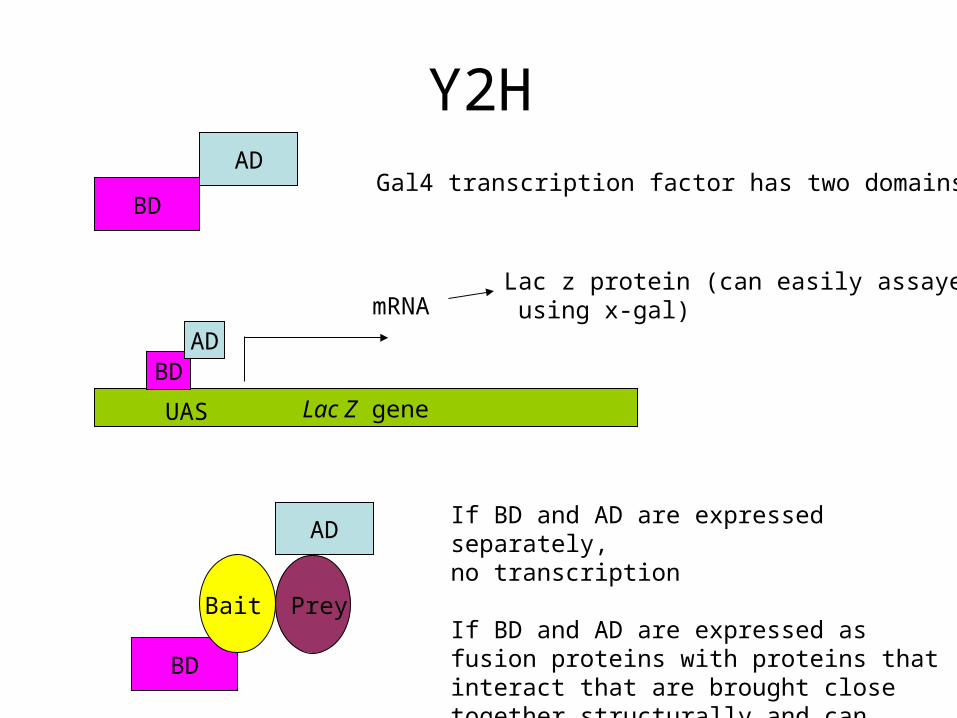
Y2H
BD
ADGal4 transcription factor has two domains
Lac Z gene
BDAD
Lac z protein (can easily assayed using x-gal)
BD
AD
Bait Prey
If BD and AD are expressed separately, no transcription
If BD and AD are expressed as fusion proteins with proteins that interact that are brought close together structurally and can bring about transcription of LacZ
mRNA
UAS
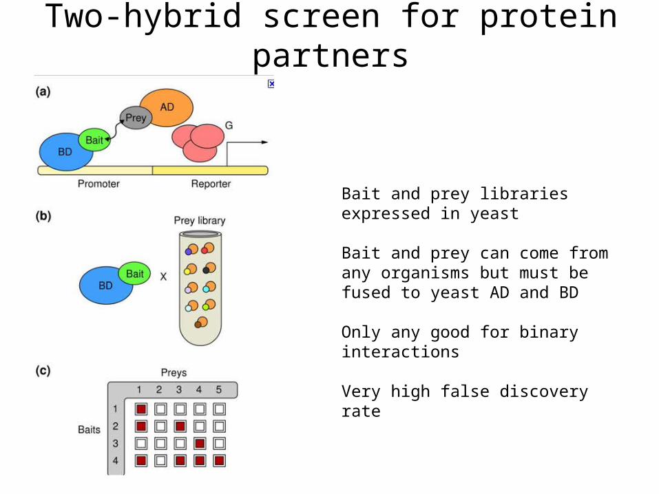
Two-hybrid screen for protein partners
Bait and prey libraries expressed in yeast
Bait and prey can come from any organisms but must be fused to yeast AD and BD
Only any good for binary interactions
Very high false discovery rate

Protein Complex Purification
• Methods mostly based around fusion of bait protein and selective purification of complex by immunoprecipitation
• Methods can be ‘dirty’
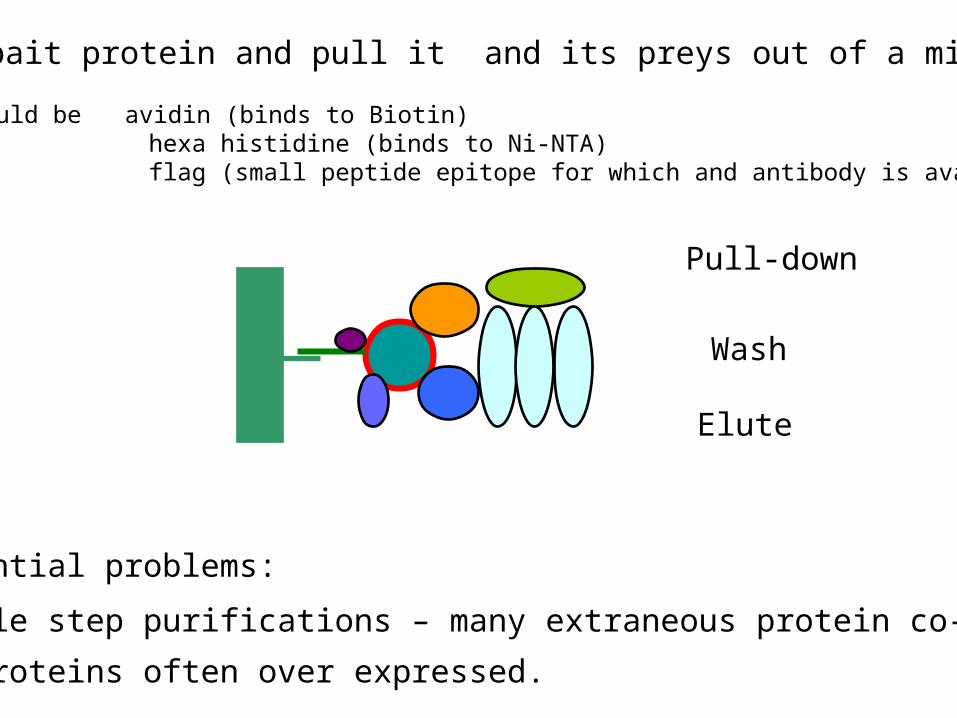
Pull-down
Wash
Elute
Two potential problems:
All single step purifications – many extraneous protein co-purified.
Tagged proteins often over expressed.
Tag bait protein and pull it and its preys out of a mixture
Tag could be avidin (binds to Biotin) hexa histidine (binds to Ni-NTA) flag (small peptide epitope for which and antibody is available)
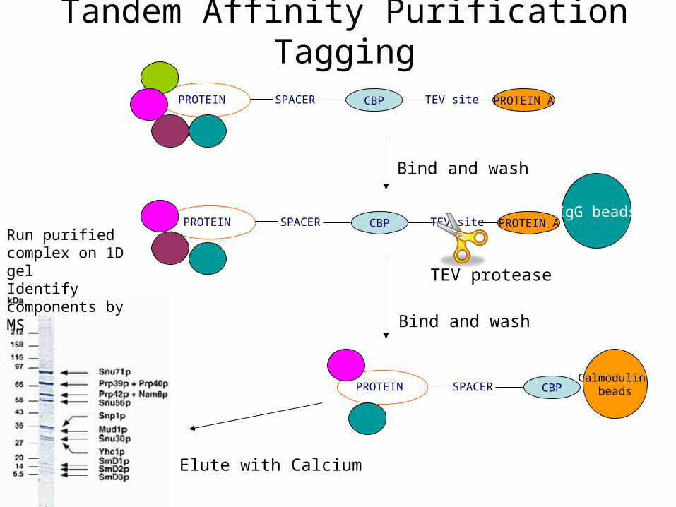
Tandem Affinity Purification Tagging
PROTEIN SPACER CBP TEV site PROTEIN A
IgG beadsPROTEIN SPACER CBP TEV site PROTEIN A
TEV protease
Calmodulin beadsPROTEIN SPACER CBP
Bind and wash
Bind and wash
Elute with Calcium
Run purified complex on 1D gel Identify components by MS
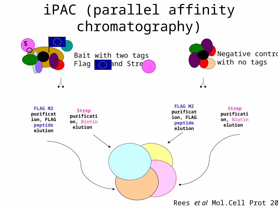
iPAC (parallel affinity chromatography)
S
F
FLAG M2 purification,
FLAG peptide elution
Strep purification,
Biotin elution
FLAG M2 purification,
FLAG peptide elution
Strep purification,
Biotin elution
Rees et al Mol.Cell Prot 2011
Bait with two tagsFlag and Strep
Negative control with no tags
Types of protein analysis
Proteins present PPI SCL Function
Mass Spectrometry
Western blotting
GFP tagging
Immuno- histochemistry
Enzyme assay
Arrays
Abundance
Quantitative Mass Spec
Western blotting
GFP tagging
Immuno- histochemistry
Enzyme assay
Arrays
Isoform status
Mass Spectrometry
Western blotting
Functional arrays
Y2H
Tagging + Mass Spectrometry
Western blotting
Structural studies
Protein Arrays
Biophysical assays (e.g.ITC,
AUC)
Mass Spectrometry
GFP tagging
Immuno- histochemistry
Enzyme assay
Functional arrays
Enzyme assay
Genetic approaches
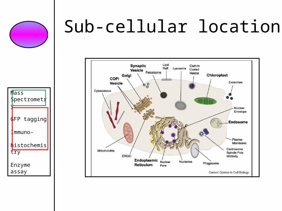
Sub-cellular location
Mass Spectrometry
GFP tagging
Immuno- histochemistry
Enzyme assay
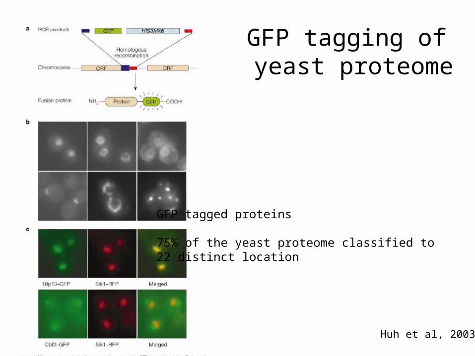
GFP tagging of yeast proteome
Huh et al, 2003
GFP tagged proteins
75% of the yeast proteome classified to 22 distinct location
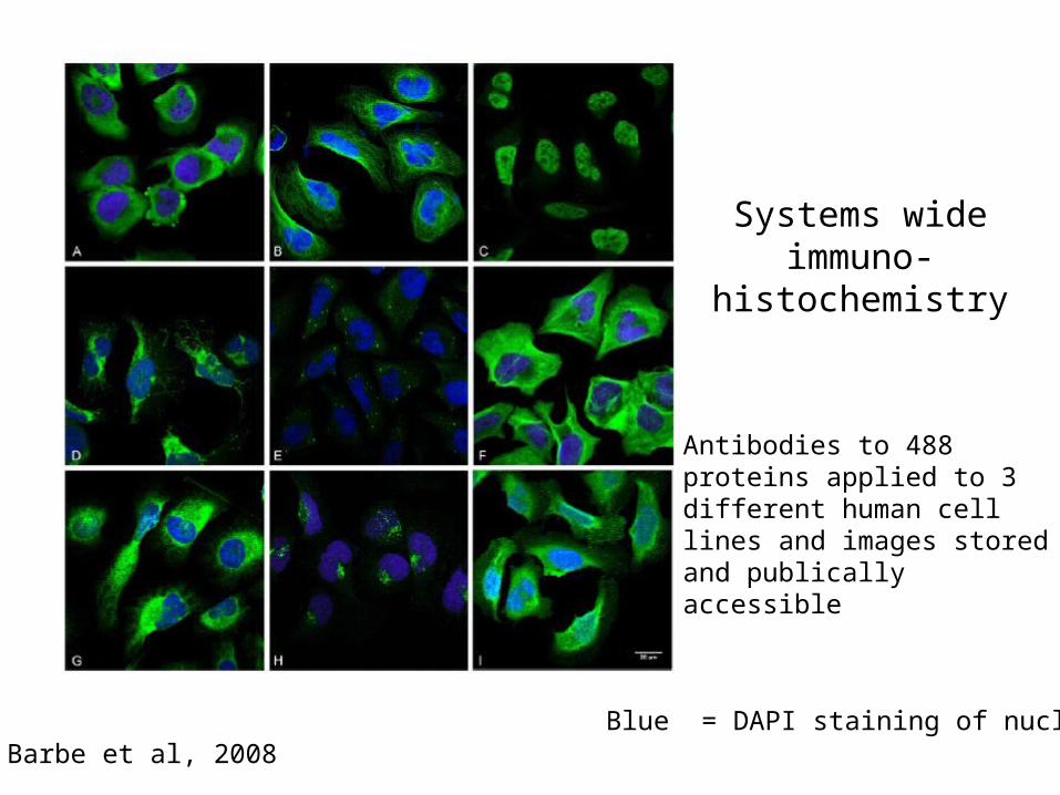
Systems wide immuno-
histochemistry
Barbe et al, 2008
Antibodies to 488 proteins applied to 3 different human cell lines and images stored and publically accessible
Blue = DAPI staining of nucleus
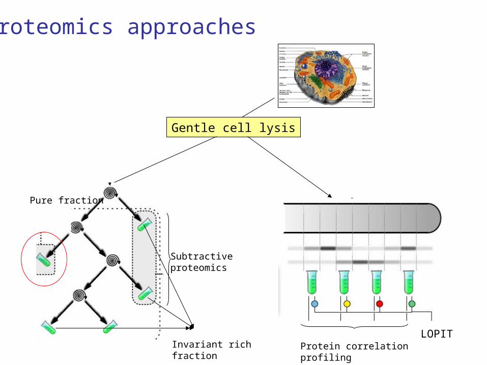
Gentle cell lysis
Pure fraction
Protein correlation profiling
LOPIT
Subtractive proteomics
Invariant rich fraction
Proteomics approaches
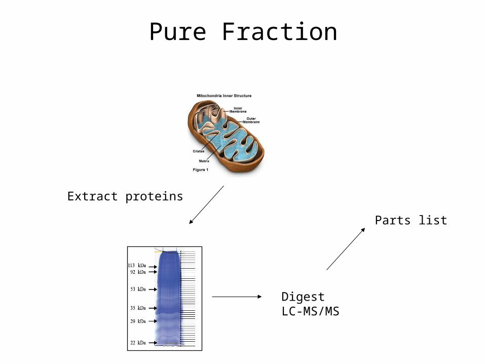
Pure Fraction
Extract proteins
DigestLC-MS/MS
Parts list
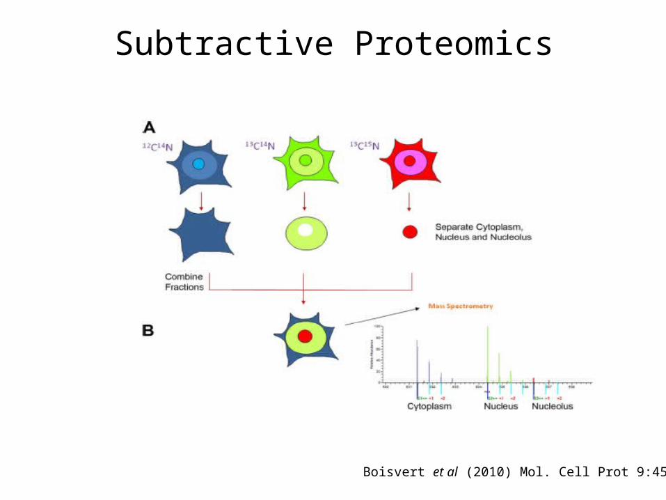
Subtractive Proteomics
Boisvert et al (2010) Mol. Cell Prot 9:457
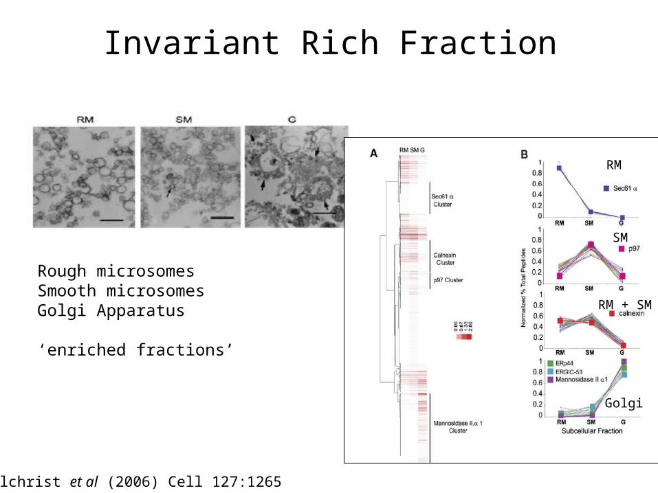
Invariant Rich Fraction
Rough microsomesSmooth microsomesGolgi Apparatus
‘enriched fractions’
RM
RM + SM
SM
Golgi
Gilchrist et al (2006) Cell 127:1265
2
4
6
8
10
12
14
16
18
20
1
3
5
7
9
11
13
15
17
19
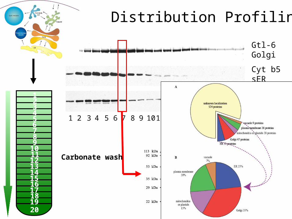
Gtl-6 Golgi
Cyt b5sER
Sec12ER
2 4 6 8 10 12 14 16 18 201 3 5 7 9 11 13 15 17 19
Carbonate wash
Tom Dunkley
Distribution Profiling
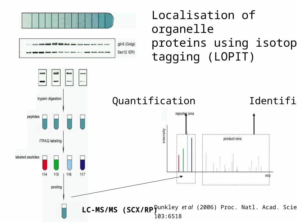
LOPIT Protocol
LC-MS/MS (SCX/RP)
Localisation of organelle proteins using isotope tagging (LOPIT)
Quantification Identification
Dunkley et al (2006) Proc. Natl. Acad. Sciences
103:6518
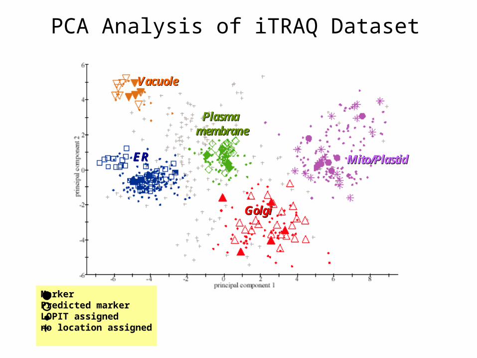
PCA Analysis of iTRAQ Dataset
GolgiGolgi
ERER
VacuoleVacuole
Plasma Plasma membranemembrane
Mito/PlastidMito/Plastid
GolgiGolgi
ERER
VacuoleVacuole
Plasma Plasma membranemembrane
Mito/PlastidMito/Plastid
MarkerPredicted markerLOPIT assignedno location assigned+
Types of protein analysis
Proteins present PPI SCL Function
Mass Spectrometry
Western blotting
GFP tagging
Immuno- histochemistry
Enzyme assay
Arrays
Abundance
Quantitative Mass Spec
Western blotting
GFP tagging
Immuno- histochemistry
Enzyme assay
Arrays
Isoform status
Mass Spectrometry
Western blotting
Functional arrays
Y2H
Tagging + Mass Spectrometry
Western blotting
Structural studies
Protein Arrays
Biophysical assays (e.g.ITC,
AUC)
Mass Spectrometry
GFP tagging
Immuno- histochemistry
Enzyme assay
Functional arrays
Enzyme assay
Genetic approaches
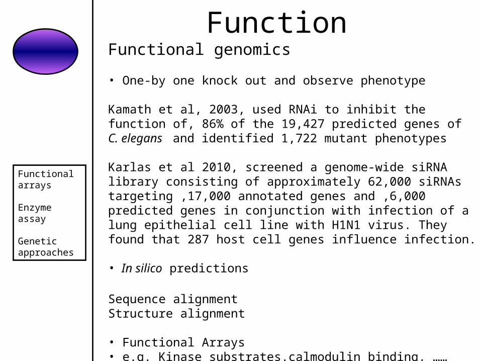
Function
Functional arrays
Enzyme assay
Genetic approaches
Functional genomics
• One-by one knock out and observe phenotype
Kamath et al, 2003, used RNAi to inhibit the function of, 86% of the 19,427 predicted genes of C. elegans and identified 1,722 mutant phenotypes
Karlas et al 2010, screened a genome-wide siRNA library consisting of approximately 62,000 siRNAs targeting ,17,000 annotated genes and ,6,000 predicted genes in conjunction with infection of a lung epithelial cell line with H1N1 virus. They found that 287 host cell genes influence infection.
• In silico predictions
Sequence alignment Structure alignment
• Functional Arrays• e.g. Kinase substrates.calmodulin binding, ……
Protein Arrays
Three types:
Functional Arrays
Analytical/Diagnostic Arrays
Tissue Arrays
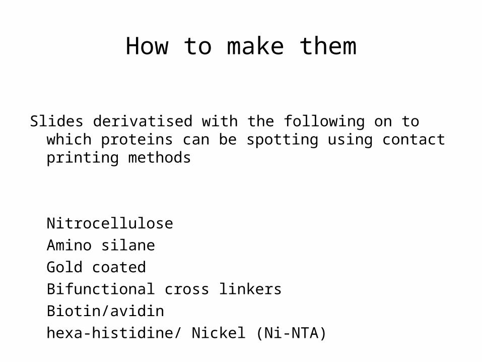
How to make them
Slides derivatised with the following on to which proteins can be spotting using contact printing methods
Nitrocellulose
Amino silane
Gold coated
Bifunctional cross linkers
Biotin/avidin
hexa-histidine/ Nickel (Ni-NTA)
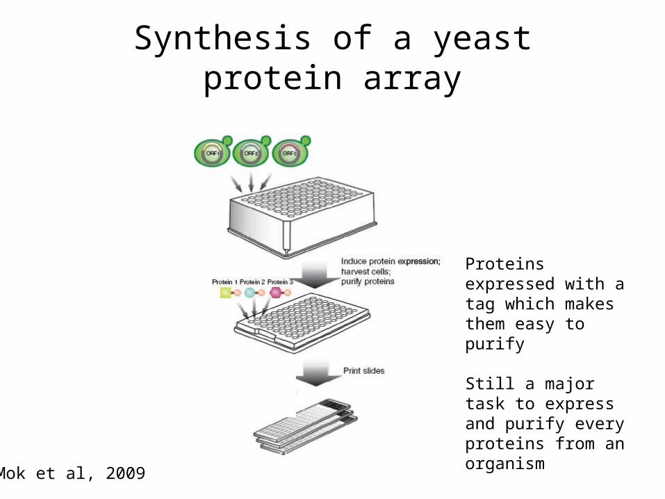
Synthesis of a yeast protein array
Mok et al, 2009
Proteins expressed with a tag which makes them easy to purify
Still a major task to express and purify every proteins from an organism
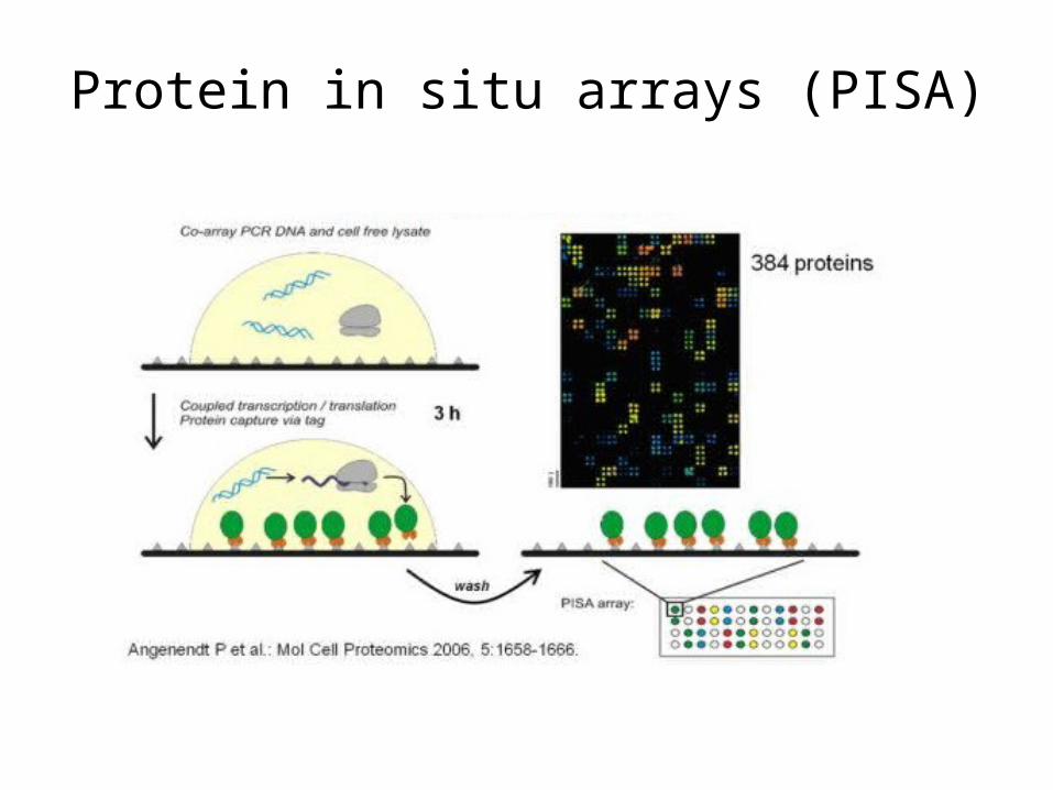
Protein in situ arrays (PISA)
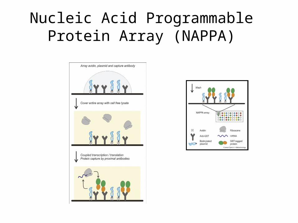
Nucleic Acid Programmable Protein Array (NAPPA)
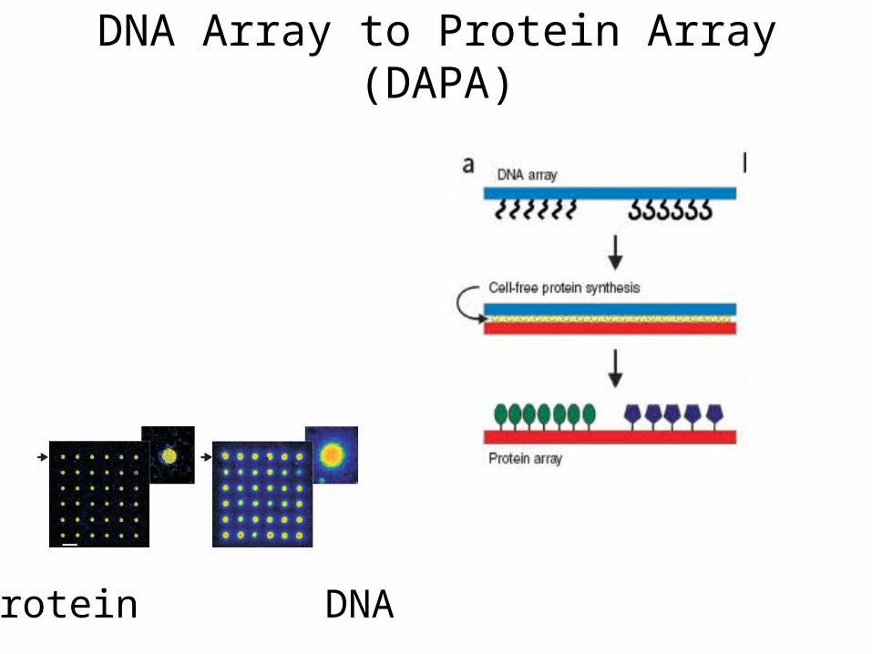
DNA Array to Protein Array (DAPA)
Protein DNA
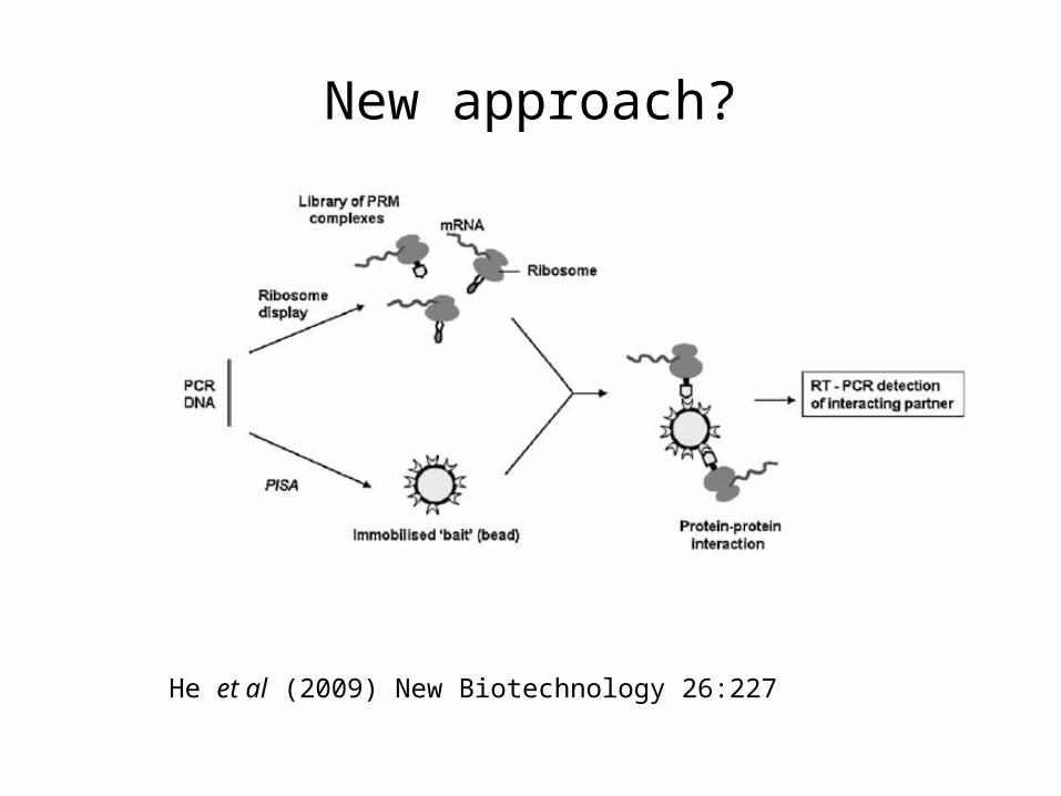
New approach?
He et al (2009) New Biotechnology 26:227
Detection
Fluorescence
Radioactivity
Quantum dots
Surface plasmon resonance
Mass spectrometry
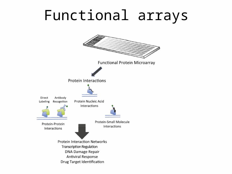
Functional arrays
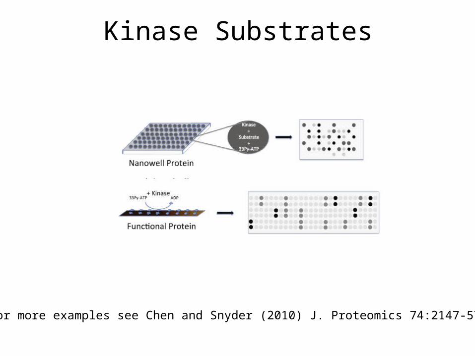
Kinase Substrates
For more examples see Chen and Snyder (2010) J. Proteomics 74:2147-57
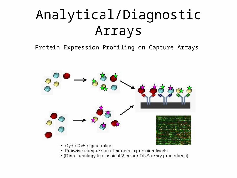
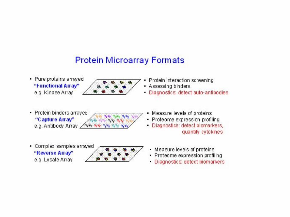
Analytical/Diagnostic Arrays
Protein Expression Profiling on Capture Arrays
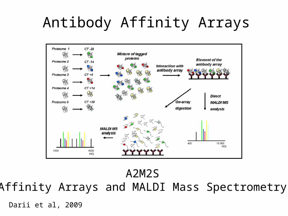
Antibody Affinity Arrays
A2M2SAffinity Arrays and MALDI Mass Spectrometry
Darii et al, 2009
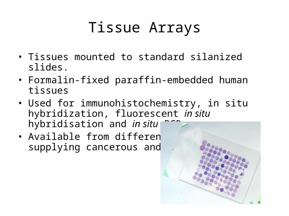
Tissue Arrays
• Tissues mounted to standard silanized slides.• Formalin-fixed paraffin-embedded human tissues• Used for immunohistochemistry, in situ hybridization,
fluorescent in situ hybridisation and in situ PCR• Available from different companies supplying cancerous
and normal tissues
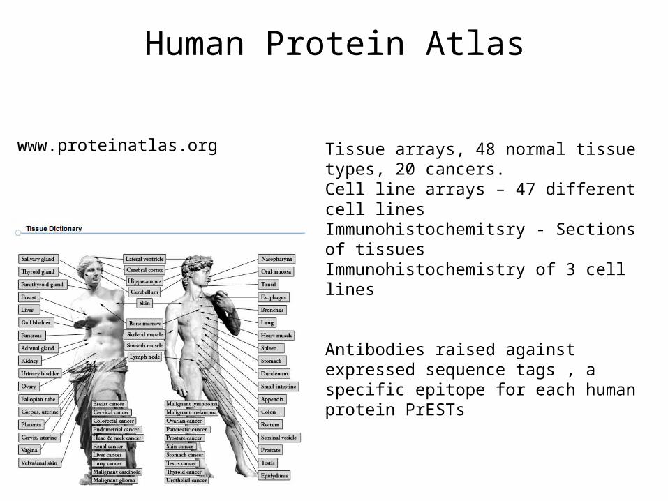
Human Protein Atlas
Tissue arrays, 48 normal tissue types, 20 cancers. Cell line arrays – 47 different cell lines Immunohistochemitsry - Sections of tissuesImmunohistochemistry of 3 cell lines
Antibodies raised against expressed sequence tags , a specific epitope for each human protein PrESTs
www.proteinatlas.org

Strengths
Under represented proteins present
Functional information
High throughput
Weaknesses
Proteins need to be in correct orientation and in vivo relevant structure
Unknown splicing variants not detected
Mixed PTMs not detected
Co-interacting partners not detected
Off target interactions
High cost of manufacture
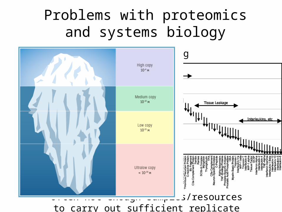
Problems with proteomics and systems biology
Tip of ice berg
Combinatorial PTMs
Keeping complexes together
Tissue specificity
Synchronisation of cells
Singel cell proteomics
Whole proteome is highly dynamic
Often not enough samples/resources to carry out sufficient replicate experiments

Single Cell Proteomics
Bendall et al (2011) Science
ICPMS
Inductively coupled plasma MS