Embed Size (px)
Citation preview

DearColleague,
Fornearly30years,theAdelaideTuitionCentrehaspublishedresourcestosupportteachersofBiologyinSouthAustralia.InitiallythiswasintheformofaStage2Workbook(1989),thenaStage2Textbook(1999),Stage1Textbook(2010)andmorerecentlytheSACE1AustralianCurriculumBiologyWorkbook(2016).
AswehavedonewiththeSACE1ACBiologyWorkbooklastyear;wearepleasedtoannounceourSACE2ACBiologyWorkbookwhichisbeingproducedbythesameteam.ThesebooksarewelladvancedandwillbeavailableforsaleinNovemberthisyear.
TheauthorteamconsistsofAlanCrierieandDavidGreig(whohavepreviouslypreparedtheStage2WorkbookandTextbook)andSimonRuthven(whohaspreviouslypreparedtheStage1Textbook).Togethertheyhaveover100years’experienceintheclassroomand40yearsasauthors!
Asyoucanseefromthis(draft)samplebooklet,thefeaturesoftheseSACE2ACBiologyWorkbooksinclude:
1. AllfourTopicsandcoversthewholecourseindetail.2. EachTopicisfurtherdividedintoupto8Chapters,eachcorrespondingto
approximatelyoneweek’swork.3. EachChapterconsistsof:a. textmaterial,supportedbyfullcolourillustrationsandphotographsb. alistofKeyTermsc. avarietyofstudentexercises,gradedindifficultyd. an‘InquirySkillsPractical’(whererelevant)e. a‘HumanEndeavourAssignment’(whereappropriate)f. implicitandexplicitopportunitiestodevelopCapabilitiesasdetailedinthe
SubjectOutline.4. Numerous‘HelpfulOnlineResources’andQRcodeswhichwillprovideimmediate
accesstorelevantwebsitesandourowncustommade‘VideoAnimations’and‘ConceptVideos’.Yourphoneortabletwillneeda‘QRcodereader’Apptoaccessthese.
5. Inaddition,Exam-style‘TopicTests’,answerstoallexercises,laboratorynotesforthepracticals,anindex,acomprehensiveglossaryoftermsandotherteacherresourcematerialwillbeprovidedinageneralappendix.PleaseusetheOrderFormatthebackofthissamplebookletorvisitourwebsiteorcontacttheofficeformoreinformation.
Kindregards,
SamBarry|Director
2
TABLE OF CONTENTS STAGE 2 BIOLOGY WORKBOOK
Topic 1 DNA and proteinsChapter 1.1 DNA structure
Chapter 1.2 DNA and protein synthesis
Chapter 1.3 Structure and function of proteins
Chapter 1.4 Enzymes
Chapter 1.5 Gene expression
Chapter 1.6 Mutations
Chapter 1.7 Biotechnology: tools and techniques
Chapter 1.8 Biotechnology: applications
Chapter Appendices (Answers and Laboratory Notes)
Topic 1 Test Yourself and suggested answers
Topic 2 Cells As the Basis of LifeChapter 2.1 The cell theory
Chapter 2.2 Two major types of cells
Chapter 2.3 Energy transformations
Chapter 2.4 Movement of substances
Chapter 2.5 Cell metabolism
Chapter 2.6 Asexual cell division
Chapter 2.7 Sexual cell division
Chapter 2.8 The cell cycle and cell culturing
Chapter Appendices (Answers and Laboratory Notes)
Topic 2 Test Yourself and suggested answers
Topic 3 HomeostasisChapter 3.1 Maintaining the internal environment
Chapter 3.2 The nervous sysem
Chapter 3.3 The endocrine system
Chapter 3.4 The nervous and endocrine systems
Chapter Appendices (Answers and Laboratory Notes)
Topic 3 Test Yourself and suggested answers
Topic 4 EvolutionChapter 4.1 The origin of life on Earth
Chapter 4.2 Comparative genomics
Chapter 4.3 Species
Chapter 4.4 Genetic Variation
Chapter 4.5 Evolutionary change
Chapter 4.6 Types of evolution
Chapter 4.7 Human impacts
Chapter Appendices (Answers and Laboratory Notes)
Topic 4 Test Yourself and suggested answers
General Appendix
Glossary of Key Terms
Index

Topic 1opic 1
1.1 DNA structure
1.2 DNA and protein synthesis
1.3 Structure and function of proteins
1.4 Enzymes
1.5 Gene expression
1.6 Mutations
1.7 Biotechnology: tools and techniques
1.8 Biotechnology: applications
Answers and Laboratory Notes
Topic 1 DNA and proteins

4
TOPIC 1 DNA AND PROTEINS
© Essentials Education 2017
Chapter 1.7 Biotechnology: tools and techniquesScience UnderstandingDNA sequencing enables mapping of species genomes; DNA profiling identifies the unique genetic makeup of individuals. Human beings can sequence even small amounts of DNA.
Segments of DNA can be multiplied using the polymerase chain reaction (PCR); their base sequences can then be identified.
Describe PCR, including the roles of:
• heating and cooling• primers• free nucleotides• heat-resistant enzymes• electropherograms.Explain how differences in DNA fragments, identified by DNA profiling, can be used; for example, in forensic science.
Biotechnology can involve the use of bacterial enzymes, plasmids as vectors, and techniques including gel electrophoresis, bacterial transformations and PCR.
DNA can be extracted from cells.
• Describe how particular genes can be selected and removed using probes and restriction enzymes.• Describe how selected genes can be transferred between species; for example, using bacterial
pasmids, viruses, and microinjection. © SACE 2016
In this chapter students improve their numeracy capability by solving problems using calculations and by learning about the impact of ICT on the development of genome sequencing to enrich their ICT capability. Students who study the text on genetic testing will have the opportunity to appreciate why respecting and engaging with different cultural views is essential to ensuring the technology is fully inclusive, thus acquiring intercultural understanding.
BiotechnologyThe use or application of science to provide solutions to problems is called technology. Biotechnology refers to forms of technology that are based around biology. It is not new, humans have been using biological techniques and processes for thousands of years to make bread, wine, cheese and other products. In the last few decades, with the advancement of scientific discoveries, new and continually developing ranges of tools, processes and technologies are emerging that enhance human well-being and potentially help the planet and its ecosystems.
This chapter will present and discuss a range of the most commonly used tools and techniques used in biotechnology and the following chapter (1.8) will examine a range of applications, along with ethical, economic and other issues associated with the technologies.
Extracting DNA from cellsGenetic material can be manipulated. In other words, it can be deliberately altered by scientists e.g. as a result of transferring DNA from one species to the DNA of another species. It is possible to move DNA between species as the genetic code is universal. This means that, for every organism, the same mRNA codon codes for the same amino acid. This makes it possible; for example, to introduce a human gene that can code for insulin to a bacterium which can then make the human protein insulin. As has already been discussed, the human genome has 46 chromosomes and around 21,000 protein-coding genes. Given that the average size of a gene is many thousand bases on the template strand, it is not hard to appreciate how difficult it is to locate or select a particular gene that scientists wish to work with.
Helpful Online RESOURCES about extracting DNA from human cellsTo learn how DNA is extracted from human cells, use a ‘QR code reader’ App on your phone or tablet to scan this QR code to visit a virtual laboratory:
<http://learn.genetics.utah.edu/content/labs/extraction/>

5
CHAPTER 1.7BIOTECHNOLOGY: TOOLS AND TECHNIQUES
© Essentials Education 2017
Gene probesGene probes are single-stranded DNA or mRNA segments that have been constructed in the laboratory. They can be used to select particular genes because they have radioactive or fluorescent markers that enable them to be readily located. The probes work by binding in a complementary manner to the ‘gene of interest’. When it binds to the probe, the gene of interest is said to have been hybridised with the radioactive probe. Refer to Figure 171.
G
A
A A
T
T
T
G
G
G
AT
C
C
C
A
G G
G G
A
T
TC
radioactive gene probe
single stranded DNA
TAC C
A
G
TAC C
A
base pairing identifies the ‘gene of interest’
mix with single stranded DNA from genomic library
no binding
binding
Figure 171 The use of gene probes
Figure 172 reveals a grid of DNA fragments making up the human chromosome 17. Scientists have isolated this to be the site of a defective gene responsible for many cases of inherited breast cancer. The grid represents pieces of DNA which have been spotted onto filter paper and following this, an X-ray plate has been superimposed showing fragments tagged with a radioactive marker. These tagged fragments, some of which correspond to genes, have been hybridised to help researchers map genes on a chromosome.
Figure 172 Using hybridisation to locate the gene for breast cancer

6
TOPIC 1 DNA AND PROTEINS
© Essentials Education 2017
Restriction enzymesBacteria produce restriction enzymes (endonucleases) which cut DNA naturally and thus act as a defence mechanism against viral DNA that may invade the bacteria. These enzymes were discovered in 1970 and not long after they were used in a type of biotechnology called genetic engineering to cut DNA at specific sites and thus help remove or isolate genes. Most restriction enzymes recognise short nucleotide sequences; around 4 to 8 and these sites are called restriction sites. They work by making an incision or cut through the sugar phosphate backbone of the DNA. The enzyme named EcoR1 was extracted from the bacteria E Coli and cuts DNA in a staggered fashion as can be seen in Figure 173.
G A A T T C
C T T A A G
cut
cut
G
C T T A A
A A T T C
G
EcoRI
to check layout and spacing Figure 173 The action of a restriction enzyme ‘EcoR1’
As seen in Figure 173, the enzyme cuts between the G and A bases and the effect is that the DNA is cut leaving overlapping ends. When preparing to transfer DNA, the same restriction enzyme is used for the host and the donor so that the cuts are made in the same way. When this occurs, the overlapping sections are referred to as ‘sticky ends’ and the sticky ends enable complementary binding between host DNA and donor DNA. An enzyme, DNA ligase, is used to join the host and donor DNA. The incorporation of DNA into bacteria like this is called bacterial transformation.
Refer to Figure 174 which shows the joining of bacterial plasmid DNA (host) and human DNA (donor) using a restriction enzyme to cut the DNA and a ligase enzyme to join the two pieces together; the resulting DNA is termed recombinant DNA. Techniques that permit or produce recombinant DNA are called recombinant DNA technology.
GCA
A
A A A GC
CG
ATT
TT
T
T T TA A AT
A GTTA A A AC TT T
A GTT CT T TA A A
A A A AC TT T G A T
G
GC CGG G
A A A G
C
CC C T T
T TA A
G CGCGA A ACC C T T G
GG G C GC CT TA A
GG CGC CT TA A
Plasmid DNA Human DNA
Recombinant DNA
DNA ligase joins DNA at these points
restriction enzyme cuts DNA at these points
restriction enzyme cuts DNA at these points
DNA fragments
Figure 174 The production of recombinant DNA
7
CHAPTER 1.7BIOTECHNOLOGY: TOOLS AND TECHNIQUES
© Essentials Education 2017
Helpful Online RESOURCES about restriction enzymesTo learn more about restriction enzymes, use the QR code to visit:
<https://www.youtube.com/watch?v=pDHCHa1C85Y>
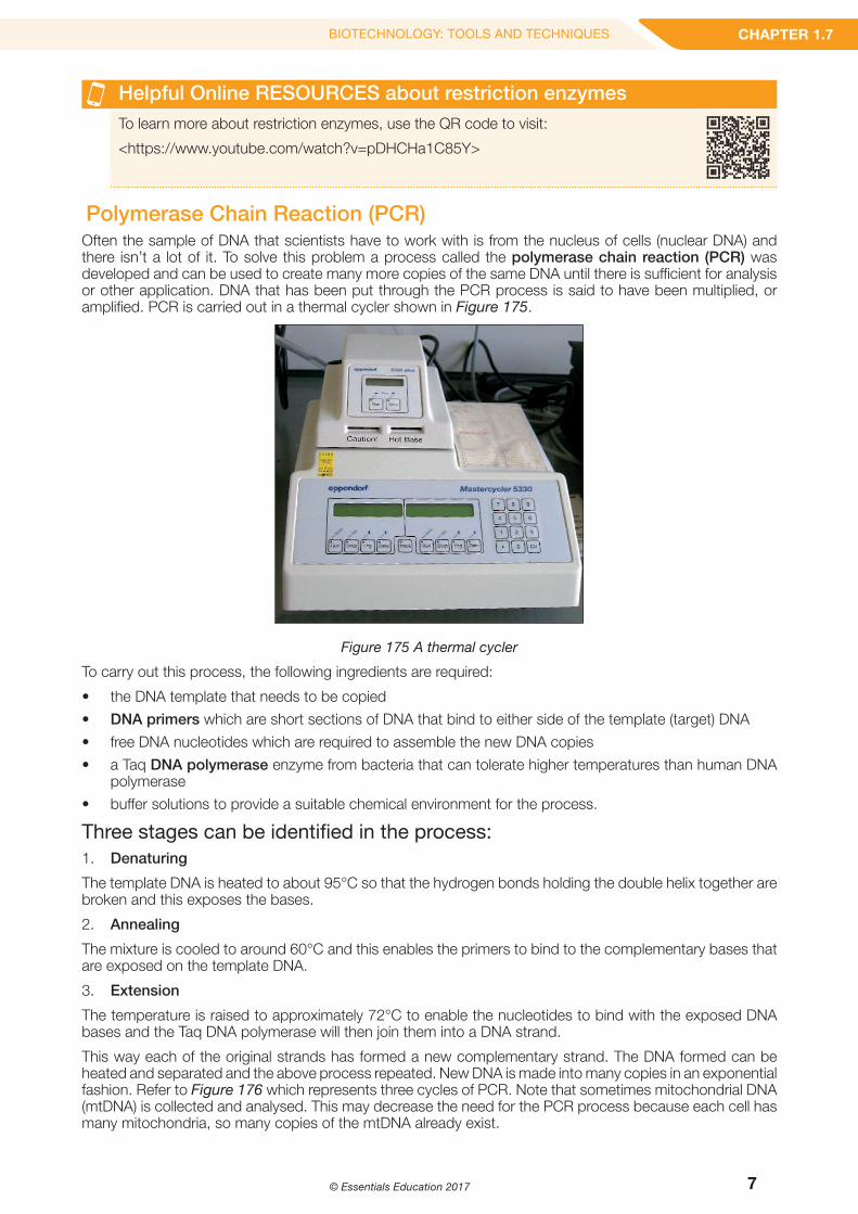
Polymerase Chain Reaction (PCR)Often the sample of DNA that scientists have to work with is from the nucleus of cells (nuclear DNA) and there isn’t a lot of it. To solve this problem a process called the polymerase chain reaction (PCR) was developed and can be used to create many more copies of the same DNA until there is sufficient for analysis or other application. DNA that has been put through the PCR process is said to have been multiplied, or amplified. PCR is carried out in a thermal cycler shown in Figure 175.
Figure 175 A thermal cycler
To carry out this process, the following ingredients are required:
• the DNA template that needs to be copied
• DNA primers which are short sections of DNA that bind to either side of the template (target) DNA
• free DNA nucleotides which are required to assemble the new DNA copies
• a Taq DNA polymerase enzyme from bacteria that can tolerate higher temperatures than human DNA polymerase
• buffer solutions to provide a suitable chemical environment for the process.
Three stages can be identified in the process:1. Denaturing
The template DNA is heated to about 95°C so that the hydrogen bonds holding the double helix together are broken and this exposes the bases.
2. Annealing
The mixture is cooled to around 60°C and this enables the primers to bind to the complementary bases that are exposed on the template DNA.
3. Extension
The temperature is raised to approximately 72°C to enable the nucleotides to bind with the exposed DNA bases and the Taq DNA polymerase will then join them into a DNA strand.
This way each of the original strands has formed a new complementary strand. The DNA formed can be heated and separated and the above process repeated. New DNA is made into many copies in an exponential fashion. Refer to Figure 176 which represents three cycles of PCR. Note that sometimes mitochondrial DNA (mtDNA) is collected and analysed. This may decrease the need for the PCR process because each cell has many mitochondria, so many copies of the mtDNA already exist.

8
TOPIC 1 DNA AND PROTEINS
© Essentials Education 2017
cycle 1 cycle 2 cycle 3
target DNA sequence to be amplified
two copies of the DNA
four copies of the DNA
eight copies of the DNA
original DNA
new DNA
Figure 176 The process of PCR
C Intercultural understanding: Inclusive genetic testingIt is now possible to test for hundreds of human genetic disorders. In many cases this involves using PCR with primers that bind to the genes associated with them. The presence or absence of the disease-causing mutation in the amplified DNA can then be determined by DNA sequencing. Common genetic tests include those for sickle-cell disease, cystic fibrosis and the blood clotting disorder called haemophilia. The use of such tests permits the identification of the disease before the onset of symptoms. Many can also be used to identify carriers of faulty-disease causing genes who do not have the disease but have the potential to have children with the disease. In some cases, identification of a genetic disease can even be confirmed before birth. Many genetic tests are in development and new ones are introduced every year; for example, a more accurate test for bowel cancer called Colvera that became available in Australia for the first time in 2017.
Until recently, genetic testing required the patient to go to a clinic or hospital, have the test there, and then have a follow-up with a doctor or genetic counsellor to accurately interpret the results. People begin to come to terms with what can be confronting information. Genetic tests do not always provide ‘clear-cut’ results. And the implications of confirmation of a serious genetic disease go well beyond the self – they have implications for family members, employment and decisions about how lifestyle should or can be adjusted. It is possible to advise and counsel patients like this in a way that is culturally inclusive as well. This is important because people of different cultural origin may hold different views and customs that influence how they receive, process and react to information like genetic test results and related advice that may be about having blood tests, decisions about family planning, or the termination of a pregnancy.
Direct-to-consumer (DCT) genetic testing provides a more patient-independent way to get genetic information about the risks of a genetic disease developing at some point in the future. On paying for the test, a person sends a sample (usually saliva or an inner cheek swab) which is analysed by a laboratory overseas who then send the results directly back to the person who sent the sample. There is no involvement of a health or genetic professional which promotes inaccurate processing of the information and incorrect decisions about how it should be used. While DCT genetic testing may appeal to some whose cultural views forbid for instance blood tests (a blood test is not required), transmission of genetic information like this is very unlikely to be respectful of or engage with broader cultural views thereby undermining its inclusiveness.

9
CHAPTER 1.7BIOTECHNOLOGY: TOOLS AND TECHNIQUES
© Essentials Education 2017
Helpful Online RESOURCES for PCRTo learn more about the Polymerase Chain Reaction, use this QR code to visit:
<https://www.youtube.com/watch?v=IaKXv1JqBtY>
Helpful Online RESOURCES - an EVA for PCRTo view a (draft) Essentials Video Animation (EVA) on the topic of PCR, scan this QR code:
<http://essentialseducation.com.au/resources/sace2/biology/eva-polymerase-chain-reaction/>
Gel electrophoresisGel electrophoresis is a technique used to separate proteins or fragments of DNA according to their size and charge. A gel is prepared in a thin layer and placed in a container as shown in Figures 177(a) and (b).
gel layer
buffer solution
+ ve- ve wells
Figure 177 (a) The principle of gel electrophoresis
If samples of DNA fragments are loaded into the wells and the electrophoresis tank is connected to a power supply to generate an electric field, the DNA fragments (-ve charge) will be repelled away from the wells (-ve charge) - the smaller the DNA fragments (less DNA base pairs) the further the fragment moves (refer to SIS1.7). An electropherogram is a plot of results from separation done by electrophoresis.
Figure 177 (b) An actual gel electrophoresis tray
Figure 178 illustrates a ladder of known sizes of DNA fragments, (in base pairs) that can be used to identify unknown fragments. Sometimes chemicals can be added to make these fragments easier to observe e.g. fluorescent dyes.
2000900
15001000
800700
600500
400300
200100
100 bp Ladder DNA Marker
base pairs
- ve + ve
Figure 178 A sample ladder of DNA fragments

10
TOPIC 1 DNA AND PROTEINS
© Essentials Education 2017
Helpful Online RESOURCES for gel electrophoresisTo conduct your own virtual gel electrophoresis experiment, use the QR code to visit a virtual laboratory:
<http://learn.genetics.utah.edu/content/labs/gel/>
DNA profilingDNA profiling (or DNA fingerprinting as it was once called) is a technique that identifies the unique genetic make-up of individuals or compares DNA from different sources without mapping the entire genome. An application is in forensic science where DNA bands from a suspect match those found from samples (e.g. blood, semen, hair) at a crime scene; refer to Figure 179.
Figure 179 An example of the use of DNA profiling for forensic purposes
Helpful Online RESOURCES for DNA profiling (or fingerprinting) To learn how DNA fingerprinting is used to identify a criminal use the QR code to visit:
<https://www.youtube.com/watch?v=AkBUriMK9u8>
Another use here includes paternity testing whether DNA of a father can be compared with a potential offspring’s DNA.
The technique has proven to be successful in comparing what are termed short tandem repeats (STR) from one individual to another. STR’s are short sections of the DNA, usually around 2 to 6 base pairs, that are repeated and the pattern has been found to be highly variable between individuals and thus a good technique for identification. An example of a profile using gel electrophoresis showing the fluorescent markers is seen in Figure 1710.
Figure 1710 Gel electrophoresis using fluorescent markers

11
CHAPTER 1.7BIOTECHNOLOGY: TOOLS AND TECHNIQUES
© Essentials Education 2017
DNA sequencingDNA sequencing involves techniques that are used to determine the actual sequence of DNA bases (A, T, G, C) in the gene segment of DNA and the entire genome of an organism (in which case it is often called genome sequencing). The process started with small, simple organisms such as bacteria and also viral particles and culminated in the first sequence published of the entire human genome through the human genome project that was completed in 2003.
This work continues in laboratories around the globe as scientists continue to map the genomes of different species. Collating and analysing this information continues to advance our understanding of genetics, gene control and evolutionary relationships between organisms. It also enhances our ability to use biotechnology and genetic techniques to enhance the well-being of humans and other species.
DNA sequencing has advanced from its first appearance in 1977 to the latest computer generated large-scale automated analyses. Refer to Figure 1711 of a circular genome map showing shared genetic material between humans, chimpanzee, mouse, rat, dog, chicken and zebra fish chromosomes (from outside inwards). This map was created using ‘Circos’ software which visualises complex genetic comparisons between different species. Note: specific details of this diagram are not required knowledge in this course.
Figure 1711 A map comparing genomes of different species.

12
TOPIC 1 DNA AND PROTEINS
© Essentials Education 2017
C ICT: The impact of ICT on biology and its application in societyThe development of the technique of genome sequencing provides a good example of the impact of ICT on the development of biology. The first genome sequencing methods took a long time and were very expensive. The determination and then publication of the human genome sequence in 2003 was a major achievement but it took over 14 years and cost about $3 billion. By 2013, the development of advanced high speed ICT-driven automated DNA sequencers was yielding human genome sequences in only three days for as little as $1000. In December 2016, the UK-based company Oxford Nanopore Technologies announced genome-sequencing-in-a-day capability using an easy to operate portable DNA sequencer called a MinION.
The applications for super-fast, cheap genome sequencing are numerous and very wide-ranging. It is already possible for biotechnology companies to sequence thousands of genomes a year. This greatly increases the ability of them to contribute to major research projects; for example, those looking into understanding and developing new treatments for cancers or genetic disorders. The technologies also make possible more personalised medicines that, informed by an individual’s own genome, respond more effectively to drug treatments with less side-effects. Even more ground-breaking is the potential ability to sequence genomes from a huge number of people so that in the not too distant future analysis of them would usher in what might be called prognostic genomics – an individual’s genome will be used to discover vulnerability to disease and to inform the steps to be taken before it develops, or to assist treatment choices if it has developed
Transferring genes between speciesGene transfer is a technique in which genetic material is transferred from one organism to another. Where this is done by scientists it is for the purpose of modifying the genetic information of the organism receiving the genes so that it may acquire a new, improved characteristic or produce a different protein of benefit. The process is called transgenesis (and often GM or genetic modification) and organisms formed in this manner are called transgenic organisms. Gene transfer can be achieved chemically, physically or by using a virus or plasmids from a bacterium. Viruses and plasmids used in this way are called vectors. These methods are explained in more detail in the sections below.
MicroinjectionMicroinjection is a physical method of gene transfer where DNA from a donor is injected directly inside the cell and into the cell nucleus through a very thin micro pipette. This technique is highly flexible but is slow and requires highly skilled technicians. This method is also used in vitro fertilisation to improve the chance of fertilisation of an ovum with a sperm (the technique is called intra-cytoplasmic sperm injection).
Refer to Figure 1712 which illustrates the technique of microinjection.
Bacterial plasmidsIt has already been stated that plasmids are small, circular DNA structures capable of independent replication and found in prokaryotes such as bacteria. These chromosomes are able to transfer DNA between bacteria and this characteristic has been utilised by scientists so that plasmids can be used as a vector (plasmid vectors) in transmitting genes to other species. The desirable gene for transfer is incorporated into the plasmid for further incorporation into a recipient or host cell.
The use of a virus Viruses naturally infect plant and animal cells and in the process transfer their DNA into the DNA in the nucleus of the cells they are invading. If the viral DNA is modified to contain a ‘desired’ gene this can also be transferred into a host or target cell. These are called viral vectors.
The aim of these and other gene transfer technologies is to integrate selected genes into host DNA so that the host cell can express the gene and make a desirable gene product, or this leads to production of an organism that does this, or one that has an altered phenotype that is desirable.
Figure 1712 Microinjection

13
CHAPTER 1.7BIOTECHNOLOGY: TOOLS AND TECHNIQUES
© Essentials Education 2017
Figure 1713 represents a brief summary of three gene transfer techniques:
a) On the left (a) microinjection; note that the red strand represents a gene of interest and the two sets of scissors the restriction enzyme used to remove it from the surrounding DNA.
b) In the middle (b) using a plasmid vector; this (recombinant) plasmid may be introduced into plant cells directly or indirectly by placing it in the cytoplasm of bacteria that naturally infect the plant cells.
c) On the right (c) another technique using metallic particles where DNA is fired into the nucleus of plant cells by a gene gun.
(a) (b) (c)
Figure 1713 (a), (b) and (c) Gene transfer techniques
Helpful Online RESOURCES - An EVA to show the Cloning of genesTo view a (draft) video animation of how genes can be cloned, scan this QR code:
<http://essentialseducation.com.au/resources/sace2/biology/eva-cloning-genes/>

14
TOPIC 1 DNA AND PROTEINS
© Essentials Education 2017
CRISPRClustered regularly interspaced short palindromic repeat (or CRISPR, pronounced ‘crisper’) technology is a genomics editing tool that has the capacity to edit parts of the gene by removing, adding or altering sections of DNA.
There are two major molecules involved in the CRISPR process:
• an enzyme Cas 9 which acts as a pair of molecular scissors to cut DNA at specific sites
• guide RNA which ensures that the Cas 9 cuts at the right spots on the DNA.
This technology is currently the fastest and best system for editing genes. It can also be applied to the DNA of virtually any species. Chapter 1.8 will examine some further aspects of CRISPR technology. More information about this technique is given in the following chapter.
Helpful Online RESOURCES about how CRISPR worksTo learn more about how CRISPR works, use this QR code to access the following:
<https://www.youtube.com/watch?v=0Mkie4R7haA>
Key Concepts1. Using biotechnology involves an impressive array of ever developing tools and techniques to
manipulate DNA.
2. Gene probes are short pieces of DNA or RNA that bind to, and thus help scientists to select particular genes (or locate the position of genes on chromosomes).
3. Restriction enzymes were discovered in bacteria and provide a very useful DNA (cutting tool) that bind to specific sites on DNA and cut it, allowing particular genes to be removed from chromosomes.
4. The polymerase chain reaction (PCR) enables multiple copies of DNA to be made from a small, original sample that can then be used in laboratory work and forensic science.
5. DNA profiling identifies the unique genetic make-up of individuals. This enables DNA from different sources to be compared e.g. in forensic science and paternity testing.
6. Gel electrophoresis can separate fragments of DNA based on their charge and size. It has many applications from DNA profiling, DNA sequencing and evolutionary comparisons of DNA.
7. DNA sequencing determines the actual base sequence in segments of DNA and nowadays is carried out by high-speed automated DNA sequencing machines.
8. There are a range of techniques used to transfer genes between species. They include using vectors e.g. plasmids or viruses or a physical method such as micro injection.
9. CRISPR is a new technology that can be used to edit and/or transfer genes. It is potentially a very powerful process, the development of which is only in its infancy.
Helpful Online RESOURCES - Essentials Concept Videos (ECV)<http://essentialseducation.com.au/resources/sace2/biology/ecv-dna-replication/>
Editor’s note to teachers: in addition to the features seen in this sample chapter, we are also developing a series of relevant ‘Essentials Concept Videos’ (ECV) which will stream from our website when the Workbook is published. An example of a (draft) ECV, together with a QR Code, although relevant to Chapter 1.1, is provided here for your information:

15
CHAPTER 1.7BIOTECHNOLOGY: TOOLS AND TECHNIQUES
© Essentials Education 2017
What have you learned?Key Terms
biotechnology
genetic engineering
gene probe
hybridisation
restriction enzyme
DNA ligase
recombinant DNA
polymerase chain reaction (PCR)
DNA primer
DNA polymerase
gel electrophoresis
electropherogram
DNA profiling
short tandem repeats (STR)
gene transfer
transgenesis
microinjection
plasmid vector
viral vector
CRISPR
Knowledge and Understanding
1. Briefly describe how it is possible for a bacterium to produce a human protein. (No details of processes are required.)
2. State two characteristics of molecules that are able to act as gene probes.
a)
b)
3. Often, during PCR, the DNA enzyme and nucleotide mixture is heated to temperatures well in excess of 60-70°C.
a) State why it is necessary to heat the DNA in this manner.
b) Explain why this heating would prevent the use of human DNA polymerase to catalyse a reaction.

16
TOPIC 1 DNA AND PROTEINS
© Essentials Education 2017
4. Describe the benefit in genetic engineering when the same restriction enzyme is used to cut the DNA of both the host and the donor.
5. In DNA profiling, bands or segments of DNA appear at different spots in the electrophoretic gel. Explain why segments appear at different spots after electrophoresis.
6. Microinjection is not always successful in altering the genetic information of a host. State why, giving a likely reason.
7. PCR is carried out ‘in cycles’ in a thermal cycler machine. As a result of each cycle the amount of DNA is doubled; in other words, after one cycle, there are 2 copies of the sample of DNA to be amplified, after two cycles there are 4 copies, and so on. How many copies of the original sample of DNA will have been produced after 10 PCR cycles? Show your working.
8. A cattle industry carried out a study using DNA profiling to determine if beef from various sources was contaminated. Refer to the DNA profile below from the study.
a)
Contaminated beef
Source A
Source B
Source C
Source D
How many sources of beef were contaminated?
b) What percentage of the total number of sources used in the DNA profile above were contaminated? Show your working.

17
CHAPTER 1.7BIOTECHNOLOGY: TOOLS AND TECHNIQUES
© Essentials Education 2017
Application, Analysis and Evaluation
9. State and briefly give reasons regarding which technique would most likely be used in the following situations:
a) Multiplying or amplifying the DNA of a suspected criminals hair follicle cells found at a crime scene.
b) Comparing human DNA with chimpanzee DNA, looking for differences in coding for a particular protein.
c) Analysing DNA in an embryo, looking for susceptibility to a particular genetic abnormality.
10. In order to answer this question, you will need to refer back to Chapter 1.2 and Figures128 (a) and (b) indicating which codons code for particular amino acids.
One of the techniques used to locate genes of interest is to use the protein product to trace the gene.
If the beginning of an amino acid sequence was: serine - glycine - proline - methionine
a) Give a sample DNA sequence that could have coded for this.
b) If a complementary probe was made to help locate the gene, what would its DNA sequence be for the given section?
11. In a hypothetical experiment, a gene for silk produced by a species of spider is incorporated into the DNA of female sheep. It is discovered that these sheep then go on to produce 5.0g of the spider silk per litre of milk per day.
a) The sheep above makes 8 litres of milk per day. How much spider silk would this contain? Show your working.
b) Clones of the sheep are produced. How many of these sheep would be needed to make 2 kg of the spider silk per day? Show your working.

18
TOPIC 1 DNA AND PROTEINS
© Essentials Education 2017
12. When a restriction enzyme cuts a DNA molecule, the cuts are not even and leave single stranded ends. Suggest why this is an advantage in recombinant DNA technology.
13. Refer to the diagram below showing a procedure that can be used to compare DNA and answer the questions below.
a)
Cut with restriction enzyme
Gel
ele
ctro
phor
esis
DNA from individual 1
DNA from individual 2
Stage A Stage B Stage C
Specialmembrane
Autoradiography
DNAfragments
DNA transferred from gel to special membrane
DNA fragments made single stranded and hybridised with a radioactivelabelled RNA probe
Explain why this procedure could be used to identify a suspect at the scene of the crime.
b) In Stage C the DNA of the gel has been hybridized with a radioactive probe. State the purpose of this step in the procedure.

19
CHAPTER 1.7BIOTECHNOLOGY: TOOLS AND TECHNIQUES
© Essentials Education 2017
14. Short tandem repeats (STR’s), found in segments of DNA have been found to be very useful in discriminating between individuals in the process of DNA profiling. The segments have very low mutation rates. Explain why these low mutation rates are also an advantage in DNA profiling.
15. Compare the techniques and applications of DNA sequencing and profiling.
16. Cystic fibrosis is caused by a single faulty gene. Gene therapy trials have been undertaken trying to deliver the functional gene into the cells of the respiratory tract using viruses.
a) Explain how such a technique might work.
b) This technique has experienced quite a high failure rate and also comes with risks to the patient. Suggest possible reasons for this.
17. CRISPR is a recent advance in gene editing and modification.
a) State the function of the protein ‘Cas 9’ in the process.
b) Explain how ‘guide RNA’ works in ensuring that the action of ‘Cas 9’ occurs in the right position.
c) Predict how the ‘guide RNA’ might end up in the wrong position.
18. These two segments of DNA were produced by the action of a specific restriction enzyme.
a)
A GTT CT T TA A A
A A A AC TT T G A T
Segment 1 Segment 2
On the left side, draw a diagram of the single DNA segment prior to the action of the enzyme.
b) Indicate on the diagram, or below, the specific recognition site for this enzyme and where the cuts occur.
20
TOPIC 1 DNA AND PROTEINS
© Essentials Education 2017
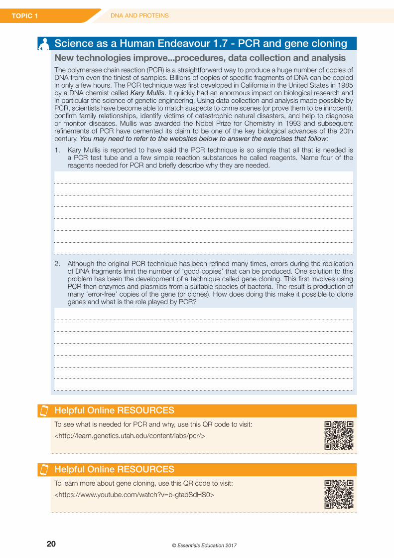
Science as a Human Endeavour 1.7 - PCR and gene cloningNew technologies improve...procedures, data collection and analysisThe polymerase chain reaction (PCR) is a straightforward way to produce a huge number of copies of DNA from even the tiniest of samples. Billions of copies of specific fragments of DNA can be copied in only a few hours. The PCR technique was first developed in California in the United States in 1985 by a DNA chemist called Kary Mullis. It quickly had an enormous impact on biological research and in particular the science of genetic engineering. Using data collection and analysis made possible by PCR, scientists have become able to match suspects to crime scenes (or prove them to be innocent), confirm family relationships, identify victims of catastrophic natural disasters, and help to diagnose or monitor diseases. Mullis was awarded the Nobel Prize for Chemistry in 1993 and subsequent refinements of PCR have cemented its claim to be one of the key biological advances of the 20th century. You may need to refer to the websites below to answer the exercises that follow:
1. Kary Mullis is reported to have said the PCR technique is so simple that all that is needed is a PCR test tube and a few simple reaction substances he called reagents. Name four of the reagents needed for PCR and briefly describe why they are needed.
2. Although the original PCR technique has been refined many times, errors during the replication of DNA fragments limit the number of ‘good copies’ that can be produced. One solution to this problem has been the development of a technique called gene cloning. This first involves using PCR then enzymes and plasmids from a suitable species of bacteria. The result is production of many ‘error-free’ copies of the gene (or clones). How does doing this make it possible to clone genes and what is the role played by PCR?
Helpful Online RESOURCESTo see what is needed for PCR and why, use this QR code to visit:
<http://learn.genetics.utah.edu/content/labs/pcr/>
Helpful Online RESOURCESTo learn more about gene cloning, use this QR code to visit:
<https://www.youtube.com/watch?v=b-gtadSdHS0>

21
CHAPTER 1.7BIOTECHNOLOGY: TOOLS AND TECHNIQUES
© Essentials Education 2017
? Science Inquiry Skills 1.7 – Using agarose gel electrophoresis
IntroductionGel electrophoresis is a laboratory method that is used to separate very small volumes of mixtures of molecules e.g. proteins and DNA, according to their size and charge.
Minute quantities of mixtures are provided by using a special pipette called a micro-pipette. Molecules to be separated are moved by an electric field through a gel that contains small spaces between molecules. The gel is submerged in a liquid called an electrophoretic buffer to facilitate the movement of electric current, and thus the movement of molecules to be separated. Small molecules travel further through the pores away from the negative end (or cathode) of the gel than larger ones do. One commonly used gel is made using a polysaccharide called agarose.
Part A Setting up the agarose gelAimTo learn how to set up an agarose gel, and use it to separate a mixture of dyes using electrophoresis.
Materials (per group or for a class demonstration)• Electrophoresis tank or chamber with lid and power leads attached
• Suitable power supply – this may be used to run two tanks at once
• Gel tray containing pre-prepared agarose gel (1% agarose in electrophoresis buffer) and a well former (or comb)
• 250 mL electrophoresis buffer solution (TAE - Tris Acetate EDTA)
• Black card
Method1. Place the electrophoresis tank on
the bench at your work station in a position away from the edge of the bench. Refer to Figure 1.
2. Put the lid on (with power leads attached) and then remove it from the tank to become familiar with how it feels to attach it correctly; it is important to be able to do this carefully and with a minimum of vibration (so as to not disturb the gel in Part B).
3. Collect the agarose gel containing the well former (or comb).
4. Remove the masking tape or ‘dams’ at the ends of the gel tray.
5. Place the tray down on the bench and gently remove the well former as shown by your teacher – this will expose the wells for use in Part B.
6. Place the gel tray into the electrophoresis tank.
7. Carefully fill the electrophoresis tank with buffer until the gel is completely submerged. (There should be no gel above the surface of the buffer). If this is done too quickly or roughly the gel may lift off the tray making loading samples into the wells very difficult.
8. Place the black card under the tank to make the wells easier to see.
Figure 1 The equipment

22
TOPIC 1 DNA AND PROTEINS
© Essentials Education 2017
? Science Inquiry Skills 1.7 – Using agarose gel electrophoresis (cont.)
Part B Running the gelAimTo use the provided dye samples to demonstrate separation of different molecules by electrophoresis.
Materials (per group or for demonstration)• 100 mL beaker containing about 50 mL of distilled water
• Micropipette and tips
• 100 mL beaker for used tips
• Dye samples
Method1. Before you load the dye samples into the gel, practice how to use a micropipette using a small
beaker of distilled water as shown by your teacher.
2. Using the micropipettes and dye samples provided, load the samples one at a time into the wells as shown by your teacher.
3. Place the lid onto the electrophoresis tank being very careful to not disturb the gel.
4. Connect the power leads of the lid to the power supply.
5. Turn the power supply to the voltage recommended by your teacher and leave the gel to run – it will probably take 10-20 minutes for clear separation to be achieved. Ensure the tank is not moved in any way and keep a close watch while the gel it is running to make sure that your samples do not end up running off the end of the gel (refer to Figure 2)
6. Once the electrophoresis is completed, turn off the power supply, and carefully remove the lid from the tank.
7. Gently remove the gel tray and gel from the tank. Be careful to keep the gel tray horizontal or the gel may slide off.
8. Place the gel tray on the bench and observe the results of the electrophoresis.
9. Take a photograph of the gel and the separated dye to keep a record of your results. This is important as once the electric current is removed (when the power supply is turned off) the dyes will diffuse into the gel and ‘the bands’ will become less distinct. Refer to Figure 3.
Figure 2 The process Figure 3 The product

23
CHAPTER 1.7BIOTECHNOLOGY: TOOLS AND TECHNIQUES
© Essentials Education 2017
? Science Inquiry Skills 1.7 – Using agarose gel electrophoresis (cont.)
Discussion1. The agarose gel needs to be completely submerged in an electrophoresis buffer solution. Why is
an electrophoresis buffer solution needed for electrophoresis to be successful ?
2. What is a micropipette, and why does one need to be used in agarose gel electrophoresis?
3. Electrophoresis is a separation technique that relies on an electric field. What is an electric field, and why is one needed in electrophoresis?
4. The agarose gel acts as a type of tiny filter. What is meant by this statement?
5. Were all the samples of dyes negatively charged? How do you know?
6. Which dye contained the largest molecules, and which one contained the smallest ones? Use your results to support your answer.
Editor’s note: more information about the suppliers and cost of this equipment, as well as further advice to teachers and technicians, will be provided in the Laboratory Notes for this SIS practical activity.
Helpful Online RESOURCES about using agarose gelTo watch how DNA samples are loaded and run an agarose gel, use the QR code to visit: <https://www.youtube.com/watch?v=vq759wKCCUQ>
More Helpful Online RESOURCES about using agarose gelTo carry out a simulation of making then running a gel, use the QR code to visit: <http://learn.genetics.utah.edu/content/labs/gel/>


![Dear Colleague, - R.M. Hoffman Company · Dear Colleague, Thank you for ... [STAC6-Si] controllers ... input / output monitor screen capture motor selection screen capture instruction/Tool](https://img.pdfslide.net/doc/110x75/5b15f1677f8b9a472e8c0a7c/dear-colleague-rm-hoffman-dear-colleague-thank-you-for-stac6-si.jpg)