Embed Size (px)
Citation preview

Decidual cytokines and pregnancy
complications: focus on spontaneous
miscarriage
Gendie E. Lash and Jan Ernerudh
Linköping University Post Print
N.B.: When citing this work, cite the original article.
Original Publication:
Gendie E. Lash and Jan Ernerudh, Decidual cytokines and pregnancy complications: focus on
spontaneous miscarriage, 2015, Journal of Reproductive Immunology, (108), 83-89.
http://dx.doi.org/10.1016/j.jri.2015.02.003
Copyright: Elsevier
http://www.elsevier.com/
Postprint available at: Linköping University Electronic Press
http://urn.kb.se/resolve?urn=urn:nbn:se:liu:diva-118252

1
Decidual cytokines and pregnancy complications: focus on spontaneous miscarriage
Gendie E Lash1* and Jan Ernerudh2
1Reproductive and Vascular Biology Group, Institute of Cellular Medicine, Newcastle
University, Newcastle upon Tyne, UK; 2Clinical Immunology, Department of Clinical and
Experimental Medicine, Faculty of Health Sciences, Linköping University, Linköping, Sweden.
*Address for correspondence:
Dr Gendie Lash
Reproductive and Vascular Biology Group
Institute of Cellular Medicine
3rd Floor, William Leech Building
Newcastle University
Newcastle upon Tyne
NE2 4HH, UK
Email: [email protected]
Phone: +44 191 208 8578

2
Abstract
The establishment of pregnancy requires the co-ordinated implantation of the embryo into
the receptive decidua, placentation, trophoblast invasion of the maternal decidua and
myometrium as well as remodelling of the uterine spiral arteries. Failure of any of these
steps can lead to a range of pregnancy complications including miscarriage, pre-eclampsia,
fetal growth restriction, placenta accreta and pre-term birth. Cytokines are small
multifunctional proteins often derived from leucocytes and have primarily been described
through their immunomodulatory actions. The maternal-fetal interface is considered to be
immunosuppressed to allow development of the semi-allogeneic placental-fetal unit.
However, cytokine profiles of the decidua and different decidual cell types suggests that the
in vivo situation is more complex. Data suggests that decidual derived cytokines not only
play roles in immunosuppression but also in other aspects of the establishment of
pregnancy including regulation of trophoblast invasion and spiral artery remodelling. This
review focuses on the potential role of decidual derived cytokines in the aetiology of
unexplained spontaneous miscarriage.
Keywords: decidua; cytokines; miscarriage; trophoblast invasion; spiral artery remodelling

3
1 Introduction
Several pregnancy pathologies, including pre-eclampsia (PE), fetal growth restriction (FGR)
and miscarriage, are associated with deficient invasion of extravillous trophoblast cells (EVT)
into the decidua and myometrium as well as incomplete remodelling of the uterine spiral
arteries. EVT invasion and remodelling of the uterine spiral arteries are highly complex
processes that require the dynamic interplay of many different biological signals from
different cellular sources, including the decidua. Of particular note are the decidual
cytokines and how their dysregulation may play roles in the aetiology of complications of
pregnancy. This review will discuss the pathophysiology of spontaneous miscarriage, key
processes in the establishment of pregnancy that may be causative of this pregnancy
complication as well as decidual cytokines shown to be dysregulated in miscarriage, their
cellular sources, known functions and how they may contribute to the pathogenesis of
miscarriage.
2 Cytokines
Cytokines are small (5-20kDa) signalling proteins produced by a wide range of cells. They
show diverse functions, including regulation of cellular invasion and both humoral and cell-
based immune responses. These cell-based and humoral immune responses are mediated
by different sets of cytokines that can be grouped according to their main function for
example; pro- and anti-inflammatory, or associated with different T helper (Th) subsets
referred to as Th1, Th2, Th17 and regulatory (Treg) cells. Major pro-inflammatory cytokines
include TNF, IL-1β, IL-6 and CXCL8/IL-8, while anti-inflammatory cytokines include IL-10 and

4
TGF-β. Signature cytokines of Th-associated responses are; interferon (IFN)-γ (Th1); IL-4, IL-
5, IL-9 and IL-13 (Th2); IL-17A and IL-17E (Th17); IL-10 and TGF-β (Treg). Several other
cytokines also exist that do not precisely fit into the described groups, like interferons,
chemokines, growth factors and many others. The area is complex because of a redundancy
in cytokines with antagonistic, synergistic, similar and sometimes identical effects; for
instance, there are at least 37 interleukins and approximately 50 chemokines. In this review
we will focus on a limited number of established and well-known cytokines in reproductive
biology, but also include recent advances that confirm or challenge our understanding of
this complex area of biology.
Cytokines in normal pregnancy
In the early era after the discovery of the Th1/Th2 dichotomy, Wegmann et al. (1993)
proposed that during pregnancy a Th2 response prevails, whereas a Th1 response is
detrimental to pregnancy. From a general perspective and when considering systemic
changes, this view is supported by observations like the improvement of Th1-associated
autoimmune disease multiple sclerosis (Confavreux et al., 1998) and rheumatoid arthrirtis
(Ostensen and Villiger, 2007) during pregnancy. However, this view is likely too simplistic as
Th1 cytokines have also been shown to be essential for maintenance of pregnancy, e.g. IFN-
γ is essential for successful pregnancy as it is pivotal in spiral artery remodelling and
successful pregnancy outcome (Ashkar et al., 2000; Croy et al., 2003; Robson et al., 2012).
Furthermore, a study using a Th2 knockout (IL-4, IL-5, IL-9 and IL-13 ko) mouse showed
normal reproductive outcome (Fallon et al., 2002), although different systems may be
involved in maintenance of mouse and human pregnancies. Additional cytokines, e.g. IL-15

5
and IL-18, which are not classed as Th1 or Th2 type cytokines, have been observed at the
maternal-fetal interface (Chaouat et al., 2002; Murphy et al., 2009), and the broadening of
the Th subset families to include Th17 and Treg has indeed challenged the paradigm and
adding to its complexity. Although IL-17 has been mostly associated with hyper-
inflammation in autoimmunity, it was recently suggested that stromal cell-derived IL-17 was
present in the first trimester and had a positive role in sustaining human pregnancy by
recruiting Th17 cells, and promoting trophoblast invasion and inhibiting trophoblast
apoptosis (Wu et al., 2014). Conversely, Th17 cells have been reported to be rare and
outnumbered by an enrichment of Treg cells (Mjösberg et al., 2010).
The human endometrium produces a wide variety of cytokines throughout the proliferative
and secretory phase of the menstrual cycle (Tabibzadeh, 1991). These cytokines are
believed to play a significant role in the modulation of the uterine environment preparing
the uterus for implantation of the developing conceptus and formation of a functional
placenta during the establishment of pregnancy.
Prevention of maternal rejection of the fetus requires a regulated environment, which
occurs primarily at the maternal-fetal interface and the uterine tissues. Classically, naive
CD4+ T-cells are the major producers of cytokines divided into the subsets of Th1, Th2, Th17
and Treg. However, at the maternal-fetal interface a plethora of Th-associated cytokines
are also produced by trophoblast cells, stromal cells, epithelial cells, maternal T
lymphocytes, macrophages, natural killer (NK) cells and other maternal leucocytes (Vince
and Johnson, 2000), suggesting that maintenance and development of the fetal-placental
unit is dependent on these cytokines. The presence of cytokines at the maternal-fetal
interface may influence the environment by regulating processes such as implantation,

6
placental development, cytotrophoblast proliferation, angiogenesis, extravillous trophoblast
cell (EVT) invasion, spiral artery remodelling, cellular growth and apoptosis, as well as
induction of fetal tolerance (Piccinni et al., 2000; Drake et al., 2001; Dimitriadis et al., 2005;
Lash et al., 2006; Murphy et al., 2009). However, this is a complex area of research due to
the pleiotropy and redundancy of the cytokine network.
3 Miscarriage
Miscarriage is the commonest gynaecological emergency (70000-90000 per year in England
and Wales) (Everett, 1997) and has huge financial and personal implications. The vast
majority of miscarriages occur in healthy women during the first trimester of pregnancy.
Between 11 and 20% of all clinically recognised pregnancies are lost before the 20th week of
gestation. Most miscarriages are sporadic, that is they are non-recurring although a sub-set
of women do suffer from recurrent miscarriage defined as three or more consecutive
miscarriages. It is likely that the aetiology of sporadic and recurrent miscarriage differs and
therefore this article will focus on sporadic or spontaneous miscarriage.
Approximately 50% of miscarriages are associated with chromosomal abnormalities
(aneuploidy) (Jauniaux and Hustin, 1992). However, the cause in the remaining 50% is
unknown and the mechanisms involved in sporadic first trimester miscarriages are poorly
understood (Hustin et al., 1996), and it is these idiopathic cases that we will concentrate on
in this review.
Approximately 70% of early miscarriages have been associated with premature and
continuous intervillous blood flow, evidence arising from flow patterns using gray scale

7
Doppler. This was linked to oxidative stress in villous trophoblast, an effect of a thin and
disrupted trophoblast shell (Jauniaux et al., 1994; Jauniaux et al., 2000). There is also
evidence suggesting that deficient trophoblast invasion may be linked with first trimester
miscarriage; using a morphological method (Hustin et al., 1990) demonstrated reduced
trophoblast invasion in early aneuploid miscarriages. It was this observation that led to the
suggestion that trophoblast invasion and spiral artery remodelling are primarily reduced in
miscarriage, although reports are inconsistent. Ball et al. (2006a) assessed trophoblast
subpopulations and spiral artery transformation in placental bed biopsies from sporadic
miscarriage cases (n=73) compared with controls (n=179), using an immunohistochemical
approach. In contrast to Hustin et al. (1990) there was no evidence of failure of trophoblast
invasion and abnormal spiral artery remodelling in the decidua or myometrium in early
(≤12+6 weeks gestational age) miscarriage, suggesting that these processes do not have a
pivotal role in the pathogenesis of early miscarriage. The varying results between the
different studies may be due to the tissue sampled; the abnormalities in early miscarriages
were detected in studies generally using aspirated gestational sacs which include
cytotrophoblast cell columns and the cytotrophoblast shell (Hustin et al., 1990); whereas,
placental bed biopsies extend deeper into the uterine wall (Ball et al., 2006a). However,
reduced trophoblast invasion and spiral artery remodelling in late (≥13 weeks gestational
age) miscarriage was observed (Ball et al., 2006b).
4 Decidua
Symbiotic signalling between the blastocyst and mother is necessary for successful
pregnancy and requires a highly receptive endometrium (decidua) during the window of

8
implantation (Aplin, 2000). The decidua is comprised of luminal and glandular epithelium,
stromal cells, spiral arteries, lymphatics and leucocytes. In the first trimester of pregnancy,
approximately 30-40% of the cells in the decidual stroma are leucocytes, primarily uterine
natural killer (uNK) cells, macrophages and T lymphocytes (Bulmer et al., 1991) although
other less abundant but functionally important endometrial leucocyte populations are also
present including dendritic cells (Gardner and Moffett, 2003), natural killer T (NKT) (Tsuda et
al., 2001) cells and regulatory T cells (Mjösberg et al., 2010; Ernerudh et al., 2011). The
decidua is a rich source of cytokines, growth factors and proteases that may contribute to
regulation of EVT invasion and spiral artery remodelling.
5 Trophoblast invasion
EVT are naturally highly invasive although their invasion into the decidua and inner third of
the myometrium is tightly regulated. In simple terms there are three general features of
cellular invasion; attachment to the extracellular matrix (ECM), proteolytic breakdown of
the ECM and then movement into that cleared space prior to reattachment. EVT
invasiveness is associated with their phenotype, for example EVT express a unique
repertoire of cell surface integrins, distinct from those expressed by villous cytotrophoblast
(CTB). In particular, EVT are characterised by the expression of α1β1 and α5β1 integrins,
while CTB express α6β4 integrin (Damsky et al., 1992). This switch in integrin expression
appears to be essential for the invasive phenotype of EVT (Damsky et al., 1994). Many
different factors (growth factors, cytokines and chemokines) have been shown to regulate
trophoblast invasion in in vitro models, and it is likely that in vivo relative local
concentrations of these different compounds play roles in stimulation or inhibition of the

9
invasive process. Whether the natural invasive capacity of EVT is altered in miscarriage has
not been investigated and studies have concentrated on alterations in decidual factors that
regulate this process.
6 Spiral artery remodelling
Remodelling of the uterine spiral arteries is one of the most important maternal adaptations
to pregnancy (Pijnenborg et al., 2006). During spiral artery remodelling the blood vessels
supplying the uterus undergo significant alterations that result in the decidual and
superficial myometrial portions of the vessels losing their musculoelastic wall, which is
replaced by fibrinoid and intramural EVT (Pijnenborg et al., 2006). This remodelling process
allows for maternal blood that is not under vasoactive control to be delivered to the fetal-
placental unit. The underlying pathology of pre-eclampsia and fetal growth restriction is
associated with reduced spiral artery remodelling, likely from reduced EVT invasion (Khong
et al., 1987; Pijnenborg et al., 1991). Spiral artery remodelling is often described in terms of
its sequential morphological features which include vascular smooth muscle cell (VSMC)
separation, endothelial cell swelling, vessel dilatation, endovascular and/or interstitial EVT
invasion, transient loss of endothelial cells, VSMC loss, fibrinoid deposition, presence of
intramural EVT and regeneration of the endothelium (Pijnenborg et al., 2006). While EVT
are absolutely required for completion of successful spiral artery remodelling the initial
steps occur in the absence of EVT, and may be mediated by uNK cells. However, the
molecular triggers of spiral artery remodelling are not known and exactly how EVT
contribute to this process is also not known.

10
7 Homeostasis and tolerance
The remodelling process involves the production of waste material that needs to be
smoothly taken care of. Decidual macrophages and their scavenger receptors are key
players in this homoeostatic process (Svensson-Arvelund and Ernerudh, 2014). Along this
line, decidual macrophages also take care of threatening microbes without creating hyper-
inflammation. Decidual macrophages are induced by M-CSF and IL-10 (Svensson et al.,
2011); while deficiency of IL-10 has been associated with missed abortion (Plevyak et al.,
2002), the role of M-CSF has not been explored in the context of spontaneous miscarriage.
Decidual macrophages themselves also contribute by secretion of IL-10 (Lidström et al.,
2003, Heikkinen et al., 2003) as well as CCL18 (Gustafsson et al., 2008), and as being the
most abundant antigen presenting cells (APC) at the fetal maternal interface they also
interact with T cells thereby contributing to fetal tolerance. In this process, Regulatory T
cells are regarded as the main contributors, with several direct and indirect effects
(Ernerudh et al., 2011); IL-6/TGF-β and TGF-β/IL-10 being the key cytokines in the induction
and effector process, respectively.
8 Altered decidual cytokine expression and spontaneous miscarriage
Altered decidual cytokine expression, involved in the central processes that have been
described above, has been shown to be associated with spontaneous miscarriage. However,
due to the timing of sample collection it is never fully possible to conclude that this altered
expression is causative of the miscarriage or a consequence of fetal and placental demise.

11
Some of the major cytokine candidates associated with spontaneous miscarriage are
described below (Table 1).
IL-6
IL-6 is a pleiotropic cytokine that is an important mediator in acute-phase responses to
injury and infection. It signals through the IL-6 receptor and the gp130 receptor subunit and
the Stat pathway to activate target genes involved in cellular differentiation, proliferation,
apoptosis and cell survival.
IL-6 is present in human endometrium throughout the menstrual cycle as well as in early
pregnancy decidua (Tabibzadeh et al., 1995). It has also been reported that EVT cells
express IL-6, and it has been suggested this cytokine can influence trophoblast invasion by
up-regulating MMPs (Jauniaux et al., 1996; Meisser et al., 1999; Salamonsen et al., 2007).
The role of IL-6 in regulation of EVT invasion remains unclear as different studies report
different effects. Jovanovic and Vicovac (2009) reported that both endogenous IL-6 and
exogenous IL-6 increased the migration and invasive potential of the trophoblast-like cell
line HTR-8/SVneo due to the up-regulation of the trophoblast integrin receptors α1, α5 and
β5, receptors which bind to components within the extracellular matrix. However,
Champion et al. (2012) reported no effect on EVT invasion from first trimester placental
explants. In addition, IL-6 has been shown to induce vascular smooth muscle
disorganisation in an in vitro model of spiral artery remodelling (Pitman et al., 2013).
Studies in mouse have demonstrated that increased levels of IL-6 at the maternal-fetal
interface were associated with fetal loss (Zenclussen et al., 2003) and more recent data has

12
demonstrated increased levels of serum IL-6 in women with second trimester miscarriage
compared with levels from first trimester miscarriage (Galazios et al., 2011). However
reduced uNK cell and decidual macrophage secreted IL-6 was reported in women with first
trimester miscarriage compared with controls, while altered decidual cell expression of IL-6
and its receptors (as determined by immunohistochemistry) was predominantly shown in
the placental bed of women with second trimester miscarriage (Pitman et al., 2013).
CXCL8/IL-8
CXCL8, formerly known as IL-8, is classified as a pro-inflammatory multifunctional CXC
chemokine, that signals through the CXCR1 and CXCR2 receptors. CXCL8 is known for its
ability to recruit CXCR1 and CXCR2 expressing granulocytes, although these receptors are
also expressed on monocytes, NK cells and subpopulations of CD8 cells (Takata et al., 2004).
In the early pregnant human decidua, uNK cells are a major producer of CXCL8 (Saito et al.,
1994; De Oliveira et al., 2010). There are also reports describing expression of CXCL8 in the
pregnant decidua and it is likely to be involved in leucocyte trafficking to the spiral arteries
during the first trimester of pregnancy (Hornung et al., 1997; Jones et al., 2004). CXCL8 has
been shown to be produced by cells of the placenta, including trophoblast, placental
macrophages (Hofbauer cells) and fibroblasts, and it has been suggested that CXCL8
mediates the recruitment and accumulation of neutrophils at the maternal-fetal interface as
a defence mechanism against invading bacteria (Shimoya et al., 1999).
CXCL8 has been shown to stimulate trophoblast invasion in in vitro models and contribute to
the uNK cell stimulation of trophoblast invasion (Hanna et al., 2006; De Oliveira et al., 2010).
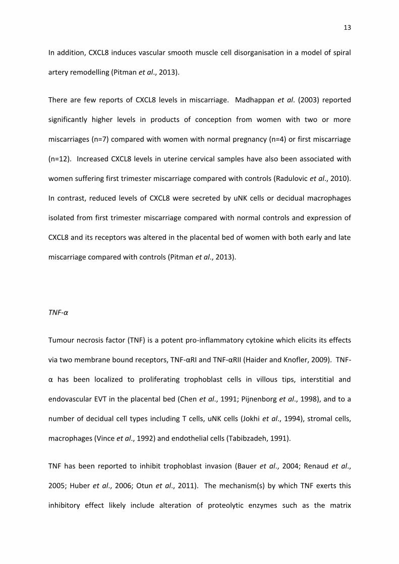
13
In addition, CXCL8 induces vascular smooth muscle cell disorganisation in a model of spiral
artery remodelling (Pitman et al., 2013).
There are few reports of CXCL8 levels in miscarriage. Madhappan et al. (2003) reported
significantly higher levels in products of conception from women with two or more
miscarriages (n=7) compared with women with normal pregnancy (n=4) or first miscarriage
(n=12). Increased CXCL8 levels in uterine cervical samples have also been associated with
women suffering first trimester miscarriage compared with controls (Radulovic et al., 2010).
In contrast, reduced levels of CXCL8 were secreted by uNK cells or decidual macrophages
isolated from first trimester miscarriage compared with normal controls and expression of
CXCL8 and its receptors was altered in the placental bed of women with both early and late
miscarriage compared with controls (Pitman et al., 2013).
TNF-α
Tumour necrosis factor (TNF) is a potent pro-inflammatory cytokine which elicits its effects
via two membrane bound receptors, TNF-αRI and TNF-αRII (Haider and Knofler, 2009). TNF-
α has been localized to proliferating trophoblast cells in villous tips, interstitial and
endovascular EVT in the placental bed (Chen et al., 1991; Pijnenborg et al., 1998), and to a
number of decidual cell types including T cells, uNK cells (Jokhi et al., 1994), stromal cells,
macrophages (Vince et al., 1992) and endothelial cells (Tabibzadeh, 1991).
TNF has been reported to inhibit trophoblast invasion (Bauer et al., 2004; Renaud et al.,
2005; Huber et al., 2006; Otun et al., 2011). The mechanism(s) by which TNF exerts this
inhibitory effect likely include alteration of proteolytic enzymes such as the matrix

14
metalloproteinases (MMP’s) (Bischof et al., 1995) and the urokinase plasminogen activator
system (Queenan et al., 1987) as well as altering EVT apoptosis or proliferation (von Rango
et al., 2003).
Few studies have investigated TNF in human miscarriage although it has been reported to
induce spontaneous abortion in rodents (Chaouat et al., 1990). More recently increased
decidual stromal cell secreted levels of TNF have been reported in women with spontaneous
miscarriage (Sun et al., 2013).
Other cytokines
Several other cytokines, such as IL-2, IL-1β, IL-17A, have also been shown to be dysregulated
in the decidua of women miscarriage (Sun et al., 2013; Garzia et al., 2013). However, their
roles in establishment of pregnancy have not been well defined, although they do play roles
in immune responses. Therefore, it is difficult to speculate whether they have a causative
role in the aetiology of miscarriage or are a consequence of this devastating condition.
9 Conclusions
Reduced decidual IL-6 and CXCL8/IL-8 as well as increased TNF-α levels may negatively
impact on the normal processes of pregnancy establishment by dysregulating control of
trophoblast invasion as well as aspects of spiral artery remodelling and therefore contribute
to the aetiology of both early and late miscarriage. However, these are very complex

15
processes and likely regulated by several different factors, leading to a multifactorial disease
state.
Miscarriage remains one of the most prevalent obstetrical complications with little known
about risk factors and aetiology. We review here the role that decidual cytokines may play
in this process, but owing to the lack of known risk factors or biomarkers for this condition,
there is a need for deeper insights into mechanisms before we will be able to translate this
knowledge into preventative therapies.

16
References
Aplin, J.D., 2000. The cell biological basis of human implantation. Baillieres Best Pract. Res.
Clin. Obstet. Gynaecol. 14, 757-764.
Ashkar, A.A., Di Santo, J.P., Croy, B.A., 2000. Interferon gamma contributes to initiation of
uterine vascular modification, decidual integrity, and uterine natural killer cell maturation
during normal murine pregnancy. J. Exp. Med. 192, 259-70.
Ball, E., Robson, S.C., Ayis, S., Lyall, F., Bulmer, J.N., 2006a. Early embryonic demise: no
evidence of abnormal spiral artery transformation or trophoblast invasion. J. Pathol. 208,
528-534.
Ball, E., Bulmer, J., Ayis, S., Lyall, F., Robson, S., 2006b. Late sporadic miscarriage is
associated with abnormalities in spiral artery transformation and trophoblast invasion. J.
Pathol. 208, 535-542.
Bauer, S., Pollheimer, J., Hartmann, J., Husslein, P., Aplin, J.D., Knofler, M., 2004. Tumor
necrosis factor-alpha inhibits trophoblast migration through elevation of plasminogen
activator inhibitor-1 in first-trimester villous explant cultures. J. Clin. Endocrinol. Metab. 89,
812-822.
Bischof, P., Martelli, M., Campana, A., Itoh, Y., Ogata, Y., Nagase, H., 1995. Importance of
matrix metalloproteinases in human trophoblast invasion. Early Pregnancy. 1, 263-269.
Bulmer, J.N., Morrison, L., Longfellow, M., Ritson, A., Pace, D., 1991. Granulated
lymphocytes in human endometrium: histochemical and immunohistochemical studies.
Hum. Reprod. 6, 791-798.

17
Champion, H., Innes, B.A., Robson, S.C., Lash, G.E., Bulmer, J.N., 2012. Effects of interleukin-
6 on extravillous trophoblast invasion in early human pregnancy. Mol. Hum. Reprod. 18,
391-400.
Chaouat, G., Menu, E., Kinsky, R., Brezin, C., 1990. Immunologically mediated abortions: one
or several pathways? Res. Immunol. 141, 188-195.
Chaouat, G., Zourbas, S., Ostojic, S., Lappree-Delage, G., Dubanchet, S., Ledee, N., Martal, J.,
2002. A brief review of recent data on some cytokine expressions at the materno-foetal
interface which might challenge the classical Th1/Th2 dichotomy. J. Reprod. Immunol. 53,
241-256.
Chen, H.L., Yang, Y.P., Hu, X.L., Yelavarthi, K.K., Fishback, J.L., Hunt, J.S., 1991. Tumor
necrosis factor alpha mRNA and protein are present in human placental and uterine cells at
early and late stages of gestation. Am. J. Pathol. 139, 327-335.
Confavreux, C., Hutchinson, M., Hours, M.M., Cortinovis-Tourniaire, P., Moreau, T., 1998.
Rate of pregnancy-related relapse in multiple sclerosis. Pregnancy in Multiple Sclerosis
Group. N. Engl. J. Med. 339, 285-291.
Croy, B.A., Esadeg, S., Chantakru, S., Van Den Heuvel, M., Paffaro, V.A., He, H., Black, G.P.,
Ashkar, A.A., Kiso, Y., Zhang, J., 2003. Update on pathways regulating the activation of
uterine Natural Killer cells, their interactions with decidual spiral arteries and homing of
their precursors to the uterus. J. Reprod. Immunol. 59, 175-191.
18
Damsky, C.H., Fitzgerald, M.L., Fisher, S.J., 1992. Distribution patterns of extracellular matrix
components and adhesion receptors are intricately modulated during first trimester
differentiation along the invasive pathway, in vivo. J. Clin. Invest. 89, 210-222.
Damsky, C.H., Librach, C., Lim, K.H., Fitzgerald, M.L., McMaster, M.T., Janatpour, M., Zhou,
Y., Logan, S.K., Fisher, S.J., 1994. Integrin switching regulates normal trophoblast invasion.
Development. 120, 3657-3666.
De Oliveira, L.G., Lash, G.E., Murray-Dunning, C., Bulmer, J.N., Innes, B.A., Searle, R.F., Sass,
N., Robson, S.C., 2010. Role of interleukin 8 in uterine natural killer cell regulation of
extravillous trophoblast cell invasion. Placenta. 31, 595-601.
Dimitriadis, E., White, C.A., Jones, R.L., Salamonsen, L.A., 2005. Cytokines, chemokines and
growth factors in endometrium related to implantation. Hum. Reprod. Update. 11, 613-630.
Drake, P.M., Gunn, M.D., Charo, I.F., Tsou, C.L., Zhou, Y., Huang, L., Fisher, S.J., 2001. Human
placental cytotrophoblasts attract monocytes and CD56(bright) natural killer cells via the
actions of monocyte inflammatory protein 1alpha. J. Exp. Med. 193, 1199-1212.
Ernerudh, J., Berg, G., Mjösberg, J., 2011. Regulatory T helper cells in pregnancy and their
roles in systemic versus local immune tolerance. Am. J. Reprod. Immunol. 66, 31-43.
Fallon, P.G., Jolin, H.E., Smith, P., Emson, C.L., Townsend, M.J., Fallon, R., Smith, P.,
McKenzie, A.N., 2002. IL-4 induces characteristic Th2 responses even in the combined
absence of IL-5, IL-9, and IL-13. Immunity. 17, 7-17.

19
Galazios, G., Tsoulou, S., Zografou, C., Tripsianis, G., Koutlaki, N., Papazoglou, D., Tsikouras,
P., Maltezos, E., Liberis, V., 2011. The role of cytokines IL-6 and IL-8 in the pathogenesis of
spontaneous abortions. J. Matern. Fetal Neonatal. Med. 24, 1283-1285.
Gardner, L., Moffett, A., 2003. Dendritic cells in the human decidua. Biol. Reprod. 69, 1438-
1446.
Garzia, E., Clauser, R., Persani, L., Borgato, S., Bulfamante, G., Avagliano, L., Quadrelli, F.,
Marconi, A.M., 2013. Prolactin and proinflammatory cytokine expression at the
fetomaternal interface in first trimester miscarriage. Fertil. Steril. 100, 108-115.
Gustafsson, C., Mjösberg, J., Matussek, A., Geffers, R., Matthiesen, L., Berg, G., Sharma, S.,
Buer, J., Ernerudh, J., 2008. Gene expression profiling of human decidual macrophages:
evidence for immunosuppressive phenotype. PLoS One. 3, e2078.
Haider, S., Knöfler, M. 2009. Human tumour necrosis factor: physiological and pathological
roles in placenta and endometrium. Placenta. 30, 111-123.
Hanna, J., Goldman-Wohl, D., Hamani, Y., Avraham, I., Greenfield, C., Natanson-Yaron, S.,
Prus, D., Cohen-Daniel, L., Arnon, T.I., Manaster, I. et al., 2006. Decidual NK cell regulate key
developmental processes at the human fetal-maternal interface. Nat. Med. 12, 1065-1074.
Heikkinen, J., Möttönen, M., Komi, J., Alanen, A., Lassila, O., 2003. Phenotypic
characterization of human decidual macrophages. Clin. Exp. Immunol. 131, 498-505.
Hornung, D., Ryan, I.P., Chao, V.A., Vigne, J.L., Schriock, E.D., Taylor, R.N., 1997.
Immunolocalization and regulation of the chemokine RANTES in human endometrial and
endometriosis tissues and cells. J. Clin. Endocrinol. Metab. 82, 1621-1628.

20
Huber, A.V., Saleh, L., Bauer, S., Husslein, P., Knofler, M., 2006. TNFalpha-mediated
induction of PAI-1 restricts invasion of HTR-8/SVneo trophoblast cells. Placenta. 27, 127-
136.
Hustin, J., Jauniaux, E., Schaaps, J.P., 1990. Histological study of the materno-embryonic
interface in spontaneous abortion. Placenta. 11, 477-486.
Hustin, J., Kadri, R., Jauniaux, E., 1996. Spontaneous and habitual abortion--a pathologist's
point of view. Early Pregnancy. 2, 85-95.
Jauniaux, E., Hustin, J., 1992. Histological examination of first trimester spontaneous
abortions: the impact of materno-embryonic interface features. Histopathology. 21, 409-
414.
Jauniaux, E., Zaidi, J., Jurkovic, D., Campbell, S., Hustin, J., 1994. Comparison of colour
Doppler features and pathological findings in complicated early pregnancy. Hum. Reprod. 9,
2432-2437.
Jauniaux, E., Gulbis, B., Schandene, L., Collette, J., Hustin, J., 1996. Distribution of
interleukin-6 in maternal and embryonic tissues during the first trimester. Mol. Hum.
Reprod. 2, 239-243.
Jauniaux, E., Watson, A.L., Hempstock, J., Bao, Y.P., Skepper, J.N., Burton, G.J., 2000. Onset
of maternal arterial blood flow and placental oxidative stress. A possible factor in human
early pregnancy failure. Am. J. Pathol. 157, 2111-2122.

21
Jokhi, P.P., King, A., Sharkey, A.M., Smith, S.K., Loke, Y.W., 1994. Screening for cytokine
messenger ribonucleic acids in purified human decidual lymphocyte populations by the
reverse-transcriptase polymerase chain reaction. J. Immunol. 153, 4427-4435.
Jones, R.L., Hannan, N.J., Kaitu’u, T.J., Zhang, J., Salamonsen, L.A., 2004. Identification of
chemokines important for leukocyte recruitment to the human endometrium at the times
of embryo implantation and menstruation. J. Clin. Endocrinol. Metab. 89, 6155-6167.
Jovanovic, M., Vicovac, L., 2009. Interleukin-6 stimulates cell migration, invasion and
integrin expression in HTR-8/SVneo cell line. Placenta. 30, 320-328.
Khong, T.Y., Liddell, H.S., Robertson, W.B., 1987. Defective haemochorial placentation as a
cause of miscarriage: a preliminary study. Br. J. Obstet. Gynaecol. 94, 649-655.
Lash, G.E., Schiessl, B., Kirkley, M., Innes, B.A., Cooper, A., Searle, R.F., Robson, S.C., Bulmer,
J.N., 2006. Expression of angiogenic growth factors by uterine natural killer cells during early
pregnancy. J. Leukoc. Biol. 80, 572-580.
Lidström, C., Matthiesen, L., Berg, G., Sharma, S., Ernerudh, J., Ekerfelt, C., 2003. Cytokine
secretion patterns of NK cells and macrophages in early human pregnancy decidua and
blood: implications for suppressor macrophages in decidua. Am. J. Reprod. Immunol. 50,
444-452.
Madhappan, B., Kempuraj, D., Christodoulou, S., Tsapikidis, S., Boucher, W., Karagiannis, V.,
Athanassiou, A., Theoharides, T.C., 2003. High levels of intrauterine corticotropin-releasing
hormone, urocortin, tryptase, and interleukin-8 in spontaneous abortions. Endocrinology.
144, 2285-2290.

22
Meisser, A., Cameo, P., Islami, D., Campana, A., Bischof, P., 1999. Effects of interleukin-6 (IL-
6) on cytotrophoblastic cells. Mol. Hum. Reprod. 5, 1055-1058.
Mjösberg, J., Berg, G., Jenmalm, M.C., Ernerudh, J., 2010. FOXP3+ regulatory T cells and T
helper 1, T helper 2, and T helper 17 cells in human early pregnancy decidua. Biol. Reprod.
82, 698-705.
Murphy, S.P., Tayade, C., Ashkar, A.A., Hatta, K., Zhang, J., Croy, B.A., 2009. Interferon
gamma in successful pregnancies. Biol. Reprod. 80, 848-859.
Ostensen, M., Villiger, P.M., 2007. The remission of rheumatoid arthritis during pregnancy.
Semin. Immunopathol. 29, 185-191.
Otun, H.A., Lash, G.E., Innes, B.A., Bulmer, J.N., Naruse, K., Hannon, T., Searle, R.F., Robson,
S.C., 2011. Effect of tumour necrosis factor-alpha in combination with interferon-gamma on
first trimester extravillous trophoblast invasion. J. Reprod. Immunol. 88, 1-11.
Piccinni, M.P., Scaletti, C., Maggi, E., Romagnani, S., 2000. Role of hormone-controlled Th1-
and Th2-type cytokines in successful pregnancy. J. Neuroimmunol. 109, 30-33.
Pijnenborg, R., Anthony, J., Davey, D.A., Rees, A., Tiltman, A., Vercruysse, L., van Assche, A.,
1991. Placental bed spiral arteries in the hypertensive disorders of pregnancy. Br. J. Obstet.
Gynaecol. 98, 648-655.
Pijnenborg, R., McLaughlin, P.J., Vercruysse, L., Hanssens, M., Johnson, P.M., Keith, J.C. Jr.,
Van Assche, F.A., 1998. Immunolocalization of tumour necrosis factor-alpha (TNF-alpha) in
the placental bed of normotensive and hypertensive human pregnancies. Placenta. 19, 231-
239.

23
Pijnenborg, R., Vercruysse, L., Hanssens, M., 2006. The uterine spiral arteries in human
pregnancy: facts and controversies. Placenta. 27, 939-958.
Pitman, H., Innes, B.A., Robson, S.C., Bulmer, J.N., Lash, G.E., 2013. Altered expression of
interleukin-6, interleukin-8 and their receptors in decidua of women with sporadic
miscarriage. Hum. Reprod. 28, 2075-2086.
Plevyak, M., Hanna, N., Mayer, S., Murphy, S., Pinar, H., Fast, L., Ekerfelt, C., Ernerudh, J.,
Berg, G., Matthiesen, L., Sharma, S., 2002. Deficiency of decidual IL-10 in first trimester
missed abortion: a lack of correlation with the decidual immune cell profile. Am. J. Reprod.
Immunol. 47, 242-250.
Queenan, J.T. Jr., Kao, L.C., Arboleda, C.E., Ulloa-Aguirre, A., Golos, T.G., Cines, D.B., Strauss,
J.F. 3rd., 1987. Regulation of urokinase-type plasminogen activator production by cultured
human cytotrophoblasts. J. Biol. Chem. 262, 10903-10906.
Radulovic, N.V., Ekerhovd, E., Abrahamsson, G., Norstrom, A., 2010. Cervical tissue changes
in women with miscarriage: a morphological and biochemical investigation. Acta. Obstet.
Gynecol. Scand. 89, 54-64.
Renaud, S.J., Postovit, L.M., Macdonald-Goodfellow, S.K., McDonald, G.T., Caldwell, J.D.,
Graham, C.H., 2005. Activated macrophages inhibit human cytotrophoblast invasiveness in
vitro. Biol. Reprod. 73, 237-243.
Robson, A., Harris, L.K., Innes, B.A., Lash, G.E., Aljunaidy, M.M., Aplin, J.D., Baker, P.N.,
Robson, S.C., Bulmer, J.N., 2012. Uterine natural killer cells initiate spiral artery remodeling
in human pregnancy. FASEB J. 26, 4876-4885.

24
Saito, S., Kasahara, T., Sakakura, S., Enomoto, M., Umekage, H., Harada, N., Morii, T.,
Nishikawa, K., Narita, N., Ichijo, M., 1994. Interleukin-8 production by CD16-CD56bright
natural killer cells in the human early pregnancy decidua. Biochem. Biophys. Res. Commun.
200, 378-383.
Salamonsen, L.A., Hannan, N.J., Dimitriadis, E., 2007. Cytokines and chemokines during
human embryo implantation: roles in implantation and early placentation. Semin. Reprod.
Med. 25, 437-444.
Shimoya, K., Moriyama, A., Matsuzaki, N., Ogata, I., Koyama, M., Azuma, C., Saji, F., Murata,
Y., 1999. Human placental cells show enhanced production of interleukin (IL)-8 in response
to lipopolysaccharide (LPS), IL-1 and tumour necrosis factor (TNF)-alpha, but not to IL-6.
Mol. Hum. Reprod. 5, 885.
Sun, C., Zhang, Y.Y., Tang, C.L., Wang, S.C., Piao, H.L., Tao, Y., Zhu, R., Du, M.R., Li, D.J., 2013.
Chemokine CCL28 induces apoptosis of decidual stromal cells via binding CCR3/CCR10 in
human spontaneous abortion. Mol. Hum. Reprod. 19, 676-686.
Svensson, J., Jenmalm, M.C., Matussek, A., Geffers, R., Berg, G., Ernerudh, J., 2011.
Macrophages at the fetal-maternal interface express markers of alternative activation and
are induced by M-CSF and IL-10. J. Immunol. 187, 3671-3682.
Svensson-Arvelund, J., Ernerudh, J., 2014. The Role of Macrophages in Promoting and
Maintaining Homeostasis at the Fetal-Maternal Interface. Am. J. Reprod. Immunol. In Press.
Tabibzadeh, S., 1991. Human endometrium: an active site of cytokine production and
action. Endocr. Rev. 12, 272-290.

25
Tabibzadeh, S., Kong, Q.F., Babaknia, A., May, L.T., 1995. Progressive rise in the expression
of interleukin-6 in human endometrium during menstrual cycle is initiated during the
implantation window. Hum. Reprod. 10, 2793-2799.
Takata, H., Tomiyama, H., Fujiwara, M., Kobayashi, N., Takiguchi, M. 2004. Cutting edge:
expression of chemokine receptor CXCR1 on human effector CD8+ T cells. J. Immunol. 173,
2231-2235.
Tsuda, H., Sakai, M., Michimata, T., Tanebe, K., Hayakawa, S., Saito, S., 2001.
Characterization of NKT cells in human peripheral blood and decidual lymphocytes. Am. J.
Reprod. Immunol. 45, 295-302.
Vince, G., Shorter, S., Starkey, P., Humphreys, J., Clover, L., Wilkins, T., Sargent, I., Redman,
C., 1992. Localization of tumour necrosis factor production in cells at the materno/fetal
interface in human pregnancy. Clin. Exp. Immunol. 88, 174-180.
Vince, G.S., Johnson, P.M., 2000. Leucocyte populations and cytokine regulation in human
uteroplacental tissues. Biochem. Soc. Trans. 28, 191-195.
Von Rango, U., Classen-Linke, I., Raven, G., Bocken, F., Beier, H.M., 2003. Cytokine
microenvironments in human first trimester decidua are dependent on trophoblast cells.
Fertil. Steril. 79, 1176-1186.
Wegmann, T.G., Lin, H., Guilbert, L., Mosmann, T.R., 1993. Bidirectional cytokine
interactions in the maternal-fetal relationship: is successful pregnancy a TH2 phenomenon?
Immunol. Today. 14, 353-356.

26
Wu, H.X., Jin, L.P., Xu, B., Liang, S.S., Li, D.J., 2014. Decidual stromal cells recruit Th17 cells
into decidua to promote proliferation and invasion of human trophoblast cells by secreting
IL-17. Cell. Mol. Immunol. 11, 253-262.
Zenclussen, A.C., Blois, S., Stumpo, R., Olmos, S., Arias, K., Malan Borel, I., Roux, M.E.,
Margni, R.A., 2003. Murine abortion is associated with enhanced interleukin-6 levels at the
feto-maternal interface. Cytokine. 24, 150-160.

27
Table 1: Summary of the potential role of altered decidual cytokines in the
pathophysiology of spontaneous miscarriage.
Cytokine Role in early pregnancy Levels in spontaneous miscarriage
IL-6 EVT invasion – variable results from
in vitro assay systems.
Spiral artery remodelling – induces
VSMC disorganisation.
Serum - increased.
Decidual leucocytes (uNK cells and
macrophages) - decreased.
CXCL8/IL-8 EVT invasion – stimulatory.
Spiral artery remodelling – induces
VSMC disorganisation.
Products of conception - increased.
Decidual leucocytes (uNK cells and
macrophages) - decreased.
TNF-α EVT invasion – inhibitory. Decidual stromal levels – increased.


![Inflammatory cytokines as predictive markers for early detection … · development of diabetic microvascular complications in-cluding nephropathy [10–12]. Accordingly, it is suggested](https://img.pdfslide.net/doc/110x75/5f896f573ef90e24204795c8/inflammatory-cytokines-as-predictive-markers-for-early-detection-development-of.jpg)