Embed Size (px)
Citation preview
Decoding Humor Experiences from Brain Activity ofPeople Viewing Comedy MoviesYasuhito Sawahata*, Kazuteru Komine, Toshiya Morita, Nobuyuki Hiruma
Science and Technology Research Laboratories, NHK (Japan Broadcasting Corporation), Setagaya, Tokyo, Japan
Abstract
Humans naturally have a sense of humor. Experiencing humor not only encourages social interactions, but also producespositive physiological effects on the human body, such as lowering blood pressure. Recent neuro-imaging studies haveshown evidence for distinct mental state changes at work in people experiencing humor. However, the temporalcharacteristics of these changes remain elusive. In this paper, we objectively measured humor-related mental states fromsingle-trial functional magnetic resonance imaging (fMRI) data obtained while subjects viewed comedy TV programs.Measured fMRI data were labeled on the basis of the lag before or after the viewer’s perception of humor (humor onset)determined by the viewer-reported humor experiences during the fMRI scans. We trained multiple binary classifiers, ordecoders, to distinguish between fMRI data obtained at each lag from ones obtained during a neutral state in whichsubjects were not experiencing humor. As a result, in the right dorsolateral prefrontal cortex and the right temporal area,the decoders showed significant classification accuracies even at two seconds ahead of the humor onsets. Furthermore,given a time series of fMRI data obtained during movie viewing, we found that the decoders with significant performancewere also able to predict the upcoming humor events on a volume-by-volume basis. Taking into account the hemodynamicdelay, our results suggest that the upcoming humor events are encoded in specific brain areas up to about five secondsbefore the awareness of experiencing humor. Our results provide evidence that there exists a mental state lasting for a fewseconds before actual humor perception, as if a viewer is expecting the future humorous events.
Citation: Sawahata Y, Komine K, Morita T, Hiruma N (2013) Decoding Humor Experiences from Brain Activity of People Viewing Comedy Movies. PLoS ONE 8(12):e81009. doi:10.1371/journal.pone.0081009
Editor: Masahiko Sumitani, The University of Tokyo Hospital, Japan
Received May 9, 2013; Accepted October 9, 2013; Published December 4, 2013
Copyright: � 2013 Sawahata et al. This is an open-access article distributed under the terms of the Creative Commons Attribution License, which permitsunrestricted use, distribution, and reproduction in any medium, provided the original author and source are credited.
Funding: The authors have no support or funding to report.
Competing Interests: The authors have declared that no competing interests exist.
* E-mail: [email protected]
Introduction
A sense of humor is a common human characteristic that people
in many cultures experience. Humor not only encourages social
interaction, but also produces positive effects for the human body,
such as healthy physiological changes [1]. Comedians and movie
directors work hard to stimulate humor in audiences. Would a
better understanding of the mechanisms of humor perception help
comedians and movie directors better amuse audiences? Previous
psychological studies have suggested that there are mental stages
associated with experiencing humor [2,3]. Furthermore, recent
neuro-imaging studies have revealed physiological evidence for a
relationship between mental stages and experiencing humor [4–
18]. For example, Chan et al. examined fMRI activity while
audiences listened to several humorous short stories, and showed
that humor-related mental states correlated with activity in distinct
brain areas; incongruity detection, resolution and elaboration of
humor were respectively involved in the right middle temporal
gyrus and right medial frontal gyrus, the left superior frontal gyrus
and left inferior parietal lobule, and the left ventromedial
prefrontal cortex, the bilateral parahippocampal gyri and the
bilateral amygdale [16,18]. Such physiological measurements for
experiencing humor should be of value to the creators of comedy
shows and movies, because it would help them to know the
detailed reactions of audiences and the objective value of their
products.
It is a challenging task to objectively measure the dynamic
mental events underlying such subjective experiences during
movie watching. With advances in neuro-imaging technologies,
a great number of studies [4–18] have successfully measured brain
activity related to the perception of humor. However, these studies
did not focus on a single humorous event but on multiple
humorous events within intentionally designed experiments, as
they sought only to the distinguish brain activity in the humorous
trials from that in the non-humorous trials. If it is possible to
monitor the viewer’s mental states on a volume-by-volume basis of
fMRI images, we can expect that the results will not only be of
value to the creators of comedies but also improve our
understanding of the dynamic processes in the brain related to
humor.
To predict humor-related mental events, we analyzed fMRI
data using a decoding approach [19–22] that classifies given brain
activity patterns into pre-defined brain states by using a statistical
machine learning algorithm. A general linear model (GLM)-based
approach [23], which is often used in neuro-imaging studies about
humor, is not suitable for mental event prediction, since it is an
encoding model that predicts brain activity from stimuli,
experimental or task variables [24]; the modeling direction is
opposite from ours. By using a decoding approach, once a
mapping from brain activity patterns to humor-related mental
states is learned, the mental states of viewers can be predicted from
individual fMRI volumes taken while they are watching comedic
PLOS ONE | www.plosone.org 1 December 2013 | Volume 8 | Issue 12 | e81009
situations. It is reasonable to take the decoding approach to
measure the subjective mental state changes that cannot be
externally observed.
Recent neuro-imaging studies [16,18] have shown that the
neural correlates of humor processing have two stages: compre-
hension and elaboration. These studies suggested that there should
be humor-related mental state changes before and after humorous
events. However, the time scale of the dynamic humor processing
remains elusive. Since a mental state can be predicted from single-
trial brain activity data by using the decoding approach, we can
expect that the characteristics of the mental state changes can be
extracted with the same temporal resolution as that of the brain
activity data.
In the present study, we investigated the dynamic mental state
changes by applying the decoding approach to single-trial fMRI
data obtained while subjects viewed comedy TV programs. We
hypothesized that each single-trial observation data observed
during humor experiences may have distinct information from
that observed during ‘neutral’ states in which a viewer did not
experience humor. We conducted an fMRI experiment in which a
viewer watched comedy TV programs in an fMRI scanner and
reported the humor levels that he or she experienced. Regions of
interest (ROIs) were defined in each subject by dividing the whole
brain into sub-regions based on anatomical landmarks. We
constructed multiple decoders for each ROI at each lag before
and after the onset of a humor report in order to investigate
whether brain activity patterns obtained before and/or after the
humor perception and during neutral states can be classified on a
trial-by-trial basis. By testing the performance of each trained
decoder, we could predict where in the brain and when the
humor-related information would be elicited while viewing
comedy TV shows.
Materials and Methods
SubjectsTen healthy adults (mean age 28.267.99 years, range 20–44; 7
men and 3 women) participated in the movie-viewing experiment.
Ten healthy adults (Four of them also participated in the movie-
viewing experiment; mean age 27.766.77 years, range 20–44; 8
men and 2 women) participated in the control experiment. All the
subjects were native Japanese speakers, and had no history of
psychiatric or neurological disorders. The subjects gave written
informed consent and the study was approved by the Ethics
Committee of the NHK Science and Technology Research
Laboratories.
StimuliTen stand-up comedies, or manzai in Japanese, in which small
groups of comedians performed funny dialogues were selected
from comedy TV shows, called ‘On-air Battle’, broadcast on NHK
TV in Japan. Laughter of audiences in the recording hall was also
included in the movies. Each movie was about 4.5 minutes long.
Visual stimuli were presented within 10u610u visual angles, rear-
projected onto a screen placed in the scanner bore using an LCD
projector and viewed via a mirror mounted on the head coil.
Audio stimuli were given by MRI compatible head-phones.
Design and TasksMovie-viewing experiment. In each run, a movie was presented
between a 32 s initial rest period and a 30 s closing rest period in
which a fixation cross on a gray background was shown at the
center of the screen. Hence, each run had about 5.5 minutes of
stimulus presentation in total. During the rest periods, subjects
fixated on the cross. During the movie-viewing periods, subjects
viewed stimuli (the standup comedies) without fixation and
reported the magnitude of humor they were experiencing by
manipulating a slider-style response device (fORP; Cambridge
Research Systems, Rochester, Kent, UK). Subjects were instructed
not to move their head even if they wanted to laugh.
Control experiment. To investigate whether decoding results
were derived from humor processing or manipulations of the
response device, we conducted control experiments in which
subjects manipulated the response device without viewing the
comedy movies. Subjects were instructed to manipulate the slider
device so as to mimic the motion of a cartoon slider tab shown in a
screen. The control experiment involved a 32 s initial rest block
followed by a 16 s manipulation block and a 16 s rest block
repeated six times per run. In total, eight runs were conducted. In
each manipulation block, the cartoon slider was moved period-
ically at 0.5 Hz.
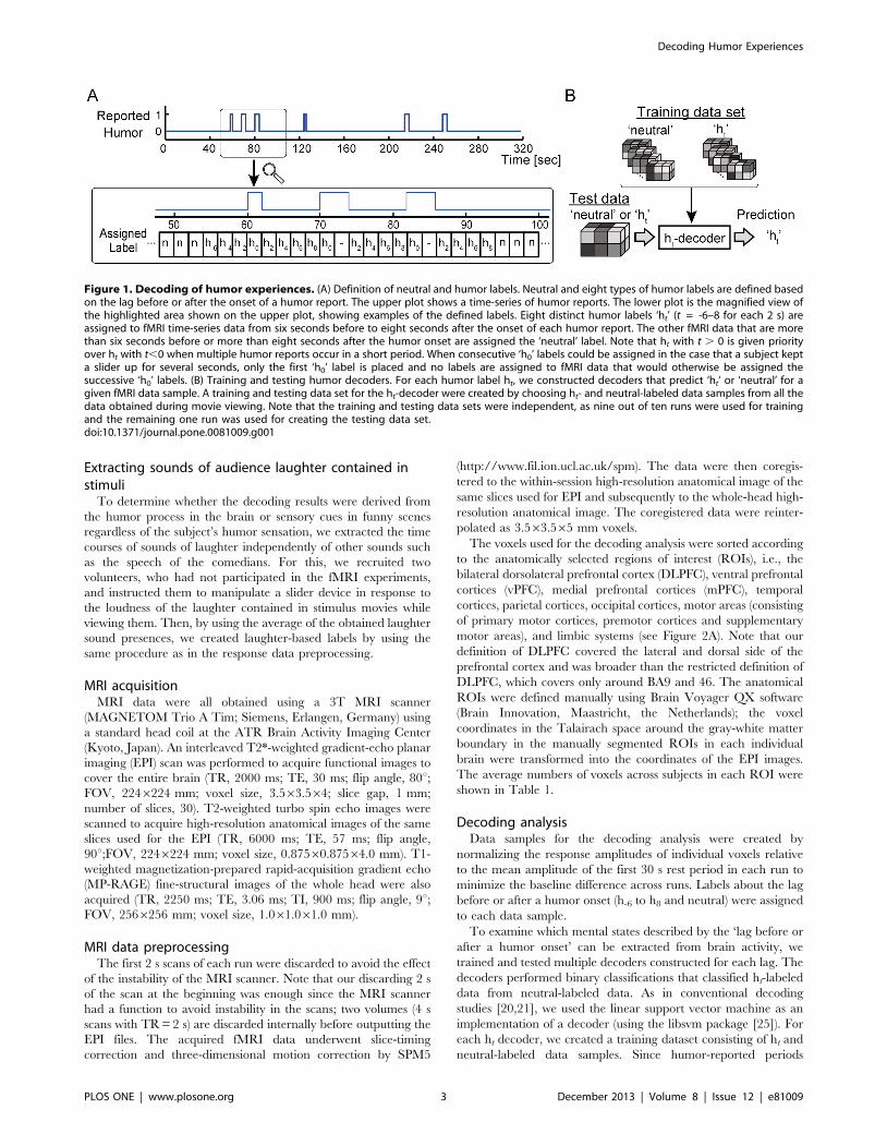
Response data preprocessingResponses from a slider device were converted into labels for
fMRI data based on the lag before or after a humor onset (Figure
1). First, measured response values (sampled with 100 Hz) were
binarized into two clusters, ‘‘presence’’ or ‘‘non-presence’’ of slider
manipulation, by applying k-means clustering with k = 2 to the
response values of all runs. Next, to associate response data with
the simultaneously observed fMRI images, we sorted the binarized
responses into 2 s long bins and regarded a bin with at least one
slider manipulation as a slider-manipulated bin. The slider-
manipulated bins were thus defined as humor onsets and assigned
‘h0’ labels (0 s to the humor onset). Subsequently, time points that
were t s to the nearest humor onset were assigned ‘ht’ labels (t s to
the humor onset; -6 # t # 8 for each 2 s). ‘Neutral’ labels were
assigned to bins that were temporally far away from the humor
onset (more than 6 s before or 8 s after the humor onset). Note that
ht with t . 0 is given priority over ht with t,0 when the labeling
criteria indicated that multiple labels could be assigned to a bin.
Furthermore, in the cases that consecutive ‘h0’ labels could be
assigned to data samples by the above labeling criteria when
subjects kept the slider up for several seconds, only the first ‘h0’
label was assigned and no labels were assigned to the samples that
were supposed to be assigned the subsequent ‘h0’ labels. Hence,
the fMRI data associated with ht labels (t # 0) were always the
ones obtained before or simultaneously with the humor onsets.
Monitoring changes in facial expressionTo take into account artifacts caused by facial expressions or
head motions, four light-reflective markers were attached around
the subject’s mouth (both corners of the mouth and edges of the
lower lip). By using an infrared camera (SONY HDR-CX700V;
used in the night shot mode), marker motions were recorded with
192061080 pixels at 60 frames per second from the side of the
scanner-bed through the mirror mounted on the head-coil (i.e. two
mirrors were mounted on the head-coil: one for stimulus-viewing
and another for marker motion recording).
We created the alternative labels from the time series of the
marker positions. First, each coordinate value of markers was
normalized relative to the averaged coordinate value over a run.
Next, the relative values of the marker positions were converted
into one-dimensional signals by computing their squared sum at
each time point. Then, the signals underwent linear trend removal
within each run. The motion-based labels were extracted using the
same procedure described in the response data preprocessing, but
the response data were substituted with the computed time series
data of the marker positions.
Decoding Humor Experiences
PLOS ONE | www.plosone.org 2 December 2013 | Volume 8 | Issue 12 | e81009
Extracting sounds of audience laughter contained instimuli
To determine whether the decoding results were derived from
the humor process in the brain or sensory cues in funny scenes
regardless of the subject’s humor sensation, we extracted the time
courses of sounds of laughter independently of other sounds such
as the speech of the comedians. For this, we recruited two
volunteers, who had not participated in the fMRI experiments,
and instructed them to manipulate a slider device in response to
the loudness of the laughter contained in stimulus movies while
viewing them. Then, by using the average of the obtained laughter
sound presences, we created laughter-based labels by using the
same procedure as in the response data preprocessing.
MRI acquisitionMRI data were all obtained using a 3T MRI scanner
(MAGNETOM Trio A Tim; Siemens, Erlangen, Germany) using
a standard head coil at the ATR Brain Activity Imaging Center
(Kyoto, Japan). An interleaved T2*-weighted gradient-echo planar
imaging (EPI) scan was performed to acquire functional images to
cover the entire brain (TR, 2000 ms; TE, 30 ms; flip angle, 80u;FOV, 2246224 mm; voxel size, 3.563.564; slice gap, 1 mm;
number of slices, 30). T2-weighted turbo spin echo images were
scanned to acquire high-resolution anatomical images of the same
slices used for the EPI (TR, 6000 ms; TE, 57 ms; flip angle,
90u;FOV, 2246224 mm; voxel size, 0.87560.87564.0 mm). T1-
weighted magnetization-prepared rapid-acquisition gradient echo
(MP-RAGE) fine-structural images of the whole head were also
acquired (TR, 2250 ms; TE, 3.06 ms; TI, 900 ms; flip angle, 9u;FOV, 2566256 mm; voxel size, 1.061.061.0 mm).
MRI data preprocessingThe first 2 s scans of each run were discarded to avoid the effect
of the instability of the MRI scanner. Note that our discarding 2 s
of the scan at the beginning was enough since the MRI scanner
had a function to avoid instability in the scans; two volumes (4 s
scans with TR = 2 s) are discarded internally before outputting the
EPI files. The acquired fMRI data underwent slice-timing
correction and three-dimensional motion correction by SPM5
(http://www.fil.ion.ucl.ac.uk/spm). The data were then coregis-
tered to the within-session high-resolution anatomical image of the
same slices used for EPI and subsequently to the whole-head high-
resolution anatomical image. The coregistered data were reinter-
polated as 3.563.565 mm voxels.
The voxels used for the decoding analysis were sorted according
to the anatomically selected regions of interest (ROIs), i.e., the
bilateral dorsolateral prefrontal cortex (DLPFC), ventral prefrontal
cortices (vPFC), medial prefrontal cortices (mPFC), temporal
cortices, parietal cortices, occipital cortices, motor areas (consisting
of primary motor cortices, premotor cortices and supplementary
motor areas), and limbic systems (see Figure 2A). Note that our
definition of DLPFC covered the lateral and dorsal side of the
prefrontal cortex and was broader than the restricted definition of
DLPFC, which covers only around BA9 and 46. The anatomical
ROIs were defined manually using Brain Voyager QX software
(Brain Innovation, Maastricht, the Netherlands); the voxel
coordinates in the Talairach space around the gray-white matter
boundary in the manually segmented ROIs in each individual
brain were transformed into the coordinates of the EPI images.
The average numbers of voxels across subjects in each ROI were
shown in Table 1.
Decoding analysisData samples for the decoding analysis were created by
normalizing the response amplitudes of individual voxels relative
to the mean amplitude of the first 30 s rest period in each run to
minimize the baseline difference across runs. Labels about the lag
before or after a humor onset (h-6 to h8 and neutral) were assigned
to each data sample.
To examine which mental states described by the ‘lag before or
after a humor onset’ can be extracted from brain activity, we
trained and tested multiple decoders constructed for each lag. The
decoders performed binary classifications that classified ht-labeled
data from neutral-labeled data. As in conventional decoding
studies [20,21], we used the linear support vector machine as an
implementation of a decoder (using the libsvm package [25]). For
each ht decoder, we created a training dataset consisting of ht and
neutral-labeled data samples. Since humor-reported periods
Figure 1. Decoding of humor experiences. (A) Definition of neutral and humor labels. Neutral and eight types of humor labels are defined basedon the lag before or after the onset of a humor report. The upper plot shows a time-series of humor reports. The lower plot is the magnified view ofthe highlighted area shown on the upper plot, showing examples of the defined labels. Eight distinct humor labels ‘ht’ (t = -6–8 for each 2 s) areassigned to fMRI time-series data from six seconds before to eight seconds after the onset of each humor report. The other fMRI data that are morethan six seconds before or more than eight seconds after the humor onset are assigned the ‘neutral’ label. Note that ht with t . 0 is given priorityover ht with t,0 when multiple humor reports occur in a short period. When consecutive ‘h0’ labels could be assigned in the case that a subject kepta slider up for several seconds, only the first ‘h0’ label is placed and no labels are assigned to fMRI data that would otherwise be assigned thesuccessive ‘h0’ labels. (B) Training and testing humor decoders. For each humor label ht, we constructed decoders that predict ‘ht‘ or ‘neutral’ for agiven fMRI data sample. A training and testing data set for the ht-decoder were created by choosing ht- and neutral-labeled data samples from all thedata obtained during movie viewing. Note that the training and testing data sets were independent, as nine out of ten runs were used for trainingand the remaining one run was used for creating the testing data set.doi:10.1371/journal.pone.0081009.g001
Decoding Humor Experiences
PLOS ONE | www.plosone.org 3 December 2013 | Volume 8 | Issue 12 | e81009
tended to be short compared to the entire duration of a movie and
the ratio between the number of ht and neutral-labeled data
samples in a training dataset was very biased, we equalized the
number of samples with both labels by omitting randomly chosen
samples with neutral labels. Although we could have created a
training dataset without equalizing the sample sizes, we chose to
equalize them because our preliminary analysis showed that
equalized datasets performed better in terms of prediction
accuracy and processing time. Taking into account the variability
of the training dataset created by the random sampling, we
obtained a decoder in three steps. First, we created 500 different
training datasets by random sampling. Next, we trained multiple
decoders independently for each training dataset. Then, we
determined a single decoder by averaging the weight parameters
of the trained multiple decoders. To evaluate the prediction
accuracy of each ht decoder, we created a test dataset consisting of
ht and neutral-labeled samples. It should be noted that the label
information was only used to create a test dataset and it was
hidden from the decoders. To evaluate the decoding accuracy
using a test dataset with a biased number of samples in each class,
we computed the receiver operating characteristic (ROC) curve on
the basis of decision values for test samples and obtained the area
under the ROC curve (AUC), which represents the performance
of a pattern classifier by a value between 0 and 1 (chance level,
AUC = 0.5; perfect classification, AUC = 1.0). The decoding
accuracy was computed in a cross validation manner whereby
data samples in one run were used to test a decoder trained with
the data samples from all other runs, and this training-test set was
repeated for all runs.
Subsequently, to determine whether the decoders could predict
the viewer’s mental state even from the observed time-series fMRI
data, all data samples in a test run were given to the decoders that
showed above chance-level performance in the former analysis.
Predictions were made in a similar way to the cross-validation
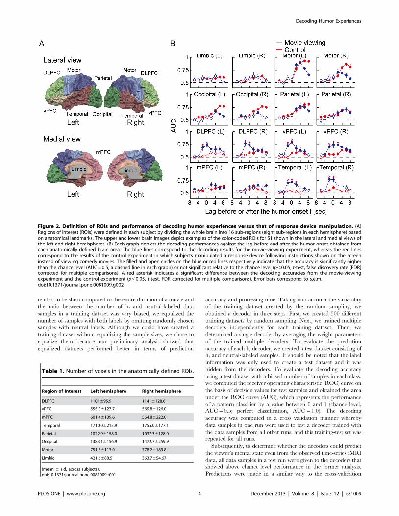
Figure 2. Definition of ROIs and performance of decoding humor experiences versus that of response device manipulation. (A)Regions of interest (ROIs) were defined in each subject by dividing the whole brain into 16 sub-regions (eight sub-regions in each hemisphere) basedon anatomical landmarks. The upper and lower brain images depict examples of the color-coded ROIs for S1 shown in the lateral and medial views ofthe left and right hemispheres. (B) Each graph depicts the decoding performances against the lag before and after the humor-onset obtained fromeach anatomically defined brain area. The blue lines correspond to the decoding results for the movie-viewing experiment, whereas the red linescorrespond to the results of the control experiment in which subjects manipulated a response device following instructions shown on the screeninstead of viewing comedy movies. The filled and open circles on the blue or red lines respectively indicate that the accuracy is significantly higherthan the chance level (AUC = 0.5; a dashed line in each graph) or not significant relative to the chance level (p,0.05, t-test, false discovery rate [FDR]corrected for multiple comparisons). A red asterisk indicates a significant difference between the decoding accuracies from the movie-viewingexperiment and the control experiment (p,0.05, t-test, FDR corrected for multiple comparisons). Error bars correspond to s.e.m.doi:10.1371/journal.pone.0081009.g002
Table 1. Number of voxels in the anatomically defined ROIs.
Region of Interest Left hemisphere Right hemisphere
DLPFC 1101695.9 11416128.6
vPFC 555.06127.7 569.86126.0
mPFC 601.46109.6 564.86222.0
Temporal 1710.06213.9 1755.06177.1
Parietal 1022.96158.0 1037.36128.0
Occpital 1383.16156.9 1472.76259.9
Motor 751.56113.0 778.26189.8
Limbic 421.6688.5 363.7654.67
(mean 6 s.d. across subjects).doi:10.1371/journal.pone.0081009.t001
Decoding Humor Experiences
PLOS ONE | www.plosone.org 4 December 2013 | Volume 8 | Issue 12 | e81009

manner described above; data samples in one run were input into
a decoder trained with the data samples from all the other runs,
and this training-prediction set was repeated for all combinations.
In a separate analysis to determine whether the decoding
accuracies were from humor processes in the brain or from other
artifacts, such as facial expression changes or head motion elicited
by laughing, we conducted decoding analyses with the alternative
labels extracted from facial marker motions instead of the original
labels extracted from the humor reports of the subjects. Finally, the
prediction accuracies of the original decoding and the decoding
using marker motion were compared.
Furthermore, we conducted an additional decoding analysis
with labels identifying audience laughter in the movie stimuli to
see if we could determine whether the original decoding results
were derived from the subjective humor process or just from
specific visual or auditory cues embedded in funny scenes
independent of individual humor sensations. The prediction
accuracies of the original decoding and the decoding with the
laughter sound-based labels were compared in the same way as the
decoding using facial marker motion explained above.
Results
Behavioral resultsSubjects reported their humor experiences using a slider device.
According to the interviews after the fMRI scans, all the subjects
said they were able to respond without a large delay in their
awareness of humor. The number of humor reports for each
movie is shown in Table 2. We labeled data samples on the basis of
the humor reports. The number of data samples for each label
across the subjects is shown in Table 3.
Decoding resultsWe first trained the ht-decoders using voxels from the
anatomically selected ROIs and validated the performance of
each decoder. Compared with the decoding accuracies obtained
from the control experiment, we found humor-specific information
was encoded two seconds before or at the same time as a humor
onset in the brain regions including bilateral DLPFC, vPFC and
temporal areas (Figure 2). Although significant prediction accura-
cies above the chance level occurred two or more seconds after
humor reports in the brain regions including bilateral motor and
parietal areas, these results could not reject the possibility of the
relevance of the motor manipulation, since significant perfor-
mances were also found in the control experiment. Although we
found the bilateral motor activation induced by a motor action of
the right hand, it is likely because the laterality is not always
maintained, as reported by [26]. Also, although significant
prediction accuracies were found in the occipital areas, we could
not reject the possibility that they were derived from the
differences in the visual stimuli under the ht and neutral conditions
and not from subjective states related to humor processing,
because we did not control the content of the original movie
stimuli. Hence, taking into account the hemodynamic delay of the
BOLD responses, which was about three seconds after the neural
activity, the predictive neural process in broad areas in the PFC
and temporal areas should be preceded three or five seconds by
the awareness of experiencing humor.
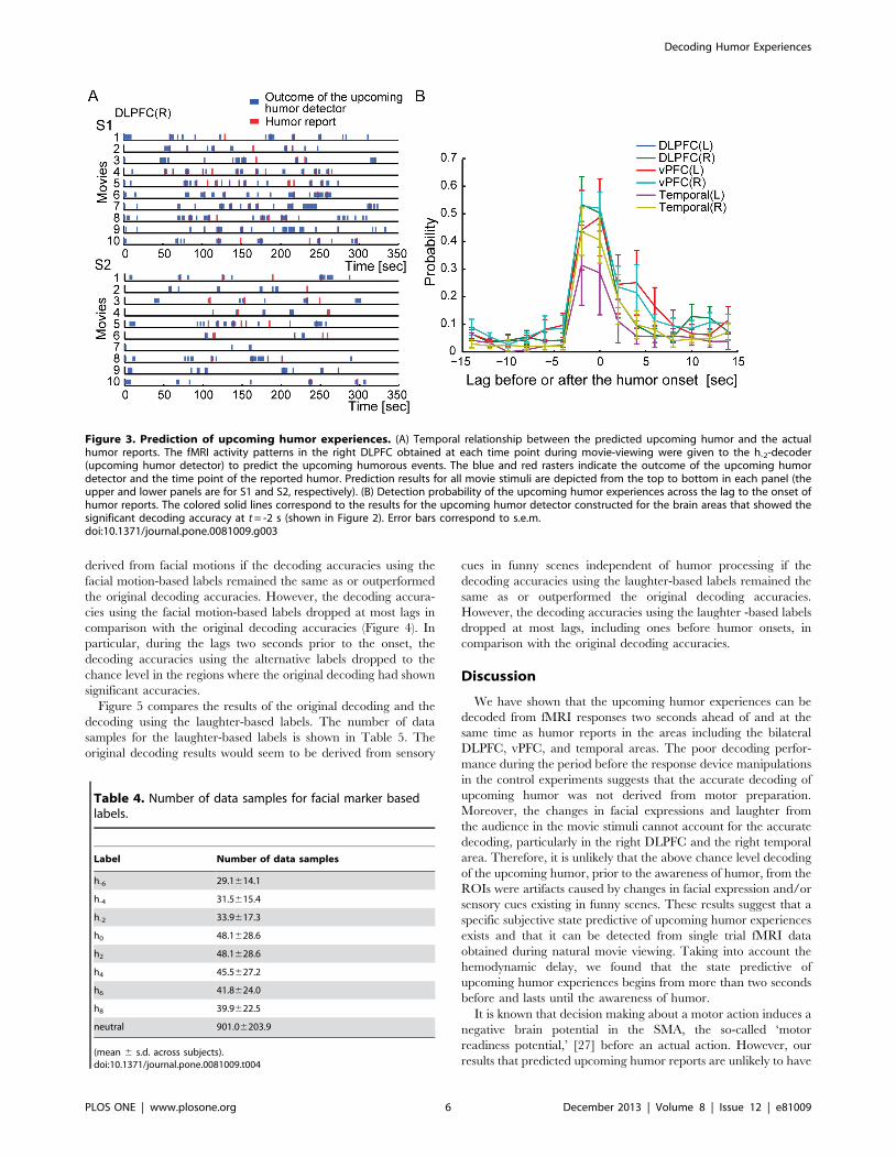
Figure 3 illustrates the decoding outcomes of the h-2-decoder for
the sequences of single volume fMRI data in each run. Figure 3A
illustrates the outcomes of the h-2-decoder, denoted by upcoming
humor detector, for the right DLPFC of S1 and S2. Since we
chose the h-2-decoder that showed significant decoding accuracy
using fMRI data obtained two seconds before the humor onset, the
high detection probability of the upcoming humor at the same lag
is not a surprising result. However, the results show that the
positive outcomes of the h-2-decoder were found not only two
seconds prior to the humor onsets but also at the subsequent time
points. By sorting the predictions of upcoming humor relative to
the humor onsets, we found that the h-2-decoders constructed for
the bilateral DLPFC, vPFC, and temporal areas predicted the
upcoming humor not only two seconds before the humor onsets
but also at the same time as them (Figure 3B). A similar tendency
was also observed from the h0-decoders that showed the significant
accuracies in Figure 2B. Hence, the information extracted by the
h-2-decoders was the same as that extracted by the h0-decoders.
Therefore, it is likely that the fMRI activity elicited at two seconds
before and at the same time as the humor onset represented the
same information that anticipates the upcoming humor events.
To validate that the decoding results were not derived from
artifacts, such as facial expression changes or head motions, we
reanalyzed fMRI data by assigning the alternative labels extracted
from facial motion changes instead of the humor reports to the
data samples (see Materials and Methods). Humor onsets were
determined on the basis of large changes in the facial expression
whereas they were determined on the basis of humor reports given
by a response device in the original analysis. The number of data
samples for each facial motion based label across the subjects is
shown in Table 4. The original decoding results would seem to be
Table 2. Number of humor reports for each movie.
Movie ID Number of humor reports
1 3.2062.82
2 2.3061.82
3 2.2061.75
4 3.5064.37
5 4.0064.32
6 2.9063.03
7 3.5064.00
8 3.4063.37
9 5.1063.63
10 2.8062.86
(mean 6 s.d. across subjects).doi:10.1371/journal.pone.0081009.t002
Table 3. Number of data samples for each label.
Label Number of data samples
h-6 21.8612.8
h-4 23.3614.4
h-2 24.6615.7
h0 32.9625.9
h2 32.9625.9
h4 31.8624.1
h6 29.9621.8
h8 28.8620.2
neutral 1038.16173.3
(mean 6 s.d. across subjects).doi:10.1371/journal.pone.0081009.t003
Decoding Humor Experiences
PLOS ONE | www.plosone.org 5 December 2013 | Volume 8 | Issue 12 | e81009
derived from facial motions if the decoding accuracies using the
facial motion-based labels remained the same as or outperformed
the original decoding accuracies. However, the decoding accura-
cies using the facial motion-based labels dropped at most lags in
comparison with the original decoding accuracies (Figure 4). In
particular, during the lags two seconds prior to the onset, the
decoding accuracies using the alternative labels dropped to the
chance level in the regions where the original decoding had shown
significant accuracies.
Figure 5 compares the results of the original decoding and the
decoding using the laughter-based labels. The number of data
samples for the laughter-based labels is shown in Table 5. The
original decoding results would seem to be derived from sensory
cues in funny scenes independent of humor processing if the
decoding accuracies using the laughter-based labels remained the
same as or outperformed the original decoding accuracies.
However, the decoding accuracies using the laughter -based labels
dropped at most lags, including ones before humor onsets, in
comparison with the original decoding accuracies.
Discussion
We have shown that the upcoming humor experiences can be
decoded from fMRI responses two seconds ahead of and at the
same time as humor reports in the areas including the bilateral
DLPFC, vPFC, and temporal areas. The poor decoding perfor-
mance during the period before the response device manipulations
in the control experiments suggests that the accurate decoding of
upcoming humor was not derived from motor preparation.
Moreover, the changes in facial expressions and laughter from
the audience in the movie stimuli cannot account for the accurate
decoding, particularly in the right DLPFC and the right temporal
area. Therefore, it is unlikely that the above chance level decoding
of the upcoming humor, prior to the awareness of humor, from the
ROIs were artifacts caused by changes in facial expression and/or
sensory cues existing in funny scenes. These results suggest that a
specific subjective state predictive of upcoming humor experiences
exists and that it can be detected from single trial fMRI data
obtained during natural movie viewing. Taking into account the
hemodynamic delay, we found that the state predictive of
upcoming humor experiences begins from more than two seconds
before and lasts until the awareness of humor.
It is known that decision making about a motor action induces a
negative brain potential in the SMA, the so-called ‘motor
readiness potential,’ [27] before an actual action. However, our
results that predicted upcoming humor reports are unlikely to have
Figure 3. Prediction of upcoming humor experiences. (A) Temporal relationship between the predicted upcoming humor and the actualhumor reports. The fMRI activity patterns in the right DLPFC obtained at each time point during movie-viewing were given to the h-2-decoder(upcoming humor detector) to predict the upcoming humorous events. The blue and red rasters indicate the outcome of the upcoming humordetector and the time point of the reported humor. Prediction results for all movie stimuli are depicted from the top to bottom in each panel (theupper and lower panels are for S1 and S2, respectively). (B) Detection probability of the upcoming humor experiences across the lag to the onset ofhumor reports. The colored solid lines correspond to the results for the upcoming humor detector constructed for the brain areas that showed thesignificant decoding accuracy at t = -2 s (shown in Figure 2). Error bars correspond to s.e.m.doi:10.1371/journal.pone.0081009.g003
Table 4. Number of data samples for facial marker basedlabels.
Label Number of data samples
h-6 29.1614.1
h-4 31.5615.4
h-2 33.9617.3
h0 48.1628.6
h2 48.1628.6
h4 45.5627.2
h6 41.8624.0
h8 39.9622.5
neutral 901.06203.9
(mean 6 s.d. across subjects).doi:10.1371/journal.pone.0081009.t004
Decoding Humor Experiences
PLOS ONE | www.plosone.org 6 December 2013 | Volume 8 | Issue 12 | e81009

a connection with motor decision making, because the time scale
of the activity involved in motor decision making is much shorter
than the upcoming-humor prediction; a few milliseconds precedes
the actual action in the decision making whereas a few seconds
preceded the upcoming humor reports in our results. It should be
difficult to obtain the readiness potential-related activity by using a
measurement device with poor temporal resolution such as fMRI.
In our experiment, ten subjects participated in each movie-
viewing and control experiment, but four individuals participated
in both experiments (i.e. 16 individuals participated in total).
Hence, the subjects could be categorized strictly into three groups:
subjects who participated only in the movie-viewing experiments,
those who participated in the control experiments, and those who
participated in both. Analyses under this categorization would be
appropriate if participation in a movie-viewing experiment affects
the results of a control experiment. However, since the task that
the subjects conducted in the control experiment was just a simple
motor task, we considered that it was valid to assume there were
no cross interactions between the two experiments. In fact, there
were no significant differences between the mean decoding
accuracies of the subjects who participated only in the control
experiments and those who participated in both experiments.
Thus, even with the original categorization of subjects, we believe
that we could reliably determine whether the decoding accuracies
were derived by humor experiences or response device manipu-
lations.
Previous fMRI studies on humor processing [4,9,14–16,18]
have been based mostly on the incongruity resolution theory [2] or
its related theories [3], in which humor processing can be divided
into two or more stages, such as incongruity detection, incongruity
resolution, humor appreciation, etc. These studies divided
humorous episodes into two phases, a setup line and a punch
line, and compared BOLD responses for punch lines with ones for
various baseline conditions to examine the neural basis of
incongruity detection and resolution [4,15,16,18]. There is a
study [14] that compared the responses to unfunny and funny
episodes, without dividing them into setup and punch lines.
However, the present study found a humor-related brain
representation lasting for a few seconds in the setup phase. Since
incongruity detection and incongruity resolution processes could
be done in a moment at the end of a setup line, it is unlikely that
the evidence of the present study fits incongruity theory. Indeed,
the humor processing identified in the present study appears to be
done before the incongruity processes. Thus, the present results
suggest that there are periods in which a subject is preparing for or
expecting humor events before they perceive them.
The present study found that the broad brain areas were
involved in the prediction of upcoming humor perception.
Particularly, brain areas, such as prefrontal and temporal areas,
were shown to be involved mainly in the prediction of upcoming
humor perception. The inferior frontal areas have previously been
shown to be involved in understanding or inferring others’ mental
states [28] and understanding semantic context in speech [29,30].
The temporal poles and superior temporal sulcus (STS) included
in the temporal ROIs have been shown to be involved in the
inference of others’ intentions and recalling socially relevant
Figure 4. Facial skin motion did not explain the performance of the upcoming humor detectors. (A) Four light-reflective markers putaround the mouth. (B) An image of the markers taken by an infrared video camera through a mirror mounted on the head coil. (C) Temporal changesin the marker positions and the determined motion labels. In the upper panel, each coordinate value of the markers is normalized relative to theaveraged value over a run and plotted against time. The lower panel depicts the determined motion labels corresponding to the large facial markermotion shown in the upper panel. (D) Performance of decoding humor experiences versus that of facial marker motion. The fMRI activity patterns ineach brain area were re-analyzed by labeling them with detected facial marker motion whereas the original activity patterns were labeled with humorreports. Each graph depicts the decoding performances obtained from each anatomically defined brain area across the latency to the humor ormotion onset. The magenta and blue solid lines correspond to the results of the decoding of facial marker motion and the humor experiences,respectively. The filled and open circles on the magenta or blue lines respectively indicate that the accuracy is significantly higher than chance level(AUC = 0.5; a dashed line in each graph) or not significant relative to the chance level (p,0.05, t-test, FDR corrected for multiple comparisons). A redasterisk indicates a significant difference between the accuracies of the original decoding and the decoding with facial marker motion (p,0.05,t-test,FDR corrected for multiple comparisons). Error bars correspond to s.e.m.doi:10.1371/journal.pone.0081009.g004
Decoding Humor Experiences
PLOS ONE | www.plosone.org 7 December 2013 | Volume 8 | Issue 12 | e81009

memories [28,31,32]. These basic characteristics in the prefrontal
and temporal areas may support our finding that there is mental
processing for preparing or expecting upcoming humor before the
perception of it.
Neuro-imaging studies of humor have indicated that the
temporoparietal junction (TPJ) plays an important role in
understanding jokes [5,7,10,12,13,17]. In our analysis, areas
corresponding to the TPJ were included in the parietal ROIs.
Although our results cannot be used to prove the unique
involvement of the TPJ in humor processing since data with
information relevant to motor processing was also extracted, the
parietal area indeed showed the significant decoding performance
after the humor onset that was consistent with the previous studies.
Further efforts, e.g., obtaining subjective reports from people
viewing movies without contaminating humor processing, are
needed to clarify the involvement of the TPJ in humor processing
of natural and dynamic humor stimuli.
In the decoding analyses, successful decoding of upcoming
humor was performed and the mean values of AUC across
subjects were more than 0.7 but did not reach 0.8, which is
considered to be a threshold of good accuracy. Although higher
accuracies would make the results more reliable, a sufficient value
of the prediction accuracy should be determined on the basis of
the purpose of study and/or the requirements of the applications.
Our results showed significantly higher accuracies than chance
level and than in the control experiments. They at least suggest
that there exists meaningful information related to upcoming
humor in the fMRI activity patterns. Development of more
sophisticated decoding algorithms would improve the prediction
performance.
Although our method could predict upcoming humor experi-
ences from single-trial fMRI data, it has a limitation when it comes
to identifying precise brain regions involved in the mental
processing. We roughly defined ROIs based on the anatomical
landmarks of the brain and used a statistical learning algorithm to
decode information from each ROI. This method can identify
informative ROIs but needs more work to identify informative
sub-regions in a ROI. In a linear classifier, since a classification is
performed by thresholding a weighted sum of voxel activities,
voxels with bigger absolute weight values could be interpreted as
relatively informative voxels in a ROI. Although we examined the
biased distribution of informative voxels shared across subjects by
mapping weight values onto the surface of the normal brain, we
could not find a significant tendency. The searchlight decoding
method [33], in which a decoding is applied to small spherical
ROIs centered at each location in the brain, could be used, but we
conducted ROI-based decoding analyses because searchlight
decoding is very computationally expensive. Since our framework
involves iteratively choosing samples in a random fashion for
creating training datasets, searchlight decoding would not be able
to finish all the analyses within a realistic time frame. An algorithm
that automatically selects the relevant voxels for decoding from
many voxels [34] would also not be feasible because it has similar
computational issues. Future improvements to these methods and/
or new computational technologies may be able to overcome this
limitation.
Finally, our results on predicting upcoming humor, or
anticipating humorous events, suggest that it is important to make
a viewer expect a humorous event and then give him or her a
punch line within a few seconds to induce laughter efficiently. It
would make sense that, for example, if a stern professor suddenly
says a joke in a serious lecture, no student would be able to follow
the joke. Of course, since our experiments did not cover all kinds
of humor, further investigation is needed to determine whether or
not our findings are valid in various kinds of humors. The
objective measurement of such expectations of humorous events
may be extended so that it can be used to evaluate the
performance of humorous movies. Movie producers and comedi-
ans would be able to improve their products and performances if
they had a means to improve movie scenes in which viewers
Figure 5. Audience laughter did not explain the performanceof the upcoming humor detectors. The performance of decodinghumor experiences versus that of laughter from the audience is shown.The fMRI activity patterns in each brain area were re-analyzed bylabeling them with presence of sounds of laughter from the audiencecontained in movie stimuli whereas the original activity patterns werelabeled with humor reports. Each graph depicts the decodingperformances obtained from each anatomically defined brain areaacross the latency to the humor or motion onset. The cyan and bluesolid lines correspond to the results of the decoding of laughter soundsand the humor experiences, respectively. The filled and open circles onthe cyan or blue lines respectively indicate that the accuracy issignificantly higher than chance level (AUC = 0.5; a dashed line in eachgraph) or not significant relative to the chance level (p,0.05, t-test, FDRcorrected for multiple comparisons). A red asterisk indicates asignificant difference between the accuracies of the original decodingand the decoding with laughter sounds (p,0.05, t-test, FDR correctedfor multiple comparisons). Error bars correspond to s.e.m.doi:10.1371/journal.pone.0081009.g005
Table 5. Number of data samples for laughter-based labels.
Label Number of data samples
h-6 40
h-4 46
h-2 62
h0 151
h2 151
h4 127
h6 111
h8 89
neutral 414
doi:10.1371/journal.pone.0081009.t005
Decoding Humor Experiences
PLOS ONE | www.plosone.org 8 December 2013 | Volume 8 | Issue 12 | e81009
expected an upcoming humor event but could not reach the level
of laughter. The ability to extract mental states during movie
viewing would also lead to more convincing critiques of movie
content; so far, movies are evaluated mainly on the basis of
opinions of experienced professionals. Further development of a
means to extract mental states during movie viewing may
eventually allow evaluations not only of comedies but also of
dramatic movies that should be rated by affective perspectives.
Acknowledgments
The authors thank K. Aizawa for helpful comments; M. Takemiya and Y.
Tsushima for manuscript editing; Y. Furukawa and T. Horikawa for
arrangements of subjects.
Author Contributions
Conceived and designed the experiments: YS KK TM NH. Performed the
experiments: YS KK TM. Analyzed the data: YS. Contributed reagents/
materials/analysis tools: YS KK TM. Wrote the paper: YS KK NH.
References
1. McCreaddie M, Wiggins S (2008) The purpose and function of humour in
health, health care and nursing: a narrative review. J Adv Nurs 61: 584–595.2. Suls JM (1972) A two-stage model for the appreciation of jokes and cartoons: An
information processing analysis. In: P M, editor. The Psychology of Humor:
Theoretical Perspectives and Empirical Issues. New York: Academic Press. pp.81–100.
3. Wyer RS, Collins JE (1992) A theory of humor elicitation. Psychol Rev 99: 663–688.
4. Goel V, Dolan RJ (2001) The functional anatomy of humor: segregatingcognitive and affective components. Nat Neurosci 4: 237–238.
5. Mobbs D, Greicius MD, Abdel-Azim E, Menon V, Reiss AL (2003) Humor
modulates the mesolimbic reward centers. Neuron 40: 1041–1048.6. Berns GS (2004) Something funny happened to reward. Trends Cogn Sci 8:
193–194.7. Moran JM, Wig GS, Adams RB, Janata P, Kelley WM (2004) Neural correlates
of humor detection and appreciation. Neuroimage 21: 1055–1060.
8. Mobbs D, Hagan CC, Azim E, Menon V, Reiss AL (2005) Personality predictsactivity in reward and emotional regions associated with humor. Proc Natl Acad
Sci USA 102: 16502–16506.9. Bartolo A, Benuzzi F, Nocetti L, Baraldi P, Nichelli P (2006) Humor
comprehension and appreciation: an FMRI study. J Cogn Neurosci 18: 1789–1798.
10. Wild B, Rodden FA, Rapp A, Erb M, Grodd W, et al. (2006) Humor and
smiling: cortical regions selective for cognitive, affective, and volitionalcomponents. Neurology 66: 887–893.
11. Goel V, Dolan RJ (2007) Social regulation of affective experience of humor. JCogn Neurosci 19: 1574–1580.
12. Samson AC, Zysset S, Huber O (2008) Cognitive humor processing: different
logical mechanisms in nonverbal cartoons--an fMRI study. Soc Neurosci 3: 125–140.
13. Samson AC, Hempelmann CF, Huber O, Zysset S (2009) Neural substrates ofincongruity-resolution and nonsense humor. Neuropsychologia 47: 1023–1033.
14. Franklin RG, Adams RB (2011) The reward of a good joke: neural correlates ofviewing dynamic displays of stand-up comedy. Cogn Affect Behav Neurosci 11:
508–515.
15. Bekinschtein TA, Davis MH, Rodd JM, Owen AM (2011) Why clowns tastefunny: the relationship between humor and semantic ambiguity. J Neurosci 31:
9665–9671.16. Chan Y-C, Chou T-L, Chen H-C, Liang K-C (2012) Segregating the
comprehension and elaboration processing of verbal jokes: an fMRI study.
Neuroimage 61: 899–906.17. Neely MN, Walter E, Black JM, Reiss AL (2012) Neural correlates of humor
detection and appreciation in children. J Neurosci 32: 1784–1790.
18. Chan Y-C, Chou T-L, Chen H-C, Yeh Y-C, Lavallee JP, et al. (2013) Towards a
neural circuit model of verbal humor processing: An fMRI study of the neural
substrates of incongruity detection and resolution. Neuroimage 66: 169–176.
19. Haxby JV, Gobbini MI, Furey ML, Ishai A, Schouten JL, et al. (2001)
Distributed and overlapping representations of faces and objects in ventral
temporal cortex. Science 293: 2425.
20. Cox D, Savoy L (2003) Functional magnetic resonance imaging (fMRI) ‘‘brain
reading’’: detecting and classifying distributed patterns of fMRI activity in
human visual cortex. Neuroimage 19: 261–270.
21. Kamitani Y, Tong F (2005) Decoding the visual and subjective contents of the
human brain. Nat Neurosci 8: 679–685.
22. Haynes J-D, Rees G (2005) Predicting the orientation of invisible stimuli from
activity in human primary visual cortex. Nat Neurosci 8: 686–691.
23. Friston KJ, Holmes AP, Worsley KJ, Poline JP, Frith CD, et al. (1995) Statistical
parametric maps in functional imaging: a general linear approach. Hum Brain
Mapp 2: 189–210.
24. Naselaris T, Kay KN, Nishimoto S, Gallant JL (2011) Encoding and decoding in
fMRI. Neuroimage 56: 400–410.
25. Chang C-C, Lin C-J (2011) LIBSVM. ACM Trans Intell Syst Technol 2: 1–27.
26. Horenstein C, Lowe MJ, Koenig KA, Phillips MD (2009) Comparison of
unilateral and bilateral complex finger tapping-related activation in premotor
and primary motor cortex. Hum Brain Mapp 30: 1397–1412.
27. Libet B, Gleason CA, Wright EW, Pearl DK (1983) Time of conscious intention
to act in relation to onset of cerebral activity (readiness-potential). The
unconscious initiation of a freely voluntary act. Brain 106 (Pt 3): 623–642.
28. Frith CD, Frith U (1999) Interacting minds--a biological basis. Science 286:
1692–1695.
29. Rodd JM, Davis MH, Johnsrude IS (2005) The neural mechanisms of speech
comprehension: fMRI studies of semantic ambiguity. Cereb Cortex 15: 1261–
1269.
30. Rothermich K, Kotz SA (2013) Predictions in speech comprehension: fMRI
evidence on the meter-semantic interface. Neuroimage 70: 89–100.
31. Allison T, Puce A, McCarthy G (2000) Social perception from visual cues: role of
the STS region. Trends Cogn Sci 4: 267–278.
32. Gallagher HL, Frith CD (2003) Functional imaging of ‘‘theory of mind’’. Trends
Cogn Sci 7: 77–83.
33. Kriegeskorte N, Goebel R, Bandettini P (2006) Information-based functional
brain mapping. Proc Natl Acad Sci USA 103: 3863–3868.
34. Yamashita O, Sato M, Yoshioka T, Tong F, Kamitani Y (2008) Sparse
estimation automatically selects voxels relevant for the decoding of fMRI activity
patterns. Neuroimage 42: 1414–1429.
Decoding Humor Experiences
PLOS ONE | www.plosone.org 9 December 2013 | Volume 8 | Issue 12 | e81009