Embed Size (px)
Citation preview

1
Decreased aerobic capacity in ANO5-muscular dystrophy1
2
Emil Ylikallioa,b*,+, Mari Auranena,b,*, Ibrahim Mahjnehc,d, Antti Lamminene, Maria Kousif,3
Ann-Liz Träskelinf, Tiina Muurineng, Mervi Löfbergb, Tapani Salmih, Anders Paetaui,4
Anna-Elina Lehesjokia,f,j, Päivi Piiriläg, Sari Kiuru-Enarib5
6
a Research Programs Unit, Molecular Neurology, Biomedicum Helsinki, University of7
Helsinki, Helsinki, Finland8
b Clinical Neurosciences, Neurology, University of Helsinki and Helsinki University9
Hospital, Finland10
c Division of Neurology, Pietarsaari District Hospital, Pietarsaari, Finland11
d Department of Neurology, MRC Oulu, Oulu University Hospital and University of Oulu,12
Finland13
e Department of Radiology, HUS Medical Imaging Center, Helsinki, Finland;14
f Folkhälsan Institute of Genetics, Helsinki, Finland15
g Unit of Clinical Physiology, HUS Medical Imaging Center, Helsinki University Hospital,16
Helsinki, Finland17
h Department of Clinical Neurophysiology, Medical Imaging Center, Helsinki University18
Hospital, Helsinki, Finland19
I Department of Pathology, HUSLAB and University of Helsinki, Helsinki, Finland20
j Neuroscience Center, University of Helsinki, Finland21
22
Running title: Exercise in ANO5 dystrophy23

2
+Correspondence to: Emil Ylikallio, Biomedicum r.C526b, Haartmaninkatu 8, 002901
Helsinki, Finland. E-mail: [email protected], Telephone: +358-50-448-6380, fax:2
+ 358-91-912-5610. *Equal contribution.3
4
5
6
7
8
9
10
11
12
13
14
15
16
17
18
19
20
21
22
23
3
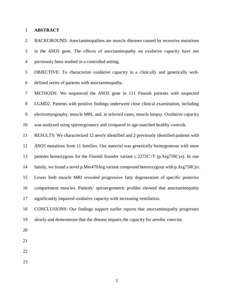
ABSTRACT1
BACKGROUND: Anoctaminopathies are muscle diseases caused by recessive mutations2
in the ANO5 gene. The effects of anoctaminopathy on oxidative capacity have not3
previously been studied in a controlled setting.4
OBJECTIVE: To characterize oxidative capacity in a clinically and genetically well-5
defined series of patients with anoctaminopathy.6
METHODS: We sequenced the ANO5 gene in 111 Finnish patients with suspected7
LGMD2. Patients with positive findings underwent close clinical examination, including8
electromyography, muscle MRI, and, in selected cases, muscle biopsy. Oxidative capacity9
was analyzed using spiroergometry and compared to age-matched healthy controls.10
RESULTS: We characterized 12 newly identified and 2 previously identified patients with11
ANO5 mutations from 11 families. Our material was genetically homogeneous with most12
patients homozygous for the Finnish founder variant c.2272C>T (p.Arg758Cys). In one13
family, we found a novel p.Met470Arg variant compound heterozygous with p.Arg758Cys.14
Lower limb muscle MRI revealed progressive fatty degeneration of specific posterior15
compartment muscles. Patients’ spiroergometric profiles showed that anoctaminopathy16
significantly impaired oxidative capacity with increasing ventilation.17
CONCLUSIONS: Our findings support earlier reports that anoctaminopathy progresses18
slowly and demonstrate that the disease impairs the capacity for aerobic exercise.19
20
21
22
23
4
KEYWORDS1
Muscular dystrophies, limb-girdle; Muscular Diseases; Inborn Genetic Diseases; Aerobic2
Exercise3
4
ABBREVIATIONS5
AT anaerobic threshold6
BE base excess7
CK creatine kinase8
EMG electromyography9
ExAC Exome Aggregation Consortium10
FetCO2 fraction of end tidal CO211
FVC forced vital capacity12
LGMD limb-girdle muscular dystrophy13
MMD3 Miyoshi myopathy14
RPE rate of perceived exertion15
RQ gas exchange ratio V´CO2/V’O216
SE spin echo17
TI inversion time18
VECO2 ventilatory equivalent for CO2 production19
VEO2 ventilatory equivalent for O2 uptake20
V’O2max maximal oxygen uptake21
Wmax/3min mean workload during the last 3 minutes of exercise22
Wmax/V’O2max mechanical efficiency23

5
INTRODUCTION1
Anoctaminopathies are muscle diseases caused by recessively inherited ANO5 mutations2
first identified in proximal limb-girdle muscular dystrophy type 2L (LGMD2L) and distal3
non-dysferlin Miyoshi myopathy (MMD3) [1, 2]. The phenotypic heterogeneity of4
anoctaminopathy has since expanded, with the mildest cases presenting as the5
asymptomatic elevation of creatine kinase (‘hyperCK-emia’) [1, 3]. The symptoms and6
signs are restricted to skeletal muscle in most cases, but occasionally patients present with7
a suspicion of heart involvement [4, 5]. Using muscle MRI, we and others have shown that8
anoctaminopathy typically involves the posterior compartments of the thighs and calves9
rather than the anterior compartments [6–8].10
11
Vissing et al recently reported positive effects from a 10-week supervised aerobic exercise12
regimen in patients with anoctaminopathy [9]. However, little controlled data exist13
measuring the effects of the disease on baseline oxidative capacity. Spiroergometric testing14
(breathing gas analysis during exercise) with the simultaneous analysis of blood lactate and15
ammonia concentrations provides not only an objective means of evaluating pulmonary,16
skeletal, and heart muscle involvement, but can also underline biochemical abnormalities17
through the monitoring of blood metabolites [10].18
19
This study aimed to characterize a series of anoctaminopathy patients specifically20
emphasizing oxidative performance, an aspect that has been little studied previously.21
Patients were examined in detail, including by muscle MRI, changes in muscle pathology,22
and spiroergometric testing.23

6
MATERIALS AND METHODS1
Patients2
Patients were examined at Helsinki University Hospital. All patients and healthy controls3
subjects provided their informed consent to participate in the study, and investigations were4
conducted in accordance with the 1975 Declaration of Helsinki. Furthermore, the ethics5
committee of the Hospital District of Helsinki and Uusimaa approved the study protocol.6
Total DNA was extracted from peripheral blood, and the coding exons and flanking intron7
sequences of the ANO5 gene (NM_213599.2) were sequenced. Primer sequences are8
available upon request from the authors. Variants whose pathogenicity were previously9
unknown were scored using PolyPhen-2 v.2.2.5 (http://genetics.bwh.harvard.edu/pph2/)10
[11] and Mutation Taster 2 (http://www.mutationtaster.org/) [12]. The population11
frequencies for variants were taken from the Exome Aggregation Consortium (ExAC,12
http://exac.broadinstitute.org/, accessed August 2015). Novel variants were submitted to13
the ClinVar database (http://www.ncbi.nlm.nih.gov/clinvar/).14
15
Spiroergometry, control subjects, laboratory specimens, and statistical methods16
Spiroergometric exercise testing was completed for 12 ANO5 variant–positive patients and17
24 age- and gender-adjusted controls using follow-up venous lactate and ammonium18
samples (Table 1).19
20
Spiroergometry was performed as described earlier [13]. A cannula was inserted into the21
left cubital vein, and the blood specimens for venous ammonia and lactate were drawn at22
nine time points: at rest, low exercise, maximal exercise, and at 2, 4, 6, 10, 20, and 30 min23

7
after exercise. Blood specimens were collected using the vacuum technique. Following a1
waste specimen of 2 ml, the lactate and ammonia specimens were placed into fluoride2
oxalate and EDTA syringes, respectively, centrifuged, and analyzed with a Cobas Integra3
400+ analyzer (Roche Diagnostics, Mannheim, Germany). Lactate and ammonia were4
assayed by enzymatic methods using lactate dehydrogenase and glutamate dehydrogenase,5
respectively. Next, 3-ml samples for the analysis of the venous blood gases were placed in6
Ca-titrated Lithium heparin syringes and analyzed using a Radiometer ABL800 analyzer7
(Radiometer Medical, Bronshoj, Danmark). The test started with a 40-W workload8
increased by 40 W in 3-min steps in women and increased by 50 W in 3-min steps in men.9
If the patient’s reported physical condition was low, we used 20-W increases in 2-min steps10
or 30 W in 3-min steps. We attained a rate of perceived exertion (RPE) of 17 to 19 on the11
Borg scale and a gas exchange ratio V´CO2/V’O2 (RQ) of more than 1.0 for all participants.12
13
We used an unpaired t-test to analyze the spiroergometric results as well as the results of14
the venous blood specimen between patients and controls; a nonparametric Mann-Whitney15
U test was used for parameters not normally distributed. Because of the multiple16
comparisons, we used a Bonferroni correction to the significance level applied. Due to the17
slight difference in the weight and BMI between the groups, we controlled the results of18
the spiroergometric variables for BMI.19
20
The patient and control groups had slight differences in maximal level of exercise in the21
spiroergometric test. To control for this difference in the comparison of spiroergometric22
data, we normalized the data according to blood lactate levels. This is because the lactate23

8
level increases with the level of anaerobic metabolism and is therefore a measure of the1
level of exercise during the test.2
3
Increase of lactate increases ventilation, which can be measured as increase of ventilatory4
equivalent for O2 and CO2 (VE/O2% and VE/CO2) and decrease of fraction of end tidal5
CO2 (FetCO2%). In the patients, VE/O2% and VE/CO2% were increased and FetCO2%6
decreased. This could have been interpreted as increased anaerobic metabolism, suggesting7
patients’ exercise tests to be more maximal than those of the controls. By controlling8
spiroergometric variables for maximal lactate levels in addition to BMI, we could see that9
the level of exercise of the patients was not greater than in the controls. Before these10
calculations, we had analyzed the behavior of lactate and ammonia associated with exercise,11
and no specific findings were seen. The respiratory quotient, RQ, could also have been12
used to control for the level of exercise. However, maximal lactate level serves here better13
as adjustment because, in contrast to RQ, lactate is not a spiroergometric variable itself.14
15
Muscle MRI16
MRI was performed using a 1.5 T Siemens system. Axial images were acquired for the17
pelvic, thigh, and leg muscles. The pulse sequences were T1-weighted spin echo (SE) with18
TR of 600 to 700 ms, TE of 15 ms, and a STIR fat suppression sequence with TR of 320019
to 4300 ms, TE of 33 ms, and an inversion time (TI) of 160 ms. Slices of 7 mm were used.20
The involvement of each muscle was scored on a scale of 0 to 3, where 0 indicated normal,21
1 indicated minor involvement, 2 indicated moderate involvement, and 3 indicated22
maximal involvement. Aggregate scores were computed for the muscles of the thigh23

9
(quadriceps femoris, adductors, hamstrings, sartorius, and gracilis) and lower leg (tibialis1
posterior, peroneus, deep posterior compartment, soleus, and gastrocnemius). Based on2
five leg and thigh muscles, the maximum aggregated score was 15. Follow-up scans were3
performed using the same scanner and identical parameters for patients 1 (2 years between4
scans), 2 (4 years between scans), 4 (2 years between scans), 7 (2 years between scans), 95
(2 years between scans), and 13 (12 years between scans).6
7
RESULTS8
Screening of the ANO5 gene9
We sequenced the ANO5 coding regions and flanking intron sequences in 111 patients with10
suspected myopathy of an undetermined cause based on symptoms and clinical findings,11
an elevated CK, myopathic electromyography (EMG), muscle biopsy, or muscle MRI. We12
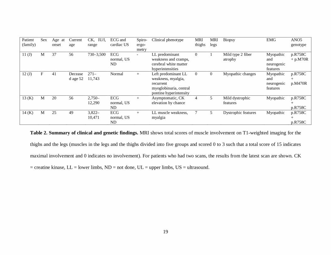
identified 12 patients from 10 families as positive for ANO5 variants (Table 2). Clinical13
and genetic data from patients 13 and 14 were previously reported [1, 2, 8].14
15
The most common genetic finding was homozygous ANO5 c.2272C>T (p.Arg758Cys).16
Eight patients were homozygous for this variant, while the remaining four were compound17
heterozygous for p.Arg758Cys in combination with either c.191dupA (p.Asn62LysfsX15),18
c.1409T>G (p.Met470Arg), or c.1664G>T (p.Ser555Ile) (Table 2). The latter two variants19
have not previously been confirmed as pathogenic in anoctaminopathy. Both were20
predicted as ‘disease causing’ by Mutation Taster, whereas PolyPhen-2 rated p.Met470Arg21
as ‘possibly damaging’ and p.Ser555Ile as ‘probably damaging’. The population frequency22

10
of p.Ser555Ile was 1.2x10⁻⁴ in European populations, whereas p.Met470Arg was not found1
in the ExAC database.2
3
Clinical, neurophysiologic, and pathologic findings4
Despite being relatively genetically homogeneous, our anoctaminopathy series showed5
some variability in clinical features (Table 2). Six of our 14 patients were female. The age6
of reported symptom onset ranged from 12 to 62 years, with an average age of 33 for female7
patients (SD 5.9 years) and 31 for male patients (SD 16 years). The disease duration ranged8
from 6 to 58 years. At onset, three patients complained of muscle pain, unpleasant muscle9
sensations in the calves, and cramps. Six patients initially experienced difficulty walking10
on their tiptoes, two patients complained of difficulty climbing stairs, and two patients11
complained of difficulty running and climbing stairs. One patient reported no muscle12
symptoms and an increased CK was found incidentally. During the course of the disease,13
muscle pain, stiffness, or cramps were common among all patients. Clinical examination14
showed distal lower limb muscle involvement in six patients, proximal involvement in two15
patients, and both distal and proximal involvement in six patients.16
Plasma CK ranged from 240 to 12,290 IU/l. EMG findings were myopathic in 7 out of 1217
patients tested. Four patients exhibited both neurogenic and myopathic features, whereas18
one patient had a normal EMG (Table 2). Muscle biopsies were obtained from 13 patients.19
The findings ranged from necrotizing myopathy or strong dystrophic changes to mild20
atrophy of type 2 fibers. Three out of nine patients who underwent cardiac ultrasound had21
slightly abnormal findings primarily attributed to hypertension (Table 2). ECGs showed22
normal findings for all patients (Table 4).23

11
Skeletal muscle imaging1
Table 3 summarizes the muscle MRI findings. Muscle T1-weighted MRI scans showed2
fatty replacement predominantly in the adductors, hamstrings, gastrocnemius, and soleus.3
Muscle MRI STIR-weighted scans showed hyperintensities suggesting myoedema4
primarily in the hamstrings, quadriceps, adductors, gastrocnemius, and soleus. These5
findings were more evident at the early stages of disease.6
7
In six patients who completed two follow-up MRI scans, the total T1 score increased by8
an average of 0.6 (SD 0.7) points per year in the thighs and 0.6 (SD 0.3) points per year in9
the legs based on the 0 to 15 scoring scale. The STIR score does not show any significant10
change over time. Thus, the progression of the disease is reflected in any sequential11
increases in fatty degeneration observable in T1 imaging. Representative images from one12
patient are shown in Figures 1A–F.13
14
Spiroergometric testing15
Table 1 shows the anthropometric and spirometric characteristics for patients and controls.16
During the exercise test, patients with anoctaminopathy exhibited a significantly lower17
maximal working capacity (Wmax/3min), maximum oxygen uptake (VO2max), oxygen18
pulse (V’O2/HR), and mechanical efficiency (Wmax/V’O2max) than those for controls19
(Table 4). However, the respiratory quotient (V’CO2/V’O2) tended to be higher in patients,20
and signs of increased ventilation demonstrated by a significantly increased ventilatory21
equivalent for O2 (VE/V’O2) and CO2 (VE/V’CO2) in addition to a decreased fraction of22
end-tidal CO2 (FetCO2) were seen in patients compared to controls.23

12
In the blood gas analyses, patients exhibited the lowest blood pH of 7.3 at 2 to 6 min and1
the lowest base excess (BE) -5.5 mmol/l at 6 min after exercise, while the corresponding2
values in controls were pH of 7.26 at 2 min and BE -7.77 mmol/l at 4 min after exercise.3
For the BE values, we found a significant difference between patients and controls at 2 and4
4 min after exercise (p = 0.039 and 0.013, respectively), and a significant difference in the5
pH values at 2 min after exercise (p = 0.048). In addition, we found significantly lower6
lactate readings in patients compared to controls (p = 0.049) at 2 and 4 min after exercise.7
However, we found no difference between patients and controls in the levels of ammonia8
(Figure 2).9
10
DISCUSSION11
We studied a cohort of patients with ANO5-related dystrophy, providing a detailed clinical12
analysis. Anoctaminopathy has been reported in several countries, including Canada [2],13
the UK and Germany [6, 14], Finland [1, 3], the Netherlands [2, 15], Denmark [5], the14
Czech Republic [16], and Italy and other countries in Europe and worldwide [17–19]. We15
found that 11% of our suspected LGMD patients had ANO5 mutations, supporting that they16
are a common cause of myopathy in northern Europe.17
18
We found the variant c.2272C>T (p.Arg758Cys) in all of our patients, appearing as19
homozygous in 10 patients from 8 families and as compound heterozygous with another20
variant in 4 patients from 3 families. This finding is not surprising since c.2272C>T has a21
carrier frequency of 0.35% in Finland, thus explaining most known Finnish cases [3].22
Among the compound heterozygous patients, one had the c.191dupA (p.Asn62Lysfs15X)23

13
variant, which is common in other Northern European populations [14]. One patient had1
the c.1664G>T (p.Ser555Ile) variant, which was recently found in the heterozygous state2
in a patient sequenced as part of a cohort of patients with LGMD or unspecified myopathy3
[17]. Bioinformatic predictions support the pathogenicity of this variant. However, since4
carriers are found in European populations, it is likely that a second hit is necessary on the5
other allele for this variant to cause disease, as was the case in our patient. Finally, two6
siblings (Family J, patients 11 and 12) presented with previously unknown variant7
c.1409T>G (p.Met470Arg). This variant received high scores for pathogenicity using8
bioinformatics tools, and was not found in public exome databases.9
10
The clinical findings in our anoctaminopathy patients correspond to previous descriptions,11
featuring asymmetric proximal or distal muscle involvement and myalgia [1–3, 5, 6, 14–12
20]. We found a tendency towards asymmetric muscle involvement, a feature of13
anoctaminopathy not typically reported for other forms of LGMD [2, 21] with the14
exception of facioscapulohumeral muscle dystrophy. Echocardiographic abnormalities15
were documented in only three patients, all associated with hypertension.16
17
Our findings provide further confirmation of the variable age of onset of anoctaminopathy.18
Even in the presence of the homozygous p.Arg758Cys variant, initial symptoms were19
reported between the ages of 12 and 62 years. The variable age of onset suggests the20
presence of genetic or environmental disease modifiers. Previous reports have well-21
documented an association between being male and anoctaminopathy [3, 14, 15, 22].22

14
However, we found no significant difference in the average age of disease onset based on1
gender, although the variance for the age of onset was greater among the men in our study.2
3
Earlier radiologic studies by us and others demonstrated that anoctaminopathy affects4
lower leg muscles, suggesting predominantly posterior compartment involvement in both5
the thighs and the legs. The tibialis anterior, gracilis, and sartorius are better spared, similar6
to other LGMDs, indicating that MRI may not be the best diagnostic tool [1, 6–8]. The7
results presented here further demonstrate that fatty degeneration tends to increase as the8
disease progresses. Therefore, T1-weighted MRI may be useful as a follow-up tool or as9
an assessment tool for potential treatment. The STIR technique is a sensitive tool in early10
diagnostics, but not in the later stages of disease. Furthermore, EMG results remain11
variable, featuring both myopathic and neurogenic findings.12
13
To our knowledge, this is the first study to explore the oxidative capacity of14
anoctaminopathy patients compared to an anthropometrically matched control population.15
Patients with anoctaminopathy showed significantly lower oxygen uptake than controls,16
which may serve as a primary feature of the disease or secondary to deconditioning17
provoked by muscle weakness and pains. Additionally, the patients’ oxygen pulse18
(V’O2/HR) was lower than controls, most likely associated with the diminished capacity19
of the peripheral muscles [23, 24]. The level of maximal oxygen uptake reported here20
corresponds to that measured by Vissing et al [9] before regular aerobic training.21
22

15
In patients, we found an excess of ventilation accompanied an increase in the ventilatory1
equivalents for O2 and CO2. In addition, more than half (7/12 patients) had FetCO2 lower2
than 4.5% during maximal exercise suggesting slight hyperventilation. By contrast,3
controls showed normoventilation, although we expected them to display a stronger4
respiratory compensation due to stronger metabolic acidosis associated with exercise5
compared to patients. The spirometric findings did not explain this inconsistency. The6
forced vital capacity (FVC) was slightly lower in patients than in healthy controls, but only7
one patient (patient 9) developed restrictive ventilatory impairment (FVC < 80% of the8
predicted value), suggesting that ventilatory function was not the cause of the increased9
ventilation. The increased ventilation might be explained by the lowered aerobic exercise10
capacity of the muscles leading to secondary hyperventilation during exercise. In addition,11
the reduced mechanical efficiency (Wmax/V’O2max) most likely reflected this finding,12
indicating an increased need for oxygen uptake related to the attained workload.13
14
Patients’ lactate and ammonia levels tended to be lower than those among controls. This15
might be explained by the lower exercise capacity due to muscle disease. However, the RQ16
levels of patients tended to be higher than those among controls indicating at least the same17
level of aerobic capacity among both patients and healthy controls. We should note that an18
increase in ventilation may partly explain the elevated RQ levels we observed in patients.19
We found no specific findings or indications of metabolic muscular myopathy.20
21
In conclusion, our report confirms the importance of the recessive ANO5 variants as causes22
of muscle disease with significant variability in clinical severity. In addition, we found a23

16
new disease-associated variant, and provide an initial and detailed case–control1
comparison suggesting a decreased oxidative capacity. Our results support aerobic training2
as a useful intervention to mitigate the symptoms of anoctaminopathy. Further studies3
featuring a larger sample of patients are needed to confirm our findings.4
5
ACKNOWLEDGEMENTS6
We thank all individuals who participated in this study. The authors wish to thank the7
following for their financial support of this research: Hospital District of Helsinki and8
Uusimaa (for S.K-E., M.A., and E.Y.), the Academy of Finland (for E.Y.), and the9
Folkhälsan Research Foundation (for A.-E.L.).10
11
CONFLICTS OF INTEREST12
The authors have no conflicts of interest to report.13
14
15
16
17
18
19
20
21
22

17
Patients with an ANO5deficiency(N = 12)
Healthy controls(N = 24)
Sex (m/f) 6/6 12/12Weight (kg) 81.7 (18.8) 75.1 (14.8)Height (cm) 169.8 (10.9) 165.3 (36.4)BMI 28.2 (5.4) 23.9 (5.8)Age 50.6 (10.5) 45.7 (10.6)FVC (l) 4.05 (0.87) 4.6 (1.3)FVC % of pred. 94.3 (10.4) 102.5 (12.3)FEV1 (l) 3.33 (0.73) 3.63 (0.21)FEV1 % of pred. 95.2 (11.6) 98.9 (11.4)
1
Table 1. Anthropometric characteristics of patients and controls. FVC = forced vital2
capacity, FEV1 = forced expiratory volume in 1 s, pred. = predicted value. Means and3
standard deviations (in parenthesis) are presented.4
5
6
7
8
9
10
11
12
13

18
Patient(family)
Sex Age atonset
Currentage
CK, IU/l,range
ECG andcardiac US
Spiro-ergo-metry
Clinical phenotype MRIthighs
MRIlegs
Biopsy EMG ANO5genotype
1 (A) M 17 65 1,337–3,279
Normal + Distal weakness ofright UL, later LLfatigue, myalgia,cramps
5 3 Chronic necrotizingmyopathy withdystrophic features
Myopathic p.R758C+p.R758C
2 (A) M 62 68 650–4,542 Mildlyhypertrophicleft ventricle
+ Mild distal LLweakness
6 3 Chronic necrotizingmyopathy withdystrophic features
Myopathic p.R758C+p.R758C
3 (B) F 31 43 272–2,023 Normal + LL myalgia, crampsand weakness
0 2 No diagnosticchanges, mild fibertype 2 atrophy
Myopathic p.R758C+p.R758C
4 (C) M 12 52 1,027–60,000
Milddiastolicrelaxationabnormality
+ Distal LL cramps,exercise intolerance,later UL weakness
3 4 Nonspecific myopathy Myopathic p.R758C+p.R758C
5 (D) F 34 53 1,367–3,363
Normal + Distal LL weakness,stiffness, myalgia, ULweakness (marginal)
6 6 No diagnosticchanges, mild fibertype 2 atrophy
Myopathic p.R758C+p.R758C
6 (E) M 40 60 766–2,517 ECGnormal, USND
- Progressive proximalLL weakness, laterproximal right ULweakness
9 6 Dystrophic andsecondaryinflammatory changes
Myopathic,LLneuropathy
p.R758C+p.N62Kfs15X
7 (F) F 38 51 921–5,119 Slight apicalseptalthickening
+ Restless legs, CKelevation, later LLmyalgia
0 3 Mild chronicmyopathy
Myopathic p.R758C+p.R758C
8 (G) M 38 69 1,300 Normal + Proximal LL and ULweakness, myalgia
14 3 Dystrophic features Myopathic p.R758C+p.R758C
9 (H) F 29 56 2,000–3,000
ECGnormal, USND
+ LL myalgia and mildweakness
6 3 Mild necrotizingmyopathy withdystrophic features
Myopathicandneurogenicfeatures
p.R758C+p.R758C
10 (I) F 25 31 240–2,973 Normal + LL myalgia, fatigue 0 0 Myopathic changes Normal p.R758C+p.S555I
19
Patient(family)
Sex Age atonset
Currentage
CK, IU/l,range
ECG andcardiac US
Spiro-ergo-metry
Clinical phenotype MRIthighs
MRIlegs
Biopsy EMG ANO5genotype
11 (J) M 37 56 730–3,500 ECGnormal, USND
- LL predominantweakness and cramps,cerebral white matterhyperintensities
0 1 Mild type 2 fiberatrophy
Myopathicandneurogenicfeatures
p.R758C+ p.M70R
12 (J) F 41 Deceased age 52
271–11,743
Normal + Left predominant LLweakness, myalgia,recurrentmyoglobinuria, centralpontine hyperintensity
0 0 Myopathic changes Myopathicandneurogenicfeatures
p.R758C+p.M470R
13 (K) M 20 56 2,750–12,290
ECGnormal, USND
+ Asymptomatic, CKelevation by chance
4 5 Mild dystrophicfeatures
Myopathic p.R758C+p.R758C
14 (K) M 25 49 3,822–10,471
ECGnormal, USND
+ LL muscle weakness,myalgia
7 5 Dystrophic features Myopathic p.R758C+p.R758C
Table 2. Summary of clinical and genetic findings. MRI shows total scores of muscle involvement on T1-weighted imaging for the
thighs and the legs (muscles in the legs and the thighs divided into five groups and scored 0 to 3 such that a total score of 15 indicates
maximal involvement and 0 indicates no involvement). For patients who had two scans, the results from the latest scan are shown. CK
= creatine kinase, LL = lower limbs, ND = not done, UL = upper limbs, US = ultrasound.

20
Muscle group T1 STIR
Thighs
Adductors 1.7 0.5
Hamstrings 1.5 1.0
Quadriceps 0.7 0.7
Gracilis 0.2 0.2
Sartorius 0.1 0.2
Legs
Gastrocnemius 2.1 1.1
Soleus 0.7 1.1
Anterior compartment 0.2 0.1
Peroneal compartment 0 0
Deep posterior
compartment0 0
Table 3. Muscle imaging. Muscle involvement on MRI imaging is scored 0 to 3, where 0
indicates no involvement and 3 indicates maximal involvement. Average scores are shown
for T1 and STIR images.

21
Parameter ANO5,N = 12
Healthycontrols,N = 24
P-Value
Maximum heart rate 172 (12.5) 162.3 (30.3) NSMaximum heart rate % 89.1 (15.8) 93.1 (8.7) NSAT% of expected maximal V’O2 51.6 (16.8) 66.7 (20.2) NSWmax/3 min (W) 108.92 (46.9) 208.54
(74.74)0.00
Wmax/3 min % of predicted 67.8 (28.1) 114.36 (26.2) <0.001V’O2 max (maximum oxygenuptake) (l/min)
1.86 (0.61) 3.7 (4.04) NS
V’O2max % of predicted 86.1 (29.5) 122.1 (26.8) 0.001V’O2max (ml/min/kg) 22.9 (5.7) 38.4 (11.9) 0.002V’O2max/kg of predicted (%) 76.5 (22.7) 120.0 (27.0) 0.002V’O2/HR % of predicted (%) 101.8 (33.1) 136.3 (25.6) 0.001Respiratory quotient(V’CO2max/V’O2max, RQ)
1.22 (0.08) 1.13 (0.64) 0.001
VEO2 % of predicted (%) 136.7 (16.6) 110.8 (15.5) <0.001VECO2 % of predicted (%) 124.1 (14.3) 109.7 (14.0) 0.002Fraction of end-tidal CO2(FetCO2)(%)
4.65 (0.48) 5.3 (0.51) <0.001
Wmax/VO2max (%) 16.3 (4.8) 20.8 (1.8) 0.004Breathing frequency (1/min) 37.92 (10.7) 37.45 (8.9) NSTidal volume % or predicted (%) 93.4 (28.6) 112.1 (18.2) NSSubjective maximum strengthperceived
18.8 (0.87) 18.39 (0.90) NS
Table 4. Spiroergometric data for patients with ANO5 and healthy controls. AT =
anaerobic threshold, Wmax/3 min = mean workload during the last 3 min of exercise,
maximal V’O2max = maximal oxygen uptake, VECO2 = ventilatory equivalent for CO2
production, VEO2 = ventilatory equivalent for O2 uptake, Wmax/V’O2 max = mechanical
efficiency. Comparisons were controlled for the maximal lactate levels and BMI. Level of
significance after Bonferroni correction is P < 0.003.

22
FIGURE LEGENDS
Figure 1. Muscle imaging. Lower limb MRI from patient 9 shows the disease progression
over 2 years. The first images for the thighs (A–B) are compared to later images (D–E),
illustrating that the most prominent changes can be found in the progressive fatty
infiltration symmetrically in the adductors, biceps femoris, quadriceps (vastus lateralis and
intermedius, and rectus femoris), and left semimembranosus muscles. In the legs (C and
F), selective and progressive involvement of the medial head of the gastrocnemius muscle
can be found, with more prominent changes visible on the left-hand side.
Figure 2. Results from exercise testing. Spiroergometric results showing the levels of
ammonia and lactate in 12 patients and 24 healthy controls. Significantly lower lactate
levels are visible in patients compared to controls (P = 0.049) at 2 and 4 min after exercise.
No differences exist in the levels of ammonia between patients and controls. Mean and
standard deviation, *P < 0.05.

23
Figure 1.

24
Figure 2.

25
REFERENCES
[1] Mahjneh I, Jaiswal J, Lamminen A, Somer M, Marlow G, Kiuru-Enari S, Bashir R. A
new distal myopathy with mutation in anoctamin 5. Neuromuscul Disord 2010; 20: 791-
795. doi 10.1016/j.nmd.2010.07.270.
[2] Bolduc V, Marlow G, Boycott KM, Saleki K, Inoue H, Kroon J, Itakura M, Robitaille
Y, Parent L, Baas F, Mizuta K, Kamata N, Richard I, Linssen WH, Mahjneh I, de Visser
M, Bashir R, Brais B. Recessive mutations in the putative calcium-activated chloride
channel Anoctamin 5 cause proximal LGMD2L and distal MMD3 muscular dystrophies.
Am J Hum Genet 2010; 86: 213-221. doi 10.1016/j.ajhg.2009.12.013.
[3] Penttila S, Palmio J, Suominen T, Raheem O, Evila A, Muelas Gomez N, Tasca G,
Waddell LB, Clarke NF, Barboi A, Hackman P, Udd B. Eight new mutations and the
expanding phenotype variability in muscular dystrophy caused by ANO5. Neurology
2012; 78: 897-903. doi 10.1212/WNL.0b013e31824c4682.
[4] Wahbi K, Behin A, Becane HM, Leturcq F, Cossee M, Laforet P, Stojkovic T, Carlier
P, Toussaint M, Gaxotte V, Cluzel P, Eymard B, Duboc D. Dilated cardiomyopathy in
patients with mutations in anoctamin 5. Int J Cardiol 2013; 168: 76-79. doi
10.1016/j.ijcard.2012.09.070.
[5] Witting N, Duno M, Petri H, Krag T, Bundgaard H, Kober L, Vissing J. Anoctamin 5
muscular dystrophy in Denmark: prevalence, genotypes, phenotypes, cardiac findings,
and muscle protein expression. J Neurol 2013; 260: 2084-2093. doi 10.1007/s00415-013-
6934-y.
26
[6] Sarkozy A, Deschauer M, Carlier RY, Schrank B, Seeger J, Walter MC, Schoser B,
Reilich P, Leturq F, Radunovic A, Behin A, Laforet P, Eymard B, Schreiber H, Hicks D,
Vaidya SS, Glaser D, Carlier PG, Bushby K, Lochmuller H, Straub V. Muscle MRI
findings in limb girdle muscular dystrophy type 2L. Neuromuscul Disord 2012; 22 Suppl
2: S122-9. doi 10.1016/j.nmd.2012.05.012.
[7] Ten Dam L, van der Kooi AJ, Rovekamp F, Linssen WH, de Visser M. Comparing
clinical data and muscle imaging of DYSF and ANO5 related muscular dystrophies.
Neuromuscul Disord 2014; 24: 1097-1102. doi 10.1016/j.nmd.2014.07.004.
[8] Mahjneh I, Bashir R, Kiuru-Enari S, Linssen W, Lamminen A, Visser M. Selective
pattern of muscle involvement seen in distal muscular dystrophy associated with
anoctamin 5 mutations: a follow-up muscle MRI study. Neuromuscul Disord 2012; 22
Suppl 2: S130-6. doi 10.1016/j.nmd.2012.02.007.
[9] Vissing CR, Preisler N, Husu E, Prahm KP, Vissing J. Aerobic training in patients
with anoctamin 5 myopathy and hyperckemia. Muscle Nerve 2014; 50: 119-123. doi
10.1002/mus.24112.
[10] Volpi L, Ricci G, Orsucci D, Alessi R, Bertolucci F, Piazza S, Simoncini C,
Mancuso M, Siciliano G. Metabolic myopathies: functional evaluation by different
exercise testing approaches. Musculoskelet Surg 2011; 95: 59-67. doi 10.1007/s12306-
011-0096-9.
27
[11] Adzhubei IA, Schmidt S, Peshkin L, Ramensky VE, Gerasimova A, Bork P,
Kondrashov AS, Sunyaev SR. A method and server for predicting damaging missense
mutations. Nat Methods 2010; 7: 248-249. doi 10.1038/nmeth0410-248.
[12] Schwarz JM, Cooper DN, Schuelke M, Seelow D. MutationTaster2: mutation
prediction for the deep-sequencing age. Nat Methods 2014; 11: 361-362. doi
10.1038/nmeth.2890.
[13] Mustelin L, Pietilainen KH, Rissanen A, Sovijarvi AR, Piirila P, Naukkarinen J,
Peltonen L, Kaprio J, Yki-Jarvinen H. Acquired obesity and poor physical fitness impair
expression of genes of mitochondrial oxidative phosphorylation in monozygotic twins
discordant for obesity. Am J Physiol Endocrinol Metab 2008; 295: E148-54. doi
10.1152/ajpendo.00580.2007
[14] Hicks D, Sarkozy A, Muelas N, Koehler K, Huebner A, Hudson G, Chinnery PF,
Barresi R, Eagle M, Polvikoski T, Bailey G, Miller J, Radunovic A, Hughes PJ, Roberts
R, Krause S, Walter MC, Laval SH, Straub V, Lochmuller H, Bushby K. A founder
mutation in Anoctamin 5 is a major cause of limb-girdle muscular dystrophy. Brain 2011;
134: 171-182. doi 10.1093/brain/awq294.
[15] van der Kooi AJ, Ten Dam L, Frankhuizen WS, Straathof CS, van Doorn PA, de
Visser M, Ginjaar IB. ANO5 mutations in the Dutch limb girdle muscular dystrophy
population. Neuromuscul Disord 2013; 23: 456-460. doi 10.1016/j.nmd.2013.03.012.
[16] Stehlikova K, Skalova D, Zidkova J, Mrazova L, Vondracek P, Mazanec R,
Vohanka S, Haberlova J, Hermanova M, Zamecnik J, Soucek O, Oslejskova H,

28
Dvorackova N, Solarova P, Fajkusova L. Autosomal recessive limb-girdle muscular
dystrophies in the Czech Republic. BMC Neurol 2014; 14: 154-014-0154-7. doi
10.1186/s12883-014-0154-7.
[17] Savarese M, Di Fruscio G, Tasca G, Ruggiero L, Janssens S, De Bleecker J, Delpech
M, Musumeci O, Toscano A, Angelini C, Sacconi S, Santoro L, Ricci E, Claes K,
Politano L, Nigro V. Next generation sequencing on patients with LGMD and
nonspecific myopathies: Findings associated with ANO5 mutations. Neuromuscul Disord
2015; 25: 533-541. doi 10.1016/j.nmd.2015.03.011.
[18] Bohlega S, Monies DM, Abulaban AA, Murad HN, Alhindi HN, Meyer BF.
Clinical and genetic features of anoctaminopathy in Saudi Arabia. Neurosciences
(Riyadh) 2015; 20: 173-177. doi 10.17712/nsj.2015.2.20140547.
[19] Magri F, Del Bo R, D'Angelo MG, Sciacco M, Gandossini S, Govoni A, Napoli L,
Ciscato P, Fortunato F, Brighina E, Bonato S, Bordoni A, Lucchini V, Corti S, Moggio
M, Bresolin N, Comi GP. Frequency and characterisation of anoctamin 5 mutations in a
cohort of Italian limb-girdle muscular dystrophy patients. Neuromuscul Disord 2012; 22:
934-943. doi 10.1016/j.nmd.2012.05.001.
[20] Liewluck T, Winder TL, Dimberg EL, Crum BA, Heppelmann CJ, Wang Y, Bergen
HR,3rd, Milone M. ANO5-muscular dystrophy: clinical, pathological and molecular
findings. Eur J Neurol 2013; 20: 1383-1389. doi 10.1111/ene.12191.

29
[21] Jarry J, Rioux MF, Bolduc V, Robitaille Y, Khoury V, Thiffault I, Tetreault M,
Loisel L, Bouchard JP, Brais B. A novel autosomal recessive limb-girdle muscular
dystrophy with quadriceps atrophy maps to 11p13-p12. Brain 2007; 130: 368-380.
[22] Sarkozy A, Hicks D, Hudson J, Laval SH, Barresi R, Hilton-Jones D, Deschauer M,
Harris E, Rufibach L, Hwang E, Bashir R, Walter MC, Krause S, van den Bergh P, Illa I,
Penisson-Besnier I, De Waele L, Turnbull D, Guglieri M, Schrank B, Schoser B, Seeger
J, Schreiber H, Glaser D, Eagle M, Bailey G, Walters R, Longman C, Norwood F, Winer
J, Muntoni F, Hanna M, Roberts M, Bindoff LA, Brierley C, Cooper RG, Cottrell DA,
Davies NP, Gibson A, Gorman GS, Hammans S, Jackson AP, Khan A, Lane R,
McConville J, McEntagart M, Al-Memar A, Nixon J, Panicker J, Parton M, Petty R,
Price CJ, Rakowicz W, Ray P, Schapira AH, Swingler R, Turner C, Wagner KR,
Maddison P, Shaw PJ, Straub V, Bushby K, Lochmuller H. ANO5 gene analysis in a
large cohort of patients with anoctaminopathy: confirmation of male prevalence and high
occurrence of the common exon 5 gene mutation. Hum Mutat 2013; 34: 1111-1118. doi
10.1002/humu.22342.
[23] Myers J, Froelicher VF. Hemodynamic determinants of exercise capacity in chronic
heart failure. Ann Intern Med 1991; 115: 377-386.
[24] Wasserman K. Diagnosing cardiovascular and lung pathophysiology from exercise
gas exchange. Chest 1997; 112: 1091-1101.