Embed Size (px)
Citation preview

The Decreased apical dominance1/Petunia hybridaCAROTENOID CLEAVAGE DIOXYGENASE8 Gene AffectsBranch Production and Plays a Role in Leaf Senescence,Root Growth, and Flower Development
Kimberley C. Snowden,a Andrew J. Simkin,b Bart J. Janssen,a Kerry R. Templeton,a Holly M. Loucas,c
Joanne L. Simons,a Sakuntala Karunairetnam,a Andrew P. Gleave,a David G. Clark,c and Harry J. Kleeb,1
a HortResearch, Private Bag 92169, Mt. Albert, Auckland, New Zealandb Plant Molecular and Cellular Biology Program, University of Florida, Gainesville, Florida 32611-0690c Environmental Horticulture, University of Florida, Gainesville, Florida 32611-0670
Carotenoids and carotenoid cleavage products play an important and integral role in plant development. The Decreased
apical dominance1 (Dad1)/PhCCD8 gene of petunia (Petunia hybrida) encodes a hypothetical carotenoid cleavage
dioxygenase (CCD) and ortholog of the MORE AXILLARY GROWTH4 (MAX4)/AtCCD8 gene. The dad1-1 mutant allele was
inactivated by insertion of an unusual transposon (Dad-one transposon), and the dad1-3 allele is a revertant allele of dad1-1.
Consistent with its role in producing a graft-transmissible compound that can alter branching, the Dad1/PhCCD8 gene is
expressed in root and shoot tissue. This expression is upregulated in the stems of the dad1-1, dad2, and dad3 increased
branching mutants, indicating feedback regulation of the gene in this tissue. However, this feedback regulation does not
affect the root expression of Dad1/PhCCD8. Overexpression of Dad1/PhCCD8 in the dad1-1 mutant complemented the
mutant phenotype, and RNA interference in the wild type resulted in an increased branching phenotype. Other differences in
phenotype associated with the loss of Dad1/PhCCD8 function included altered timing of axillary meristem development,
delayed leaf senescence, smaller flowers, reduced internode length, and reduced root growth. These data indicate that the
substrate(s) and/or product(s) of the Dad1/PhCCD8 enzyme are mobile signal molecules with diverse roles in plant
development.
INTRODUCTION
The architecture of plants is diverse and ranges from a simple
unbranched shoot system (e.g., a monopodial palm tree) to the
complex multiply branched shoot systems, classified into 23
distinct architectural patterns (Halle, 1999), seen in most trees.
The development of complex shoot systems and branching
patterns is a dynamic process that continues throughout the life
of the plant by progressive addition of new shoot modules. In
flowering plants, additional shoots originate from meristematic
centers that form within the leaf axils through a process called
lateral branching (Steeves and Sussex, 1989). The diversity and
architectural complexity found in plants are the result of several
processes. Distribution of lateral shoots is an important first step
in establishing the overall architecture of the plant. However,
placement of branches alone is not sufficient to produce the
diversity of branch patterns seen in nature. Other developmental
programs play important roles in determining the final form of the
plant. These include both initiation and duration of shoot growth,
differential growth rates of lateral and main shoots, angle
(orthotropic versus plagiotropic) of branch growth, and branch
ontogeny (monopodial versus sympodial development; Steeves
and Sussex, 1989; Sussex and Kerk, 2001). These developmen-
tal processes can be summarized in terms of meristem de-
velopment as position, identity (or potential identity), and timing
of meristem activity (Bell, 1991). It is important to note that these
developmental processes do not occur in isolation but rather as
a dynamic interactive network responding to genetic and envi-
ronmental cues (Bell, 1991).
Mutational approaches to the study of lateral branch devel-
opment have been undertaken in multiple plant species (Napoli
et al., 1999). Relatively nonpleiotropic mutants include the de-
creased apical dominance (dad) mutants of petunia (Petunia
hybrida; Napoli, 1996; Napoli and Ruehle, 1996; Napoli et al.,
1999; Snowden and Napoli, 2003), the ramosus (rms) mutants of
pea (Pisum sativum; Beveridge, 2000; Morris et al., 2001;
Rameau et al., 2002; Beveridge et al., 2003), and the more
axillary growth (max) mutants of Arabidopsis thaliana (Stirnberg
et al., 2002; Turnbull et al., 2002; Sorefan et al., 2003). TheMAX2,
MAX3, MAX4, and RMS1 genes have been cloned (Stirnberg
et al., 2002; Sorefan et al., 2003; Booker et al., 2004), withMAX4
(AtCCD8) and RMS1 being orthologous members of a family of
1 To whom correspondence should be addressed. E-mail [email protected]; fax 352-846-2063.The authors responsible for distribution of materials integral to thefindings presented in this article in accordance with the policy describedin the Instructions for Authors (www.plantcell.org) are: Kimberley C.Snowden ([email protected]) and Harry J. Klee ([email protected]).Article, publication date, and citation information can be found atwww.plantcell.org/cgi/doi/10.1105/tpc.104.027714.
The Plant Cell, Vol. 17, 746–759, March 2005, www.plantcell.orgª 2005 American Society of Plant Biologists

carotenoid cleavage dioxygenases (CCDs) of which MAX3
(AtCCD7) is also a member. The isolation of multiple types of
branching mutants indicates that control of lateral branching is
both plastic and multigenic.
TheCCD family includes proteins fromboth plants and animals
that are known to act upon carotenoid substrates. In animals,
examples include CCDs involved in synthesis of vitamin A (von
Lintig and Vogt, 2000; Redmond et al., 2001; von Lintig et al.,
2001; Lindqvist and Anderson, 2002) and RPE65, which binds
all-trans-retinyl esters and is involved in regeneration of 11-cis-
retinol in the visual cycle (Redmond et al., 1998; Gollapalli et al.,
2003; Mata et al., 2004). In bacteria, enzymes in this family
have substrates other than carotenoids (e.g., lignostilbene
dioxygenases [LSDs]; Kamoda and Saburi, 1993a, 1993b). In
plants, the founding member of the CCD family was Vp14,
a 9-cis-epoxycarotenoid dioxygenase (NCED). This and closely
related dioxygenases catalyze the 11,12 double bond cleavage
of 9-cis-violaxanthin and 9-cis-neoxanthin to produce xanthoxin
(Schwartz et al., 1997; Tan et al., 1997), which is subsequently
converted to the phytohormone abscisic acid (ABA). Several
additional plant CCDs that cleave carotenoids into apocarote-
noids at different double bonds also have been identified
(Schwartz et al., 2001; Bouvier et al., 2003a, 2003b; Simkin
et al., 2004a, 2004b). Of particular significance are CCD7/MAX3
and CCD8/MAX4. The former enzyme cleaves multiple carote-
noids at the 9,10 double bond (Booker et al., 2004). The latter
enzyme has been shown to further cleave 109-apo-b-carotenal,
the 9,10 cleavage product of b-carotene, to yield 13-apo-
b-carotenone (Schwartz et al., 2004). These data are consistent
with CCD7 and CCD8 acting in the same pathway to produce
a novel apocarotenoid phytohormone.
In petunia, growth is monopodial before flowering, with lateral
branching occurring in two different patterns (Snowden and
Napoli, 2003). During vegetative growth, basal lateral branches
develop acropetally from nodes in a defined zone that is pre-
ceded and followed by zones where lateral branching is inhib-
ited. After flowering, the main axis of growth is continued with
a series of sympodial branches. In addition, apical branching
develops in a basipetal direction and is restricted to the distal-
most nodes of the monopodial axis. When grown in short days,
the main axis is retarded and basal branching extended. The
dad1-1 mutant has increased lateral branching compared with
wild-type V26. There is no initial zone of branch inhibition, the
branch potential (percentage of nodes with a branch) is in-
creased, and there is no change in lateral branch development in
short days (Napoli, 1996; Napoli andRuehle, 1996; Snowden and
Napoli, 2003).
Formation of lateral branches has historically been considered
in terms of the repression of lateral bud outgrowth by auxin
derived from the apical meristem (Napoli et al., 1999). However,
analysis of the dad mutants in petunia (Napoli, 1996; Napoli and
Ruehle, 1996) and the rms mutants in pea (Beveridge, 2000;
Morris et al., 2001) provides strong evidence for the role of other
plant organs, such as roots, generating other signal molecules
that control axillary bud outgrowth. Some of the petunia (dad1
and dad3) and pea (rms1, rms2, and rms5) mutants can be
reverted to the wild type by grafting to wild-type rootstocks
(Beveridge et al., 1994; Napoli, 1996; Foo et al., 2001; Morris
et al., 2001; C.A. Napoli and K.C. Snowden, unpublished data),
and in the case of petunia by an interstock of wild-type tissue
placed between mutant roots and scion (Napoli, 1996). Similar
results have been reported for the Arabidopsis max1, max4/
AtCCD8, and max3/AtCCD7 mutants (Turnbull et al., 2002;
Sorefan et al., 2003; Booker et al., 2004). These results suggest
that wild-type roots can inhibit lateral branch outgrowth by
production of a novel unidentified inhibitor of branching that is
transported through graft unions. However, the observation by
Napoli (1996) that mutant dad1-1 root development above the
graft union blocks restoration of the wild-type phenotype sug-
gests an alternative model; roots produce an inducer of lateral
shoot growth that is degraded by the Dad1 gene product. In
either case, identification of the novel modulator(s) of lateral
branch outgrowth remains a tantalizing goal.
Similar branching phenotypes between the dad1-1mutant and
themax4 and rms1mutants and, in particular, the ability to revert
the mutant phenotype to the wild type by grafting of mutant
scions ontowild-type rootstocks suggested that a homolog of the
MAX4/RMS1genesmight bemutated indad1-1. In this article,we
report the cloning and characterization of the gene responsible for
the dad1mutation in petunia. Sequencing of three mutant alleles
of dad1, complementation of the dad1-1 mutant in transgenic
plants, as well as the phenotypes of RNA interference (RNAi)
transgenic plants all confirmed that theDad1 gene is amember of
the CCD gene family with a role in branch development.
RESULTS
Phenotypes of dad1 Alleles
The dad1-1 mutant was isolated and described by Napoli and
Ruehle (1996) and further characterized with respect to its
branching phenotype (Napoli, 1996; Snowden and Napoli,
2003). This mutant has a highly branched growth habit that can
be reversed by micrografting (Napoli, 1996). We have isolated
two additional alleles of dad1 (Figure 1A) and characterized their
growth in comparison with the dad1-1 allele. The dad1-2 allele
was discovered in the background of thedad2branchingmutant.
A third dad1 allele (dad1-3) was generated by ethyl methane-
sulfonate (EMS) mutagenesis of dad1-1, performed to look for
second-site suppressors. During screening of this population,
a line that had reverted to a near wild-type phenotype was
isolated. Allelism tests showed that this line was a new allele of
dad1, with a phenotype very similar to the wild type (a potential
revertant of dad1-1).
Quantitative comparisons between wild-type V26 and the
dad1 alleles are shown in Figure 1B. A diagram of the patterns
of lateral branching in V26 and the dad1 mutant alleles is shown
in Figure 1C. Under the growth conditions used for these experi-
ments, V26 plants produced four branches from nodes 2 to 7
(where the cotyledonary node is counted as node 0, i.e., V26
branches from the second node above the cotyledons) with
no branching from the cotyledonary node. As described pre-
viously by Napoli (1996), the dad1-1 allele produced branches
from every node up to node 10, including both axils at the
cotyledonary node. For each of the branching traits measured,
Dad1 Encodes a Hypothetical Carotenoid Cleavage Dioxygenase 747

dad1-1 had the most extreme phenotype with more branches,
more nodes that produced branches, and a greater percentage
of nodes with branches.
Although it is possible to distinguish dad1-3 branching from
V26 in some growth conditions, there was no significant differ-
ence in branching pattern between dad1-3 and V26 in the
controlled growth experiments shown in Figures 1A and 1B.
The dad1-2 allele displayed a branching phenotype interme-
diate between dad1-1 and V26. As found for V26, dad1-2 did not
produce branches from nodes higher than node 7. However,
branches were observed at the cotyledonary node in many (10
out of 16) of the dad1-2 plants (Figure 1C). In addition, the dad1-2
allele had a greater percentage of nodes with a branch than V26,
the combination of both factors resulting in more branches in the
dad1-2 mutant allele than in V26.
All three dad1 alleles reduced the overall height of the plant,
although in some experiments the difference between V26 and
dad1-3 was not significant. The flowering time, as measured by
the number of nodes to first flower, was increased for dad1-1
compared with V26. The dad1-2 allele showed no significant
delay in flowering, and the dad1-3 showed a slight but statisti-
cally significant reduction in flowering time.
While comparing the growth habit of the dad1 alleles, we
noticed that leaf senescence appeared to be altered for the
dad1-1 and dad1-2 alleles, with older (lower node) leaves having
delayed senescence (Figure 1A). In V26 plants, leaves at lower
nodes went through a rapid browning and collapse of leaf
structure; this leaf senescence was delayed in dad1-1 and
dad1-2 mutants and was distinct from the slight chlorosis
reported previously by Napoli (1996) for all dad1-1 leaves. A
comparison of the node number of the oldest green leaf showed
a significant difference between V26, in which the oldest green
leaf was at node 15.66 0.3, comparedwith dad1-1 at node 8.66
0.5 and dad1-2 at node 12.7 6 0.3.
V26 and dad1-1 Cotyledonary Branching
The early stages of branch development in dad1-1 and V26
plants were examined using scanning electronmicroscopy of the
cotyledonary axils of seedlings. Seedlings of both dad1-1
mutants and V26 were examined at the same stage of de-
velopment when the first true leaves had emerged and were;2
mm in length. Altogether 25 dad1-1 cotyledonary axils and 27
wild-type cotyledonary axils were examined (Table 1). For 21 of
27 V26 axils, ameristem-like structurewas observed, and in 11 of
those cases the meristem showed shoot or leaf development as
well. The meristems seen in V26 axils were located on the main
stem and were aligned with the midrib of the subtend-
ing cotyledon (Figure 1D). Interestingly, in 14 of 25 dad1-1
Figure 1. Phenotype of dad1 Alleles.
(A) Wild-type V26, dad1-1, dad1-2, and dad1-3 plants after 12 weeks of
growth.
(B) Quantitative phenotypic analysis of V26 and dad1 mutant alleles.
Plants were grown for 12 weeks as described in Methods. Number of
branches, overall height, the node of the highest branch, and the num-
ber of nodes to the first flower were measured for the wild type, dad1-1
(d1-1), dad1-2 (d1-2), and dad1-3 (d1-3; n ¼ 16 for each genotype).
Mean values 6 SE are shown.
(C) Branch distribution within wild-type and dad1 alleles. The colored
bars represent the proportion of plants (n ¼ 16 for each genotype) that
had a branch at that node. The value for the cotyledonary node (cot) is
>1.0 when there is a branch at >1 cotyledonary axil (1 node) for the
majority of the plants.
(D) Scanning electron micrographs of V26 and dad1-1 plants. Seedlings
were germinated in vitro and grown until the first true leaves were;2mm
in length. The cotyledonary axils are shown. The gray scale bars in each
photograph are 0.1 mm in length. S, main shoot; C, cotyledon. Arrow-
head on the wild type (V26) indicates axillary meristem.
748 The Plant Cell
cotyledonary axils, nomeristem-like structure was observed. For
seedlings for which it was possible to score all cotyledonary
axils, the mean number of meristems observed for V26 was
1.54 6 0.18 (n ¼ 13) and for dad1-1 was 0.75 6 0.18 (n ¼ 12).
This result contrasts with the observation that V26 does not
produce branches from the cotyledonary nodes,whereasdad1-1
does produce branches from the cotyledonary nodes.
Isolation of PhCCD8
The phenotype of the petunia dad1-1mutant (Napoli and Ruehle,
1996) is similar to that found for the Arabidopsis max4 (Stirnberg
et al., 2002) and the pea rms1 mutants (Rameau et al., 1997).
Based on sequence comparisons of the Arabidopsis MAX4 gene
and pea RMS1 gene (Sorefan et al., 2003), degenerate oligonu-
cleotidesweredesignedandused to amplify aDNA fragment from
V26 petunia root and stem cDNA. A 1030-bp fragmentwith strong
sequence similarity to the family of CCDs that includesMAX4 and
RMS1wasamplified. A combination ofPCRand inversePCR from
genomic DNA was used to obtain 5240 bp of sequence of the
PhCCD8 gene including intron sequences, 1050 bp of sequence
upstreamof the start codon, and 280bpof sequencedownstream
of the stop codon.RT-PCR fromamixture of stemand rootmRNA
wasused toamplify a full-length cDNAclone (1736bp including an
open reading frame [ORF] of 1668 bp), which was sequenced to
confirm exon-intron boundaries (Figure 2A).
A copy of this gene also was isolated from wild-type Mitchell
Diploid (MD) petunia using a similar approach with degenerate
oligonucleotides to amplify a gene fragment from MD genomic
DNA and using genome walker technology to obtain the full
genomic sequence. Comparison of the predicted amino acid
sequences from the two petunia lines showed 98% amino acid
identity. The predicted PhCCD8 amino acid sequence from V26
(and MD) was 60% (61%) identical to MAX4 and 74% (74%)
identical to RMS1 (MAX4 and RMS1 are 62% identical).
The PhCCD8 gene belongs to a family of related sequences
present in the animal, plant, and bacterial kingdoms. A series of
iterative BLAST searches (Altschul et al., 1990) was used to
identify sequences related to the PhCCD8 gene. The PhCCD8
gene falls into a large family of related genes,many of which have
CCD activity (Figure 2E). As expected, the PhCCD8 gene falls in
a well-supported clade with the Arabidopsis gene MAX4/
AtCCD8, the pea gene RMS1, and two rice (Oryza sativa) genes.
Using DNA gel blot analysis, PhCCD8 was determined to be
a single copy gene in petunia (data not shown); from these data
we conclude that PhCCD8 is the ortholog of MAX4 and RMS1.
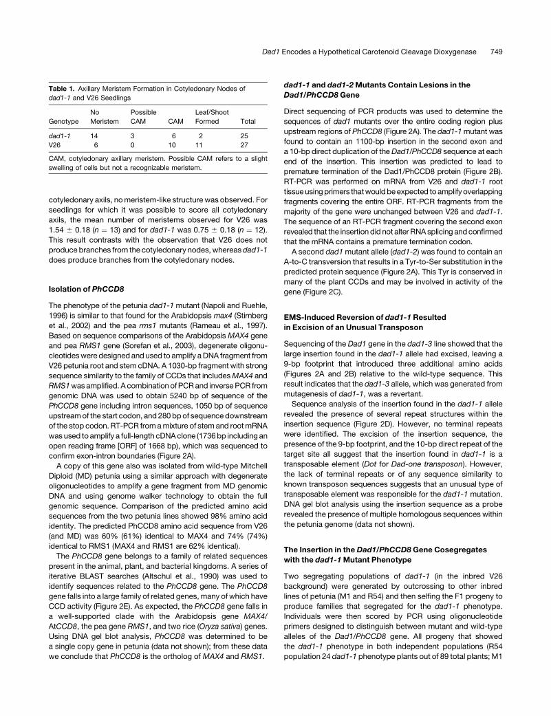
dad1-1 and dad1-2Mutants Contain Lesions in the
Dad1/PhCCD8 Gene
Direct sequencing of PCR products was used to determine the
sequences of dad1 mutants over the entire coding region plus
upstream regions of PhCCD8 (Figure 2A). The dad1-1mutant was
found to contain an 1100-bp insertion in the second exon and
a 10-bp direct duplication of theDad1/PhCCD8 sequence at each
end of the insertion. This insertion was predicted to lead to
premature termination of the Dad1/PhCCD8 protein (Figure 2B).
RT-PCR was performed on mRNA from V26 and dad1-1 root
tissueusingprimers thatwouldbeexpected to amplifyoverlapping
fragments covering the entire ORF. RT-PCR fragments from the
majority of the gene were unchanged between V26 and dad1-1.
The sequence of an RT-PCR fragment covering the second exon
revealed that the insertion did not alter RNA splicing andconfirmed
that the mRNA contains a premature termination codon.
A second dad1mutant allele (dad1-2) was found to contain an
A-to-C transversion that results in a Tyr-to-Ser substitution in the
predicted protein sequence (Figure 2A). This Tyr is conserved in
many of the plant CCDs and may be involved in activity of the
gene (Figure 2C).
EMS-Induced Reversion of dad1-1 Resulted
in Excision of an Unusual Transposon
Sequencing of the Dad1 gene in the dad1-3 line showed that the
large insertion found in the dad1-1 allele had excised, leaving a
9-bp footprint that introduced three additional amino acids
(Figures 2A and 2B) relative to the wild-type sequence. This
result indicates that the dad1-3 allele, which was generated from
mutagenesis of dad1-1, was a revertant.
Sequence analysis of the insertion found in the dad1-1 allele
revealed the presence of several repeat structures within the
insertion sequence (Figure 2D). However, no terminal repeats
were identified. The excision of the insertion sequence, the
presence of the 9-bp footprint, and the 10-bp direct repeat of the
target site all suggest that the insertion found in dad1-1 is a
transposable element (Dot for Dad-one transposon). However,
the lack of terminal repeats or of any sequence similarity to
known transposon sequences suggests that an unusual type of
transposable element was responsible for the dad1-1 mutation.
DNA gel blot analysis using the insertion sequence as a probe
revealed the presence of multiple homologous sequences within
the petunia genome (data not shown).
The Insertion in the Dad1/PhCCD8 Gene Cosegregates
with the dad1-1Mutant Phenotype
Two segregating populations of dad1-1 (in the inbred V26
background) were generated by outcrossing to other inbred
lines of petunia (M1 and R54) and then selfing the F1 progeny to
produce families that segregated for the dad1-1 phenotype.
Individuals were then scored by PCR using oligonucleotide
primers designed to distinguish between mutant and wild-type
alleles of the Dad1/PhCCD8 gene. All progeny that showed
the dad1-1 phenotype in both independent populations (R54
population 24 dad1-1 phenotype plants out of 89 total plants; M1
Table 1. Axillary Meristem Formation in Cotyledonary Nodes of
dad1-1 and V26 Seedlings
Genotype
No
Meristem
Possible
CAM CAM
Leaf/Shoot
Formed Total
dad1-1 14 3 6 2 25
V26 6 0 10 11 27
CAM, cotyledonary axillary meristem. Possible CAM refers to a slight
swelling of cells but not a recognizable meristem.
Dad1 Encodes a Hypothetical Carotenoid Cleavage Dioxygenase 749

Figure 2. Structure of the Dad1/PhCCD8 Gene.
(A) Structure of the Dad1/PhCCD8 gene showing the locations of the mutant alleles. Open boxes represent the ORF within the exons, with introns
represented by thin black lines. The promoter and 59 and 39 UTRs are denoted by shaded bars. The sequence associated with the dad1-1 allele
represents the Dad1/PhCCD8 sequence that was duplicated by the insertion of a transposon (the actual transposon sequence is not shown), and the
sequence associated with the dad1-3 allele is the 9-bp footprint left by the excision of the transposon. For dad1-2, the single base change is shown.
750 The Plant Cell

population 30 out of 148) contained PCR products consistent
with the presence of the dad1-1 allele. These results provided
genetic confirmation that theDad1/PhCCD8 gene is linked to the
dad1-1phenotype.
Expression of Dad1/PhCCD8 Complements the dad1-1
Mutation in Transgenic Plants
To determine whether expression of Dad1/PhCCD8 transgenes
could complement dad1 mutants, we used Agrobacterium-
mediated transformation into dad1-1 mutant petunia plants to
introduce a cDNA copy of the V26 Dad1 gene fused to the 35S
promoter of the Cauliflower mosaic virus (CaMV). In 7 out of 10
plants transformed with the 35S cDNA construct, the highly
branched phenotype was significantly reduced and the height of
the plants was increased, resulting in plants indistinguishable
from wild-type V26 (Figure 3A).
Downregulation of Dad1/PhCCD8 in Wild-Type Plants
Phenocopies the dad1Mutant
To test whether we could phenocopy the dad1 mutation, we
transformed V26 with an RNAi construct under the control of
the CaMV 35S promoter. In six out of eight wild-type plants
transformed with this construct, the number of branches was
increased and the plant height was reduced, resulting in a phe-
notype very similar to the dad1-1 mutant (Figure 3A).
Wild-type MD plants were also transformed with an RNAi
construct under the control of the constitutively expressed 35S
promoter of the Figwort mosaic virus. Gene expression in Dad1/
PhCCD8 RNAi lines decreased to undetectable levels in root
tissue of selected lines (Figure 4A). Three independent trans-
genic lines showing the greatest degree of RNAi were selected
for further study.
Transgenic and control lines were grown for phenotypic
characterization. The RNAi lines had a highly branched pheno-
type visibly different from the wild type. The branching pheno-
type was visible 3 weeks postgermination (data not shown) and
became more noticeable throughout development (Figure 3B).
At 14 weeks postgermination, a significant difference between
the RNAi lines and the wild type was observed (Figure 3C). This
difference is more easily observed in plants stripped of their
leaves (Figure 3D).
In addition to altered lateral shoot growth, several other
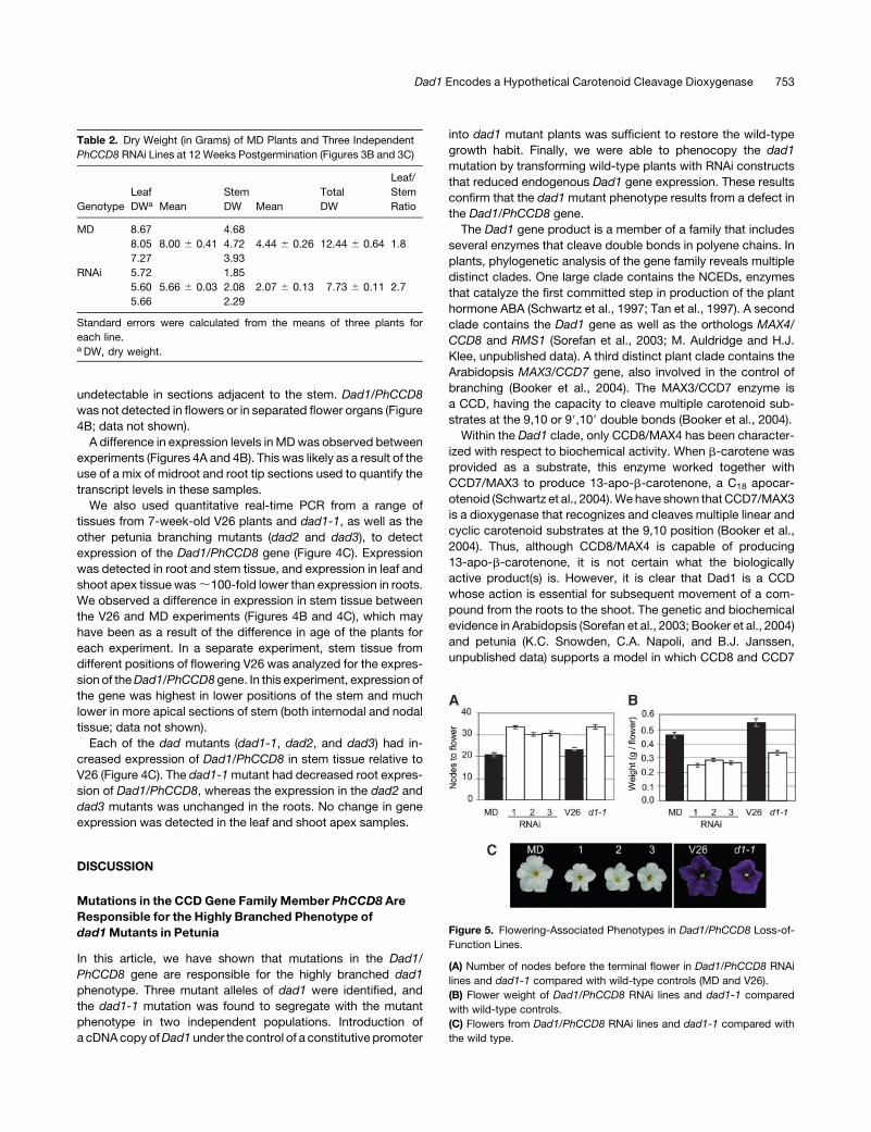
phenotypic changes were observed. The dry weights of leaves
and stems were determined for each of the three RNAi lines at
14 weeks postgermination (Table 2). A significant decrease in
the dry weight of the RNAi lines compared with MD was
observed. Furthermore, a significant difference in the ratio of
leaf to stem matter was observed, increasing from a ratio of 1.8
to 2.7.
MD plants had an average of 21 nodes between the cotyledon
and the terminal flower of the primary stem. Suppression of
Dad1/PhCCD8 led to a significant increase in node number
before flowering, increasing to 31 to 34 nodes, similar to the
result observed for dad1-1 (Figures 1B and 5A). The flowering
time for RNAi plants increased from 14 to 18 weeks (data not
shown). The MD RNAi lines showed reduced flower weight
(Figure 5B) and size (Figure 5C). This difference in flower size also
was observed in dad1-1 (Figures 5B and 5C).
Because Dad1/PhCCD8 transcripts are primarily found in the
root tissue, we examined how Dad1/PhCCD8 interference af-
fected root development.Dad1/PhCCD8 RNAi led to a reduction
in root mass 12 weeks postgermination (Figure 6A). A reduction
in root mass in the dad1-1 mutant compared with V26 was also
observed (Table 3). However, no obvious changes in root
structure were observed (Figure 6B).
Figure 2. (continued).
(B) Alignment of the predicted amino acid sequence of the Dad1 wild-type and mutant alleles around the position of the insertion in dad1-1, in
comparison with other members of the CCD8 clade. The consensus sequence shows amino acids that are absolutely conserved in the sequences
shown (the mutant alleles were not used in consensus generation). The differences between the wild-type Dad1 sequence and the mutant alleles are
underlined. In dad1-1, the asterisk indicates a stop codon.
(C) Alignment of the Dad1 and dad1-2 predicted amino acid sequence in comparison with one member of each clade of the CCD gene family. The
consensus shows amino acids for which at least three members have the sequence shown (the dad1-2 sequence was not used in the consensus
generation). The changed amino acid in the dad1-2 allele is underlined.
(D) Repeat structure of the transposon (Dot) inserted in the dad1-1 allele, including the 10-bp direct duplication of the Dad1 sequence. The key shows
the consensus sequence for each motif found in the transposon sequence with the maximum number of mismatches allowed for inclusion in the figure.
The longer CT-rich region is composed of 93% CT and the shorter region is 100% CT, whereas the GA-rich region is 89% GA.
(E) Unrooted phylogenetic tree of the CCD gene family. Only full-length members of the family are included, and not all members in the NCED clade are
included for clarity (though all known Arabidopsis members of the gene family are included). The predicted protein sequences were initially clustered
using ClustalX (Thompson et al., 1997) and the alignment refined manually with the program MacClade (Maddison and Maddison, 2000). Phylogenetic
relationships between the family members were determined using the neighbor-joining method and bootstrap analysis with 1000 replicates using the
Phylip package of programs (Felsenstein, 2004). Bootstrap values are shown as percentages. Accession numbers for sequences used from the
following species are as follows: Arabidopsis AtCCD1 (At3g63520), AtNCED2 (At4g18350), AtNCED3 (At3g14440), AtCCD4 (At4g19170), AtNCED5
(At1g30100), AtNCED6 (At3g24220), AtCCD7 (At2g44990), AtCCD8 (At4g32810), and AtNCED9 (At1g78390); Bixa orellana BoLCD (AJ489277.1);
Crocus sativus CsZCD (CAD33262.1); Zea mays ZmVP14 (U95953.1); rice OsCCD7 (AL663000.4), OsCCD8a (AP003296.3), and OsCCD8b
(AP003376.3); petunia Dad1/PhCCD8 (AY743219); pea RMS1 (AY557341.1); Sphingomonas paucimobilis PpLSD1 (S80637.1), PpLSD2 (S65040.1),
and PpLSD3 (AB073227.1); Streptomyces antibioticus SaSIM14 (AF322256.1); Synechocystis sp SynLSD1 (D90914.1) and SynLSD2 (AP005369.1);
Anabaena sp AnaLSD (AP003595.1); Homo sapiens HsRPE65 (AF039855.1), HsCCD (AJ290393.1), and HsCCD3 (AF294900.1); Drosophila
melanogaster DmCCD (AY121617.1); Caenorhabditis elegans CeCCD3 (AL110485.5) and CeCCD4 (AF098992.2).
Dad1 Encodes a Hypothetical Carotenoid Cleavage Dioxygenase 751

Stem cuttings (;8 cm in length) were also taken from
sympodial shoots. After 6 weeks, wild-type cuttings showed
the development of roots, but RNAi cuttings showed significantly
less root development (Figure 6C). Paradoxically, mature RNAi
plants developed adventitious roots around the lower stems at
later stages of development (Figure 6D). Adventitious root
formation was not observed on stems of wild-type MD or V26
plants. However, the formation of similar roots on the lower
stems of dad1-1 plants has been observed previously (Napoli,
1996).
The Dad1/PhCCD8 Gene Is Expressed in Root
and Stem Tissue
From micrograft analysis of the dad1-1 mutant (Napoli, 1996;
K.C. Snowden and C.A. Napoli, unpublished data), the Dad1
gene was predicted to be expressed in root and stem tissue.
Dad1/PhCCD8 transcripts are present at very low levels and are
not easily detectable by RNA gel blots in either V26 or MD plants.
Expression of the gene was therefore examined using real-time
PCR. We quantified the expression of theDad1/PhCCD8 gene in
the leaves, stem, roots, and flowers (Figure 4B). Dad1/PhCCD8
mRNA was detected at very low levels in the stem and leaves.
The highest transcript levels were consistently detected in the
roots representing 1.8 3 10�3% total mRNA. Transcript levels
decrease in roots segments further from the root tip, becoming
Figure 3. Complementation and Phenocopying of the dad1-1 Mutant
Phenotype.
(A) Complementation of dad1-1 with Dad1/PhCCD8 and phenocopying
of the mutant phenotype in V26 with a Dad1/PhCCD8 RNAi construct.
Plants of similar ages with a control transgene are shown for comparison.
(B) Branching phenotypes of control (MD) and three independent Dad1/
PhCCD8 RNAi lines 8 weeks postgermination.
(C) The same plants as shown in (B) 12 weeks postgermination.
(D) Plants from (C) shown stripped of their leaves.
Figure 4. Expression of the Dad1/PhCCD8 Gene in Petunia.
(A)Dad1/PhCCD8 expression in roots and leaves ofDad1/PhCCD8 RNAi
plants as determined by TaqMan real-time RT-PCR.
(B) Dad1/PhCCD8 expression in the tissues of wild-type MD plants
as determined by TaqMan real-time RT-PCR. Roots were taken from
12-week-old plants separated into tip, middle, and base sections and
analyzed independently. Stem tissue included the entire stem and nodes
from mature flowering plants ranging from 30 to 35 cm in height from the
base to the flower.
(C) Real-time RT-PCR analysis of Dad1/PhCCD8 transcripts in 7-week-
old V26 and dad mutant plants. The shoot apex sample included the
apex of the plant, including leaves smaller than 1.5 cm in length. The root
and stem samples included all root or stem tissue (except for that
included in the shoot apex samples) from a pool of six plants. The plants
were not flowering at the time of harvest.
752 The Plant Cell
undetectable in sections adjacent to the stem. Dad1/PhCCD8
was not detected in flowers or in separated flower organs (Figure
4B; data not shown).
A difference in expression levels in MDwas observed between
experiments (Figures 4A and 4B). This was likely as a result of the
use of a mix of midroot and root tip sections used to quantify the
transcript levels in these samples.
We also used quantitative real-time PCR from a range of
tissues from 7-week-old V26 plants and dad1-1, as well as the
other petunia branching mutants (dad2 and dad3), to detect
expression of the Dad1/PhCCD8 gene (Figure 4C). Expression
was detected in root and stem tissue, and expression in leaf and
shoot apex tissue was;100-fold lower than expression in roots.
We observed a difference in expression in stem tissue between
the V26 and MD experiments (Figures 4B and 4C), which may
have been as a result of the difference in age of the plants for
each experiment. In a separate experiment, stem tissue from
different positions of flowering V26 was analyzed for the expres-
sion of theDad1/PhCCD8 gene. In this experiment, expression of
the gene was highest in lower positions of the stem and much
lower in more apical sections of stem (both internodal and nodal
tissue; data not shown).
Each of the dad mutants (dad1-1, dad2, and dad3) had in-
creased expression of Dad1/PhCCD8 in stem tissue relative to
V26 (Figure 4C). The dad1-1mutant had decreased root expres-
sion of Dad1/PhCCD8, whereas the expression in the dad2 and
dad3 mutants was unchanged in the roots. No change in gene
expression was detected in the leaf and shoot apex samples.
DISCUSSION
Mutations in the CCD Gene Family Member PhCCD8 Are
Responsible for the Highly Branched Phenotype of
dad1Mutants in Petunia
In this article, we have shown that mutations in the Dad1/
PhCCD8 gene are responsible for the highly branched dad1
phenotype. Three mutant alleles of dad1 were identified, and
the dad1-1 mutation was found to segregate with the mutant
phenotype in two independent populations. Introduction of
a cDNAcopy ofDad1 under the control of a constitutive promoter
into dad1 mutant plants was sufficient to restore the wild-type
growth habit. Finally, we were able to phenocopy the dad1
mutation by transforming wild-type plants with RNAi constructs
that reduced endogenous Dad1 gene expression. These results
confirm that the dad1mutant phenotype results from a defect in
the Dad1/PhCCD8 gene.
The Dad1 gene product is a member of a family that includes
several enzymes that cleave double bonds in polyene chains. In
plants, phylogenetic analysis of the gene family reveals multiple
distinct clades. One large clade contains the NCEDs, enzymes
that catalyze the first committed step in production of the plant
hormone ABA (Schwartz et al., 1997; Tan et al., 1997). A second
clade contains the Dad1 gene as well as the orthologs MAX4/
CCD8 and RMS1 (Sorefan et al., 2003; M. Auldridge and H.J.
Klee, unpublished data). A third distinct plant clade contains the
Arabidopsis MAX3/CCD7 gene, also involved in the control of
branching (Booker et al., 2004). The MAX3/CCD7 enzyme is
a CCD, having the capacity to cleave multiple carotenoid sub-
strates at the 9,10 or 99,109 double bonds (Booker et al., 2004).
Within the Dad1 clade, only CCD8/MAX4 has been character-
ized with respect to biochemical activity. When b-carotene was
provided as a substrate, this enzyme worked together with
CCD7/MAX3 to produce 13-apo-b-carotenone, a C18 apocar-
otenoid (Schwartz et al., 2004).We have shown that CCD7/MAX3
is a dioxygenase that recognizes and cleaves multiple linear and
cyclic carotenoid substrates at the 9,10 position (Booker et al.,
2004). Thus, although CCD8/MAX4 is capable of producing
13-apo-b-carotenone, it is not certain what the biologically
active product(s) is. However, it is clear that Dad1 is a CCD
whose action is essential for subsequent movement of a com-
pound from the roots to the shoot. The genetic and biochemical
evidence in Arabidopsis (Sorefan et al., 2003; Booker et al., 2004)
and petunia (K.C. Snowden, C.A. Napoli, and B.J. Janssen,
unpublished data) supports a model in which CCD8 and CCD7
Table 2. Dry Weight (in Grams) of MD Plants and Three Independent
PhCCD8 RNAi Lines at 12 Weeks Postgermination (Figures 3B and 3C)
Genotype
Leaf
DWa Mean
Stem
DW Mean
Total
DW
Leaf/
Stem
Ratio
MD 8.67 4.68
8.05 8.00 6 0.41 4.72 4.44 6 0.26 12.44 6 0.64 1.8
7.27 3.93
RNAi 5.72 1.85
5.60 5.66 6 0.03 2.08 2.07 6 0.13 7.73 6 0.11 2.7
5.66 2.29
Standard errors were calculated from the means of three plants for
each line.a DW, dry weight.
Figure 5. Flowering-Associated Phenotypes in Dad1/PhCCD8 Loss-of-
Function Lines.
(A) Number of nodes before the terminal flower in Dad1/PhCCD8 RNAi
lines and dad1-1 compared with wild-type controls (MD and V26).
(B) Flower weight of Dad1/PhCCD8 RNAi lines and dad1-1 compared
with wild-type controls.
(C) Flowers from Dad1/PhCCD8 RNAi lines and dad1-1 compared with
the wild type.
Dad1 Encodes a Hypothetical Carotenoid Cleavage Dioxygenase 753

act in the same pathway to produce a novel, graft-transmissible
apocarotenoid hormone either as sequential steps, where the
intermediate is not mobile, or at the same step. The conservation
of the CCD8 gene and the mutant phenotypes between petunia,
pea, and Arabidopsis indicates that branch development is
controlled by a similar mobile signaling molecule, which in turn
suggests that the mechanism of control of branch development
may be widely conserved.
Phenotypic Analysis of the dad1 Alleles Suggests That
Partial Loss of Function of the DAD1 Protein Is Possible
The dad1-1 allele is predicted to produce a truncated protein that
is unlikely to have any biological activity because it is missing
amino acids that are completely conserved in all of the CCDs.
The dad1-1mutant allele produces branches at every node up to
the 10th node and exhibits reduced height, delayed flowering,
and reduced leaf senescence (Figures 1A and 1B; Snowden and
Napoli, 2003). The dad1-2 allele has a single amino acid sub-
stitution that replaces a Tyr at amino acid 456 with a Ser. This
amino acid is highly conserved, being either a Phe or a Tyr in
everymember of the Arabidopsis gene family, and substitution of
Ser apparently compromises the activity of the enzyme. This
amino acid substitution results in a phenotype intermediate
between dad1-1 and the wild type for some but not all traits.
An intermediate phenotype has not been reported for any alleles
of themax4 or rms1mutants (Sorefan et al., 2003). These results
suggest that, in dad1-2 mutants, the translocated substance is
present at reduced levels and affects the different developmental
processes to varying degrees. However, we cannot exclude the
possibility that the Dad1/PhCCD8 protein acts on multiple
substrates and the dad1-2 mutation alters substrate specificity.
The phenotype of dad1-3 plants is almost indistinguishable from
the wild type, suggesting that inclusion of an additional three
amino acids in this region of the protein has a minimal effect on
the biological activity of the protein.
The dad1-1Mutation Was Caused by Insertion of a
Novel Transposon (Dot)
Sequencing of the dad1-1 allele revealed the presence of
a 1.1-kb insertion in exon 2 that was not present in the wild-
type gene. This was unexpected because the dad1-1 allele was
generated by EMS mutagenesis (Napoli and Ruehle, 1996). This
insertion has some features characteristic of a transposon: at
each end of the insertion is a 10-bp direct repeat of the Dad1
sequence, and the insertion contains a highly repetitive structure
(Figure 2D). In addition, DNA gel blot analysis using the insertion
sequence as a probe revealed multiple copies of related
sequences in the petunia genome, as would be expected for
an active transposon. However, no terminal repeats (inverted or
Figure 6. Root Phenotypes Associated with Loss of Dad1/PhCCD8.
(A) Roots of wild-type (MD) and Dad1/PhCCD8 RNAi plants 12 weeks postgermination.
(B) Roots of wild-type (V26) and dad1-1 plants 13 weeks postgermination.
(C) The development of roots on cuttings from MD and Dad1/PhCCD8 RNAi lines after 6 weeks.
(D) Adventitious roots developing on the lower stems of a Dad1/PhCCD8 RNAi line but not on wild-type (MD) stems.
Table 3. Fresh Weight of Root and Shoot (All Stem and Leaf Tissue)
of V26 and dad1-1 Mutants at 9 and 13 Weeks
Postgermination (Figure 6B)
Tissue V26 dad1-1 LSD
Root FW (g)a 2.24 1.60 0.480
Shoot FW (g) 38.5 38.7 7.97
Data was analyzed by two-way ANOVA using the GenStat (version 7)
statistical software package. Values given are means. No interactions
were detected between genotype and age of plants at harvest. LSD,
least significant differences of means (5% level). If means differ by more
than the LSD shown, then they are significantly different.a FW, fresh weight.
754 The Plant Cell

direct) could be found. No instability of the dad1-1 mutant had
been observed over more than 12 generations and thousands of
individual plants grown. However, when seeds of the dad1-1
mutant were treated with EMS to identify second-site suppres-
sors of the dad1-1 mutation, the dad1-3 revertant allele was
isolated. This mutagenesis appears to have resulted in excision
of the insertion found in the dad1-1 allele, presumably as a result
of activation of a transposable element, perhaps in response to
genomic stress (McClintock, 1984; Kunze, 1996). Sequencing of
theDad1 gene in this revertant allele revealed excision of the 1.1-
kb insertion and the presence of a 9-bp footprint corresponding
to part of the insertion sequence and the remaining target site
duplication seen in the dad1-1 sequence. The target site dupli-
cation, combined with the EMS-induced excision of this 1.1-kb
insertion and the presence of the footprint, provides strong
evidence that the insertion sequence found in the dad1-1 allele is
a transposon, which we have namedDot. The identification of an
additional transposon in petunia may prove useful for isolation of
genes in the future, particularly because transposition may be
inducible by EMS mutagenesis.
The lack of any terminal repeat structures in an active trans-
poson is unusual. The structure of this insertion element sug-
gests that it would form a unique transposon class. Many
characterized transposon families (e.g., En/Spm or Ac/Ds) have
repetitive elements, particularly in the subterminal regions.
However, the transposon found in the dad1-1 allele has a differ-
ent structure. The major repetitive elements are located in the
middle of the element, with multiple degenerate copies of an 18-
bp palindrome. Searches within sequence databanks have not
found other sequences with homology to this element. Although
the element does contain some ORFs, all those that are larger
than 30 amino acids are found within the middle repetitive region
or within the regions with high TC or GA composition. None of the
putative ORFs present within the element has any homology to
known proteins. It is likely that if transposition of this element
requires protein(s), are derived from elsewhere in the genome
(i.e., this element is probably nonautonomous in the manner of
transposons such as Ac/Ds [Kunze, 1996]).
Axillary Meristems Are Produced Early in Wild-Type
V26 but Do Not Develop into Branches
Scanning electronmicroscopy of cotyledonary axils fromboth V26
and dad1-1 mutants revealed that, in wild-type plants, cotyledon
axil meristems and axillary shoots appear to form at an earlier
stage of development than in dad1-1. However, these shoots do
not continue growth to form branches in thewild type. By contrast,
branches are formed from both cotyledonary nodes in dad1-1, yet
scanning electron micrographs showed no detectable meristem
development for the majority of the dad1-1 axils at the stage that
was examined. One question is whether the meristems formed in
the cotyledonary nodes of dad1-1 mutants are fundamentally
different from normal axillary meristems that develop in the wild
type. It may be that dad1-1 mutants fail to form normal axillary
meristems at all, and the branches that do form may result from
adventitious meristems that form as a result of an abnormal signal
present in the mutant. Alternatively, meristem formation may
simply be delayed in the dad1-1mutant, but, once initiated, shoot
growth proceeds without dormancy. This result contrasts with the
observation of the max1 and max2 mutants, in which timing of
meristem initiation is not changed but development of primordia
by the axillary meristem is advanced relative to the wild type
(Stirnberg et al., 2002). Thus, it may be that the imposition of dor-
mancy of axillary meristems occurs at different stages in petunia
and Arabidopsis and warrants further investigation.
The Dad1 Gene Is Involved in Leaf Senescence
While examining the branching phenotype of the dad1 mutants,
we observed that leaf senescence was delayed in dad1-1 and
dad1-2mutants, with dad1-2 having an intermediate phenotype.
The difference observed between dad1-1 and the wild type may
be a consequence of the difference in flowering time between
these two genotypes. However, the dad1-2 allele also shows
delayed leaf senescence without any alteration in flowering time.
A relationship between branching phenotype and senescence
has been shown previously for the oresara9 (ore9)/max2 mutant
of Arabidopsis (Woo et al., 2001; Stirnberg et al., 2002). This
mutant was originally isolated in a screen for mutants with
delayed leaf senescence (Oh et al., 1997). Subsequent positional
cloning of the ore9 gene (Woo et al., 2001) showed that it is
a member of the Leu-rich repeat–type F-box gene family, in-
volved in ubiquitin-dependent degradation of proteins. Posi-
tional cloning of the branching mutant max2 (Stirnberg et al.,
2002) identified the same F-box gene, identifying a linkage
between branch development and leaf senescence.
The finding that mutations in Dad1 also affect both branching
and leaf senescence suggests that the Dad1/PhCCD8 and
ORE9/MAX2 genes could affect the same signaling pathway.
A direct interaction between the F-box protein and Dad1/
PhCCD8 protein is unlikely because ubiquitin-mediated degra-
dation of Dad1/PhCCD8byORE9/MAX2would result in opposite
phenotypes for mutations in these genes. Further, Dad1/
PhCCD8 is likely to be located in the plastid, based on its
homology to the chloroplast-localized Arabidopsis ortholog
AtCCD8 (M. Auldridge and H.J. Klee, unpublished data).
Ubiquitin-dependent protein turnover is not believed to occur
in plastids (Beers et al., 1992).
It is possible that the translocated hormone has a role in
initiation of leaf senescence. The apocarotenoid hormone ABA
has been shown to promote leaf abscission, flower senescence,
and senescence of excised leaf discs (Smart, 1994), and the
enzymes from the NCED clade of the gene family have been
shown to be responsible for the first committed step in ABA
production (Schwartz et al., 1997). Thus, it seems reasonable to
suggest that the Dad1/PhCCD8 protein is involved in production
of a similar compound that has a role in leaf senescence.
However, the ore9/max2 mutant has a reduced response to
inducers of senescence, including ABA, methyl jasmonate, and
ethylene (Woo et al., 2001), suggesting that ore9/max2 acts at
a point downstream or independent of the ABA senescence
signal. Furthermore, the role of cytokinin in leaf senescence (Gan
and Amasino, 1995) and branch development (Napoli et al.,
1999) suggests that the role ofDad1/PhCCD8 in leaf senescence
may be indirect.
Dad1 Encodes a Hypothetical Carotenoid Cleavage Dioxygenase 755

Loss of Dad1 Expression Results in Reduced
Root Development
In transgenic MD plants expressing an RNAi construct and in
the dad1-1 mutant, root mass is reduced. In RNAi lines, the for-
mation of adventitious roots on cuttings also is inhibited. By
contrast, in both wild-type plants transformed with an RNAi
construct (Figure 6D) and in dad1-1, adventitious roots are
observed on the lower portions of the stems (Napoli, 1996).
Whether adventitious root formation is a consequence of re-
duced root growth is not clear. It also is not clear whether the
Dad1 gene product has a normal role in root development, or
whether the altered root growth observed is a consequence of
a build up of the substrate of the Dad1 gene product or even
a consequence of altered shoot growth. We do not, however,
believe that the shoot morphology is simply a result of reduced
root mass because when dad1-1mutant scions are grafted onto
wild-type roots, the presence of dad1-1 mutant roots above the
graft union induces the mutant branching phenotype despite the
presence of a wild-type (normal-sized) root mass.
Expression of Dad1/PhCCD8 Suggests That the Gene
Is under Feedback Control
The expression of theDad1/PhCCD8 gene observed in roots and
stems of petunia plants is consistent with a role in control of
a graft-transmissible substance that can alter growth of axillary
branches. The increased levels of expression in the stems of
dad1-1, dad2, and dad3 mutants relative to the wild type are
indicative of a feedback control mechanism that is responding to
a change in the amount or detection of a branching signal. This
increased expression is not seen in the root tissue of the dad
mutants, suggesting that the feedback mechanism is restricted
to the stem tissue, and expression in the roots may be inde-
pendently controlled. Although the level ofDad1/PhCCD8RNA in
the roots of the dad2 and dad3 mutants is unchanged, it is
significantly lower in the roots of the dad1-1 mutant than in the
wild type. The insertion in the dad1-1mutant may lead to mRNA
instability and a high level of mRNA turnover. In dad1-1 stems,
any increase in mRNA turnover may be masked by positive
feedback.
The Role of Dad1/PhCCD8 in Branch Signaling
Observations made in petunia by Napoli (1996) suggested that
a mobile signal molecule is transported from the roots acrope-
tally to the axillary meristems, where branch development
occurs. From graft analysis, such a signal must be produced in
the roots and transported across graft unions. These observa-
tions in petunia have been replicated in pea (Beveridge et al.,
1994, 2000; Foo et al., 2001) and Arabidopsis (Turnbull et al.,
2002). Interstock grafts in which a small section ofwild-type stem
is inserted above dad1 mutant roots and below dad1 mutant
scion also revert the mutant scion to wild-type phenotype
(Napoli, 1996). This result indicates that the Dad1/PhCCD8
gene product is also present and active in stem tissue, which
we have confirmed by expression analysis (Figure 4C). Because
only a small piece of wild-type stem interstock tissue is required
for reversion, the Dad1/PhCCD8 gene product must be active to
a high enough level to produce the wild-type phenotype.
Although these observations strongly suggest a mobile signal
that is acted upon in some way by the Dad1/PhCCD8 gene
product, they do not define whether the mobile signal is a re-
pressor or promoter of branching. Two possibilities exist. First,
the Dad1/PhCCD8 gene product acts to produce or transport
a repressor of branching, and in the absence of Dad1/PhCCD8,
lack of this repressor results in branching. Alternatively, the
substrate of the Dad1/PhCCD8 gene product (possibly sub-
sequently processed) is a promoter of branching that is pro-
cessed or in some way inactivated by the Dad1/PhCCD8 gene
product. In the absence of Dad1/PhCCD8, this promoter of
branching is transported to the shoot and induces branching.
However, in petunia, when dad1 mutant scions are grafted onto
wild-type rootstocks and dad1 mutant roots form above the
graft, the plant does not revert to thewild type (Napoli, 1996; K.C.
Snowden, unpublished data). When these roots are removed,
subsequent nodes revert to the wild type and do not branch. This
result is consistent with the presence of a promoter of branching
that is produced from dad1 mutant roots and transported to the
shoot, where branches are induced. These two possibilities are
not mutually exclusive. The Dad1/PhCCD8 gene product may
produce an inhibitor of branching in addition to the substrate
being a promoter of branching. However, in the grafts described
above, this putative inhibitor is not sufficient to prevent branch
development in the presence of the promoter of branching being
produced by the dad1mutant roots. If indeed theDad1/PhCCD8
gene product converts a promoter of branching into an inhibitor,
it is possible that in other species the balance between pro-
motion and inhibition of branch development is altered.
METHODS
Genetic Stocks and Plant Growth Conditions
Inbred cv MD petunia (Petunia axillaris 3 [P. axillaris 3 Petunia integ-
rifolia]) plants were grown under greenhouse conditions with a day/night
temperature regime of 258C/188C in commercial potting medium (Fafard
2B; Conrad Fafard, Agawam,MA) in 15-cm, 1.5-L pots andwere fertilized
at each irrigation with 150mg L�1 nitrogen from 15:7:14.1 soluble fertilizer
(Peter’s Fertilizer Products, Fogelsville, PA).
Petunia (Petunia hybrida) Vilm inbred genetic stock V26 and the dad
mutants derived from V26 (Napoli and Ruehle, 1996) were grown under
greenhouse conditions with a minimum/maximum temperature regime of
208C to 308Cand aminimumof 12 h light, supplementedwhen necessary.
Plants for RNA isolation and the characterization of the dad1 alleles were
grown in commercial potting medium (Dalton’s potting mix; Matamata,
New Zealand) in six-packs (100-mL size); transgenic plants were initially
grown in 400-mL pots and then transferred to 1.5-liter pots after 6 to
8 weeks. All plants were fertilized at each irrigation with 80 mg L�1
nitrogen, 80 mg L�1 phosphorus, and 60 mg L�1 potassium (from Wuxal
Super 8-8-6 plus micro liquid fertilizer; Aglukon, Dusseldorf, Germany).
Alleles of dad1 were grown until they were 12 weeks of age. Nodes
were recorded and branches defined as by Snowden and Napoli (2003).
Gene Isolation and Vector Construction
RNA was isolated from combined root and stem tissue of V26 using an
RNeasy plant mini kit (Qiagen, Hilden, Germany). cDNA was synthesized
756 The Plant Cell

using Superscript II reverse transcriptase (Invitrogen, Carlsbad, CA) and
used as a template for PCR using primers designed from the amino
acid sequence of the Arabidopsis thaliana MAX4, pea (Pisum sativum)
RMS1, and rice (Oryza sativa) CCD8 genes, forward primer
59-TTCCGGCATCTCTTCGACGGCTAC-39 and reverse primer 59-GGA-
TCCAGCAACCATGCAAGCC-39. This PCR was used as template in a
second nested PCR using redundant primers, forward 59-GTI-
ATGTGYYTNACNGARAC-39 and reverse 59-TAIGGNARICCRTANGG-
RAA-39. A 1-kb fragment was isolated and cloned into the pGEM-T Easy
vector (Promega, Madison, WI). Sequence of this clone was used to
design primers for inverse PCR from genomic DNA. Inverse PCR
(Snowden and Napoli, 1998) from V26 genomic DNA was used to isolate
the 59 and 39 ends of the gene. After obtaining a full-length sequence for
the gene, proofreading PCR was used to isolate overlapping fragments
of the gene fromV26 and dad1. PCRproducts were sequenced directly to
obtain confirmed sequences of the gene and the mutants.
To generate a vector for the overexpression of Dad1/PhCCD8, a cDNA
clone containing the full ORF of Dad1/PhCCD8 was generated using
primers 59-CAAACTTAGCAGACCATAAAGAGTGCAC-39 and 59-GTC-
TGGGATGTGAGGTAACTA-39 and ligated into pGEM-T Easy. PCR
amplification was performed using this clone as template with primers
SP33 (59-attB2-CCGGGCCCCCCCTCGAG-39) and SP30 (59-attB1-
CCCCGGGCTGCAGGAATTC-39). The resulting 1667-bp PCR amplifica-
tion product was recombined via an attB 3 attP Gateway reaction with
pDONR201 to generate pENTRY_DAD1. A Gateway attL3 attR reaction
with pENTRY_DAD1 and the pHEX1 destination vector was performed to
generate pHEX1S_DAD1. All Gateway reactions were performed as
recommended by the manufacturer (Invitrogen). pHEX1 is a Gateway-
adapted version of the pART7/pART27 plant transformation vector
system (Gleave, 1992) with the Gateway 1711-bp RfA cassette cloned
into the SmaI site between the CaMV 35S promoter and the octopine
synthase transcriptional terminator (35S-attR2-ccdB-CmR-attR1-ocs39).
To generate the Dad1/PhCCD8 RNAi vector used in the V26 experi-
ments, a 388-bp region of the Dad1/PhCCD8 ORF (corresponding to
bases 435 to 822 of the ORF) was PCR amplified from V26 using prim-
ers oKCS1 (59-attB1-ATCTGATGCATACAAGGCAG-39) and oKCS2
(59-attB2-CCTCTCATTTGTTCCTGCTTC-39). The resulting 437-bp PCR
product was recombined via an attB 3 attP Gateway reaction with
pDONR201 to generate pENTRY_DAD1(ko). A Gateway attL 3 attR
reaction with pENTRY_DAD1(ko) and the destination vector pTKO2 was
performed to generate pTKO2S_DAD1.
To construct pTKO2, the 443-bp intron from the 59 untranslated region
(UTR) of the ArabidopsisColumbia Actin2 (ACT2) genewasPCRamplified
fromgenomicDNAusing primers INT1 (59-GCCCGTGGATCCCTCGAGT-
CCGGAACTAGTCTGCAGGTAATAGGAACTTTCTGGAT-39; underlined
regions denote homology to ACT2) and INT2 (59-GCCCGTAAGCTTGTC-
GACACCGGTGCTAGCATGCATCCTGCAAACACACAAAAAGAG-39). A
658-bp region of the 39 UTR and transcriptional terminator of the
Arabidopsis ACT2 gene was PCR amplified from Columbia genomic
DNA using primers ACT3-1 (59-GGGCCTAAGCTTGAGGCTCTCAAGAT-
CAAAGG-39) and ACT3-2 (59-GGGCCTGGTACCGCGGCCGCACTTTT-
TGGAATCAAGATGTA-39). A triple-enhanced version of the CaMV 35S
promoter (Que et al., 1997) was PCR amplified from pCHS3061 using
primers 3335S-1 (59-GGGCCGTATCTAGAATTCAATCCCACCAAAA-
CC-39) and 3335S-2 (59-GCAAAGAGCTCTTATACTCGAG-39). The
515-bp ACT2 intron PCR product was cloned as a BamHI-HindIII frag-
ment into pBluescript SK� (Stratagene, La Jolla, CA). The 693-bp ACT2
transcriptional terminator PCRproductwas then cloneddistal to the intron
as a HindIII-KpnI fragment. Subsequently, the 942-bp CaMV 35S pro-
moter PCR product was cloned proximal to the intron as an XbaI-XhoI
fragment. This created pPAN1 with an organizational structure of 35S-
intron-ACT39. To convert pPAN1 into a Gateway compatible vector, the
1711-bpGatewayRfA cassette (attR1-CmR-ccdB-attR2) was first cloned
into the PvuII site of pSP72 (Promega) and then excised as a XhoI-
XbaI fragment and cloned into SalI-NheI sites of pPAN1, placing the
RfA cassette between the ACT2 intron and transcriptional terminator.
To reduce sequence duplication in the final vector, a BamH1 deletion
was then produced, removing 703 bp from the RfA cassette, including the
entire chloramphenicol resistance gene. A second intact RfA cassette
was then cloned as an XhoI-XbaI fragment into the XhoI-SpeI sites,
placing the RfA cassette between the 35S promoter and the intron and in
the inverse orientation to the first RfA cassette introduced. The resulting
vector, referred to as pTKO1, has an organizational structure of 35S-
attR1-CmR-ccdB-attR2-intron-attR2-ccdB-attR1-Act2. pTKO1 was di-
gested with XbaI, followed by a partial NotI digestion, and the 4838-bp
Gateway-adapted 35S-intron-ACT2 39 cassette was cloned into SpeI-
NotI sites of the plant transformation vector pART27 (Gleave, 1992),
generating pTKO2. The presence of the two Gateway RfA-derived
cassettes, in an inverse orientation, facilitates the introduction of two
copies of a selected gene fragment into the vector in a single Gateway
attL3 attR reaction, generating an inverted repeat of the gene fragment,
flanking the intron sequence. When transcribed, this produces a self-
complementary RNA able to efficiently induce gene silencing.
A 2-kb fragment of PhCCD8 genomic sequence from MD from
positions 359 bp to 1462 bp of the coding sequence was cloned using
degenerate forward (59-GAGACCACGCTTCCGGCATCTCTTYGACGG-
STAC-39) and reverse (59-AAYTTCCCAATACTCTGACTAA-39) primers
corresponding to the GDHDFRHLFDGY and NFPNALSK conserved
protein sequences of the AtCCD8 and PsRMS1 orthologs. The remaining
sequencewas recovered byGenewalker (BDBiosciences, Palo Alto, CA).
A 643-bp partial coding sequence of PhCCD8 from positions 359 to 1002
was recovered by PCRwith the addition of anAscI restriction site at the 39
end. A second 316-bp partial cDNA fragment from positions 359 to 675
was recovered by PCR with an AscI site at the 39 end. The two fragments
were ligated at the AscI site and introduced into the pDESTOE vector
(Booker et al., 2004) by recombination from the pENTR/D vector
(Invitrogen). The Dad1/PhCCD8 RNAi construct used in transgenic MD
plants was under transcriptional control of the Figwort mosaic virus
promoter (Richins et al., 1987) followed by the nos 39 terminator.
Transgenic petunia plants were produced using Agrobacterium-
mediated transformation of 5-week-old petunia seedlings or tissue
culture–grown plants according to the method of Jorgensen et al.
(1996). Introduction and inheritance of the transgenes were confirmed
by PCR using primers specific for the neomycin phosphotransferase II
marker gene. All phenotypic characterizations of transgenic RNAi lines
were performed on PCR-positive T1 plants.
Scanning Electron Microscopy
V26 and dad1-1 seedlings for scanning electron microscopy analysis
were germinated and grown on 0.53 MS (including vitamins; Duchefa,
Haarlem, The Netherlands) plates, supplemented with 15 g L�1 sucrose,
until they had one visible true leaf. Seedlings were harvested and fixed in
2.5% glutaraldehyde in 0.1 M potassium phosphate buffer, pH 7.2, under
vacuum for 1 h and then stored overnight at 48C. After fixation, tissue was
dehydrated in a graded ethanol series and then dried with a Bal-Tec CPD
030 critical point drier (Balzers, Liechtenstein). Seedlings were secured to
stubs with carbon tabs and sputter coated with gold using a Polaron
E5100 (Quorum Technologies, Newhaven, UK). Specimens were viewed
with a Philips 505 scanning electron microscope (Eindhoven, The Nether-
lands) at 15 kV.
Quantitative RT-PCR
Total RNA was isolated from tissues using the RNeasy plant mini kit
(Qiagen). RNA from V26 was treated with DNase (using the Ambion
DNA-free kit; Austin, TX). Concentration and purity of RNA was checked
Dad1 Encodes a Hypothetical Carotenoid Cleavage Dioxygenase 757

using an Agilent 2100 bioanalyzer before and after DNase treatment.
Three independent reverse transcription reactions (containing 1.5 mg of
RNA, 3.5 mM of poly(dT)23V primer, 1 mM dNTP, 10 mM DTT, 13 first-
strand RT buffer, and 200 units of Superscript III RT [Invitrogen]) were
incubated at 558C for 1 h and then pooled. Primers were designed for
Dad1/PhCCD8 (forward 59-GTGGCAAGTGTAGAAGTTCC-39, reverse
59-TCAGCGCTATGCTCACAGC-39) and the control genes ACT2 (forward
59-CCTGATGAAGATCCTCACCGA-39, reverse 59-CAAGAGCCACATAG-
GCAAGCT-39), EF1a (forward 59-TGTTCTCTGCCTTGTATGTCTGG-39,
reverse 59-TCAAAAGAGGCAGGCAGACAG-39), and Histone H4 (forward
59-ATACGCTTGCACCCACCCCTA-39, reverse 59-GGAGGAGCTAAAC-
GACACCG-39). At least three replicate real-time PCR reactions were set
up for each primer pair. An aliquot of each pooled cDNA (1/300th) was
used as template for each reaction (containing 13 Platinum Taq reaction
buffer [Invitrogen], 0.1 mM dNTP, 1.5 mMMgCl2, 0.2 mM of each primer,
0.13 SYBR Green I dye [Molecular Probes, Eugene, OR], and 0.5 units of
Platinum Taq polymerase in a total volume of 20 mL). Reaction mixtures
were denatured for 2 min at 948C and then amplified for 40 cycles (15 s at
948C, 30 s at 598C, 20 s at 728C), followed by a dissociation stage (15 s at
958C, 30 s at 608C with a slow ramp to 15 s at 958C). Amplification and
expression quantification was performed using the ABI PRISM 7900 HT
sequence detection system (Applied Biosystems, Foster City, CA). Data
were analyzed with the SDS 2.0 software (Applied Biosystems) using the
OBT (Outlier, Baseline, Threshold) principle. Relative expression was
calculated using the comparative cycle threshold method (Pfaffl, 2001)
with normalization of data to the geometric average of the internal control
genes (Vandesompele et al., 2002). PCR efficiency for the amplicon was
estimated using the absolute fluorescence (window of linearity) method
(Ramakers et al., 2003), using data from the exponential phase of each
individual amplification plot.
RNA samples from MD were treated with RNase-free DNase (Qiagen)
and purified using Qiagen minicolumns. Samples were checked for DNA
contamination by TaqMan real-time RT-PCR in a reverse-minus tran-
scription reaction. Concentration and purity of total plant RNA was
determined by spectrophotometric analysis. The quantification was
verified for all RNA samples in each experiment by formaldehyde
agarose gel electrophoresis and visual inspection of rRNA bands upon
ethidium bromide staining. TaqMan one-step real-time RT-PCR was
performed as recommended by the manufacturer (Perkin-Elmer Applied
Biosystems, Foster City, CA). All reactions contained 13 TaqMan buffer
(Perkin-Elmer Applied Biosystems); 5 mMMgCl2; 200 mM dATP, 200 mM
dCTP, and 200 mM dGTP; 400 mM dUTP; 0.625 units of AmpliTaq Gold
polymerase; and 0.25 units of MultiScribe RNA reverse transcriptase
and RNase inhibitor in a 25-mL volume. Reverse transcription was per-
formed using 250 ng of total RNA, each gene-specific primer at
500 nM (forward, 59-AATTATTCTCAGGTGCATCGTTGA-39; reverse,
59-GCAAACAACTCGGCCATCA-39), and 250 nM TaqMan probe
(59-CGATAACGCGAATACAGGAGTCGTTA-39). Reaction mixtures were
incubated for 30 min at 488C for reverse transcription, 10 min at 958C,
followed by 40 amplification cycles of 15 s at 958C/1 min at 608C.
Samples were quantified in the GeneAmp 5700 sequence detection
system (Perkin-Elmer Applied Biosystems). Absolute mRNA levels were
quantified against a standard curve of tritiated in vitro-transcribed
sense-strand RNAs. Primers and probes used in the TaqMan real-time
quantitative RT-PCR assay were designed using PRIMER EXPRESS
software (Applied Biosystems). All probes were labeled at the 59 with
fluorescent reporter dye 6-carboxyfluorescein and at the 39 with black
hole quencher-1 from Integrated DNA Technologies (Coralville, IA;
Bernacchi and Mely, 2001).
Sequence data from this article have been deposited with the EMBL/
GenBank data libraries under accession numbers AY743219, AY745251,
and AY746977.
ACKNOWLEDGMENTS
This work was supported by Marsden and FoRST grants to K.S. and by
National Science Foundation Grant IBN-0115004 to H.K. This is publi-
cation R-10632 of the Florida Agricultural Experiment Station. We thank
Carolyn Napoli for helpful discussions and generation of the dad1-3
allele and Toshi Foster and Paul Sutherland for assistance with scanning
electron microscopy.
Received September 15, 2004; accepted December 5, 2004.
REFERENCES
Altschul, S.F., Gish, W., Miller, W., Myers, E.W., and Lipman, D.J.
(1990). Basic local alignment search tool. J. Mol. Biol. 215, 403–410.
Beers, E., Moreno, T., and Callis, J. (1992). Subcellular localization of
ubiquitin and ubiquitinated proteins in Arabidopsis thaliana. J. Biol.
Chem. 267, 15432–15439.
Bell, A.D. (1991). Plant Form: An Illustrated Guide to Flowering Plant
Morphology. (Oxford: Oxford University Press).
Bernacchi, S., and Mely, Y. (2001). Exciton interaction in molecular
beacons: A sensitive sensor for short range modifications of the
nucleic acid structure. Nucleic Acids Res. 29, E62–2.
Beveridge, C.A. (2000). Long-distance signalling and a mutational
analysis of branching in pea. Plant Growth Regul. 32, 193–203.
Beveridge, C.A., Ross, J.J., and Murfet, I.C. (1994). Branching mutant
rms-2 in Pisum sativum (grafting studies and endogenous indole-
3-acetic acid levels). Plant Physiol. 104, 953–959.
Beveridge, C.A., Symons, G.M., and Turnbull, C.G.N. (2000). Auxin
inhibition of decapitation-induced branching is dependent on graft-
transmissible signals regulated by genes Rms1 and Rms2. Plant
Physiol. 123, 689–697.
Beveridge, C.A., Weller, J.L., Singer, S.R., and Hofer, J.M.I. (2003).
Axillary meristem development. Budding relationships between
networks controlling flowering, branching and photoperiod respon-
siveness. Plant Physiol. 131, 927–934.
Booker, J., Auldridge, M., Wills, S., Klee, H.J., and Leyser, O.
(2004). MAX3 is a carotenoid cleavage dioxygenase required for
the synthesis of a novel plant signalling molecule. Curr. Biol. 14,
1232–1238.
Bouvier, F., Dogbo, O., and Camara, B. (2003a). Biosynthesis of the
food and cosmetic plant pigment bixin. Science 300, 289–291.
Bouvier, F., Suire, C., Mutterer, J., and Camara, B. (2003b). Oxidative
remodeling of chromoplast carotenoids: Identification of a carotenoid
cleavage dioxygenase CsCCD and CsZCD genes involved in Crocus
secondary metabolic biogenesis. Plant Cell 15, 47–62.
Felsenstein, J. (2004). PHYLIP (Phylogeny Inference Package), Version
3.6b. (Seattle: University of Washington).
Foo, E., Turnbull, C.G., and Beveridge, C.A. (2001). Long-distance
signaling and the control of branching in the rms1mutant of pea. Plant
Physiol. 126, 203–209.
Gan, S., and Amasino, R.M. (1995). Inhibition of leaf senescence by
autoregulated production of cytokinin. Science 270, 1986–1988.
Gleave, A. (1992). A versatile binary vector system with a T-DNA
organisational structure conducive to efficient integration of cloned
DNA into the plant genome. Plant Mol. Biol. 20, 1203–1207.
Gollapalli, D.R., Maiti, P., and Rando, R.R. (2003). RPE65 operates in
the vertebrate visual cycle by stereospecifically binding all-trans-
retinyl esters. Biochemistry 42, 11824–11830.
Halle, F. (1999). Ecology of reiteration in tropical trees. In The Evolution
of Plant Architecture, M.H. Kurmann and A.R. Hemsley, eds (Surrey,
UK: Royal Botanic Gardens, Kew), pp. 93–107.
758 The Plant Cell

Jorgensen, R.A., Cluster, P.D., English, J., Que, Q., and Napoli, C.A.
(1996). Chalcone synthase cosuppression phenotypes in petunia
flowers: Comparison of sense vs. antisense constructs and single-
copy vs. complex T-DNA sequences. Plant Mol. Biol. 31, 957–973.
Kamoda, S., and Saburi, Y. (1993a). Cloning, expression and sequence
analysis of a lignostilbene-a,b-dioxygenase gene from Pseudomonas
paucimobilis TMY1009. Biosci. Biotechnol. Biochem. 57, 926–930.
Kamoda, S., and Saburi, Y. (1993b). Structural and enzymatical
comparison of lignostilbene-a,b-dioxygenase isozymes, I, II and III,
from Pseudomonas paucimobilis TMY1009. Biosci. Biotechnol. Bio-
chem. 57, 931–934.
Kunze, R. (1996). The maize transposable element Activator (Ac). Curr.
Top. Microbiol. Immunol. 204, 161–194.
Lindqvist, A., and Anderson, S. (2002). Biochemical properties of
purified recombinant human b-carotene 15,15’-monooxygenase.
J. Biol. Chem. 277, 23942–23948.
Maddison, D.R., and Maddison, W.P. (2000). MacClade 4: Analysis of
Phylogeny and Character Evaluation, Version 4.0. (Sunderland, MA:
Sinauer Associates).
Mata, N.L., Moghrabi, W.N., Lee, J.S., Bui, T.V., Radu, R.A., Horwitz,
J., and Travis, G.H. (2004). Rpe65 is a retinyl ester binding protein
that presents insoluble substrate to the isomerase in retinal pigment
epithelial cells. J. Biol. Chem. 279, 635–643.
McClintock, B. (1984). The significance of responses of the genome to
challenge. Science 226, 792–801.
Morris, S.E., Turnbull, C.G., Murfet, I.C., and Beveridge, C.A. (2001).
Mutational analysis of branching in pea. Evidence that Rms1 and
Rms5 regulate the same novel signal. Plant Physiol. 126, 1205–1213.
Napoli, C.A. (1996). Highly branched phenotype of the petunia dad1–1
mutant is reversed by grafting. Plant Physiol. 111, 27–37.
Napoli, C.A., Beveridge, C.A., and Snowden, K.C. (1999). Reevaluat-
ing concepts of apical dominance and the control of axillary bud
outgrowth. Curr. Top. Dev. Biol. 44, 127–169.
Napoli, C.A., and Ruehle, J. (1996). New mutations affecting meristem
growth and potential in Petunia hybrida Vilm. J. Hered. 87, 371–377.
Oh, S.A., Park, J.H., Lee, G.I., Paek, K.H., Park, S.K., and Nam, H.G.
(1997). Identification of three genetic loci controlling leaf senescence
in Arabidopsis thaliana. Plant J. 12, 527–535.
Pfaffl, M.W. (2001). A new mathematical model for relative quantifica-
tion in real-time RT-PCR. Nucleic Acids Res. 29, e45.
Que, Q., Wang, H.-Y., English, J., and Jorgensen, R.A. (1997). The
frequency and degree of cosuppression by sense chalcone synthase
transgenes are dependent on transgene promoter strength and are
reduced by premature nonsense codons in the transgene coding
sequence. Plant Cell 9, 1357–1368.
Ramakers, C., Ruijter, J.M., Lekanne Deprez, R.H., and Moorman,
A.F.M. (2003). Assumption-free analysis of quantitative real-time
polymerase chain reaction (PCR) data. Neurosci. Lett. 339, 62–66.
Rameau, C., Bodelin, C., Cadier, D., Grandjean, O., Miard, F., and
Murfet, I.C. (1997). New ramosus mutants at loci Rms1, Rms3 and
Rms4 resulting from the mutation breeding program at Versailles.
Pisum Genet. 29, 7–12.
Rameau, C., Murfet, I.C., Laucou, V., Floyd, R.S., Morris, S.E., and
Beveridge, C.A. (2002). Pea rms6 mutants exhibit increased basal
branching. Physiol. Plant. 115, 458–467.
Redmond, T.M., Gentleman, S., Duncan, T., Yu, S., Wiggert, B.,
Gantt, E., and Cunningham, F.X. (2001). Identification, expression
and substrate specificity of a mammalian beta-carotene 15,15’-
dioxygenase. J. Biol. Chem. 276, 6560–6565.
Redmond, T.M., Yu, S., Lee, E., Bok, D., Hamasaki, D., Chen, N.,
Goletz, P., Ma, J.X., Crouch, R.K., and Pfeifer, K. (1998). Rpe65 is
necessary for production of 11-cis-vitamin A in the retinal visual cycle.
Nat. Genet. 20, 344–351.
Richins, R.D., Scholthof, H.B., and Shepherd, R.J. (1987). Sequence
of figwort mosaic virus DNA (caulimovirus group). Nucleic Acids Res.
15, 8451–8466.
Schwartz, S.H., Tan, B.C., Gage, D.A., Zeevaart, J.A.D., and McCarty,
D.R. (1997). Specific oxidative cleavage of carotenoids by VP14 of
maize. Science 276, 1872–1874.
Schwartz, S.H., Qin, X., and Loewen, M. (2004). The biochemical
characterization of two carotenoid cleavage enzymes from Arabidop-
sis indicates that a carotenoid-derived compound inhibits lateral
branching. J. Biol. Chem. 279, 46940–46945.
Schwartz, S.H., Qin, X., and Zeevaart, J.A.D. (2001). Characterization
of a novel carotenoid cleavage dioxygenase from plants. J. Biol.
Chem. 276, 25208–25211.
Simkin, A.J., Schwartz, S.H., Auldridge, M., Taylor, M.G., and Klee,
H.J. (2004a). The tomato carotenoid cleavage dioxygenase 1 genes
contribute to the formation of the flavor volatiles b-ionone, pseudoio-
none, and geranylacetone. Plant J. 40, 882–892.
Simkin, A.J., Underwood, B.A., Auldridge, M., Loucas, H.M.,
Shibuya, K., Schmelz, E., Clark, D.G., and Klee, H.J. (2004b). Circa-
dian regulation of the PhCCD1 carotenoid cleavage dioxygenase con-
trols emissions of b-ionone, a fragrance volatile of petunia flowers.
Plant Physiol. 136, 3504–3514.
Smart, C. (1994). Gene expression during leaf senescence. New Phytol.
126, 419–448.
Snowden, K.C., and Napoli, C.A. (1998). Psl: A novel Spm-like trans-
posable element from Petunia hybrida. Plant J. 14, 43–54.
Snowden, K.C., and Napoli, C.A. (2003). A quantitative study of lateral
branching in petunia. Funct. Plant Biol. 30, 987–994.
Sorefan, K., Booker, J., Haurogne, K., Goussot, M., Bainbridge, K.,
Foo, E., Chatfield, S., Ward, S., Beveridge, C., Rameau, C., and
Leyser, O. (2003). MAX4 and RMS1 are orthologous dioxygenase-like
genes that regulate shoot branching in Arabidopsis and pea. Genes
Dev. 17, 1469–1474.
Steeves, T.A., and Sussex, I.M. (1989). Patterns in Plant Development.
(Cambridge, UK: Cambridge University Press).
Stirnberg, P., van de Sande, K., and Leyser, H.M.O. (2002).MAX1 and
MAX2 control shoot lateral branching in Arabidopsis. Development
129, 1131–1141.
Sussex, I.M., and Kerk, N.M. (2001). The evolution of plant architec-
ture. Curr. Opin. Plant Biol. 4, 33–37.
Tan, B.C., Schwartz, S.H., Zeevaart, J.A., and McCarty, D.R. (1997).
Genetic control of abscisic acid biosynthesis in maize. Proc. Natl.
Acad. Sci. USA 94, 12235–12240.
Thompson, J.D., Gibson, T.J., Plewniak, F., Jeanmougin, F., and
Higgins, D.G. (1997). The ClustalX windows interface: Flexible strat-
egies for multiple sequence alignment aided by quality analysis tools.
Nucleic Acids Res. 24, 4876–4882.
Turnbull, C.G.N., Booker, J.P., and Leyser, H.M.O. (2002). Micro-
grafting techniques for testing long distance-signalling in Arabidopsis.
Plant J. 32, 255–262.
Vandesompele, J., De Preter, K., Pattyn, F., Poppe, B., Van Roy, N.,
De Paepe, A., and Speleman, F. (2002). Accurate normalization of
real-time quantitative RT-PCR data by geometric averaging of multi-
ple internal control genes. Genome Biol. 3, RESEARCH0034.
von Lintig, J., and Vogt, K. (2000). Filling the gap in vitamin A research.
J. Biol. Chem. 275, 11915–11920.
von Lintig, J., Dreher, A., Kiefer, C., Wernet, M.F., and Vogt, K.
(2001). Analysis of the blind Drosophila mutant ninaB identifies the
gene encoding the key enzyme for vitamin A formation in vivo. Proc.
Natl. Acad. Sci. USA 98, 1130–1135.
Woo, H.R., Chung, K.M., Park, J.H., Oh, S.A., Ahn, T., Hong, S.H.,
Jang, S.K., and Nam, H.G. (2001). ORE9, an F-box protein that
regulates leaf senescence in Arabidopsis. Plant Cell 13, 1779–1790.
Dad1 Encodes a Hypothetical Carotenoid Cleavage Dioxygenase 759

DOI 10.1105/tpc.104.027714; originally published online February 10, 2005; 2005;17;746-759Plant Cell
Joanne L. Simons, Sakuntala Karunairetnam, Andrew P. Gleave, David G. Clark and Harry J. KleeKimberley C. Snowden, Andrew J. Simkin, Bart J. Janssen, Kerry R. Templeton, Holly M. Loucas,
DevelopmentGene Affects Branch Production and Plays a Role in Leaf Senescence, Root Growth, and Flower
Decreased apical dominance1/Petunia hybrida CAROTENOID CLEAVAGE DIOXYGENASE8The
This information is current as of May 31, 2018
References /content/17/3/746.full.html#ref-list-1
This article cites 51 articles, 24 of which can be accessed free at:
Permissions https://www.copyright.com/ccc/openurl.do?sid=pd_hw1532298X&issn=1532298X&WT.mc_id=pd_hw1532298X
eTOCs http://www.plantcell.org/cgi/alerts/ctmain
Sign up for eTOCs at:
CiteTrack Alerts http://www.plantcell.org/cgi/alerts/ctmain
Sign up for CiteTrack Alerts at:
Subscription Information http://www.aspb.org/publications/subscriptions.cfm
is available at:Plant Physiology and The Plant CellSubscription Information for
ADVANCING THE SCIENCE OF PLANT BIOLOGY © American Society of Plant Biologists