Embed Size (px)
Citation preview
Decreased Neural Activity in Reward CircuitryDuring Personal Reference in Abstinent
Alcoholics––A fMRI Study
Moritz de Greck,1*y Alexander Supady,1y Rene Thiemann,1 Claus Tempelmann,2
Bernhard Bogerts,1 Lukas Forschner,3 Klaus v. Ploetz,3 and Georg Northoff1
1Department of Psychiatry, Otto-von-Guericke University Magdeburg, Germany2Department of Neurology II, Otto-von-Guericke University Magdeburg, Germany
3Clinic for Dependence and Addiction Alte Oelmuehle/Magdburg, Germany
Abstract: Two of the most striking features in alcoholism are the irresistible craving for alcohol and theproceeding neglect of other activities and pleasures that were formerly relevant. Craving has beeninvestigated extensively and is commonly due to a dysfunctional reward system. The neural basis ofthe neglect of self-relevant interests, which can be described as altered personal reference, and its asso-ciation to the reward system, however, remains unclear. Using fMRI, we investigated neural activityduring a paradigm that tested for both reward and personal reference with regard to the same stimuli,i.e., alcoholic and nonalcoholic pictures, in healthy subjects and abstinent alcoholic patients. Alcoholicpatients showed slightly reduced signal changes in the brain stem adjacent to ventral tegmental area(VTA) and in the ventromedial prefrontal cortex (VMPFC) during the reward task, while we found noalterations in the right and left ventral striatum (VS). The same regions (VS, VTA, and VMPFC), how-ever, showed reduced signal changes during personal reference with lack of neural differentiationbetween high and low referenced stimuli in alcoholic patients. In summary, we demonstrate for thefirst time neurophysiological alterations in reward circuitry during personal reference in alcoholicpatients. Our results underline the important role of the reward circuitry during personal reference inthe pathophysiology of alcohol addiction. Hum Brain Mapp 30:1691–1704, 2009. VVC 2008 Wiley-Liss, Inc.
Key words: fMRI; reward; alcoholism; personal reference; brain imaging; addiction
INTRODUCTION
Patients who suffer from addiction are afflicted with a va-riety of severe symptoms, which include among others astrong yearning and craving for the substance and theinability to control its intake [American Psychiatric Press,1994; Baler and Volkow, 2006; Goldstein et al. 2007]. Recentimaging studies showed dysfunctions in reward circuitry,e.g., ventral striatum (VS) and ventromedial prefrontal cor-tex (VMPFC), as being involved in substance craving. Whileaddicted patients show less activation in these regions (andother regions as, e.g., nucleus caudatus, thalamus, dorsolat-eral prefrontal cortex, anterior cingulate cortex, orbitofron-tal cortex) during anticipation of monetary reward [Wraseet al., 2007], the same regions as well as another key region
Additional Supporting Information may be found in the onlineversion of this article.yMoritz de Greck and Alexander Supady contributed equally tothis work.*Correspondence to: Moritz de Greck, Department of Psychiatry,University of Magdeburg, Leipziger Strasse 44, 39120 Magdeburg,Germany. E-mail: [email protected]
Received for publication 17 March 2008; Revised 23 May 2008;Accepted 3 June 2008
DOI: 10.1002/hbm.20634Published online 18 August 2008 in Wiley InterScience (www.interscience.wiley.com).
VVC 2008 Wiley-Liss, Inc.
r Human Brain Mapping 30:1691–1704 (2009) r
of the reward system, the ventral tegmental area (VTA),showed, in contrast, increased neural activity during merepresentation of alcoholic stimuli independent of reward[Braus et al., 2001; George et al., 2001; Grusser et al., 2004;Kareken et al., 2004; Modell and Mountz, 1995; Myricket al., 2004; Wrase et al., 2007]. Taken together, these find-ings indicate reduced neural activity in reward circuitry inresponse to nonalcoholic reward-indicating cues andincreased activity during the presentation of alcoholic stim-uli in alcoholism. Similar findings of reduced neural activityin reward circuitry during a reward task have been madefor other addictions such as cocaine [Kalivas and Volkow,2005; Volkow et al., 2003; Volkow et al., 1997, 2004ab] orpathological gambling [Reuter et al., 2005].In addition to craving and yearning, alcoholic patients de-
velop a proceeding neglect of formerly important interestsand habits with an increasing personal reference to alcoholicstimuli. Personal reference can empirically be tested for byletting subjects decide about the level of personal associationwith alcoholic and nonalcoholic stimuli. The here appliedconcept of personal reference bears strong resemblance withthe one of self-relatedness [de Greck et al., 2008; Kelley et al.,2002; Northoff and Bermpohl, 2004; Northoff et al., 2007].Previous imaging studies in healthy subjects have demon-strated the at least partial involvement of reward circuitry inself-relatedness including the VS, VTA, and the VMPFC [deGreck et al., 2008; Northoff et al., 2006, 2007; Phan et al.,2004]. This led us to our suggestion that alcoholic patientsmight not only show abnormalities in these regions duringreward but also during personal reference.The aim of our preliminary study was to assess neural
activity in reward circuitry during both reward, i.e., theperception of monetary wins and losses, and personal ref-erence, i.e., the evaluation of high and low referencedstimuli. Based on the above-described findings, we firsthypothesized that alcoholic patients show reduced neuralactivity in VS, VTA, and VMPFC during the perception ofreward. Secondly, we hypothesized altered neural activity
in these regions during evaluation of personal reference inalcoholic patients. Moreover, we expected to finddecreased activation during the evaluation of stimuli withhigh personal reference. Using event-related fMRI, weapplied a previously established paradigm that tested forreward, i.e., win and loss of money as well as for personalreference [de Greck et al., 2008; Reuter et al., 2005]. Thelatter task included the evaluation of visual stimuli as ei-ther high or low referenced. We used the functional local-izer approach [Saxe et al., 2006], which allowed us toinvestigate neural activity during personal reference inexactly those regions recruited by reward.
METHODS AND MATERIALS
Subjects
We investigated 10 healthy right-handed subjects (2women, 8 men; mean age 5 34.3 6 7.9 SD; range: 26–50years) without any psychiatric, neurological, or medical ill-ness and 10 abstinent right-handed alcoholics matchedaccording to age and sex (2 women, 8 men; mean age 538.7 6 9.1 SD; range: 24–55 years). After a detailed expla-nation of the study design and any potential risks, all sub-jects gave their written informed consent. The study wasapproved by the institutional review board of the Univer-sity of Magdeburg, Germany.All patients were diagnosed as alcohol dependent
according to ICD-10 and DSM-IV criteria and had no otherpsychiatric axis I disorders and no past history of depend-ency or current abuse of other drugs. The alcoholics hadbeen dependent for 9.7 6 5.70 years (range: 3–23 years)and abstinent from alcohol for 5.35 6 1.49 months (range:3–8 months). The maximum amount of alcohol consumedduring dependency had been 497 6 224.76 g/day (range:225–840 g/day). Both healthy subjects and alcoholics hadbeen free of benzodiazepine and clomethiazole use for atleast one week. One patient was treated with 25 mg doxe-pin per night, but no other psychotropic drugs were taken.No subject was in a nicotine withdrawal state during thefMRI session.
Paradigm
We implemented a well-established paradigm, whichwas already successfully used in healthy subjects by ourgroup [de Greck et al., 2008]. The experiment containedthree different types of tasks. During reward trials, subjectsentered a gambling situation, in which they could eitherwin or lose money. During reference-evaluation trials, sub-jects were supposed to gauge whether a stimulus was highor low in relation to them. The third task was a control task,in which subjects had to assess the orientation of a presentedstimulus. The sequence of all trial types was designed to beas similar as possible to allow comparisons (see Fig. 1).All trials began with the presentation of a decision phase
(2-s duration), in which subjects had to perform a button
Abbreviations
AC-PC anterior commissure-posterior commissureBOLD blood oxygenation level dependantEPI echo planar imagingfMRI functional magnetic resonance imagingFoV field of viewIAPS international affective picture systemNACC nucleus accumbensNAPS normative appetitive picture systemPERL practical data extraction and reporting languageSPM2 statistical parametric mapping 2TE time to echoTR time to repeatVMPFC ventromedial prefrontal cortexVTA ventral tegmental area
r de Greck et al. r
r 1692 r
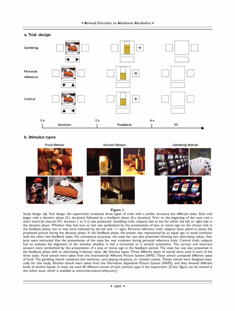
Figure 1.Study design. (a) Trial design: the experiment contained three types of trials with a similar structure but different tasks. Each trialbegan with a decision phase (2-s duration) followed by a feedback phase (2-s duration). Prior to the beginning of the next trial ashort intertrial interval (ITI, duration 1 or 2 s) was presented. Gambling trials: subjects had to bet for either the left or right side inthe decision phase. Whether they had won or lost was symbolized by the presentation of plus or minus sign on the chosen side inthe feedback phase; win or lose were indicated by the bar and 1/- signs. Personal reference trials: subjects were asked to assess thepresented picture during the decision phase. In the feedback phase, the answer was represented by an equal sign to avoid confusionwith the other two feedback tasks. For consistency purposes, the state bar was also presented showing two alternating values. Sub-jects were instructed that the presentation of the state bar was irrelevant during personal reference trials. Control trials: subjectshad to evaluate the alignment of the stimulus whether it had a horizontal or a vertical orientation. The correct and incorrectanswers were symbolized by the presentation of a plus or minus sign in the feedback period. The state bar was also presented inthe feedback phase with an alternating irrelevant value. (b) Stimulus types: Three different types of stimuli were used in each of thethree tasks. Food stimuli were taken from the International Affective Picture System (IAPS). These stimuli contained different typesof food. The gambling stimuli contained slot machines, card playing situations, or roulette scenes. Theses stimuli were designed espe-cially for this study. Alcohol stimuli were taken from the Normative Appetitive Picture System (NAPS), and they showed differentkinds of alcohol liquids. In total, we used 40 different stimuli of each stimulus type in the experiment. [Color figure can be viewed inthe online issue, which is available at www.interscience.wiley.com.]
r 1693 r
r Reward Circuitry in Abstinent Alcoholics r
press with either their the left or right hand. During thisphase, a picture was displayed in the center of the screenand two little triangles at the bottom symbolized the taskthat had to be performed. The decision phase was directlyfollowed by a feedback phase (2-s duration), in which sub-jects got a short symbolized feedback. The display con-tained a symbol on the site of their response and a statebar in the center. Every location on the screen in whichpictures or symbols could appear was surrounded by athin frame. Before each next trial, a short intertrial interval(ITI, duration 1 or 2 s) was presented in which only thefour empty location frames were presented. While the ITIwas identical in all three tasks, the specific content of thetwo phases was different for each task.In the decision phase of gambling trials, subjects were
asked to bet by deciding for the left or right site of the dis-play. In the following feedback phase, they were informedwhether they had won or lost. This was symbolized by thepresentation of a plus or minus sign on the chosen site. Addi-tionally, the state bar that reflected the actual amount of thesubject’s payment increased or decreased. In orientation onthe study performed by Reuter et al. [Reuter et al., 2005], oursubjects were made to believe that they were gamblingagainst the computer and that their luck during the gamblingtrials had a direct influence on the amount of their payment.They were not aware that, in fact, the proportion of wins andlosses was predefined and almost identical for all subjects.During the decision phase of personal reference trials,
subjects had to evaluate whether the presented picturewas of high or low personal reference. In the feedbackphase of these trials, an equality sign was presented whenthe button press was delivered in time. In contrast to bothof the other tasks, the minus sign was only presentedwhen no response occurred. We decided to present anequality sign instead of the plus sign to make sure thatthis task had no rewarding component. The state bar waspresented in these trials as well for the purpose of consis-tency. Although subjects were instructed that it had nomeaning, the actual value fluctuated around the midline.In control trials, it was the subject’s task to identify the
alignment of the presented picture during the decisionphase. All stimuli had the shape of a rectangle, half of thestimuli were horizontally aligned and half of them verti-cally. When subjects gave the right answer, a plus signwas presented in the feedback phase and a minus sign infalse trials, respectively. As described earlier, the feedbackdisplay contained the fluctuating state bar that was irrele-vant in these trials.A total of 120 stimuli were presented four times during
the experiment; once during evaluation of personal refer-ence and control trials and twice during gambling trials.The twofold presentation of stimuli during the gamblingtrials resulted from our orientation on the reward paradigmintroduced by Reuter et al. [Reuter et al., 2005], which con-tained a total number of 250 trials. To achieve the sameeffect, we also included a total number of 240 trials, whichmade the twofold presentation of our stimuli unavoidable.
The 120 stimuli included 40 food pictures, 40 alcohol pic-tures, and 40 gambling pictures. The food pictures weretaken from the International Affective Picture System [Langet al., 1999] and slightly modified. As alcohol stimuli servedpictures from the Normative Appetitive Picture System[Breiner et al., 1995; Stritzke et al., 2004] that were modifiedas well. The gambling stimuli comprised typical gamblingscenes and were developed especially for this study. Like-wise the state bar presented in the feedback phase had threedifferent colors reflecting the actual stimulus category. Theselection of stimuli was driven by our question which kindof stimulus might be best suited to investigate the relation-ship between reward and personal reference. Based on pre-vious imaging experiments, we decided to take stimuli thatshowed a strong reward value like natural reinforcers, e.g.,food stimuli [Killgore et al., 2003; Wang et al., 2004]. Clini-cal experience with alcoholic patients led us to implementpictures containing alcohol stimuli, as alcoholics report astrong reference toward these stimuli on the one hand andshow a pronounced craving for these stimuli on the other[Modell and Mountz, 1995; Park et al., 2007]. We were alsocurious as to why these patients developed alcoholism,which is a substance-related addictive disorder and not anonsubstance-related disorder such as pathological gam-bling. For this reason, we integrated pictures containinggambling stimuli.Trials were presented in rows of 10 stimuli of the same
category. Hereafter, a baseline event occurred that lastedfor 4, 5, or 6 s. The experiment was divided in eight ses-sions with the same task, i.e., four gambling sessions, twosessions with evaluation of personal reference, and twocontrol sessions. Sessions and trials were presented in apseudorandomized order. At the end of each session, ashort evaluation period was presented in which subjectswere asked to state their actual situation. They had todescribe their general contentment, hungriness, craving foralcohol, and craving for gambling by virtually moving abar on a visual analog scale.The experiment was executed on a ordinary desktop
personal computer using the software package Presenta-tion (Neurobehavioral Systems, http://www.neurobs.com). Subjects were lying inside the scanner and werewatching the projected experiment on a matt screenthrough a mirror attached on the head coil.Subsequent to the fMRI session, all subjects performed a
postscanning experiment in which they evaluated all pre-sented stimuli in respect to their personal reference andthe craving that was induced by the stimulus. After the 2-spresentation of the stimulus, subjects had to assesswhether they could consent to two displayed statements(personal reference: ‘‘The content has a lot to do with me’’;craving: ‘‘I have a strong craving for the content’’) by vir-tually moving a bar on the screen. The postscanningparadigm was likewise displayed on an ordinary desktoppersonal computer using the experimentation softwarepackage Presentation (Neurobehavioral Systems, http://www.neurobs.com).
r de Greck et al. r
r 1694 r
Behavioral Data Analysis
Behavioral data have been examined using repeatedmeasurements analysis of variance (ANOVA) and paired-samples t-tests as well as independent-samples t-tests forgroup comparisons. Different factors have been includedin the ANOVAs: Within-subjects factor condition (win,lose, reward trials with no response, high personal refer-ence, low personal reference, reference trials with noresponse, correct control trials, incorrect control trials, con-trol trials with no response), within-subjects factor per-sonal reference (high and low), within-subjects factor stim-ulus category (natural, gambling and alcohol), and thebetween-subjects factor group (healthy and alcoholics).
fMRI Data Acquisition and Analysis
Functional measurements were performed on a 3-Twhole body MRI system (Siemens Trio, Erlangen, Ger-many) with echo planar imaging (EPI) using an eight-channel head coil. The slices were acquired parallel to theAC–PC plane in an odd–even interleaved acquisitionorder. Thirty-two T2*-weighted echo planar images pervolume with blood oxygenation level-dependent (BOLD)contrast were obtained (matrix: 64 3 64; 32 slices per vol-ume; FoV: 224 3 224 mm; spatial resolution: 3.5 3 3.5 34 mm; TE 5 30 ms; TR 5 2000 ms; flip angle 5 808). Func-tional data were recorded in eight scanning sessions con-taining 210 volumes per session for each subject. The firstfour volumes were discarded. The fMRI data were prepro-cessed and statistically analyzed by the general linearmodel approach [Friston et al., 1995] using the SPM2 soft-ware package (spm2, http://www.fil.ion.ucl.ac.uk) andMATLAB 6.5 (The Mathworks, Natick, MA). All functionalimages were slice time corrected with reference to the firstslice acquired, corrected for motion artifacts by realign-ment to the last volume, and spatially normalized to astandard T2-weighted SPM template [Ashburner and Fris-ton, 1999]. The normalization was realized by warping thesubject’s last functional image to the SPM template andapplying these parameters to the other functional images.The images were resampled to 2 3 2 3 2 mm andsmoothed with an isotropic 6-mm full-width half-maxi-mum Gaussian kernel.The time-series fMRI data were filtered using a high
pass filter and cutoff of 128 s. A statistical model for eachsubject was computed by applying a canonical responsefunction [Friston et al., 1998]. All relevant periods, i.e.,the decision phase, the feedback phase, and the baselinephase were included in the SPM model. Regionally spe-cific condition effects were tested by employing linearcontrasts for each subject and different conditions. Theresulting contrast images were submitted to a second-level random-effects analysis. Here, one-sample t-testswere used on images obtained for each subjects’ volumeset and different conditions. To control for the multiple-testing problem, we performed a false discovery rate cor-
rection [Nichols and Hayasaka, 2003]. The anatomicallocalization of significant activations was assessed withreference to the standard stereotactic atlas by superimpo-sition of the SPM maps on a standard brain template pro-vided by SPM2.In a second step, we analyzed the fMRI raw data using
the Marseille Region of Interest Toolbox software package[Brett et al., 2002; MarsBaR 1.86, http://www.sourceforge.net/projects/marsbar]. Using a sphere-shaped region-of-interest (ROI, radius 5 mm) we extracted the raw datafrom activations found in the second level analysis. Tocontrol for baseline drifts, we applied a linear baselineshift correction. Mean normalized fMRI signal values fromtwo following time steps (6 and 8 s after feedback onset)of the BOLD were included in the statistical analysis usingrepeated measurements analysis of variance (ANOVA)and paired-samples t-tests as well as independent-samplest-tests for group comparisons [Dreher et al., 2006; Yarkoniet al., 2005]. Different factors have been included in theANOVAs. Within-subject’s factor reward (win and lose),within-subject’s factor personal reference (high and low),within-subject’s factor stimulus category (natural, gam-bling, and alcohol), and the between-subject’s factor group(healthy and alcoholics). As the ratio of high and low per-sonal reference trials could not be predefined as the ratioof win and lose trials, we performed Levene’s tests tocheck for possible inhomogeneity of variances.It should be mentioned at this point that our paradigm
did not allow for comparisons of activations during thereward task (i.e., monetary wins and losses) with activa-tions during the personal reference task (i.e., evaluation ofpictures with high and low personal reference), as the timepoint of the main event of both tasks was different.
RESULTS
Behavioral Data
Reaction times
Reaction times were analyzed with regard to possiblegroup differences and the effects of different tasks andconditions. Repeated measurements ANOVAs and inde-pendent-samples t-tests revealed no significant group dif-ferences. The statistical analysis for the different conditionsshowed slowest reaction times during high personal refer-ence events compared with all other conditions in healthysubjects. Alcoholics did not show any significant differencein the reaction times for different conditions (see Supple-mentary Material 1a and 1b).
Personal reference
Behavioral testing of personal reference, using independ-ent samples t-tests, revealed that alcoholic patients showeda significantly higher number of high referenced stimuli(t(18) 5 2.735; p 5 0.014) and a lower number of low
r Reward Circuitry in Abstinent Alcoholics r
r 1695 r
referenced stimuli (t(18) 5 22.703; p 5 0.015) when com-pared with healthy subjects.The effects of stimulus categories on ratings of personal
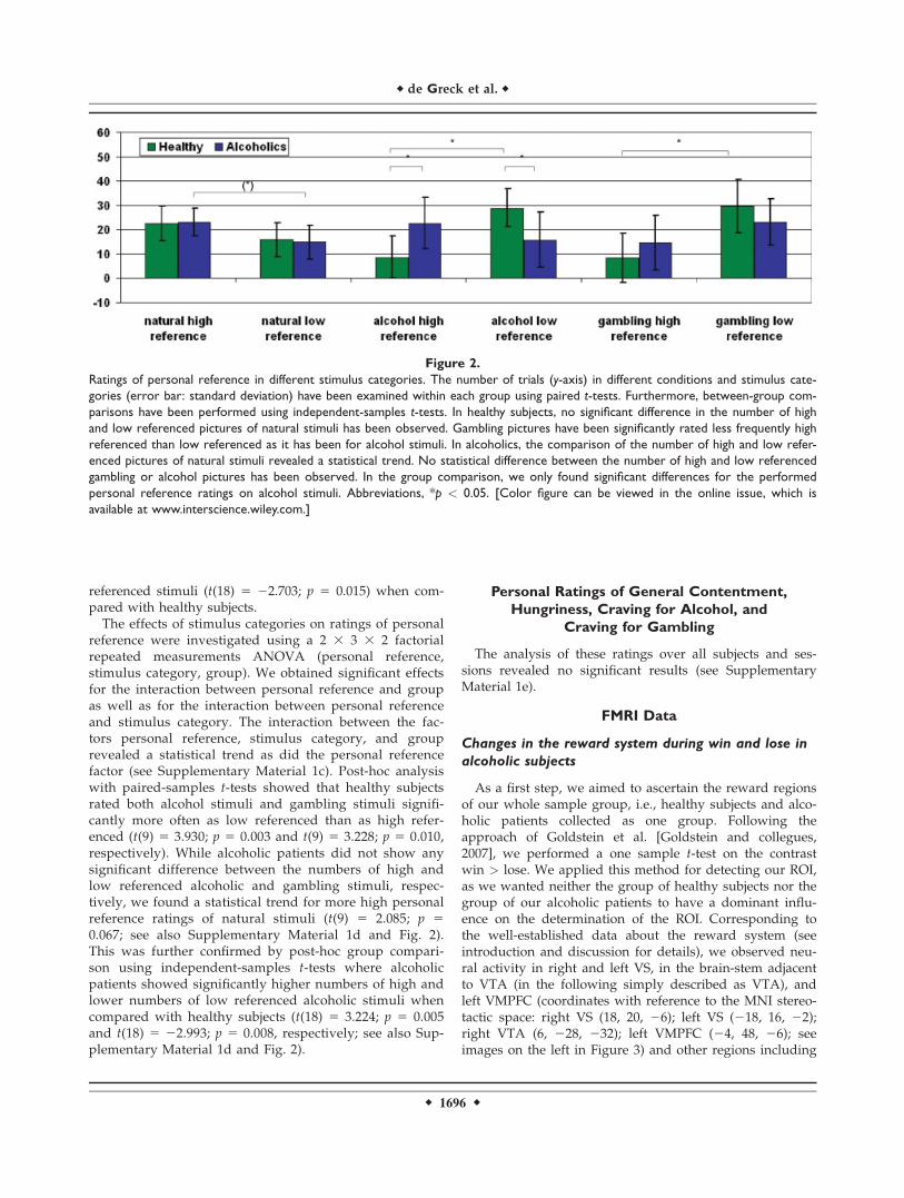
reference were investigated using a 2 3 3 3 2 factorialrepeated measurements ANOVA (personal reference,stimulus category, group). We obtained significant effectsfor the interaction between personal reference and groupas well as for the interaction between personal referenceand stimulus category. The interaction between the fac-tors personal reference, stimulus category, and grouprevealed a statistical trend as did the personal referencefactor (see Supplementary Material 1c). Post-hoc analysiswith paired-samples t-tests showed that healthy subjectsrated both alcohol stimuli and gambling stimuli signifi-cantly more often as low referenced than as high refer-enced (t(9) 5 3.930; p 5 0.003 and t(9) 5 3.228; p 5 0.010,respectively). While alcoholic patients did not show anysignificant difference between the numbers of high andlow referenced alcoholic and gambling stimuli, respec-tively, we found a statistical trend for more high personalreference ratings of natural stimuli (t(9) 5 2.085; p 50.067; see also Supplementary Material 1d and Fig. 2).This was further confirmed by post-hoc group compari-son using independent-samples t-tests where alcoholicpatients showed significantly higher numbers of high andlower numbers of low referenced alcoholic stimuli whencompared with healthy subjects (t(18) 5 3.224; p 5 0.005and t(18) 5 22.993; p 5 0.008, respectively; see also Sup-plementary Material 1d and Fig. 2).
Personal Ratings of General Contentment,
Hungriness, Craving for Alcohol, and
Craving for Gambling
The analysis of these ratings over all subjects and ses-sions revealed no significant results (see SupplementaryMaterial 1e).
FMRI Data
Changes in the reward system during win and lose in
alcoholic subjects
As a first step, we aimed to ascertain the reward regionsof our whole sample group, i.e., healthy subjects and alco-holic patients collected as one group. Following theapproach of Goldstein et al. [Goldstein and collegues,2007], we performed a one sample t-test on the contrastwin > lose. We applied this method for detecting our ROI,as we wanted neither the group of healthy subjects nor thegroup of our alcoholic patients to have a dominant influ-ence on the determination of the ROI. Corresponding tothe well-established data about the reward system (seeintroduction and discussion for details), we observed neu-ral activity in right and left VS, in the brain-stem adjacentto VTA (in the following simply described as VTA), andleft VMPFC (coordinates with reference to the MNI stereo-tactic space: right VS (18, 20, 26); left VS (218, 16, 22);right VTA (6, 228, 232); left VMPFC (24, 48, 26); seeimages on the left in Figure 3) and other regions including
Figure 2.
Ratings of personal reference in different stimulus categories. The number of trials (y-axis) in different conditions and stimulus cate-
gories (error bar: standard deviation) have been examined within each group using paired t-tests. Furthermore, between-group com-
parisons have been performed using independent-samples t-tests. In healthy subjects, no significant difference in the number of high
and low referenced pictures of natural stimuli has been observed. Gambling pictures have been significantly rated less frequently high
referenced than low referenced as it has been for alcohol stimuli. In alcoholics, the comparison of the number of high and low refer-
enced pictures of natural stimuli revealed a statistical trend. No statistical difference between the number of high and low referenced
gambling or alcohol pictures has been observed. In the group comparison, we only found significant differences for the performed
personal reference ratings on alcohol stimuli. Abbreviations, *p < 0.05. [Color figure can be viewed in the online issue, which is
available at www.interscience.wiley.com.]
r de Greck et al. r
r 1696 r
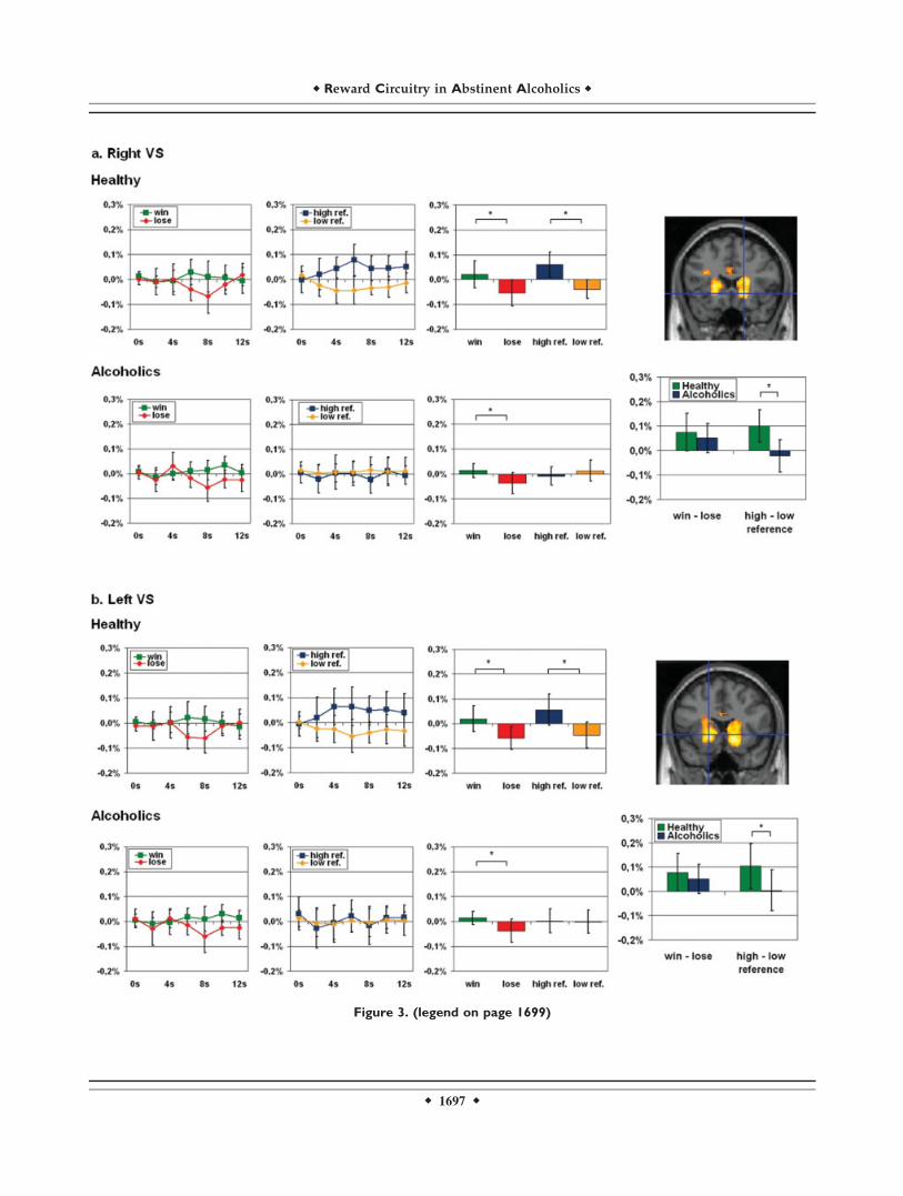
Figure 3. (legend on page 1699)
r Reward Circuitry in Abstinent Alcoholics r
r 1697 r
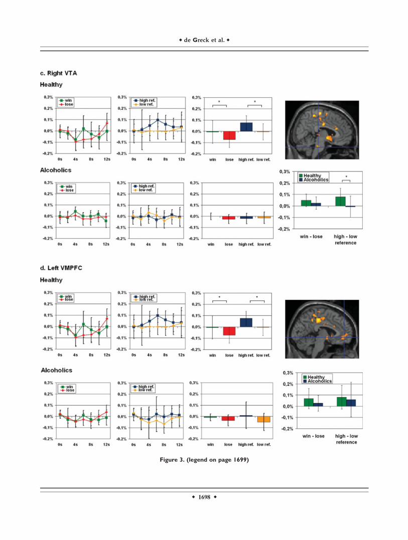
Figure 3. (legend on page 1699)
r de Greck et al. r
r 1698 r
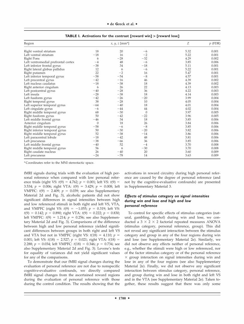
the left inferior frontal gyrus, the right lateral globus pal-lidus and the right putamen (see Table I). Further analysisfocused exclusively on the right and left VS, the VTA andthe left VMPFC, as these regions have been demonstratedto be crucially involved in the reward system in bothhealthy and alcoholic subjects (see Introduction and Dis-cussion for details).Based on the regions as elucidated in the first step (see
above and Methods for details), we compared signal inten-sities for win and lose between both groups. A 2 3 2 facto-rial ANOVA (reward, group) revealed a significant maineffect for the factor reward in each of the four regions butnot for the reward 3 group interaction (see SupplementaryMaterial 2a). Paired-samples t-tests in healthy subjectsrevealed significant differences in signal intensitiesbetween win and lose in all of the four regions (right VS:t(9) 5 3.041, p 5 0.014; left VS: t(9) 5 3.122, p 5 0.012;right VTA: t(9) 5 2.800; p 5 0.021; left VMPFC: t(9) 52.428; p 5 0.038; see also Supplementary Material 2b andFig. 3). Alcoholic patients also showed significant differen-ces between win and lose in the right and left VS, but theyfailed to show this in the VTA and the VMPFC (right VS:t(9) 5 2.673; p 5 0.025; left VS: t(9) 5 2.658; p 5 0.026;right VTA: t(9) 5 1.465; p 5 0.177; left VMPFC: t(9) 5
1.227; p 5 0.251; see also Supplementary Material 2b andbar diagrams on the right in Figure 3 for win and lose sep-arately in both groups). Despite these differences, this didnot amount to statistically significant differences betweengroups in the difference between win and lose (right VS:t(18) 5 0.778; p 5 0.447; left VS: t(18) 5 0.842; p 5 0.411;right VTA: t(18) 5 0.990; p 5 0.335; left VMPFC: t(18) 51.148; p 5 0.266; see also Supplementary Material 2b andFig. 3). Levene’s tests for equality of variances did notyield significant values for any of the comparisons.
Changes in the reward system during high and low
personal reference in alcoholic patients
We then investigated signal intensities during high andlow personal reference in those regions that were recruitedduring win and lose in both groups. A 2 3 2 factorialANOVA (evaluation of personal reference, group) revealeda significant main effect for the factor evaluation of per-sonal reference in three of the four regions (i.e. right andleft VS, left VMPFC) and for the personal reference 3group interaction in three of the four regions (i.e., rightand left VS, right VTA, see Supplementary Material 2c). Incontrast to healthy subjects who revealed significant higher
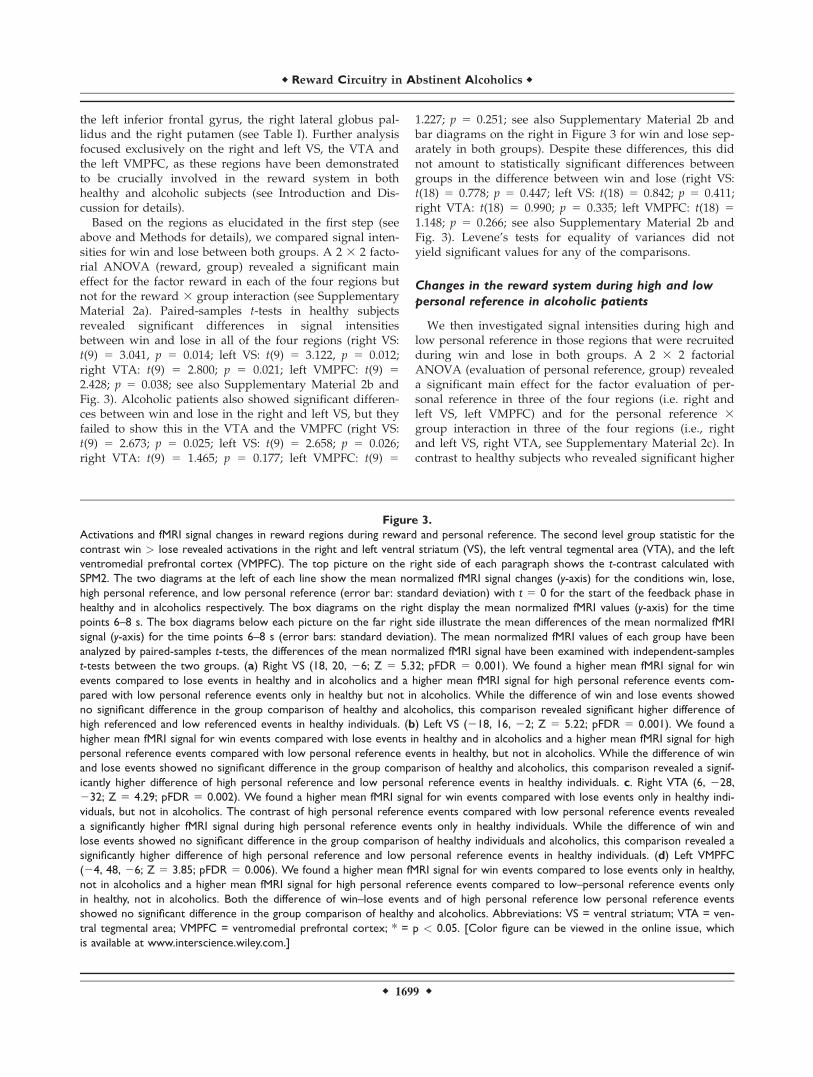
Figure 3.
Activations and fMRI signal changes in reward regions during reward and personal reference. The second level group statistic for the
contrast win > lose revealed activations in the right and left ventral striatum (VS), the left ventral tegmental area (VTA), and the left
ventromedial prefrontal cortex (VMPFC). The top picture on the right side of each paragraph shows the t-contrast calculated with
SPM2. The two diagrams at the left of each line show the mean normalized fMRI signal changes (y-axis) for the conditions win, lose,
high personal reference, and low personal reference (error bar: standard deviation) with t 5 0 for the start of the feedback phase in
healthy and in alcoholics respectively. The box diagrams on the right display the mean normalized fMRI values (y-axis) for the time
points 6–8 s. The box diagrams below each picture on the far right side illustrate the mean differences of the mean normalized fMRI
signal (y-axis) for the time points 6–8 s (error bars: standard deviation). The mean normalized fMRI values of each group have been
analyzed by paired-samples t-tests, the differences of the mean normalized fMRI signal have been examined with independent-samples
t-tests between the two groups. (a) Right VS (18, 20, 26; Z 5 5.32; pFDR 5 0.001). We found a higher mean fMRI signal for win
events compared to lose events in healthy and in alcoholics and a higher mean fMRI signal for high personal reference events com-
pared with low personal reference events only in healthy but not in alcoholics. While the difference of win and lose events showed
no significant difference in the group comparison of healthy and alcoholics, this comparison revealed significant higher difference of
high referenced and low referenced events in healthy individuals. (b) Left VS (218, 16, 22; Z 5 5.22; pFDR 5 0.001). We found a
higher mean fMRI signal for win events compared with lose events in healthy and in alcoholics and a higher mean fMRI signal for high
personal reference events compared with low personal reference events in healthy, but not in alcoholics. While the difference of win
and lose events showed no significant difference in the group comparison of healthy and alcoholics, this comparison revealed a signif-
icantly higher difference of high personal reference and low personal reference events in healthy individuals. c. Right VTA (6, 228,
232; Z 5 4.29; pFDR 5 0.002). We found a higher mean fMRI signal for win events compared with lose events only in healthy indi-
viduals, but not in alcoholics. The contrast of high personal reference events compared with low personal reference events revealed
a significantly higher fMRI signal during high personal reference events only in healthy individuals. While the difference of win and
lose events showed no significant difference in the group comparison of healthy individuals and alcoholics, this comparison revealed a
significantly higher difference of high personal reference and low personal reference events in healthy individuals. (d) Left VMPFC
(24, 48, 26; Z 5 3.85; pFDR 5 0.006). We found a higher mean fMRI signal for win events compared to lose events only in healthy,
not in alcoholics and a higher mean fMRI signal for high personal reference events compared to low–personal reference events only
in healthy, not in alcoholics. Both the difference of win–lose events and of high personal reference low personal reference events
showed no significant difference in the group comparison of healthy and alcoholics. Abbreviations: VS = ventral striatum; VTA = ven-
tral tegmental area; VMPFC = ventromedial prefrontal cortex; * = p < 0.05. [Color figure can be viewed in the online issue, which
is available at www.interscience.wiley.com.]
r Reward Circuitry in Abstinent Alcoholics r
r 1699 r
fMRI signals during trials with the evaluation of high per-sonal reference when compared with low personal refer-ence trials (right VS: t(9) 5 4.762; p 5 0.001; left VS: t(9) 53.534; p 5 0.006; right VTA: t(9) 5 3.429; p 5 0.008; leftVMPFC: t(9) 5 2.409; p 5 0.039; see also SupplementaryMaterial 2d and Fig. 3), alcoholic patients did not showsignificant differences in signal intensities between highand low referenced stimuli in both right and left VS, VTA,and VMPFC (right VS: t(9) 5 21.055; p 5 0.319; left VS:t(9) 5 0.142; p 5 0.890; right VTA: t(9) 5 0.222; p 5 0.830;left VMPFC: t(9) 5 1.214; p 5 0.256; see also Supplemen-tary Material 2d and Fig. 3). Comparisons of the differencebetween high and low personal reference yielded signifi-cant differences between groups in both right and left VSand VTA but not in VMPFC (right VS: t(18) 5 4.110; p 50.001; left VS: t(18) 5 2.527; p 5 0.021; right VTA: t(18) 52.288; p 5 0.034; left VMPFC: t(18) 5 0.346; p 5 0.734; seealso Supplementary Material 2d and Fig. 3). Levene’s testsfor equality of variances did not yield significant valuesfor any of the comparisons.To demonstrate that our fMRI signal changes during the
evaluation of personal reference are not due to nonspecificcognitive-evaluative confounds, we directly comparedfMRI signal changes from the ascertained reward regionsduring the evaluation of personal reference with thoseduring the control condition. The results showing that the
activations in reward circuitry during high personal refer-ence are caused by the degree of personal reference (andnot by the cognitive-evaluative confounds) are presentedin Supplementary Material 3.
Effects of stimulus category on signal intensities
during win and lose and high and low
personal reference
To control for specific effects of stimulus categories (nat-ural, gambling, alcohol) during win and lose, we con-ducted a 3 3 2 3 2 factorial repeated measures ANOVA(stimulus category, personal reference, group). This didnot reveal any significant interaction between the stimuluscategory and group in any of the four regions during winand lose (see Supplementary Material 2e). Similarly, wedid not observe any effects neither of personal reference,e.g., whether the stimuli were high or low referenced, norof the factor stimulus category or of the personal reference3 group interaction on signal intensities during win andlose in any of the four regions (see also SupplementaryMaterial 2e). Finally, we did not observe any significantinteraction between stimulus category, personal reference,and group during win and lose in both right and left VSand in the VTA (see Supplementary Material 2e). Taken to-gether, these results suggest that there was only some
TABLE I. Activations for the contrast [reward win] > [reward lose]
Region x, y, z [mm*] Z p (FDR)
Right ventral striatum 18 20 26 5.32 0.001Left ventral striatum 218 16 22 5.22 0.001Right Pons 6 228 232 4.29 0.002Left ventromedial prefrontal cortex 24 48 26 3.85 0.006Left inferior frontal gyrus 238 34 218 5.11 0.001Right lateral globus pallidus 12 6 26 5.22 0.001Right putamen 22 22 16 5.47 0.001Left inferior temporal gyrus 258 254 28 4.57 0.001Left precentral gyrus 242 216 46 4.39 0.002Left nucleus caudatus 218 238 18 4.39 0.002Right anterior cingulum 6 26 22 4.13 0.003Left postcentral gyrus 240 228 36 4.22 0.003Left insula 228 238 18 4.14 0.003Left fusiform gyrus 242 226 220 3.99 0.004Right temporal gyrus 38 228 10 4.05 0.004Left superior temporal gyrus 264 240 18 4.01 0.004Left cingulate gyrus 26 244 44 4.02 0.004Right middle temporal gyrus 68 230 0 3.97 0.005Right fusiform gyrus 50 242 222 3.96 0.005Left middle frontal gyrus 246 34 18 3.85 0.006Anterior cingulum 0 18 26 3.84 0.006Right middle temporal gyrus 258 26 28 3.85 0.006Right inferior temporal gyrus 50 230 220 3.82 0.006Right middle temporal gyrus 52 238 214 3.86 0.006Left paracentral lobule 218 242 48 3.81 0.006Left precuneus 238 266 36 3.85 0.006Left middle frontal gyrus 240 52 24 3.70 0.008Right middle temporal gyrus 56 6 230 3.70 0.008Right caudate nucleus 26 240 20 3.60 0.009Left precuneus 224 278 14 3.63 0.009
*Coordinates refer to the MNI stereotactic space.
r de Greck et al. r
r 1700 r
influence of different stimulus categories on signal inten-sities during win and lose in the VMPFC but not in theother three regions in both groups.We then conducted similar analyses for investigating the
effects of stimulus category on signal intensities during theevaluation of high and low personal reference. A 3 3 2factorial repeated measurements ANOVA (stimulus cate-gory, group) revealed a significant effect of the factor stim-ulus category only in the VTA (see Supplementary Mate-rial 2f and the figure of Supplementary Material 4). Inhealthy subjects, post-hoc paired t-tests for this regionrevealed a higher difference between high referenced andlow referenced stimuli in alcohol trials compared withgambling trials (T(7) 5 3.835; p 5 0.006). However, in alco-holic patients, post-hoc paired samples t-tests for thisregion did not reveal any significant difference. We, how-ever, did not observe any significant interaction betweenstimulus category and group in the VTA or any of theother regions (see also Supplementary Material 2f). Takentogether, these results indicate some effects of stimulus cat-egory in healthy subjects in the VTA, concerning the dif-ference between gambling and alcoholic stimuli.
DISCUSSION
We investigated reward function and personal referencein reward circuitry in abstinent alcoholic patients. Contraryto our first hypothesis, we were not able to replicate previ-ous findings that clearly showed reduced activity inreward regions during the perception of reward [Wraseet al., 2007]. We could not find any statistically significantreduction in signal changes in all four regions observed(VS, VTA, and VMPFC) during the perception of monetaryreward in alcoholic patients when compared with healthysubjects. However, we observed some slight (though statis-tically nonsignificant) differences of signal intensity in theVTA and the VMPFC, which is in accordance with previ-ous findings [Wrase et al., 2007]. The most striking out-come of our study was that alcoholic patients, althoughshowing no major alterations in reward function, revealedreduced signal changes during personal reference, i.e., theevaluation of high referenced stimuli, in the very samereward regions, which confirms our second hypothesis.We thus demonstrate for the first time altered neural activ-ity in reward circuitry during personal reference in alco-holic patients.Several studies found altered neural activity during
reward tasks in reward circuitry in alcoholic patients[Wrase et al., 2007] and patients suffering from other formsof addictive diseases such as cocaine [Volkow et al., 1997,2004a] and pathological gambling [Reuter et al., 2005].Although we tested for changes in reward activity using awell-established paradigm that had shown altered rewardfunction in pathological gamblers before [Reuter et al.,2005], we were not able to detect statistically significantevidence for disturbed reward function in our alcoholic
subjects. Although we observed less differentiation in sig-nal changes between win and lose in the VTA and theVMPFC during the reward task in alcoholic patients, thesedid not yield statistically significant differences when com-pared with healthy subjects. The possible reasons for thelack of reduced reward function might include the follow-ing methodological and clinical explanations. While Wraseet al. [Wrase et al., 2007], relying on the Monetary Incen-tive Delay Task (MID-Task) established by Knutson et al.[Knutson et al., 2001a], focused on the anticipatory periodof monetary gains and losses, we here investigated thegain and loss periods themselves. One may consequentlyspeculate that the VS may specifically be associated withdeficits in anticipation of reward rather than with deficitsin the perception of reward. This interpretation is sup-ported by the findings from Knutson et al. [Knutson et al.,2001ab, 2003], who associated neural activity in the VSwith the anticipation of reward, whereas the VMPFC wasactive during the gain period itself. This neural differentia-tion however needs to be investigated in future studiesthat directly compare anticipation and perception ofreward in alcoholic patients. Another difference betweenthe two studies concerns the stage during which alcoholicpatients were investigated. While the study by Wrase et al.[2007] and others [George et al., 2001; Grusser et al., 2004;Kareken et al., 2004; Myrick et al., 2004] investigatedacutely detoxified alcoholic patients, our patients were al-ready abstinent for at least three months. Braus [Brauset al., 2001] as well as others [Heinz et al., 1996; Laineet al., 1999] presented some evidence that altered neuralactivity in reward circuitry may disappear and thus nor-malize after at least three weeks of abstinence. Deficiencyin reward circuitry during the acute state may be due to adecrease in VS dopamine transporters that recovers after afew days of abstinence [Laine et al., 1999]. The long dura-tion of abstinence in our alcoholic patients (at least threemonths of abstinence) may thus possibly account for theobserved absence of abnormalities in VS (and the absenceof between-group effects in the other regions) duringreward. The hypothesized neural differentiation betweenthe acute and abstinent state suggests that deficits inreward regions may potentially be regarded as a statemarker of acute alcohol abuse.The most striking finding is that alcoholic patients
showed considerably reduced signal changes in rewardcircuitry during the evaluation high personal referencewhen compared to healthy subjects; this has not beeninvestigated before. Recent studies in healthy subjectsdemonstrated involvement of the VS and the VMPFC inpersonal reference [Northoff, 2007; Northoff and Berm-pohl, 2004; Northoff et al., 2006; Phan et al., 2004]. Thesefindings are supported by a recent study of our group,which explicitly investigated the association of reward andevaluation of personal reference [de Greck et al., 2008]. Wewere able to demonstrate that reward circuitry (VS,VMPFC, and VTA) is recruited during the evaluation ofhigh referenced stimuli. How can we explain such appa-
r Reward Circuitry in Abstinent Alcoholics r
r 1701 r
rent overlap between reward and personal reference? Wesuggest that both reward and personal reference mightpresent distinct aspects of the so-called valuation system[Montague and Berns, 2002; Montague et al., 2006]. Thevaluation system does not only assign immediate value toa stimulus, indicating the reward value, but also its long-term value for the organism reflecting what can be calledpersonal reference. The shared valuation system mightneuronally be represented in basic reward circuitry includ-ing VTA, VS, and VMPFC. The present findings indicatethat the neural basis of this valuation system may bealtered in alcoholic patients. While our patients exhibitedonly slight abnormalities (e.g., no between group differen-ces) in reward circuitry during reward function, i.e., winand loss, they showed major abnormalities in the sameregions during the evaluation of personal reference. Suchfunctional dissociation between reward and personal refer-ence in reward circuitry suggests a possible dissociationbetween the assignment of immediate and long-term valueto stimuli in alcoholic patients. While the immediate valueassignment and thus reward function may recover withthe duration of abstinence, the deficits in long-term assign-ment of personal significance, i.e., personal reference maypersist beyond the state of acute detoxification. One mayconsequently speculate that clinically neural deficits inreward function may be considered a state marker of acuteaddiction, whereas neural deficits in personal referencemay rather be a trait marker of alcoholism describing thepredisposition of a person to addiction. This rather specu-lative hypothesis, however, awaits further confirmationfrom longitudinal studies investigating both reward andpersonal reference.Clinically, deficits in personal reference may manifest in
the patient’s attribution of abnormally high personal signifi-cance to alcohol. This corresponds well to our behavioraldata showing that alcoholic patients evaluated alcoholicstimuli more high personal referenced when compared withhealthy subjects. Behavioral attribution of higher personalreference to alcoholic stimuli was however not accompaniedby higher signal changes in either region of reward cir-cuitry, e.g., VS, VMPFC, and VTA, during alcohol-relatedstimuli. In other terms, we could not observe any specificeffects of alcoholic stimuli in reward circuitry during eitherreward or personal reference in alcoholic patients. This indi-cates a basic deficit in reward circuitry remaining independ-ent of the kind of stimulus in alcoholic patients. Investigat-ing pathological gamblers, Reuter [Reuter et al., 2005] arguethat organisms try to maintain a homeostatic baseline levelof dopamine in the ventral striatum. While healthy subjectsare able to reach and maintain a sufficient baseline level ofdopamine activity in the VS by weak reinforcers found ineveryday life, addicted patients like pathological gamblerslack this ability and seek for stronger reinforcers, as e.g.gambling or drugs. This hypothesis is supported by ourdata since we showed that the simple evaluation of stimuliwith high personal reference leads to activity in VS (andother reward regions like VTA and VMPFC) only in healthy
subjects but not in abstinent alcoholic patients. In otherwords, while healthy subjects show activation in reward cir-cuitry during the evaluation of high referenced stimuli andhence can stabilize their homeostatic baseline activity in theVS, abstinent alcoholic patients lack this activation and aredependant on stronger reinforcers.Some methodological limitations of our study should be
mentioned. First, the number of patients investigated hereis rather low so that our results should be considered pre-liminary, awaiting further support from larger samples.Second, one might criticize that our concept of rewardneeds to be parsed into distinct aspects of reward asproposed by Berridge and Robinson [2003]. Unfortunately,our design does not allow us to clearly distinguish be-tween the appetitive preconsummatory aspects (wanting)on the one hand and hedonic consummatory aspects(liking) on the other. The paradigm used in our studyrelied on a similar design that was established by Reuteret al. [Reuter et al., 2005] and showed also robust effects ina previous study conducted by our group [de Greck et al.,2008]. The focus lay on the hedonic and consummatoryphase. Third, one may argue that the concept of personalreference is rather ill-defined. We here referred to previousstudies on personal reference [Northoff and Bermpohl,2004; Northoff et al. 2006, 2007] that let subjects explicitlydecide whether the presented stimulus is high or low ref-erenced. Finally one might miss that in our paradigm wedid not test for any interaction between reward and per-sonal reference. On the one hand, we intended to clearlyidentify reward-associated regions without any active ref-erence component to use them as a functional localizer. Onthe other hand, we aimed to introduce a personal referencetask without any traces of a reward component to eluci-date whether personal reference recruits reward circuitryusing the functional localizer approach [Saxe et al., 2006].To exclude any potential pictorial differences in both tasks,we presented the same stimuli. This design allowed us totest the hypothesis whether personal reference recruitsneural activity in those regions that are involved inreward. This design, however did not allow us to test forany interaction effects between personal reference andreward at all; for that a different design that includes ahigh and low reward component within the reference eval-uation task would be needed. One nevertheless has to con-sider that we cannot completely exclude some impact ofreward even during the personal reference task becausewe used primary reinforcers, i.e., food, which subjects hadto evaluate with regard to their personal reference.Although we cannot exclude such interaction completely,our observation that neural effects of personal referenceoccurred in all three stimulus categories argues againstthis interpretation. However, to completely exclude suchinteraction, a design that totally relinquishes primary rein-forcers would be needed.In conclusion, we demonstrate reduced neural activity in
reward circuitry during personal reference in abstinentalcoholic patients. Our data show that alcoholic patients
r de Greck et al. r
r 1702 r
remain unable to appropriately increase neural activity inreward circuitry during the evaluation of high referencedstimuli. This may not only contribute to better understandthe neural basis of alterations in personal reference inthese patients but also to establish deficient reward cir-cuitry as diagnostic and therapeutic marker of addiction.
ACKNOWLEDGMENTS
The authors thank Sascha Moerth and Michael Rotte fortheir comments on conception and design and Rabea Paus,Diana Moritz, Ulrike Proesch, and Ulrike Bruer for assis-tance in data collection and analysis as well as EvaStockum and Bjoern Enzi for their helpful comments onthe manuscript. The authors also thank the staff membersin Neurology II for their support and collaboration. Thestudy was supported by a Heisenberg grant from the Ger-man Research Foundation (DFG, 304/4-1 to G.N.), theSalus Foundation (to G.N), the Swiss National ResearchFoundation (3100A0-100830) to G.N.
REFERENCES
American Psychiatric Press.1994. Diagnostic and Statistical Manualof Mental Disorders. Washington, DC.
Ashburner J, Friston KJ (1999): Nonlinear spatial normalizationusing basis functions. Hum Brain Mapp 7:254–266.
Baler RD, Volkow ND (2006): Drug addiction: The neurobiologyof disrupted self-control. Trends Mol Med 12:559–566.
Berridge KC, Robinson TE (2003): Parsing reward trends Neurosci26:507–513.
Braus DF, Wrase J, Grusser S, Hermann D, Ruf M, Flor H, Mann K,Heinz A (2001): Alcohol-associated stimuli activate the ventralstriatum in abstinent alcoholics. J Neural Transm 108:887–894.
Breiner MJ, Stritzke WGK, Lang AR (1995): The Normative Appe-titive Picture System (Photographic Slides). Florida State Uni-versity. Tallahassee, FL.
Brett M, Anton JL, Valabregue R, Poline JB (2002): Region of inter-est analysis using an SPM toolbox [abstract]. Presented at the8th International Conferance on Functional Mapping of theHuman Brain, June 2–6, 2002, Sendai, Japan. Available on CD-ROM in NeuroImage, Vol 16, No 2, abstract 497.
de Greck M, Rotte M, Paus R, Moritz D, Thiemann R, Proesch U,Bruer U, Moerth S, Tempelmann C, Bogerts B, Northoff G(2008): Is our self based on reward? Self-relatedness recruits neu-ral activity in the reward system. Neuroimage 39:2066–2075.
Dreher JC, Kohn P, Berman KF (2006): Neural coding of distinctstatistical properties of reward information in humans. CerebCortex 16:561–573.
Friston KJ, Fletcher P, Josephs O, Holmes A, Rugg MD, Turner R(1998): Event-related fMRI: Characterizing differential responses.Neuroimage 7:30–40.
Friston KJ, Holmes AP, Worsley KJ, Poline JB, Frith C, FrackowiakRSJ (1995): Statistical parametric maps in functional imaging: Ageneral linear approach. Hum Brain Mapp 2:189–210.
George MS, Anton RF, Bloomer C, Teneback C, Drobes DJ, Lorber-baum JP, Nahas Z, Vincent DJ (2001): Activation of prefrontalcortex and anterior thalamus in alcoholic subjects on exposureto alcohol-specific cues. Arch Gen Psychiatr 58:345–352.
Goldstein RZ, Alia-Klein N, Tomasi D, Zhang L, Cottone LA,Maloney T, Telang F, Caparelli EC, Chang L, Ernst T et al.
(2007): Is decreased prefrontal cortical sensitivity to monetaryreward associated with impaired motivation and self-control incocaine addiction? Am J Psychiatr 164:43–51.
Grusser SM, Wrase J, Klein S, Hermann D, Smolka MN, Ruf M,Weber-Fahr W, Flor H, Mann K, Braus DF, Heinz A (2004):Cue-induced activation of the striatum and medial prefrontalcortex is associated with subsequent relapse in abstinent alco-holics. Psychopharmacology (Berl) 175:296–302.
Heinz A, Dufeu P, Kuhn S, Dettling M, Graf K, Kurten I, Rommel-spacher H, Schmidt LG (1996): Psychopathological and behav-ioral correlates of dopaminergic sensitivity in alcohol-depend-ent patients. Arch Gen Psychiatr 53:1123–1128.
Kalivas PW, Volkow ND (2005): The neural basis of addiction: Apathology of motivation and choice. Am J Psychiatr 162:1403–1413.
Kareken DA, Claus ED, Sabri M, Dzemidzic M, Kosobud AE, Rad-novich AJ, Hector D, Ramchandani VA, O’Connor SJ, Lowe Met al. (2004): Alcohol-related olfactory cues activate the nucleusaccumbens and ventral tegmental area in high-risk drinkers:preliminary findings. Alcohol Clin Exp Res 28:550–557.
Kelley WM, Macrae CN, Wyland CL, Caglar S, Inati S, HeathertonTF (2002): Finding the self? An event-related fMRI study.J Cogn Neurosci 14:785–794.
Killgore WD, Young AD, Femia LA, Bogorodzki P, Rogowska J, Yur-gelun-ToddDA (2003): Cortical and limbic activation during view-ing of high- versus low-calorie foods. Neuroimage 19:1381–1394.
Knutson B, Adams CM, Fong GW, Hommer D (2001a): Anticipa-tion of increasing monetary reward selectively recruits nucleusaccumbens. J Neurosci 21:RC159.
Knutson B, Fong GW, Adams CM, Varner JL, Hommer D (2001b)Dissociation of reward anticipation and outcome with event-related fMRI. Neuroreport 12:3683–3687.
Knutson B, Fong GW, Bennett SM, Adams CM, Hommer D (2003):A region of mesial prefrontal cortex tracks monetarily reward-ing outcomes: characterization with rapid event-related fMRI.Neuroimage 18:263–272.
Laine TP, Ahonen A, Torniainen P, Heikkila J, Pyhtinen J, RasanenP, Niemela O, Hillbom M (1999): Dopamine transportersincrease in human brain after alcohol withdrawal. Mol Psy-chiatr 4: 189–191;104–105.
Lang PJ, Bradley MM, Cuthbert BN. 1999. International AffectivePicture System. The Center for Research in Psychophysiology,University of Florida.
MarsBaR 1.86. Available at: http://www.sourceforge.net/proj-ects/marsbar.
Modell JG, Mountz JM (1995): Focal cerebral blood flow changeduring craving for alcohol measured by SPECT. J Neuropsy-chiatr Clin Neurosci 7:15–22.
Montague PR, Berns GS (2002): Neural economics and the biologi-cal substrates of valuation. Neuron 36:265–284.
Montague PR, King-Casas B, Cohen JD (2006): Imaging valuationmodels in human choice. Annu Rev Neurosci 29:417–448.
Myrick H, Anton RF, Li X, Henderson S, Drobes D, Voronin K,George MS (2004): Differential brain activity in alcoholics andsocial drinkers to alcohol cues: Relationship to craving. Neuro-psychopharmacology 29:393–402.
Nichols T, Hayasaka S (2003): Controlling the familywise errorrate in functional neuroimaging: A comparative review. StatMethods Med Res 12:419–446.
Northoff G (2007): Subcortical regions and the self. Behav BrainSci 30:100–101.
Northoff G, Bermpohl F (2004): Cortical midline structures andthe self. Trends Cogn Sci 8:102–107.
r Reward Circuitry in Abstinent Alcoholics r
r 1703 r
Northoff G, Heinzel A, de Greck M, Bermpohl F, Dobrowolny H,Panksepp J (2006): Self-referential processing in our brain—A meta-analysis of imaging studies on the self. Neuroimage31:440–457.
Northoff G, Schneider F, Rotte M, Matthiae C, Tempelmann C,Wiebking C, Bermpohl F, Heinzel A, Danos P, Heinze HJ,Bogerts B, Walter M, Panksepp J (2007): Differential paramet-ric modulation of self-relatedness and emotions in differentbrain regions. Hum Brain Mapp, Dec 6. [Epub ahead ofprint].
Park MS, Sohn JH, Suk JA, Kim SH, Sohn S, Sparacio R (2007):Brain substrates of craving to alcohol cues in subjects withalcohol use disorder. Alcohol Alcohol 42:417–422.
Phan KL, Taylor SF, Welsh RC, Ho SH, Britton JC, Liberzon I(2004): Neural correlates of individual ratings of emotionalsalience: A trial-related fMRI study. Neuroimage 21:768–780.
Reuter J, Raedler T, Rose M, Hand I, Glascher J, Buchel C (2005):Pathological gambling is linked to reduced activation of themesolimbic reward system. Nat Neurosci 8:147–148.
Saxe R, Brett M, Kanwisher N (2006): Divide and conquer: Adefense of functional localizers. Neuroimage 30:1088–1096; dis-cussion 1097-1099.
Stritzke WG, Breiner MJ, Curtin JJ, Lang AR (2004): Assessment ofsubstance cue reactivity: Advances in reliability, specificity,and validity. Psychol Addict Behav 18:148–159.
Volkow ND, Fowler JS, Wang GJ (2003): The addicted humanbrain: insights from imaging studies. J Clin Invest 111:1444–1451.
Volkow ND, Fowler JS, Wang GJ (2004a): The addicted humanbrain viewed in the light of imaging studies: Brain circuits andtreatment strategies. Neuropharmacology 47 (Suppl 1):3–13.
Volkow ND, Fowler JS, Wang GJ, Swanson JM (2004b) Dopaminein drug abuse and addiction: results from imaging studies andtreatment implications. Mol Psychiatr 9:557–569.
Volkow ND, Wang GJ, Fowler JS, Logan J, Gatley SJ, HitzemannR, Chen AD, Dewey SL, Pappas N (1997): Decreased striataldopaminergic responsiveness in detoxified cocaine-dependentsubjects. Nature 386:830–833.
Wang GJ, Volkow ND, Telang F, Jayne M, Ma J, Rao M, Zhu W,Wong CT, Pappas NR, Geliebter A, Fowler JS (2004): Exposureto appetitive food stimuli markedly activates the human brain.Neuroimage 21:1790–1797.
Wrase J, Schlagenhauf F, Kienast T, Wustenberg T, Bermpohl F,Kahnt T, Beck A, Strohle A, Juckel G, Knutson B, Heinz A(2007): Dysfunction of reward processing correlates withalcohol craving in detoxified alcoholics. Neuroimage 35:787–794.
Yarkoni T, Gray JR, Chrastil ER, Barch DM, Green L, Braver TS(2005): Sustained neural activity associated with cognitive con-trol during temporally extended decision making. Brain ResCogn Brain Res 23:71–84.
r de Greck et al. r
r 1704 r