Embed Size (px)
Citation preview
Cancer Genetics and Cytogenetics 125 (2001) 100–111
0165-4608/01/$ – see front matter © 2001 Elsevier Science Inc. All rights reserved.
PII: S0165-4608(00)00369-1
Lead article
Dedifferentiation of a well-differentiated liposarcoma to a highly malignant metastatic osteosarcoma:
amplification of 12q14 at all stages and gain of 1q22–q24associated with metastases
Anne Forus
a,
*, Marcelo L. Larramendy
b,c
, Leonardo A. Meza-Zepeda
a
, Bodil Bjerkehagen
d
, Linda H. Godager
a
, Anine B. Dahlberg
a
, Gunnar Saeter
e
, Sakari Knuutila
b
, Ola Myklebost
a
a
Department of Tumor Biology, The Norwegian Radium Hospital, 0301 Oslo, Norway
b
Department of Medical Genetics, Haartman Institute, University of Helsinki, Helsinki, Finland
c
Laboratorio de Citogenética y Cátedra de Citología, Facultad de Ciencias Naturales, Universidad Nacional de La Plata, La Plata, Argentina
d
Department of Pathology, The Norwegian Radium Hospital, 0310 Oslo, Norway
e
Department of Clinical Oncology, The Norwegian Radium Hospital, Oslo, Norway
Received 7 June 2000; received in revised form 24 August 2000; accepted 29 August 2000
Abstract
Well-differentiated liposarcomas (WDLPS), especially those located in the retroperitoneum, mayoccasionally undergo dedifferentiation. Although this process is associated with a more aggressiveclinical course, dedifferentiated liposarcomas rarely produces metastases. The case reported hereis rather uncommon: A retroperitoneal WDLPS gave lung metastases that were diagnosed ashighly malignant osteosarcomas. We used comparative genomic hybridization (CGH), fluorescencein situ hybridization (FISH), and Southern blot analyses to characterize the copy number changesand genetic aberrations occurring at different stages of the disease. In the primary tumor, the onlydetectable aberration was amplification of 12q13–q14, which was present only in a fraction of the
cells and revealed by FISH analysis. High-level amplification of 12q13–q14, involving
CDK4
,
MDM2
, and
HMGIC
, was seen both in the relapse and the metastases. The second most commonchange, gain or high-level amplification of 1q22–q24, was detectable by CGH only in the osteo-genic metastases, as was loss of the distal 2q. FISH analyses revealed considerable heterogeneityin the samples, and the percentage of cells showing aberrations was significantly higher in the met-astatic samples. In particular, increased copy numbers of 789f2, a marker for 1q21 amplification insarcomas, was observed in more than 65% of the cells in the metastatic samples, but in less than10% of the cells from the recurrent samples. These observations could indicate that 1q amplifica-tion, in particular, may be indicative of a more malignant phenotype and ability of metastasis in
WDLPS, as has also been suggested by others. © 2001 Elsevier Science, Inc. All rights reserved.
1. Introduction
Well-differentiated liposarcomas (WDLPS) are tumorsof low malignancy grade. When located in subcutis they areusually cured by local surgery and rarely recur. However,WDLPS in deep soft tissues, such as retroperitoneum, abdo-men or muscle of the extremities have a tendency to recur,but rarely develop metastases [1]. Still, the long-term prog-
nosis of retroperitoneal tumors could be poor because of re-lentlessly recurring, locally aggressive disease [2].
WDLPS may undergo dedifferentiation [3]. A dediffer-entiated liposarcoma is a tumor that contains components ofWDLPS (or atypical lipoma) and cellular, mitotically activenon-lipogenic sarcoma [4,5]. In more than two-thirds of thecases, the poorly differentiated component of the tumor re-sembles malignant fibrous histiocytoma (MFH). Less fre-quently, poorly differentiated fibrosarcoma-, leiomyosar-coma-, or rhabdomyosarcoma-like areas [2,6], as well asfocally osteosarcomatous differentiations, heterotopic ossi-fications or whorls and bone-formation have been described
* Corresponding author. Tel.
1
47-22-93-54-11; fax:
1
47-22-52-24-21.
E-mail address
: [email protected] (A. Forus).

A. Forus et al. / Cancer Genetics and Cytogenetics 125 (2001) 100–111
101
[2,7,8]. Dedifferentiation of WDLPS is encountered mostoften in tumors of the retroperitoneum, and in most cases,there is a time period of 7–11 years from the first diagnosisand dedifferentiation [3].
Several reports have suggested associations betweendedifferentiation and altered expression of specific proteins.Dei Tos et al. [9] report overexpression of
MDM2
as themost frequently observed abnormality in dedifferentiated li-posarcomas, but it is not known whether this was associatedwith amplification. In another series of WDLPS, all caseswith giant marker chromosomes showed consistent amplifi-cation of
MDM2
, and in most cases also (over)expression ofthe protein [10]. In these tumors, poorly differentiated re-gions were not reported, suggesting that
MDM2
overexpres-sion probably is not causally linked to dedifferentiation.
Interestingly, a recent study reported expression of
p53
only in the dedifferentiated areas of WDLPS with menin-gothelial-like whorls, whereas the differentiated areas werenegative [2]. Similar observations have also been reportedfor a series of dedifferentiated chondrosarcomas whereoverexpression of
p53
was consistently observed in the de-differentiated component [11]. Abnormal
p53
expression hasbeen found also in low-grade areas of such tumors [9], thus,
p53
expression is not a reliable marker for dedifferentiation.We have studied in detail a rather peculiar case of
WDLPS dedifferentiation: The primary tumor was diag-nosed as a WDLPS, the relapse was a WDLPS showing ar-eas with ectopic bone formation and areas with bizarre-looking cells, and the tumor gave rise to metastases thatwere diagnosed as highly malignant osteosarcomas. Com-parative genomic hybridization (CGH), fluorescence in situhybridization (FISH), Southern and Northern blotting anal-yses were used to determine whether the presence of spe-cific chromosomal and genetic aberrations could be corre-lated with different stages of the disease.
2. Materials and methods
2.1. Case report
A 26-year-old woman noticed an abdominal mass in1987. Clinical and radiographic investigation showed a ret-roperitoneal mass localized between the right kidney andadrenal gland. The mass was resected (P). Pathological ex-amination revealed a tumor measuring 15 cm in maximumdiameter, and a diagnosis of WDLPS with myxoid areaswas made. Postoperative radiation therapy (50 Gy) wasgiven. Five years later, in 1992, at routine follow-up, radio-graphic examination showed lesions suspected to be me-tastases in the lung, and at the same time, a local recurrencewith multiple abdominal foci was found, and was confirmedby needle biopsy. Microscopically, the biopsy of the recur-rent tumor showed WDLPS with myxoid areas and benignossification (1-LS,
2
MYX, and
2
OS, respectively). Preop-erative chemotherapy was given with etoposide and ifosfa-mide. The patient underwent resection of the abdominal tu-
mor (1992) and two resections for lung metastasis: from theleft lung early in April 1993 (samples 2a, b, c) and a fromthe right lung (samples 3a, b) a few weeks later. The varioussamples a, b, and c were selected on the basis of macro-scopic appearance of the biopsies.
Histological analysis of the abdominal recurrence showeda mixed liposarcoma. There were areas of WDLPS of scle-rosing and lipoma-like type, and areas of myxoid differenti-ation. This part did not show any histological evidence ofchemotherapy response. Other parts of the tumor revealedareas of hyalinized tissue corresponding to ectopic ossifica-tion, and parts with rather bizarre cells not seen in the nee-dle biopsy. The latter cells were thought to show changesinduced by treatment.
The lung metastases displayed highly malignant osteosar-coma with osteoblastic differentiation. Only minor areas re-vealed histological evidence of response to preoperative che-motherapy. The patient did not have any signs or symptoms ofa primary bone tumor. Scintigraphic examination (bone scan)showed high soft tissue uptake in the lung metastases consis-tent with metastases from an osteogenic sarcoma. There wereno signs of a primary bone tumor in the skeleton (not shown).
In 1994, the patient was admitted to the hospital becauseof abdominal pain, symptoms of sepsis and gastrointestinalbleeding. Anaerobe bacteria were observed in the blood cul-tures. The patient died, and postmortem examination showedno residual tumor, but evidence of disseminated intra-vascu-lar coagulation with hemorrhage and septicemia was found.
2.2. Histology
Paraffin and frozen sections were stained with hematoxy-lin and eosin according to standard procedures, and the tumorcell percentage as well as the histological subtype was evalu-ated by a specialist in pathology (BB). From the primary le-sion, 11 blocks were examined, from the recurrent 20, andfrom the lung metastases, 12 and 5 blocks, respectively.
2.3. Tumor specimens
Tumor specimens were obtained from the patient duringdifferent stages of the disease (Table 1). From the primarytumor, only paraffin embedded tissue was available. Freshtissue was obtained directly after surgery from the recur-rent sample and two different metastases to the lungs.These samples were frozen in liquid nitrogen and stored at
2
80
8
C.
2.4. Xenograft samples
Tissue from several regions of the two lung metastaseswere transplanted to immunodeficient mice and establishedas xenografts. Sections made from the xenografts were ex-amined by the pathologist (BB), and the histological sub-type was confirmed. The xenograft lines, 2bx and 3bx, re-spectively, have been passaged in nude mice 13 and 14times since they were established.
102
A. Forus et al. / Cancer Genetics and Cytogenetics 125 (2001) 100–111
2.5. DNA extraction
DNA was extracted from paraffin embedded tissue fromthe primary tumor (P) and a part of the recurrent tumor (1-LSp), and from frozen material from the different parts ofthe recurrent tumor (1-LS, 1-OS and 1-MYX), metastases inthe left and right lungs (2a, 2b, 2c, 3a, and 3b), and xe-nograft samples established from the metastases (2bx and3bx) as described earlier [12,13].
2.6. Comparative genomic hybridization (CGH) analysis
CGH was performed using direct fluorochrome-conju-gated DNAs for all samples essentially as previously de-scribed [14]. Tumor DNA was labeled with fluoresceinisothiocyanate (FITC)-dUTP (DuPont, Boston, MA, USA),and reference DNA was labeled with Texas Red-dUTP (Du-Pont), and the hybridization mixture consisted of 400 ng oflabeled tumor DNA, 400 ng of labeled female reference ge-nomic DNA, and 10
m
g of human Cot-1 DNA (GIBCO BRL).Digital image analysis was done largely as described pre-
viously [14], using the ISIS digital image analysis system(MetaSystems Hard & Software, Altlussheim, Germany).Three-color images were acquired for 8–12 metaphases persample. Chromosomes not suitable for CGH analysis wereexcluded, and chromosomal regions were interpreted asover-represented when the corresponding ratio exceeded1.17 (gains) or 1.5 (high-level amplifications), and as under-represented (loss) when the ratio was lower than 0.85. Anegative (peripheral blood DNA from normal controls) anda positive (tumor DNA with known copy number changes)control were included and run simultaneously with the testsamples (tumors). All results were confirmed using a 99%confidence interval.
2.7. Fluorescent in situ hybridization (FISH) oninterphase nuclei
Preparation of interphase nuclei was done as describedpreviously [15].
2.7.1. Preparation of probes
YAC and cosmid DNA was labeled with biotin-14-dATPor digoxygenin-11-dUTP (Boehringer-Mannheim, Germany)by nick translation. For each hybridization, 200–500 ng oflabeled DNA was prehybridized with 50- to 100-fold excessof human Cot-1 DNA and, in the case of YAC DNA, 2–5
m
g of yeast DNA.
2.7.2. In situ hybridization
Pretreatment and denaturation of the slides was done asdescribed previously [15], or as follows (Drs. E. Schröckand T.Ried, Cold Spring Harbor course “Advanced Molecu-lar Cytogenetics” 1998). Briefly, slides were treated withpepsin (4 mg/ml) for 10 min at 37
8
C and washed in 1
3
PBS, then with 1% formaldehyde in 1
3
PBS/MgCl
2
for 10min at room temperature, followed by washes in 1
3
PBS.Denaturation (1.5–2 min 74
8
C), hybridization and washeswere according to standard procedures. For detection of greenfluorescence we used fluorescein isothiocyanate (FITC)-conjugated sheep anti-digoxygenin (Boehringer Mannheim,Germany) followed by FITC- or ALEXA 488-labeleddonkey anti-sheep (Molecular Probes, Leiden, TheNetherlands), for detection of red fluorescence we used avi-din-conjugated Cy3 (Amersham Life Science, Little Chal-font, UK).
2.7.3. Evaluation of results
Hybridized slides were examined visually using a ZeissAxioplan microscope equipped with appropriate single by-pass filter for exitation of DAPI, and double bypass filtersfor DAPI/FITC and DAPI/Rhodamine (Cy3), and a triplebypass filter for excitation of DAPI/FITC/Rhodamine(Cy3).For each probe, the number of hybridization signals wascounted in at least 150 nuclei per slide.
2.8. FISH to paraffin embedded tissue sections and nuclei
2.8.1. Preparation of sections from formalin-fixed tissues
Paraffin blocks were sectioned at 5
m
m and transferred topolylysine-coated slides that were baked at 65
8
C overnight.
Table 1Tumor material
Sample % Tumor cells Location Stage Sarcoma subtype
P 85–100% Retroperitoneum Prim Well-differentiated liposarcoma1-LS 100% Kidney/colon Rec Well-differentiated liposarcoma1-LSp 90% Kidney/colon Rec Well-differentiated liposarcoma1-MYX NA Kidney/colon Rec NA1-OS 100% Kidney/colon Rec Hyalinized tissue and bizarre cells2a 100% Lung (left side) Met Osteosarcoma2b 100% Lung (left side) Met Osteosarcoma2c 95–100% Lung (left side) Met Osteosarcoma3a 75–80% Lung (right side) Met Osteosarcoma3b 95% Lung (right side) Met OsteosarcomaXenografts
2bx 100% Subcutaneous Osteosarcoma3bx 100% Subcutaneous Osteosarcoma

A. Forus et al. / Cancer Genetics and Cytogenetics 125 (2001) 100–111
103
2.8.2. Nucleus extraction from formalin-fixed tissue sections
For isolation of nuclei we used sections of 40
m
m treatedaccording to the protocol established by D. Gisselson(Lund, Sweden, personal communication) with minor modi-fications. The following steps were done in Eppendorf tubes:Paraffin was removed (3
3
10 min in xylene), washed in100% methanol and dehydrated (100, 85, and 70% ethanol).Thereafter, the sections were treated with proteinase K (5mg/ml in 50 mM Tris pH 7.5/10 mM EDTA/5 mM NaCl/1% Triton X-100) for 1–2 h at 37
8
C, disaggregated by pipet-ting, and the suspension was centrifuged and treated withprotease-K for another 30 min at 37
8
C with shaking. Thetissue was fixed by adding 1 ml of fixative (3:1 methanol:acetic acid). After centrifugation, the pellet was resus-pended in 1 ml of fixative, left at room temperature for 30min, and centrifuged again. Fixative was removed down to100
m
l, the pellet was resuspended, and drops of 10
m
l wereadded onto silanized slides (Oncor, Gaithersburg, MD, USA).The slides were dried by incubation at 60
8
C overnight.
2.8.3. Pretreatment of slides
Tissue sections: pretreatment of sections was done ac-cording to a procedure described by A. Hopman and F. Rae-makers (personal communication, “In situ hybridisation”course at the University of Brabant, April 1998). Briefly,paraffin was removed in xylene (3
3
10 min, room temper-ature) followed by washes in 100% methanol. To avoid un-specific binding of streptavidin-HRP when using TSA de-tection (see Section 2.8.4.), slides were immersed in 1%H
2
O
2
in 100% methanol for 30 min, washed in 100% meth-anol and air-dried. Thereafter, slides were immersed in 1MNaSCN for 10 min at 80
8
C, rinsed with distilled water, andtreated with pepsin (4 mg/ml) in 0.2 M HCl for 30–60 min,rinsed in water and dehydrated in ethanol (70, 90, 96, and100%).
Interphase nuclei: Slides were baked overnight at 65
8
Cand treated with 1% formaldehyde in PBS for 10 min atroom temperature. After washes in PBS, slides were treatedwith 4 mg/ml pepsin in 0.01 M HCl for 20 min at 37
8
C,washed in PBS, dehydrated, and dried. A large fraction ofthe nuclei were lost during this procedure.
2.8.4. Preparation of probes, in situ hybridization and washes
Probe-labeling, pretreatment, and hybridization was per-formed as described in Section 2.8.3. Slides were denaturedfor 2–5 min at 70
8
C. Posthybridization washes and detec-tion of digoxygenin-labeling was done as described previ-ously, using green fluorescence.
2.8.5. Detection of biotin labeling by tyramidesignal amplification
After incubation in 0.1 M Tris-HCl pH 7.5/0.15 M NaCl/0.5% blocking reagent for 30 min at room temperature, hy-bridization signals were detected by deposition of biotiny-lated tyramides [16,17] according to the instructions of thekit supplier (NEN, Life Science Products, Boston, MA,USA). The antibodies used were avidin-Cy3 (Amersham),
FITC-labeled streptavidin (NEN) and biotinylated anti-avi-din D (Vector Laboratories).
2.9. Southern blot analyses
Preparation of filter blots and hybridization was per-formed as described previously [12]. For detection of ampli-fication in 12q13–q14, copy numbers of
MDM2
,
HMGIC
,and
CDK4
were compared to the copy number of
APOB.
For detection of 9p deletions, a probe for p15
INK4B
(
CDKN2B
)was compared to the copy number of
APOB.
Southern blotswere sequentially hybridized to probes from each locus, andto the control probe (
APOB
) [18]. Quantitation of signal in-tensity was done by two-dimensional densitometry on aMolecular Dynamics laser densitometer. The net signalsfrom specific bands were corrected for unequal sampleloading by calibration relative to the signal obtained withthe
APOB
control probe. Average signals from at least threedifferent blots were used to quantitate the amplification lev-els in the tumor.
The signals were compared to signals from normal con-trol samples (leukocytes). Signals with an intensity less than50% of the signal from the normal samples were scored asdeletions.
2.10. Northern blot analyses
Total cellular RNA was isolated from frozen tissues,electrophoresed and blotted onto membranes essentially asdescribed previously [12]. Filter blots were hybridized withprobes for alkaline phosphatase (
ALP
) and lipoprotein li-pase (
LPL
), and with an 18S rRNA oligonucleotide probefor loading control. Hybridization conditions were as de-scribed previously.
2.11. Probes
2.11.1. Southern analyses
cDNA probes for
MDM2
[19] (Drs. D. L. George and B.Vogelstein),
CDK4
cDNA was the clone pSS-CDK4-5 [20](Dr. P. S. Meltzer), the
HMGIC
probe covered the 5
9
part ofthe gene including the complete protein coding region [21](Dr. W. Van de Ven), cDNA probe for
CDKN2B
[22] (Dr.Beach). Calibration probe was
APOB
clone pB27 [18] (Dr.Breslow).
2.11.2. Northern analyses
Clone pSV2Aalp (ATCC clone 59635) for
ALP
[23] andcDNA clone lpl (ATCC number 59635) for
LPL
[24]. Anoligonucleotide complementary to nucleotides 287 to 305 inhuman 18S rRNA for RNA loading control.
2.11.3. Centromere probes
Centromeric regions of chromosome 1 and 12 were re-vealed by biotin or digoxygenin-labeled human chromo-some 1
a
-satellite (D1Z5) and chromosome 12
a
-satellite(D12Z3, Oncor, Inc., Gaithersburg, MD, USA), and digoxy-genin-labeled human satellite III specific for the pericentricregion of chromosome 1 (pUC1.77, Boehringer Mannheim).

104
A. Forus et al. / Cancer Genetics and Cytogenetics 125 (2001) 100–111
2.11.4. Yeast artificial chromosome (YAC) and cosmid clones
We used YAC 789f2 from the CEPH mega-YAC libraryto detect gains in the 1q21–q23 region [25], covering the re-gion from
FLG
to D1S3620. For detection of 12q13–q14amplification we used a cosmid for
CDK4
(CDK4cos 10-3,Dr. T. Look) [26].
3. Results
3.1. Histology
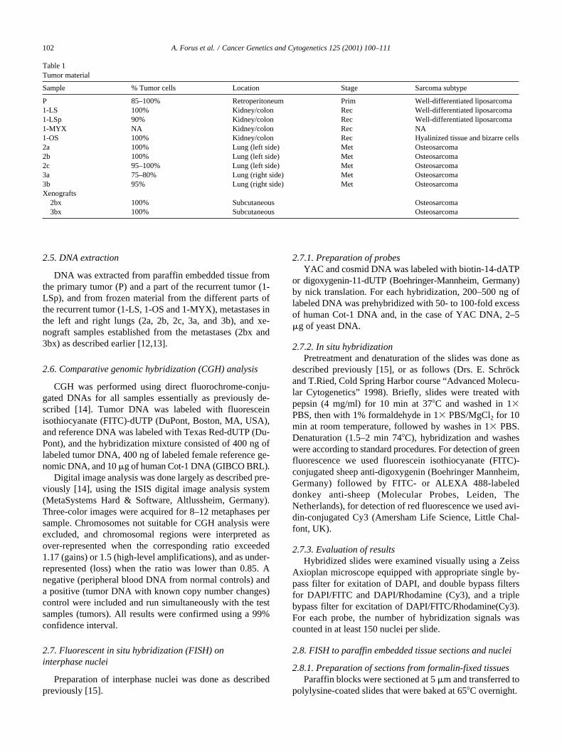
Microscopic examination of the primary tumor showed aWDLPS of a sclerosing and lipoma-like type (Figs. 1a andb). Some myxoid areas could be seen. The recurrent tumor,like the primary, was a WDLPS, but parts with benign-look-ing heterotopic ossification (Figs. 1e and f) and areas withbizarre looking cells, thought to represent chemotherapy in-duced changes, could be seen (Fig. 1g). No clear osteosar-coma component was seen. The metastases in the lung,which were detected at the same time as the abdominal re-currence, were highly malignant osteosarcomas (Figs. 1jand k). No liposarcoma components could be recognized inthese tumors. In accordance with these findings, the tumorwas diagnosed as a dedifferentiated liposarcoma.
3.2. Comparative genomic hybridization (CGH)
CGH did not detect any changes in the primary, paraffin-embedded tumor (P), but several recurrent chromosomal ab-errations were seen in all the other tumors (Table 2).
Gain of 12q material was seen in all samples except theprimary tumor (P) and the recurrent 1-LSp, and was, there-fore, the most frequently observed alteration. The specimen1-OS from the recurrent tumor had a local high-level ampli-fication of the 12q21–q22 region. This high-level amplifica-tion was detected in all samples from the metastasis in theleft lung, which was diagnosed as an osteosarcoma (2a, b,and c), and in addition, the region 12q14–q24.2 was ampli-fied at lower levels. The metastasis in the right lung, part 3b,
showed exactly the same pattern, but sample 3a had a gain of12q13–qter and not the high-level amplification of 12q21–q22.
Gain of 1q material, affecting various parts of 1q21–q32and with a minimal overlapping region at 1q23, was thesecond most common aberration. This gain was observedonly in the samples from the lung metastases, of which 2band 3b showed high-level amplification narrowed down to1q21–q31.
The two metastatic tumors also showed loss of 2q mate-rial; most of the long arm was lost, but a region from thecentromere and down to q14 or q21/22 was retained. In themetastasis from the left lung, this aberration was observedin all different parts of the tumor, but only in the 3b samplefrom the metastasis in the right lung. In the recurrent tumor(1-OS and 1-MYX) and a part of the metastasis in the leftlung (2a) loss of 9p-material was observed, but this was notseen in the other metastases, nor in any of the xenograftsfrom samples 2b and 3b established in nude mice (2bx and3bx, respectively).
The xenograft 2bx had fewer aberrations than the corre-sponding lung metastasis (2b), but the high-level amplifica-tion of 12q14–q24.2 and gain of 1q material were retained.In contrast, the xenograft 3bx had a CGH profile very similarto the other lung metastasis (3b), including the gain of 1q21–q31, but the high-level amplification of 1q22–q24 was lost.
In general, the number and complexity of aberrations in-creased as the tumor progressed towards a more malignantphenotype. Furthermore, copy number changes varied forthe different parts of the same tumor, most notably in themetastasis to the right lung where one part (3a) showed gainof 12q13–qter alone whereas the second, 3b, had this gainas well as numerous other aberrations.
3.3. FISH analyses
The two most common aberrations, gain or amplificationof regions within 1q21–q31and 12q14, were additionallyanalyzed by FISH. Copy numbers of centromere 1 and 12 as
Table 2DNA sequence copy number changes revealed by CGH
Sample Copy number changes
Patient samplesP No changes1-LS NA1-LSp No changes1-OS
2
9p21–pter,
1
12q13–q24.2(
12q21–q22
)1-MYX
1
4q,
2
9p21–pter,
1
12q14–q24.2,
2
13cen–q142a
1
1q23–q31,
2
2q22–qter,
2
9p,
112q (12q14–q24.2),117p12–pter2b 11cen21–q32(1q21–q31),22q14.2–qter,6q16–qter,112q(12q14–q24.2),114,215cen–q15,117q(17q23–q24)2c 11q21–q32,22q22–qter,211q14–q24,112q(12q14–q24.2),2133a 112q13–qter3b 1q21–q31(1q22–q24),22q14.2–qter,16p,26q,211q24–qter,112q(12q14–q24.1),213q21–qter,117,218,120,122
Xenografts2bx 11q24–q25,112q(12q14–12q24.2)3bx 11q21–q32,22q21–qter,26q21–qter,112q(12q13–q24.2),117q22–qter,218,120
Abbreviation: NA, no DNA available for analysis.Gains are marked with 1, losses with 2 and high-level amplifications are marked in bold.

A. Forus et al. / Cancer Genetics and Cytogenetics 125 (2001) 100–111 105
well as two genomic probes representing sequences knownto be highly amplified were analyzed and the results pre-sented in Fig. 1 and Table 3.
3.3.1. Centromere 1The samples from the primary tumor (P) had normal cop-
ies of centromere 12. Interestingly, several nuclei with high-
level amplification of CDK4 were observed (Fig. 1c), al-though the majority showed normal copy numbers of thisgene. In the recurrent tumor, the sample 1-OS showed twocentromere 12 signals, but here copy numbers of CDK4were even more heterogeneous (Fig. 1h). While approxi-mately 60% of the nuclei gave two signals, the remaining40% had amplification. All parts of the metastasis in the left
Table 3FISH analyses using probes from the most frequently altered regions
Patient samples Stage/Histology Cen 1 Cen 12 YAC789f2 CDK4a
P P/WDLPS 1s: 21% 1s: 25% 1s: 7% 1s: 7% 2s: 73% 2s: 71% 2s: 77% 2s: 62% 3–6s: 6% 3–6s: 4% 3–6s: 16% 3–9s: 16% >10s: 15%
1-LSp Rec/WDLPS 1s: 14% 1s: 11% 1s: 12% 2s: 82% 2s: 76% 2s: 82% 2s: 57% 3–9s: 4% 3–9s: 13% 3–9s: 6% 4s: 15% >10s: 28%
Patient samples Stage/Histology Cen1 and YAC789f2 Cen 12 and CDK4
1-LS Rec/WDLPS 2 cen and 2s: 96% Not analyzed 2 cen and 3–9s: 1% 4 cen and 4s: 3%
1-OS Rec/WDLPS, hyalinized tissue and atypical cells 2 cen and 2s: 91% 2 cen and 2s: 58% 2 cen and 3–9s: 8% 2 cen and 3–9s: 4% 4 cen and 4s: 1% 2 cen and >10s: 38%
2a Met/OS 2 cen and 2s: 19% 2 cen and 2s: 7% 3–9s of each: 43% 3–9 cen and >10s: 93% 3–9 cen and >10s: 38%
2b Met/OS 2 cen and 2s: 11% 2 cen and 2s: 6% 3–9s of each: 73% 3–9 cen and >10s: 94% >10s of each: 16%
2c Met/OS 2 cen and 2s: 7% 2 cen and 2s: 3% 2 cen and 3–9s: 5% 3–9 cen and >10s: 97% 3–9s of each: 44% 3–9 cen and >10s: 38% >10s of each: 6%
3a Met/OS 2 cen and 2s: 80% 2 cen and 2s: 72% 2 cen and 3–9s: 11% 2 cen and 3–9s: 4% 3–9s of each: 4% 2 cen and >10s: 24% 3–9 cen and >10s: 5%
3b Met/OS 2 cen and 2s: 18% 2 cen and 2s: 1% 2 cen and 3–9s: 9% 2–4 cen and >10s: 99% 3–9s of each: 62% >10s of each: 11%
Xenografts Stage/Histology Cen 1 and YAC789f2 Cen 12 1 CDK4
2bx X/OS 2 cen and 2s: 14% 2 cen and 2s: 4% 2 cen and 3–9s: 17% 2–4 cen and >10s: 96% 3–9s of each: 48% 3–9 cen and >10s: 12% >10s of each: 9%
3bx X/OS 2 cen and 2s: 12% 2 cen and 2s: 2% 2 cen and 3–9s: 17% 3–9s of each: 60% 2–4 cen and >10s: 98% 3–9 cen and $10s: 11%
FISH analysis on paraffin sections and interphase nuclei isolated from paraffin embedded primary tumor (P) and recurrent 1-LSp, and on interphase nucleifrom frozen tissue of recurrent (Rec), metastatic (Met), and xenograft (X) samples. For the frozen tissue, the centromere probes were hybridized together withYAC or cosmid from the same chromosome, and the signals were counted in the same nuclei, thus the number before “and” refers to the centromere whereasthe number after is the count for YAC789f2/CDK4, respectively. For the primary tumor (P), the probes for cen 1 and cen 12 were hybridized to separate par-affin sections. A relatively large fraction of the nuclei showed only one signal, and this is probably a result of nuclei being cut during the preparation of thesections. YAC789f2 and the CDK4 cosmid were hybridized to interphase nuclei isolated from paraffin embedded tissue. The most frequent signal pattern isshown in boldface.
Cen 5 centromeres, s 5 signals.aCDK4 signals were counted in nuclei on four different preparations to get sufficient material for analysis.

106 A. Forus et al. / Cancer Genetics and Cytogenetics 125 (2001) 100–111
lung, samples 2a, 2b and 2c, had increased numbers of cen-tromere 12 and high-level amplification of CDK4 in most ofthe nuclei (Fig. 1l). This was also the case for the 3b samplefrom the metastasis in the right lung, and the xenografts.Again, samples 3a and 3b had extremely different patterns.In 3a, only a small fraction of the nuclei showed amplifica-tion of CDK4, and aneusomy of chromosome 12 was neverobserved. In 3b, most of the nuclei showed amplification ofCDK4, and about 25% of the nuclei had more than two cen-tromeres.
3.3.2. Centromere 1 (cen1) and 1q21 markerYAC 789f2, which is so far the best marker for the 1q21-
amplification [15] was used to detect this amplicon. Thesamples from the primary tumor (P) had normal copy num-bers of YAC789f2 and the centromere in a majority of thenuclei (Fig. 1d). A relatively large fraction of the nucleishowed only one centromere signal, probably because thenuclei have been cut during the preparation of the sections.In the recurrent sample, the majority of the nuclei showednormal copy numbers of both probes (Fig. 1i). In the sam-ples from the metastases in the left lung (2a, b, c) we ob-served aneusomy of chromosome 1, and the most commonpattern was gain of both the centromere and YAC789f2(Fig. 1m). Samples 2a and 2c had a relatively large fractionof nuclei with increased copy numbers of centromere 1 andhigh-level amplification of YAC789f2, but this pattern wasless striking for 2b, although CGH revealed high-level am-plification of 1q21–q23 in this sample. Sample 3b, from themetastasis in the right lung, had a signal pattern similar to2b, whereas in 3a, almost all the nuclei analyzed showednormal copy numbers of both probes. The CGH analysesconfirmed this results, indicating that (at least) two very dif-ferent tumor cell populations are present in this sample. Inthe two xenografts (2bx and 3bx), increased copy numbersof both cen1 and YAC789f2 was most frequent, as was thecase in the corresponding tumor samples, 2b and 3b. All themetastatic samples, apart from 3a, showed gain or amplifi-cation of 1q material in more than 65% of the cells ana-lyzed, whereas the recurrent tumor showed such aberrationsin less than 10% of the cells. This could suggest that cellswith 1q aberrations are selected for during the metastaticprocess.
In general, the FISH analyses revealed considerable het-erogeneity in copy numbers among nuclei from the same tu-
mor, and there was also some variation between the differ-ent parts (a, b, and c). This was most striking for themetastasis in the right lung (3a and b), where 3a presentedmostly normal copy numbers of all probes whereas theother, 3b, showed gains and amplifications. Furthermore,the increased numbers of hybridization signals from bothcentromere 1 and 12 in the metastatic samples could indi-cate that these tumors were aneuploid.
3.4. Southern analyses
We analyzed the 12q13–q14 amplicon in more detail us-ing probes for CDK4, MDM2, and HMGIC (12q14–q15),and we used CDKN2B (p15INK4B) to examine whether a de-letion of 9p could be observed also in samples where CGHanalyses showed an apparently normal copy number of thisregion. The results are shown in Fig. 2.
We found amplification of all three genes from 12q13–q14 in all samples checked, but the amplification levels var-ied (Fig. 2). The recurrent samples generally showed loweramplification levels than the metastases, but again, sample3a from the metastasis in the right lung was an exception. Inthis part of the tumor, all three probes showed low or mod-erate amplification (2- to 4-fold or 5- to 9-fold increases),which is in keeping with CGH and FISH results.
We detected deletions of CDKN2B in sample 2a from themetastasis in the right lung. This sample showed deletion ofthe whole 9p arm by CGH (Fig. 2). Also in 2bx, this dele-tion was seen, although CGH analyses showed normal copynumbers of 9p. In contrast, CDKN2B was normal in the twoother samples, 1-OS and 1-MYX, where CGH detected de-letion of 9p21–pter. These deletions were smaller, and mightnot include this gene.
3.5. Northern analyses
RNA was available from part 1-OS of the recurrent tu-mor, from two parts of the metastasis in the left lung (2a and2b), and from the xenograft established form metastasis inthe right lung (3bx). The metastatic samples and the xe-nograft were classified as high-grade osteosarcomas, andsample 1-OS showed hyalinized tissue and heterotopic ossi-fication. All these samples expressed high levels of alkalinephosphatase (ALP), indicative of bone-forming tissue (Fig.3), whereas the unrelated liposarcoma samples on the sameblot showed no expression at all, or low levels in a few
Fig. 1. Representative histology and FISH-analyses of the primary, recurrent, and metastatic tumors. Histology: Stained paraffin-embedded tissue was evalu-ated by microscopy (320 and 340 magnification). FISH analyses: Interphase nuclei isolated from paraffin-embedded or frozen tissue was hybridized withprobes for centromere 12 and/or a cosmid for CDK4 (12q13–q14), or with YAC789f2 (1q21) as described. (a and b): Histological slides from the abdominalprimary tumor show well-differentiated liposarcoma. No osteosarcoma component was seen. (c and d): Interphase nuclei isolated from paraffin tissue hybrid-ized with CDK4 (c, green) and YAC789f2 (d, red). CDK4 is amplified whereas the YAC show normal copy numbers. (e, f, and g): Histological slides from therecurrent abdominal tumor show liposarcoma with ossification and areas of bizarre looking cells (g) thought to be chemotherapy induced. (h and i): Interphasenuclei from 1-OS hybridized with CDK4 (green) and cen12 (red, h), and YAC789f2 (red, i). CDK4 is amplified whereas the YAC show normal copy num-bers. (j and k): Histological slides from the metastasis in the left lung show a highly malignant osteosarcoma. (l and m): Interphase nuclei form the metastasisin the left lung hybridized with CDK4 (green) and cen12 (red, l) and YAC789f2 (red, m). CDK4 is highly amplified whereas the YAC show variable copynumbers.
A. Forus et al. / Cancer Genetics and Cytogenetics 125 (2001) 100–111 107
cases (Fig. 3 and results not shown). We also checked theexpression of samples lipoprotein lipase (LPL), which is ex-pressed in muscle, heart and adipose tissue, but not in bone[24]. No expression was found in the samples from me-
tastases (2a, 2b, and 3ax), and also the sample 1-OS fromthe recurrent tumor did not express this gene. LPL expres-sion was high in the other unrelated liposarcomas present onthe blot (Fig. 3), as expected [27].

108 A. Forus et al. / Cancer Genetics and Cytogenetics 125 (2001) 100–111
4. Discussion
One of the most extensive reports on dedifferentiated li-posarcomas to date is the one by Henricks et al. [7] from1997 where 155 cases were analyzed in order to define howthe extent and degree of dedifferentiation affected the clini-cal outcome. These investigators found that the majority ofdedifferentiated liposarcomas were de novo cases, and onlya minority developed from pre-existing WDLPS similar tothe one described here. There was no direct correlation be-tween the amount of dedifferentiated zones and the clinical
behavior. Rather, the location seemed to be the most impor-tant prognostic factor since retroperitoneal tumors had sig-nificantly worse survival than those located in other sites[7]. This has also been suggested by others [1,6], and seemsto be a general feature also for ordinary WDLPS in this re-gion [2].
The case presented here was first diagnosed as a WDLPSof the retroperitoneum. It was not possible to perform kary-otyping, so it is not known whether the tumor had the giantrods or ring chromosomes which are so typical for WDLPS.The recurrent tumor, which was excised from the same lo-cation 5 years later, had regions of ectopic ossification andbizarre cells thought to be induced by chemotherapy, andthe metastases to the left and the right lungs were highlymalignant osteosarcomas.
Upon scintigraphic examination of the patient, no tumorcells could be detected in the bone. It is, therefore, unlikelythat the multiple metastases seen in the lungs did not origi-nate from the WDLPS, and instead had developed from anoccult, undetectable osteogenic tumor. Moreover, the fre-quency of 12q13–q14 amplification, including MDM2 orCDK4, in sarcomas has been reported to be about 15% [28],and it is rather unlikely that two entirely independent andrare tumors should arise at the same time and both have thehigh-level amplification of 12q13–q14 (CDK4) observedhere. Additionally, an osteogenic cell population expressinghigh levels of ALP, was present in the recurrent tumor. Al-though sampling was satisfying, one cannot rule out that asmall component of osteosarcoma differentiated tumor cellswas overlooked in the primary or recurrent tumor. The partswith benign looking osteogenic metaplasia and bizarre cellswere difficult to evaluate. One cannot rule out that this partcontained small osteosarcoma foci with chemotherapy in-duced morphologic changes.
There are reports in the literature of malignant mes-enchyomas, which are sarcomas that exhibit more than two
Fig. 2. Summary of Southern blot analyses of the 12q13–q15 and 9p21 regions in recurrent, metastatic, and xenograft samples. Probes for CDK4, HMGIC,and MDM2 (12q13–q14) as well as CDKN2B (9p21) were sequentially hybridized to filter blots. Gene copy numbers relative to a reference probe was deter-mined densitometrically, and divided into five categories as indicated. Measurements were based on average signal intensities from at least three differentblots. Gene dosage values of 0.5 or lower were scored as deletions.
Fig. 3. Northern blot analyses of lipoprotein lipase (LPL) and alkalinephosphatase (ALP) in samples from the recurrent and metastatic tumors.Probes for LPL, ALP and 18srRNA were sequentially hybridized to filterblots with the recurrent 1-OS, two different parts of the left lung metastasis(2a and 2b) and the xenograft from the right lung metastasis (3ax), as wellas some unrelated liposarcoma and osteosarcoma samples. Lipoproteinlipase (LPL) was only expressed in the liposarcoma samples (LS13, andresults not shown). ALP showed high expression levels only in the osteo-genic samples (1-OS, 2a, 2b, 3ax and the osteosarcoma cell line OHS andresults not shown). Very low ALP expression could be detected in someliposarcoma samples.

A. Forus et al. / Cancer Genetics and Cytogenetics 125 (2001) 100–111 109
types of specialized differentiation [5], that could be WDLPSand osteosarcoma. Some of these may resemble the casepresented here, e.g., Geurts van Kessel et al. [29] have re-ported that such tumors may have ring chromosomes aswell as amplification of 12q13–q14.
Looking at the chromosomal and genetic aberrations oc-curring at the different stages, we found that the first, lowgrade tumor had no aberrations detectable by CGH, butFISH-analyses detected CDK4 amplification in some nu-clei. All the other samples had chromosomal and geneticchanges detectable by all the methods used. The number ofaberrations increased as the tumor became more aggressive,in keeping with the general assumption that accumulation ofgenetic aberrations is necessary for progression and me-tastasis.
Already in the primary WDLPS (P), and more profoundin the recurrent tumor (1-OS), a high-level amplification ofregions from chromosome 12, including CDK4 (alsoMDM2 and HMGIC in the recurrent tumor), was observed.All but one metastatic sample (3a) showed high-level am-plification of 12q-material by CGH, FISH and molecularanalysis, as well as extra copies of the centromere.
Many studies of amplification in WDLPS report consis-tent amplification of MDM2 [10,30–33], which probablyacts through inactivation of p53 [19]. Various other genes inthe 12q13–q14 region may be included, in particular CDK4and HMGIC [10,30–33]. In other sarcoma subtypes, such asthe more aggressive osteosarcomas, CDK4 is most consis-tently amplified, alone or together with MDM2 [34–36].Therefore, there seems to be two different kinds of 12q13–q14 amplicons in sarcomas: one associated with low-gradeor borderline tumors such as WDLPS, where MDM2 is al-ways involved, and one associated with more aggressive tu-mors, always including CDK4 [34,37].
Loss of 9p-material was detected (by CGH) in the recur-rent tumor and a part of the metastasis in the left lung (2a)but not in any of the other metastatic samples or the xe-nografts. This aberration, frequently affecting the putativetumor suppressor proteins p16/p19 and/or p15 is common inseveral cancers, including sarcomas, but seems to be un-common in liposarcomas [35]. In this series of tumors,Southern analyses showed that the CDKN2B gene was af-fected in the sample with deletion of the whole arm (2a) andin the xenograft 2bx, but not in the recurrent tumors withdeletions of a smaller part of 9p, nor in any other part of themetastatic tumors. The variable deletion pattern of 9p thatwas observed here probably reflect changes in the cellularcomposition of these tumors during the process. It alsoseems likely that other genes than CDKN2B may be the se-lective target for these deletions.
Notably, gain of chromosome 1 material was not detect-able by CGH in the primary and recurrent samples, and onlydetected in few cells (,10%) by FISH. The metastatic sam-ples and the xenografts from those had high-level amplifica-tions of 1q22–q24, and high copy numbers of the marker789f2. Previously, high-level amplifications of 1q21–q22
have been reported as a particularly frequent aberration inseveral different kinds of sarcomas [15,38–41] and havebeen detected both in primary and metastatic tumors. In thecase presented here, amplification within 1q21–q31 is a lateevent, and it seems to be associated with metastasis andtransformation to a highly malignant tumor type. Similarobservations have been made by others: Tarkkanen et al.[42] reported that osteosarcoma patients with tumors thathad gains of 1q21 showed a tendency toward short overallsurvival. In addition, in other tumor types, i.e., renal clear-cell carcinomas, gain of 1q21–q22 is particularly frequent inmetastases [43].
It is also interesting that loss of the distal 2q was ob-served only in the osteogenic lung metastases. This aberra-tion is not among the most commonly reported, and al-though observed in some cases, has not previously beenassociated specifically with osteosarcomas or metastaticdisease [42,44].
The heterogeneity within each of the tumors, as shownby variable gains and amplification patterns of cen 1 and the1q21 marker, is conserved in the xenografts. This could in-dicate that the various subpopulations of tumor cells willgrow at similar rates during and after the establishment ofthe xenograft, and that there is no selection for further am-plification of 1q material during this process. Alternatively,tumor cells with different kinds of aberrations may be se-lected for. A third, and perhaps more likely explanation, isthat there is some dynamic equilibrium, resulting in unevendistribution of amplified segments during cell division.There is little heterogeneity in the 12q amplifications com-pared to the gain or amplifications of 1q21. The presence of12q amplicons on stable extra-chromosomal segments, suchas giant rods or marker chromosomes, that would be ex-pected to be more strongly selected [45], and 1 q amplicons,e.g., on double minutes, could explain these differences.
Already in the primary tumor a percentage of the cellshad amplification of CDK4. Thus, the amplification of12q13–q14, also including HMGIC and MDM2 at least inthe recurrent and metastatic samples, was not induced bythe post-operative radiation or chemotherapy given after ex-cision of that sample. Nevertheless, we cannot rule out thatother aberrations have been induced by therapy.
There is still no good explanation as to why well-differ-entiated tumors of low malignancy may change into aggres-sive, poorly differentiated tumors. Random oncogenic acti-vation may abrogate the almost complete differentiation ofa WDLPS, and give rise to a dedifferentiated tumor. In thiscase, dedifferentiation have produced osteogenic cells thathave completely taken over during metastasis, as confirmedby the presence of osteoid cells and expression of alkalinephosphatase (ALP), and the absence of adipocytic cells ex-pressing lipoprotein lipase (LPL). How this process is regu-lated is unknown, but one may speculate that the amplifica-tion of 1q21/q22–q24 may be involved, and possibly alsoloss of 2q material, as it correlates with malignant osteo-genic differentiation as well as metastasis.
110 A. Forus et al. / Cancer Genetics and Cytogenetics 125 (2001) 100–111
Acknowledgments
We are grateful to Kjetil Boye Pedersen and Inger LivNordli for expert technical assistance. The work was sup-ported by: the Norwegian Cancer Society; The NorwegianResearch Council and the European Commission throughthe Biomedicine and Health project “Integrated analysis ofexpression and chromosomal organisation of genes local-ized on human chromosome 1q21: implications for humandisease and cancer” under BIOMED 2 (BMH4-CT96-0319);the Finnish Cancer Society in Finland; the National Univer-sity of La Plata, Argentina, the National Agency of Scien-tific and Technological Promotion (Contract grant number:BID 802/OC-AR PICT No. 01-00000-00753); the NationalCouncil of Scientific and Technological Research (CONICET);and the Nordic Cancer Union.
References
[1] Enzinger FM, Weiss SW. Soft tissue tumors. 3rd ed. Gray, SM, editor.St. Louis, MO: Mosby-Year Book Inc., 1995.
[2] Fanburg-Smith JC, Miettinen M. Liposarcoma with meningothelial-like whorls: a study of 17 cases of a distinctive histological pattern as-sociated with dedifferentiated liposarcoma. Histopathology 1998;33:414–24.
[3] Weiss SW, Rao VK. Well-differentiated liposarcoma (atypical li-poma) of deep soft tissue of the extremities, retroperitoneum, andmiscellaneous sites. A follow- up study of 92 cases with analysis ofthe incidence of “dedifferentiation”. Am J Surg Pathol 1992;16:1051–8.
[4] Evans HL. Liposarcoma: a study of 55 cases with reassessment of itsclassification. Am J Surg Pathol 1979;3:507–23.
[5] Evans HL, Khurana KK, Kemp BL, Ayala AG. Heterologous ele-ments in the dedifferentiated component of dedifferentiated liposar-coma. Am J Surg Pathol 1994;18:1150–7.
[6] McCormick D, Mentzel T, Beham A, Fletcher CD. Dedifferentiatedliposarcoma. Clinicopathologic analysis of 32 cases suggesting a bet-ter prognostic subgroup among pleomorphic sarcomas. Am J SurgPathol 1994;18:1213–23.
[7] Henricks WH, Chu YC, Goldblum JR, Weiss SW. Dedifferentiatedliposarcoma: a clinicopathological analysis of 155 cases with a pro-posal for an expanded definition of dedifferentiation. Am J SurgPathol 1997;21:271–81.
[8] Nascimento AG, Kurtin PJ, Guillou L, Fletcher CD. Dedifferentiatedliposarcoma: a report of nine cases with a peculiar neurallike whor-ling pattern associated with metaplastic bone formation. Am J SurgPathol 1998;22:945–55.
[9] Dei Tos AP, Doglioni C, Piccinin S, Maestro R, Mentzel T, Barbares-chi M, Boiocchi M, Fletcher CD. Molecular abnormalities of the p53pathway in dedifferentiated liposarcoma. J Pathol 1997;181:8–13.
[10] Pedeutour F, Forus A, Coindre JM, Berner J-M, Nicolo G, MichielsJF, Terrier P, Ranchere-Vince D, Collin F, Myklebost O, Turc-CarelC. Structure of the supernumerary ring and giant rod chromosomes inadipose tissue tumors. Genes Chromosomes Cancer 1999;24:30–41.
[11] Simms WW, Ordonez NG, Johnston D, Ayala AG, Czerniak B. p53 ex-pression in dedifferentiated chondrosarcoma. Cancer 1995;76:223–7.
[12] Forus A, Flørenes VA, Maelandsmo GM, Meltzer PS, Fodstad Ø,Myklebost O. Mapping of amplification units in the q13–14 region ofchromosome 12 in human sarcomas: some amplica do not includeMDM2. Cell Growth Differ 1993;4:1065–70.
[13] Suijkerbuijk RF, Sinke RJ, Olde Weghuis DEM, Roque L, Forus A,Stellink F, Siepman A, van de Kaa C, Soares J, Geurts van Kessel A.Amplification of chromosome subregion 12p11.2-p12.1 including the
PTHLH gene in a metastasis of an I(12p)-negative seminoma: relation-ship to tumor progression? Cancer Genet Cytogenet 1994;78:145–52.
[14] Larramendy ML, Tarkkanen M, Blomqvist C, Virolainen M, Wik-lund T, Asko-Seljavaara S, Elomaa I, Knuutila S. Comparative ge-nomic hybridization of malignant fibrous histiocytoma reveals anovel prognostic marker. Am J Pathol 1997;151:1153–61.
[15] Forus A, Berner J-M, Meza-Zepeda LA, Saeter G, Mischke D, Fod-stad Ø, Myklebost O. Molecular characterisation of a novel ampliconat 1q21–q22 frequently observed in human sarcomas. Br J Cancer1998;78:495–503.
[16] Kerstens HMJ, Poddighe PJ, Hanselaar AGJM. A novel in situ hy-bridization signal amplification method based on the deposition of bi-otinylated tyramine. J Histochem Cytochem 1995;43:347–52.
[17] Raap AK, van de Corput MPC, Vervenne RAW, van Gijlswijk RPM,Tanke HJ, Wiegant J. Ultra-sensitive FISH using peroxidase-medi-ated deposition of biotin-or fluorochrome tyramides. Hum Mol Genet1995;4:529–34.
[18] Huang LS, Bock SC, Feinstein SI, Breslow JL. Human apolipopro-tein B cDNA clone isolation and demonstration that liver apolipopro-tein B mRNA is 22 kilobases in length. Proc Natl Acad Sci USA1985;82:6825–9.
[19] Oliner JD, Kinzler KW, Meltzer PS, George DL, Vogelstein B. Am-plification of a gene encoding a p53-associated protein in human sar-comas. Nature 1992;358:80–3.
[20] Su YA, Trent JM, Guan X-Y, Meltzer PS. Direct isolation of genesencoded within a homogenously staining region by chromosome mi-crodissection. Proc Natl Acad Sci USA 1994;91:9121–5.
[21] Kools PFJ, Van de Ven WJM. Amplification of a rearranged form ofthe high-mobility group protein gene HMGIC in OSA-cl osteosar-coma cells. Cancer Genet Cytogenet 1996;91:1–7.
[22] Hannon GJ, Beach D. p15INK4B is a potential effector of TGF-beta-induced cell cycle arrest. Nature 1994;371:257–61.
[23] Weiss MJ, Ray K, Henthorn PS, Lamb B, Kadesch T, Harris H.Structure of the human liver/bone/kidney alkaline phosphatase gene.J Biol Chem 1988;263:12002–10.
[24] Wion KL, Kirchgessner TG, Lusis AJ, Schotz MC, Lawn RM. Hu-man lipoprotein lipase complementary DNA sequence. Science 1987;235:1638–45.
[25] Marenholz I, Volz A, Ziegler A, Davies A, Ragoussis I, Korge BP,Mischke D. Genetic analysis of the epidermal differentiation complex(EDC) on human chromosome 1q21: chromosomal orientation, newmarkers and a 6 Mbp YAC contig. Genomics 1996;37:295–302.
[26] Khatib ZA, Matsushime H, Valentine M, Shapiro DN, Sherr CJ,Look AT. Coamplification of the CDK4 gene with MDM2 and GLIin human sarcomas. Cancer Res 1993;53:5535–41.
[27] Myklebost O, Stenwig AE, Fodstad Ø. The analysis of gene expres-sion in liposarcoma, mesenchymal tumours resembling various stagesof adipose differentiation. J Cell Biochem Suppl 1994;18A:163.
[28] Momand J, Jung S, Wilczynski S, Niland J. The MDM2 gene amplifi-cation database. Nucl Acid Res 1998;26:3453–9.
[29] Geurts van Kessel A, Simons A, Comtesse PP, Siepman A, Janssen I,Suijkerbuijk RF, Forus A, Pruszczynski M, Veth RP. Ring chromo-somes in a malignant mesenchymoma. Cancer Genet Cytogenet1999;109:119–22.
[30] Nilbert M, Rydholm A, Willen H, Mitelman F, Mandahl N. MDM2gene amplification correlates with ring chromosomes in soft tissue tu-mors. Genes Chromosomes Cancer 1994;9:261–5.
[31] Pedeutour F, Suijkerbuijk RF, Forus A, van Gaal J, van de KlundertW, Coindre J-M, Nicolo G, Collin F, van Haelst U, Huffermann K,Turc-Carel C. Complex composition and co-amplification of SASand MDM2 in ring and giant rod marker chromosomes in well-differ-entiated liposarcoma. Genes Chromosomes Cancer 1994;10:85–94.
[32] Nilbert M, Rydholm A, Mitelman F, Meltzer PS, Mandahl N. Charac-terization of the 12q13–15 amplicon in soft tissue sarcomas. CancerGenet Cytogenet 1995;83:32–6.
[33] Pilotti S, Della Torre G, Lavarino C, Sozzi G, Minoletti F, Vergani B,Azzarelli A, Rilke F, Pierotti MA. Molecular abnormalities in lipo-

A. Forus et al. / Cancer Genetics and Cytogenetics 125 (2001) 100–111 111
sarcoma: role of MDM2 and CDK4-containing amplicons at 12q13–22.J Pathol 1998;185:188–90.
[34] Forus A, Flørenes VA, Maelandsmo GM, Fodstad Ø, Myklebost O.12q13–14 amplica in human sarcomas without MDM2 include CDK4,SAS and GADD153/CHOP. Cancer Genet Cytogenet 1994;77:200.
[35] Maelandsmo GM, Berner J-M, Flørenes VA, Forus A, Hovig E, Fod-stad Ø, Myklebost O. Homozygous deletion frequency and expres-sion levels of the CDKN2 gene in human sarcomas-relationship toamplification and mRNA levels of CDK4 and CCND1. Br J Cancer1995;72:393–8.
[36] Berner J-M, Forus A, El Kahloun A, Meltzer PS, Fodstad Ø, Mykle-bost O. Separate amplified regions encompassing CDK4 and MDM2in human sarcomas. Genes Chromosomes Cancer 1996;17:254–9.
[37] Forus A, Schmitt J-M, Pedeutour F, Turc-Carel C, Meltzer PS, Fod-stad Ø, Myklebost O. Amplification of 12q13–14 in human sarcoma:is there a common selective gene, or are different mechanisms impor-tant in different sets of tumours? Cytogenet Cell Genet 1994;67:269.
[38] Forus A, Olde Weghuis D, Smeets D, Fodstad Ø, Myklebost O,Geurts van Kessel A. Comparative genomic hybridization analysis ofhuman sarcomas: I. Occurence of genomic imbalances and identifica-tion of a novel major amplicon at 1q21–q22 in soft tissue sarcomas.Genes Chromosomes Cancer 1995;14:8–14.
[39] Forus A, Olde Weghuis D, Smeets D, Fodstad Ø, MyklebostO,Geurts van Kessel A. Comparative genomic hybridization analysisof human sarcomas: II. Identification of novel amplicons at 6p and17p in osteosarcomas. Genes Chromosomes Cancer 1995;14:15–21.
[40] Szymanska J, Tarkkanen M, Wiklund T, Virolainen M, Blomquist C,Asko-Seljavaara S, Tukiainen E, Elomaa I, Knuutila S. Gains andlosses of DNA sequences in liposarcomas evaluated by comparativegenomic hybridization. Genes Chromosomes Cancer 1996;15:89–94.
[41] Szymanska J, Virolainen M, Tarkkanen M, Wiklund T, Asko-Selja-vaara S, Tukiainen E, Elomaa I, Blomqvist C, Knuutila S. Overrepre-sentation of 1q21–q23 and 12q13–q21 in lipoma-like liposarcomasbut not in benign lipomas: A comparative genomic hybridizationstudy. Cancer Genet Cytogenet 1997;99:14–18.
[42] Tarkkanen M, Elomaa I, Blomqvist C, Kivioja AH, Kellokumpu-Lehtinen P, Bohling T, Valle J, Knuutila S. DNA sequence copynumber increase at 8q: a potential new prognostic marker in high-grade osteosarcoma. Int J Cancer 1999;84:114–21.
[43] Gronwald J, Storkel S, Holtgreve-Grez H, Hadaczek P, BrinkschmidtC, Jauch A, Lubinski J, Cremer T. Comparison of DNA gains andlosses in primary renal clear cell carcinomas and metastatic sites: Im-portance of 1q and 3p copy number changes in metastatic events.Cancer Res 1997;57:481–7.
[44] Tarkkanen M, Karhu R, KallioniemiA, Elomaa I, Kivioja AH, Nev-alainen J, Bohling T, Karaharju E, Hyytinen E, Knuutila S, Kallioni-emi O-P. Gains and losses of DNA sequences in osteosarcomas bycomparative genomic hybridization. Cancer Res 1995;55:1334–8.
[45] Gisselson D, Hoglund M, Mertens F, Mitelman F, Mandahl N. Chro-mosomal organization of amplified chromosome 12 sequences inmesenchymal tumors detected by fluorescence in situ hybridization.Genes Chromosomes Cancer 1998;23:203–12.