Embed Size (px)
Citation preview

Deep Neck Space Infections
Deep Neck Space Infections
Disclaimer: The pictures used in this presentation and its content has been obtained from a number of sources. Their use is purely for academic and teaching purposes. The contents of this presentation do not have any intended commercial use. In case the owner of any of the pictures has any objection and seeks their removal please contact at [email protected] . These pictures will be removed immediately.

The fibrous connective tissue that constitutes the cervical fascia varies from loose areolar tissue to dense fibrous bands.
This fascia serves to envelope the muscles, nerves, vessels and viscera of the neck, thereby forming planes and potential spaces that serve to divide the neck into functional units.
It functions to both direct and limit the spread of disease processes in the neck.

The cervical fascia can be divided into a simpler superficial layer and a more complex deep layer that is further subdivided into superficial, middle and deep layers.
The superficial layer of cervical fascia ensheaths the platysma in the neck and extends superiorly in the face to cover the mimetic muscles.
It is the equivalent of subcutaneous tissue elsewhere in the body and forms a continuous sheet from the head and neck to the chest, shoulders and axilla.


The superficial layer of the deep cervical fascia is also known as the investing layer.
It follows the “rule of twos”—it envelops two muscles, two glands and forms two spaces.
It originates from the spinous processes of the vertebral column and spreads circumferentially around the neck.

It covers sternocleidomastoid and trapezius muscles, encloses the submandibular and
parotid glands.
It also covers the anterior bellies of the digastrics and the mylohyoid, thereby forming the floor of the submandibular space
It forms the space of the posterior triangle on either side of the neck and the suprasternal space of Burns in the midline.


The middle layer of the deep cervical fascia is also known as the visceral fascia
It has two subdivisions, the muscular division, which surrounds the infrahyoid strap muscles, and the visceral division, which envelops the pharynx, larynx, esophagus, trachea, and thyroid gland.
The visceral division passes inferiorly into the upper mediastinum where it is continuous with the fibrous pericardium and covers the thoracic trachea and esophagus.


The deep layer of the deep cervical fascia originates from the spinous processes of the cervical vertebra and the ligamentum nuchae.
At the transverse processes of the cervical vertebra, it divides into an anterior alar layer and a posterior prevertebral layer.
The alar fascia extends from the base of the skull to the second thoracic vertebra
The prevertebral fascia lies just anterior to the vertebral bodies and extends the entire length of the vertebral column.


The carotid sheath is a fascial layer that is associated with but is anatomically separate from the previously described layers
It receives contributions from all three layers of deep cervical fascia.
It contains the carotid artery, internal jugular vein and vagus nerve
It continues from the skull base through the neck along the anterior surface of the prevertebral fascia, and enters the chest behind the clavicle


Spaces Spanning the Entire Length
of the Neck

The superficial space is located between the superficial fascia and the superficial layer of the deep fascia
This potential space lies superficial and deep to the platysma and contains loose areolar tissue, lymph nodes, nerves and vessels—the most significant of which is the external jugular vein
This space is most commonly involved with superficial cellulitis of the neck, but if abscess formation does occur, this will present with obvious fluctuance, erythema, warmth and tenderness


The deep neck spaces that run the entire length of the neck include: • Retropharyngeal space • Danger space • Prevertebral space • Visceral vascular space

Retropharyngeal Space • It occupies the space posterior to the pharynx
and esophagus. • Its anterior wall is made up of the
buccopharyngeal fascia superiorly and the visceral division of the middle fascia inferiorly
• The posterior wall is the alar layer of the deep fascia
• The lateral boundary is the carotid sheath. • This space extends from the base of the skull
to the level of the first and second thoracic vertebra


Danger Space
• Posterior to the retropharyngeal space lies the danger space
• So named because it contains loose areolar tissue and offers little resistance to the spread of infection
• It is the space between the alar layer and prevertebral layer of the deep fascia
• Runs from the skull base to the diaphragm

Visceral Vascular Space • It is the potential space within the carotid sheath. • IT contains little areolar tissue and is resistant to
the spread of infection. • Termed the “Lincoln’s highway” • It extends from the base of skull into the
mediastinum. • Receives contributions from all three layers of
deep fascia • It can become secondarily involved by infection in
any other deep neck space by direct spread.
.


Suprahyoid Spaces

The submandibular
space
The parapharyngeal
space
The peritonsillar
space
The masticator space
The Parotid space

The submandibular space
• Bounded by the mandible anteriorly and laterally
• The lingual mucosa superiorly
• The hyoid postero-inferiorly • The superficial layer of the
deep cervical fascia inferiorly


The mylohyoid muscle divides this space into a superior sublingual space and an sub-mylohyoid space
The sublingual space contains loose areolar tissue, the hypoglossal and lingual nerves, the sublingual gland and Wharton’s duct.
The submylohyoid space contains the anterior bellies of the digastrics and the submandibular glands. These two subdivisions freely communicate around the posterior border of the mylohyoid


Early appearance of patient who has Ludwig’s angina with characteristic submandibular ‘’woody’’ swelling

Parapharyngeal Space

The skull base
superiorly
Pterygo-
mandibular raphe
anteriorly
Parotid
Boundaries
Hyoid Bone
Inferiorly
Mandible
Prevertebral Fascia
Posteriorly
Bucco-pharyngeal
fascia medially
Medial Pterygoid


PPS
Submandibular Space
Masticator Space
Retropharyngeal space
Parotid Space

Pre-Styloid
Fat, muscle, lymph nodes and connective tissue
Tonsillar fossa
Medial Pterygoid plate
Post-Styloid
Carotid sheath
9th, 10th & 12th
Cranial Nerves

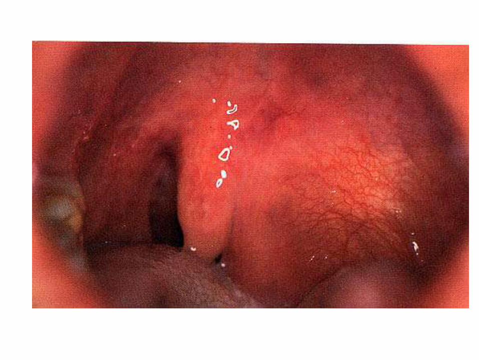
Retropharyngeal Abscess in Children

Retropharyngeal abscess

Most common deep neck abscess
Retropharyngeal lymph nodes tend to involute with age
Source of infection suppurative process in nose, nasopharynx, sinuses and adenoids
96% abscesses occur prior to 6 years of age


Symptoms
Fever
Enlarged Cervical
LNs
Post. Pharyngeal wall buldge
Trismus
Torticollis
Irritability

•Poor oral intake
•Sore throat
•Dysphagia
•Drooling
•Laryngeal oedema
•Respiratory distress

Lateral radiograph of the neck

Retropharyngel Space Abscess in
Adults

Retropharyngeal abscess in the adult is typically caused by:
• Penetrating or blunt trauma • Instrumentation such as endoscopy, • Intubation or NG tube placement • Extension of infection from an adjoining deep
neck space • Historically, the most common cause of a
prevertebral abscess was the extension of a tuberculous infection of a vertebral body, a Pott’s abscess.


Submandibular Space Infection

• In recent years, submandibular space abscess has become the most common of the deep neck space infections.
• Seventy to 85% of these cases are odontogenic in origin, the rest are caused by sialadenitis, lymphadenitis, floor of mouth lacerations or mandible fractures.
• Ludwig’s angina is the prototypical submandibular space infection, however this term should not be applied to all submandibular abscesses.


Criteria to label Ludwig's angina • A cellulitic process of the submandibular
space, not an abscess • Involvement of only the submandibular
space, although this could be bilateral • Gangrene with foul serosanguinous fluid
on incision, but no frank purulence • Involvement of the fascia, muscle and
connective tissue, • Direct spread of infection rather than
spread by lymphatics.

Ludwig’s angina
Symptoms
Tender, firm
swelling
“hot potato”
voice
Sialorrhea
Tachypnea
Dyspnea
Stridor

Early Ludwig's angina

Parapharyngeal space abscess

• Infection in the pharynx, tonsils, adenoids, teeth, parotid or lymph node chains
• Middle ear infections or mastoiditis • Extension of infection from the
nearby: • Peritonsillar space • Submandibular space • Retropharyngeal space • Masticator space


• Signs and symptoms of parapharyngeal abscess differ depending on whether the prestyloid or poststyloid compartment is involved.
• In addition to fever, chills and malaise, anterior infection will often cause pain, dysphagia and significant trismus due to medial pterygoid irritation.
• Edema in this area will cause a medial bulging of the lateral pharyngeal wall and tonsil and there will be swelling at the angle of the mandible.

• Posterior compartment infection may have no localizing signs on examination.
• Despite this, these patients do appear toxic and may receive the diagnosis “fever of unknown origin.”
• Involvement of the neurovascular structures found in this area may lead to cranial neuropathies, Horner’s syndrome, septic internal jugular thrombosis or carotid artery rupture.
• Any bleeding from the nose, mouth or ear in a patient with suspected deep neck abscess should be taken very seriously.

• Preantibiotic era—S.aureus • Currently—aerobic Strep
species and non-strep anaerobes
• Gram-negatives uncommon • Almost always polymicrobial • Remember resistance

IMAGING

CT SCAN
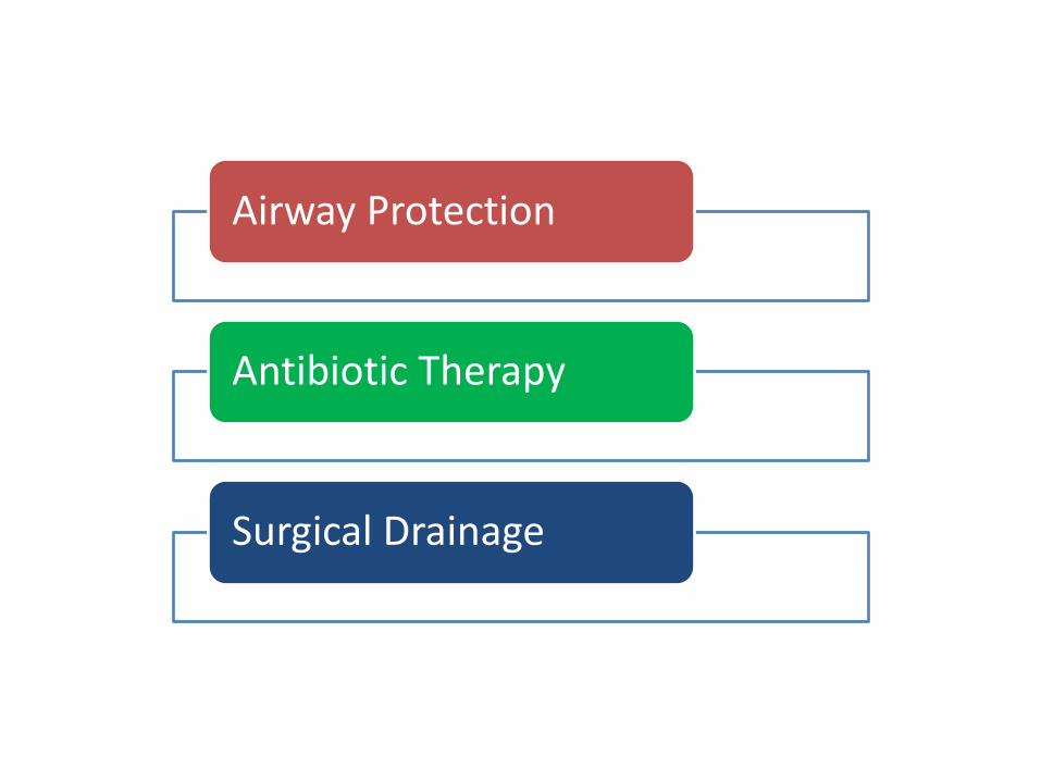
Airway Protection
Antibiotic Therapy
Surgical Drainage