Embed Size (px)
Citation preview
Defensive Armor of Potato Tubers: Nonpolar Metabolite Profiling,Antioxidant Assessment, and Solid-State NMR CompositionalAnalysis of Suberin-Enriched Wound-Healing TissuesKeyvan Dastmalchi,† Linda Kallash,† Isabel Wang,† Van C. Phan, Wenlin Huang,† Olga Serra,‡
and Ruth E. Stark*,†
†Department of Chemistry, The City College of New York, City University of New York Graduate Center and Institute forMacromolecular Assemblies, New York, New York 10031, United States
Department of Natural Sciences, Hostos Community College, 500 Grand Concourse, Bronx, New York 10451, United States‡Laboratori del Suro, Departament de Biologia, Facultat de Ciencies, University of Girona, Campus Montilivi s/n, Girona, E-17071Spain
*S Supporting Information
ABSTRACT: The cultivation, storage, and distribution of potato tubers are compromised by mechanical damage andsuboptimal healing. To investigate wound-healing progress in cultivars with contrasting russeting patterns, metabolite profilesreported previously for polar tissue extracts were complemented by GC/MS measurements for nonpolar extracts and quantitative13C NMR of interfacial solid suspensions. Potential marker compounds that distinguish cultivar type and wound-healing timepoint included fatty acids, fatty alcohols, alkanes, glyceryl esters, α,ω-fatty diacids, and hydroxyfatty acids. The abundant long-chain fatty acids in nonpolar extracts and solids from the smooth-skinned Yukon Gold cultivar suggested extensive suberinbiopolymer formation; this hypothesis was supported by high proportions of arenes, alkenes, and carbonyl groups in the solidand among the polar markers. The absence of many potential marker classes in nonpolar Atlantic extracts and interfacial solidssuggested a limited extent of suberization. Modest scavenging activities of all nonpolar extracts indicate that the majority ofantioxidants produced in response to wounding are polar.
KEYWORDS: Solanum tuberosum, closing layer, wound periderm, solid-state NMR, GC/MS, potential markers,ABTS•+ scavenging activity, suberin
■ INTRODUCTION
Potatoes can be wounded at various stages during cultivation,collection, storage, and distribution. In the absence of properhealing, the tuber surfaces are vulnerable to desiccation andbacterial infection, which can lead to significant crop losses.1
Protection is conferred by a wide range of small-moleculemetabolites and by the biopolymer suberin, which is depositedat the apoplastic barrier either during plant development or inresponse to wounding stress.2,3 Analysis of depolymerizationproducts has produced suberin structural models that include apolyphenolic domain of hydroxycinnamic acid derivatives andmonolignols derived from the phenylpropanoid pathway4,5 aswell as a polyaliphatic domain (polyester) with long-chain α,ω-diacids and ω-hydroxyacids, together with glycerol, that arethought to be esterified to the polyphenolic domain by ferulicacid.6,7 The customary “top-down” approach involves depoly-merization of suberized cell walls by chemical treatments andstudy of the resulting breakdown products to deduce themolecular constituents that comprise the suberin biopolymer.However, limitations of this technique include the incomplete-ness of depolymerization treatments and their inability torelease all classes of chemical constituents, leaving gaps in ourunderstanding of the macromolecular assembly of the suberinmacromolecular composite. A potent complementary “bottomup” strategy for understanding potato tuber defense via
suberization monitors formation of wound-induced metabolitesin potato tissues. This latter approach is also attractive becausethe cells can be induced synchronously,8 and the response inthe parenchymatic tissue below the wounding surface is initiallyrestricted to those cells that form the closing layer and later tothe cells below that form the wound periderm emerging from anew cork cambial layer.9,10 In this way, changes in the solublemetabolite profile and deposited suberin can be monitoredprogressively to build an understanding of how the protectivebiopolymer is formed.To augment GC/MS metabolite profiling reported for
wound-induced tissues from the Russet Burbank potato,8,11
we recently carried out LC−MS analyses of polar extracts fromwound tissues of four potato cultivars with differing patterns ofskin russeting (Table 1).12 These polar metabolomic analyses,conducted for Norkotah Russet, Atlantic, Chipeta, and YukonGold cultivars at two developmental time points afterwounding, were made in parallel with radical scavenging assaysusing the 2,2′-azinobis-(3-ethylbenzothiazoline-6-sulfonic acidammonium salt) free radical (ABTS•+).12 To construct a more
Received: March 18, 2015Revised: July 9, 2015Accepted: July 13, 2015Published: July 13, 2015
Article
pubs.acs.org/JAFC
© 2015 American Chemical Society 6810 DOI: 10.1021/acs.jafc.5b03206J. Agric. Food Chem. 2015, 63, 6810−6822
comprehensive chemical picture of the wound-healing processand its dependence on potato variety, the current study drawson a concurrent partitioning strategy to add cultivar-specificdifferences in the metabolite profiles and scavenging abilities ofnonpolar extracts as well as chemical compositions of thecorresponding insoluble material that remains after polar andnonpolar metabolite extraction. Contrasts are also drawnbetween the metabolites and solid materials present at early(day 3) vs late (day 7) time points after wounding, representingformation of the suberized closing layer as an initial healingresponse and when the nascent wound periderm is developing,respectively.9,10,12 The nonpolar compounds signifying thosemetabolites that distinguish the two wound-healing time points(day 3 vs day 7 in all cultivars) or that discriminate among thefour cultivars (at a given time point) are referred to as potentialwound-healing and potential cultivar markers, respectively.These compounds were detected using multivariate statisticalanalysis, then identified and elucidated by matching their GC/MS data with suitable databases. Knowledge of the markercompounds involved in the wound healing process can guidethe design of methods to expedite this process, thus improvingcrop protection during growth and after harvesting. Takentogether, these studies can promote the development of foodstaples possessing beneficial antioxidant capabilities, robustwound healing, and reduced agricultural waste.
■ MATERIALS AND METHODSReagents. The following chemicals were used for extraction and
GC/MS analysis: HPLC−MS grade water, methanol, and chloroform(J. T. Baker, Phillipsburg, NJ); hexadecyl hexadecanoate, C7−C40saturated alkane standards, and formic acid (Sigma-Aldrich, St. Louis,MO); GC/MS grade N-methyl-N-(trimethylsilyl)trifluoroacetamide(MSTFA) + 1% trimethylchlorosilane (TMCS) (Thermo Scientific,Bellefonte, PA); pyridine (EMD Millipore Corporation, Billerica,MA). Potassium peroxosulfate (Sigma-Aldrich), 2,2′-azinobis (3-ethylbenzothiazoline-6-sulfonic acid ammonium salt) (ABTS), and 6-hydroxy-2,5,7,8 tetramethylchromane-2-carboxylic acid (TCI, Tokyo,Japan) were used for the antioxidant assay.Plant Material. Potato tuber cultivars from the 2011 crop year
were provided by Joe Nunez, University of California CooperativeExtension (Davis, CA). Differences in their overall phenotypiccharacteristics are summarized in Table 1.Sample Preparation. The dormant freshly harvested potato
tubers were peeled, sectioned, and allowed to heal as describedpreviously.12 Briefly, internal flesh tissues were sectioned longitudinallywith a mandolin slicer to obtain disks about 5 mm thick. Healing in thedark at 25 °C was accomplished by placing the slices on wet cellulosefilter paper inside closed humidified plastic boxes equipped with wirenetting supports. The new brown surface layer of wound-healing tissueis identifiable because it is easily excoriated and detached, thenremoved by sweeping a flat spatula under the layer. During tissuecollection, flesh (parenchyma) contamination under the woundhealing surface was avoided to the extent possible. The sampleswere harvested at 3 and 7 days after wounding, corresponding to earlyhealing in which the suberized closing layer had formed and to thedeveloping wound periderm, respectively. Samples were frozen
immediately in liquid nitrogen and stored at −80 °C. Processinginvolved grinding under liquid nitrogen, freeze-drying, and againstoring at −80 °C.
For chemical analysis, concurrent extraction and partitioning ofpolar and nonpolar constituents were carried out using a slightlymodified version of the method introduced by Choi et al.13 formetabolomics of plant materials.14 Extraction of 10 mg samples by 1min of pan ultrasonication (Branson Ultrasonics, Danbury, CT) in 2mL of 60% (v/v) methanol−water was followed by addition of 2 mLof chloroform and an additional 1 min of sonication in a 4-mL glassvial. Extracts were then incubated in a shaker at room temperature for10 min, followed by tabletop centrifugation (Beckman Coulter,Fullerton, CA) at 1089g and 25 °C to produce three phases: uppersoluble polar, lower soluble nonpolar, and an interphase of suspendedparticulates. After removal of the liquid polar and nonpolar extractswith a glass Pasteur pipet, the remaining residue of interfacialparticulate solids was filtered, washed, and dried under a flow ofnitrogen. Six biological replicates per cultivar were extracted in parallelfor each of the day-3 and day-7 time points.
GC/MS Analysis. The samples were prepared according to Yang etal.8 For metabolite separation and identification, a 500-μL aliquot ofeach nonpolar extract was placed in a glass vial and evaporated. Eachsample was then reconstituted with 50 μL of pyridine and derivatizedusing 50 μL of N-methyl-N-(trimethylsilyl) trifluoroacetamide(MSTFA) + 1% trimethylchlorosilane (TMCS) at 50 °C for 1 h.Samples were analyzed with a QP2010 GC/MS (Shimadzu USA,Canby, RI) spectrometer with the injector oven set at 250 °C. Theinjection mode was splitless, and the volume injected for each samplewas 1 μL. A Durabond-5 column (30 m × 0.25 mm i.d., film thickness0.25 μm; Agilent Technologies, Santa Clara, CA) was used. Thetemperature program was based on the published method of Yang etal.8 with minor modifications. After an initial delay of 5 min at 70 °Cfor the solvent to clear the system, the oven temperature was raised to310 °C at a rate of 5 °C/min. The oven was kept at this temperaturefor 11 min and allowed to cool afterward to 70 °C. A 1-μL injection of0.3 μM hexadecyl hexadecanoate provided a reference standard.
Preprocessing of GC/MS Data. The GC/MS data with a .qgdextension were converted to ANDI (CDFNET) format. MZmineversion 2.4 (VTT Technical Research Center, Helsinki, Finland andTurku Center for Biotechnology, Turku, Finland) that includedfiltration, deconvolution, peak alignment, and data normalization wasused for processing.15 The region between 0 and 15 min, whichcontains peaks from derivatization artifacts, was excluded from theanalysis. These peaks were also observed in blanks consisting of thesolvent, pyridine, and derivatization reagent.
Multivariate Statistical Analysis. Simca-P+ software version 13.0(Umetricas, Umea, Sweden) was used to carry out principalcomponent analysis (PCA) of GC/MS data processed with MZMine using Pareto scaling.16 This method organizes the data byrelating the observations, cultivar types, and variables of the LC−MSdata. The method tests the consistency of each set of biologicalreplicates and discriminates among the different sample types.
Orthogonal partial least-squares discriminate analysis (OPLS-DA)of the data followed by the generation of S-plots helped to identifycompounds that account for the differences among cultivars andbetween different wounding time points, which were designated aspotential cultivar and potential wound-healing markers, respectively(Supporting Information, Figure S1). OPLS-DA is a regression andprediction method that finds information in the X data (e.g., variablesfrom the LC−MS experiments) that is related to information in the Ydata (e.g., discrete variables specifying cultivar type or wound-healingtime point). This method shows how these two sets of informationvary together and if they are dependent on each other, facilitatingclassification studies and potential marker identification. The S-plotserves to visualize OPLS-DA results in a scatter plot.17 The variables(e.g., MS ions) at the extreme ends of the S-plot had p(corr) [1] > 0.8,indicating high reliability;17 they were further examined using avariable line plot to check how specific they were to the sample typeand if they qualified as potential marker ions (Supporting Information,Figure S1).
Table 1. Potato Cultivars for Metabolite Profiling Studies
cultivar flesh periderm russeting skin
NorkotahRusset
white russeted and netted dark tan
Atlantic white lightly netted to heavily scaled whiteChipeta white small russeted areas light to
buffYukon Gold light
yellowsmooth, finely flaked, yellowishwhite
yellowish
Journal of Agricultural and Food Chemistry Article
DOI: 10.1021/acs.jafc.5b03206J. Agric. Food Chem. 2015, 63, 6810−6822
6811

The PCA model was validated by calculating the values of R2 andQ2, which indicate fitness and predictive ability, respectively.16 The Q2
value, which is reported as a result of cross validation of the model,exceeded 0.5.17 Cross validation using SIMCA-P+ divided the datainto seven groups, one of which was removed. The model was thengenerated with the remaining groups, and the deleted group waspredicted by the model. The partial predictive residual sum of squares(PRESS) was calculated. This procedure was repeated seven times tosum the partial PRESS values, obtain the overall PRESS, and calculatethe Q2 value. R2 was larger than Q2, but the difference between R2 andQ2 was small, as recommended.17 For OPLS-DA, in addition to Q2, themodel diagnostics included R2X and R2Y values calculated for the Xand Y variables, respectively.17
Metabolite Structural Identification. The ions corresponding tomarkers and other metabolites were identified using the Wiley Library(9th edition/NIST 2008). The ionic spectrum of each metabolite wasmatched against the spectra of known compounds from the libraryusing a similarity index to identify hits. In addition, the observedspectra were compared with those of the known compounds for theratio of ion intensities. C7−C40 saturated alkane standards (Sigma-Aldrich, St. Louis, MO) were injected. On the basis of their retentiontimes and m/z values and using the NIST library peaks, they wereassigned to standard reference compounds. There is a directcorrelation between retention time and chain length.ABTS•+ Scavenging. Antioxidant assessment of the nonpolar
extracts was conducted using an ABTS•+ scavenging assay essentiallyas described by Dastmalchi et al.12 Reaction of an aqueous ABTSsolution (7 mM) with K2S2O8 (2.45 mM) in the dark for 12−16 h atroom temperature yielded ABTS•+, for which the absorbance at 734nm was adjusted to 0.70 (±0.02) with ethanol. To a 2-μL aliquot ofthe extract of interest was added 198 μL of the ABTS•+ reagent; theabsorbance at 734 nm was monitored after initial mixing and at 5 minintervals up to 45 min using a Spectramax M5 microplate reader(Molecular Devices, Sunnyvale, CA). Each percentage inhibition valuewas calculated as
−×
Abs Abs
Abs100control sample
control
A calibration plot of percentage inhibition versus concentration wasmade for the reference standard, 6-hydroxy-2,5,7,8-tetramethylchro-man-2-carboxylic acid (Trolox), permitting calculation of the Troloxequivalent antioxidant capacity (TEAC, mmol Trolox/g dried sample)for each extract.Isolation of Suberin-Enriched Tissues. The interfacial solid
from the partitioned mixture was passed through a Whatman no. 4filter paper and then washed with distilled water. To removeunsuberized cell-wall materials, the sample was treated with 0.1%
(w/v) Aspergillus niger cellulase (MP Biomedicals, Illkirch, France) in a50 mM pH 5.0 acetate buffer for 48 h each at 37 and 44 °C,respectively. The residue was then treated with 0.4% (v/v) A. nigerpectinase (Sigma-Aldrich) in a 50 mM pH 4.0 acetate buffer for 24 heach at 28 and 31 °C, respectively. After the enzyme treatments, thesample was filtered, washed with deionized water, and dried at 50 °C.Soxhlet extraction was conducted under reflux conditions to removeany remaining waxes and soluble lipids, using a succession of solventsof varying polarity: methanol, chloroform, and hexane for 48 h each.The resulting solid suberin-enriched samples were reserved for solid-state NMR analysis.
Solid-State NMR Analysis. The chemical moieties present in thesuberin-enriched materials were identified and quantitated using cross-polarization and direct polarization magic-angle spinning 13C NMRexperiments (CPMAS, DPMAS) on 3−4 mg powdered samples. Afour-channel Agilent (Varian) DirectDrive I (VNMRS) NMRspectrometer (Agilent Technologies, Santa Clara, CA, USA) operatingat a 1H frequency of 600 MHz (13C at 150 MHz) and equipped with a1.6 mm HXY FastMAS probe operating at a spinning rate of 10.00kHz (±20 Hz) was used. The spectral data were typically processedwith 100 Hz line broadening and analyzed in parallel using VNMRJ(version 2.2C; Agilent) and ACD/NMR Processor Academic Edition(version 12; Advanced Chemistry Development, Inc., Toronto, ON,Canada). Chemical shifts were referenced externally to the methylene(−CH2−) group of adamantane (Sigma-Aldrich) at 38.48 ppm.
For typical CPMAS experiments used to identify the carbon-containing functional groups via their respective chemical shifts,conditions included 90° pulse durations of 1.3 and 1.2 μs for 1H and13C, respectively, a contact time of 1.5 ms, acquisition delay of 4 μs,and recycle delay of 3 s between successive acquisitions. Heteronucleardecoupling was applied with a 1H field strength of 204 kHz using theSPINAL method.18 The spectral width was 46296 Hz, and the numberof transients was 21500. For DPMAS experiments used to estimate therelative proportions of each carbon type, a 100-s recycle delay wasused to collect 2600 transients.19 Alternatively, quantitatively reliable13C NMR spectra of suberized cell walls were obtained by acquiring100 transients with the multiple-CPMAS method20 after validationwith a t-BOC alanine standard (Sigma-Aldrich) and through DPMASmeasurements for the Norkotah Russet day-7 wound suberin.Radiofrequency (rf) fields of ω1H = 204 kHz and ω1C = 189 kHzwere used for all 90° pulses. Transfer of polarization from 1H to 13Cnuclei was achieved via the condition ω1C − ω1H = ωrotation, with rffrequencies of ω1H = 53.3 kHz and ω1C = 63.4 kHz; a 10% linear rampof the 1H radiofrequency power was used during the CP period.Recycle times between successive cross-polarizations were 1.0 s; 11contact times of 1.0 ms were used to acquire each transient. SPINAL
Figure 1. PCA score plot of GC/MS data from nonpolar extracts, showing consistency of each set of biological replicates and discrimination amongthe differently russeted cultivars at each of two time points after wounding. Included are wound-healing samples at day 3 (Wd3, A), and day 7 (Wd7,B), designated as Atlantic (red, A), Chipeta (green, G), Norkotah Russet (blue, R), and Yukon Gold (gold, Y) cultivars. The x and y axes of the plotsrepresent the score values of principal components 1 and 2, respectively.
Journal of Agricultural and Food Chemistry Article
DOI: 10.1021/acs.jafc.5b03206J. Agric. Food Chem. 2015, 63, 6810−6822
6812
heteronuclear decoupling18 with ω1H = 204 kHz was applied during anacquisition period of 22 μs.The 3-day wound healing samples required 5000 transients (∼20
h), whereas 7-day samples required only 1500 transients (∼6 h).Integrated signal intensities were measured by counting pixels with theimage manipulation software GIMP (www.gimp.org) and using thefollowing chemical shift ranges to denote major structural componentsof suberin: carboxyl and amide groups (168−180 ppm, region 1);arenes and alkenes (115−160 ppm, region 2); alkoxy groups (96−108,80−92, 67−80, 59−67 ppm; regions 3−6); methoxy groups (45−59ppm, region 7); alkyl chain methylenes (15−45 ppm, region 8). Eachchemical shift range was extended by ±1 ppm to ensure that theregions were not truncated in the middle of a resonance. Each quotedquantitative estimate was the result of measurements on two biologicalreplicates, quoted as mean ± standard error.Statistical Analysis. The antioxidant and solid-state NMR data
were presented as mean values ± standard error. Significant differencesbetween the values were determined using Tukey’s pairwisecomparison21 at a level of P < 0.05, assessed with JMP softwareversion 12 (http://www.jmp.com).
■ RESULTS AND DISCUSSION
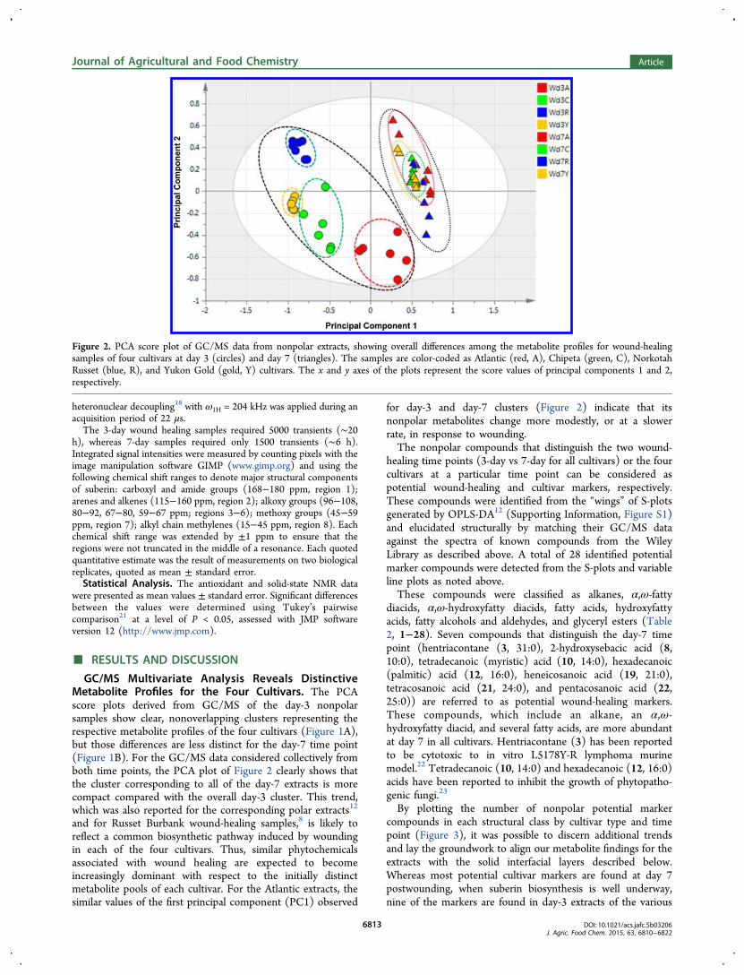
GC/MS Multivariate Analysis Reveals DistinctiveMetabolite Profiles for the Four Cultivars. The PCAscore plots derived from GC/MS of the day-3 nonpolarsamples show clear, nonoverlapping clusters representing therespective metabolite profiles of the four cultivars (Figure 1A),but those differences are less distinct for the day-7 time point(Figure 1B). For the GC/MS data considered collectively fromboth time points, the PCA plot of Figure 2 clearly shows thatthe cluster corresponding to all of the day-7 extracts is morecompact compared with the overall day-3 cluster. This trend,which was also reported for the corresponding polar extracts12
and for Russet Burbank wound-healing samples,8 is likely toreflect a common biosynthetic pathway induced by woundingin each of the four cultivars. Thus, similar phytochemicalsassociated with wound healing are expected to becomeincreasingly dominant with respect to the initially distinctmetabolite pools of each cultivar. For the Atlantic extracts, thesimilar values of the first principal component (PC1) observed
for day-3 and day-7 clusters (Figure 2) indicate that itsnonpolar metabolites change more modestly, or at a slowerrate, in response to wounding.The nonpolar compounds that distinguish the two wound-
healing time points (3-day vs 7-day for all cultivars) or the fourcultivars at a particular time point can be considered aspotential wound-healing and cultivar markers, respectively.These compounds were identified from the “wings” of S-plotsgenerated by OPLS-DA12 (Supporting Information, Figure S1)and elucidated structurally by matching their GC/MS dataagainst the spectra of known compounds from the WileyLibrary as described above. A total of 28 identified potentialmarker compounds were detected from the S-plots and variableline plots as noted above.These compounds were classified as alkanes, α,ω-fatty
diacids, α,ω-hydroxyfatty diacids, fatty acids, hydroxyfattyacids, fatty alcohols and aldehydes, and glyceryl esters (Table2, 1−28). Seven compounds that distinguish the day-7 timepoint (hentriacontane (3, 31:0), 2-hydroxysebacic acid (8,10:0), tetradecanoic (myristic) acid (10, 14:0), hexadecanoic(palmitic) acid (12, 16:0), heneicosanoic acid (19, 21:0),tetracosanoic acid (21, 24:0), and pentacosanoic acid (22,25:0)) are referred to as potential wound-healing markers.These compounds, which include an alkane, an α,ω-hydroxyfatty diacid, and several fatty acids, are more abundantat day 7 in all cultivars. Hentriacontane (3) has been reportedto be cytotoxic to in vitro L5178Y-R lymphoma murinemodel.22 Tetradecanoic (10, 14:0) and hexadecanoic (12, 16:0)acids have been reported to inhibit the growth of phytopatho-genic fungi.23
By plotting the number of nonpolar potential markercompounds in each structural class by cultivar type and timepoint (Figure 3), it was possible to discern additional trendsand lay the groundwork to align our metabolite findings for theextracts with the solid interfacial layers described below.Whereas most potential cultivar markers are found at day 7postwounding, when suberin biosynthesis is well underway,nine of the markers are found in day-3 extracts of the various
Figure 2. PCA score plot of GC/MS data from nonpolar extracts, showing overall differences among the metabolite profiles for wound-healingsamples of four cultivars at day 3 (circles) and day 7 (triangles). The samples are color-coded as Atlantic (red, A), Chipeta (green, C), NorkotahRusset (blue, R), and Yukon Gold (gold, Y) cultivars. The x and y axes of the plots represent the score values of principal components 1 and 2,respectively.
Journal of Agricultural and Food Chemistry Article
DOI: 10.1021/acs.jafc.5b03206J. Agric. Food Chem. 2015, 63, 6810−6822
6813
cultivars. All four Yukon Gold day-3 markers are fatty acids,three of which appear later as day-7 Chipeta markers (Table 2).The largest numbers of fatty acid markers are present at day 3for Yukon Gold and at day 7 for Chipeta, followed by Norkotah
Russet, respectively (Figure 3A). Fatty acids, which can besubstrates for subsequent elongation, hydroxylation, oresterification steps during suberin biosynthesis, comprise nearly40% of the nonpolar potential markers overall. Therefore, we
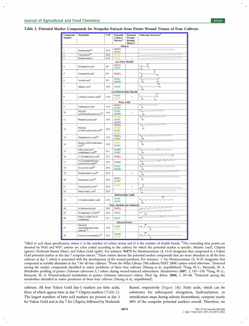
Table 2. Potential Marker Compounds for Nonpolar Extracts from Potato Wound Tissues of Four Cultivars
aAlkyl or acyl chain specification, where C is the number of carbon atoms and D is the number of double bonds. bThe wounding time points aredenoted by Wd3 and Wd7; entries are color coded according to the cultivar for which the potential marker is specific: Atlantic (red), Chipeta(green), Norkotah Russet (blue), and Yukon Gold (gold). For instance, Wd7Y for Hentriacontane (3, 31:0) designates that compound as a YukonGold potential marker in the day-7 nonpolar extract. cThese entries denote the potential marker compounds that are more abundant in all the fourcultivars at day 7, which is associated with the development of the wound periderm. For instance, ‘+’ for Hentriacontane (3, 31:0) designates thatcompound as notably abundant at day 7 for all four cultivars. dFrom the Wiley Library (9th edition/NIST 2008) unless noted otherwise. eDetectedamong the marker compounds identified in native periderms of these four cultivars (Huang et al., unpublished). fYang, W.-L.; Bernards, M. A.Metabolite profiling of potato (Solanum tuberosum L.) tubers during wound-induced suberization. Metabolomics 2007, 3, 147−159. gYang, W.-L.;Bernards, M. A. Wound-induced metabolism in potato (Solanum tuberosum) tubers. Plant Sig. Behav. 2006, 1, 59−66. hDetected among themetabolites identified in native periderms of these four cultivars (Huang et al., unpublished).
Journal of Agricultural and Food Chemistry Article
DOI: 10.1021/acs.jafc.5b03206J. Agric. Food Chem. 2015, 63, 6810−6822
6814
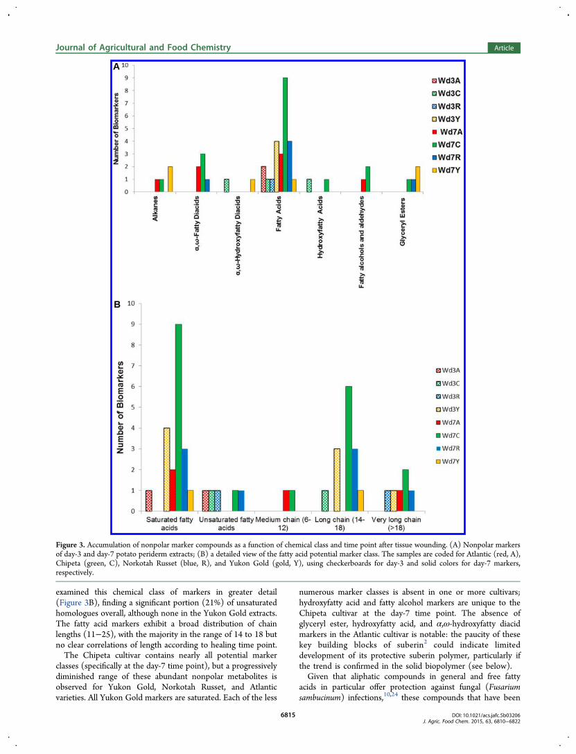
examined this chemical class of markers in greater detail(Figure 3B), finding a significant portion (21%) of unsaturatedhomologues overall, although none in the Yukon Gold extracts.The fatty acid markers exhibit a broad distribution of chainlengths (11−25), with the majority in the range of 14 to 18 butno clear correlations of length according to healing time point.The Chipeta cultivar contains nearly all potential marker
classes (specifically at the day-7 time point), but a progressivelydiminished range of these abundant nonpolar metabolites isobserved for Yukon Gold, Norkotah Russet, and Atlanticvarieties. All Yukon Gold markers are saturated. Each of the less
numerous marker classes is absent in one or more cultivars;hydroxyfatty acid and fatty alcohol markers are unique to theChipeta cultivar at the day-7 time point. The absence ofglyceryl ester, hydroxyfatty acid, and α,ω-hydroxyfatty diacidmarkers in the Atlantic cultivar is notable: the paucity of thesekey building blocks of suberin2 could indicate limiteddevelopment of its protective suberin polymer, particularly ifthe trend is confirmed in the solid biopolymer (see below).Given that aliphatic compounds in general and free fatty
acids in particular offer protection against fungal (Fusariumsambucinum) infections,10,24 these compounds that have been
Figure 3. Accumulation of nonpolar marker compounds as a function of chemical class and time point after tissue wounding. (A) Nonpolar markersof day-3 and day-7 potato periderm extracts; (B) a detailed view of the fatty acid potential marker class. The samples are coded for Atlantic (red, A),Chipeta (green, C), Norkotah Russet (blue, R), and Yukon Gold (gold, Y), using checkerboards for day-3 and solid colors for day-7 markers,respectively.
Journal of Agricultural and Food Chemistry Article
DOI: 10.1021/acs.jafc.5b03206J. Agric. Food Chem. 2015, 63, 6810−6822
6815
found abundantly in all cultivars could provide strongprotection against fungal infection. Unsaturated fatty acidmarkers such as 9-octadecenoic acid (oleic acid) (15, 18:1) and9,12-octadecadienoic acid (linoleic acid) (17, 18:2) have alsobeen implicated as active compounds against plant pathogenicfungi25 and may contribute to the antioxidant activitiesdescribed below.Diacids such as markers 4−8 provide potential sites for the
developing suberin biopolymer: for attachment to glycerolforming glyceryl esters, for esterification with long-chainalcohols forming linear esters, or for covalent linkages at eitherend.6 Thus, these metabolites can build cross-links within thesuberin polyester.4 All fatty diacid markers emerge in day-7extracts; namely, at a later time point in the healing processwhen the wound periderm is developing. Moreover, it isstriking that these diacids have relatively short acyl chainscompared with native periderm constituents (Huang et al.,unpublished). However, medium-chain (C8−C12) species withsaturated aliphatic chains have also been reported in wound-healing potato tissues as suberin triglyceride degradationproducts,26 in contrast to the typical C16, C18, and longerchain lengths found as suberin monomers.4,6 One of the diacidmarkers, 2-hydroxysebacic acid (8, 10:0), is up-regulated in allcultivars at day 7 (Table 2). Among the diacid markers, azelaicacid (6, 9:0) has been implicated in the plant immune system ofArabidopsis, offering protection against pathogens.27
Some of the potential markers identified in the current study(Table 2) have been reported previously as metabolites in
wound tissue extracts8 of potato tubers. Those markercompounds reported for the first time in the nonpolar extractsfrom potato wound tissues included α,ω-fatty diacids (4−7), anα,ω-hydroxyfatty diacid (8, 10:0), a fatty acid (9, 11:0), ahydroxyfatty acid (23, 18:0), an alcohol (26), an aldehyde (24,16:1), and an alkane (3, 31:0). Conversely, some longer-chainfatty acid (28:0−30:0) and alcohol (20:0−27:0 and 29:0)metabolites reported previously8 were not detected in thecurrent study (Supporting Information Table 1). Since ourGC/MS protocol successfully detects long-chain metabolites,we attribute this discrepancy to cultivar differences orunintended variations in tissue preparation procedures. Longerchain fatty acids, fatty alcohols, and alkanes have also beenfound previously in native potato periderms5,7 (Huang et al.,unpublished), but their absence in wound-healing tissues issensible in light of their well-established anatomical andphysiological differences.28 The identified fatty acids includedodd-numbered homologues, which have also been reportedpreviously in the wound tissues of potato tubers8 and areknown elsewhere in the plant kingdom.29 Some polarcompounds, such as phenolic acids, sugars, and amino acids,were found in the nonpolar extracts, presumably as a result ofbleed-through effects during the partitioning of the solublelayers.
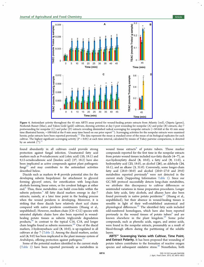
ABTS•+ Scavenging Varies with Cultivar, Time Point,and Extract Polarity. It has been proposed that wounding ofpotato tubers contributes to the formation of reactive oxygenspecies and subsequent oxidative stress.30 Nonetheless, both
Figure 4. Antioxidant activity throughout the 45 min ABTS assay period for wound-healing potato extracts from Atlantic (red), Chipeta (green),Norkotah Russet (blue), and Yukon Gold (gold) cultivars, showing activities at day-3 post wounding for nonpolar (A) and polar (B) extracts; day-7postwounding for nonpolar (C) and polar (D) extracts revealing diminished radical scavenging for nonpolar extracts (∼50-fold at the 45 min assaytime illustrated herein, ∼100-fold at the 0 min assay time based on our prior report12). Scavenging activities for the nonpolar extracts were examinedherein; polar extracts have been reported previously.12 The data represent the mean ± standard error of the mean of six biological replicates for eachcultivar. The highest significant scavenging activity (P < 0.05) at each time interval, calculated by means of Tukey pairwise comparisons, is denotedby an asterisk (*).21
Journal of Agricultural and Food Chemistry Article
DOI: 10.1021/acs.jafc.5b03206J. Agric. Food Chem. 2015, 63, 6810−6822
6816
increasing and decreasing stress responses of potato tissues byproduction of antioxidant chemical compounds have beenreported by the same authors in response to wounding.30,31 Toavoid the established limitations of the diphenylpicrylhydrazylfree radical scavenging assay used in prior studies (narrow pHrange, steric hindrance, spectral interference),32,33 we selectedan ABTS•+ assay. The latter method also permits cleancomparisons with our previous investigation12 of antioxidantactivity in polar extracts derived concurrently from the samepotato tissue samples. The end result is a more comprehensivepicture of antioxidant activity in the potato wound tissues fromthese four cultivars.Figure 4 shows that the activities of nonpolar extracts at day
7 postwounding (Figure 4C) are significantly higher than at day3 postwounding (Figure 4A), supporting an accumulation ofantioxidant constituents during the wound-healing process. Theorder of activity for day-3 nonpolar samples is Norkotah Russet> Chipeta ∼ Yukon Gold > Atlantic, whereas at day 7 the trendchanges slightly to Norkotah Russet > Chipeta > Yukon Gold∼ Atlantic. Thus, the heavily russeted Norkotah Russet cultivarexhibits the highest scavenging activity for both day-3 and day-7nonpolar extracts.As noted above, the applicability of the ABTS•+ scavenging
assay for antioxidant activity of both polar and nonpolarcompounds32,33 makes it possible to compare the respectivedata directly. As a group, the antioxidant activities after 45 minof free radical scavenging reaction for both day-3 and day-7extracts are 50- to 100-fold smaller for the nonpolar samples
(Figure 4), reflecting the relative dearth of phenolic and otherunsaturated molecular moieties among the nonpolar metabo-lites. Conversely, it can be deduced that the majority of theantioxidant compounds produced within the healing tissues inresponse to wounding stress are polar in nature, consistent withprior reports that resistance to infection directly after tuberwounding is related to the appearance of phenolic monomersthat subsequently serve as substrates for suberin polyphenolicbiosynthesis.34 The increase in antioxidant activity for nonpolarextracts as healing progresses from day 3 to 7 is in contrast withour observations for the polar portions, which tend to maintaintheir antioxidant activity levels, except for the Yukon Goldcultivar (Figures 4B and D).4
In addition, the ABTS•+ assay allows us to evaluate thecapacity of both fast- and slow-acting antioxidants.32 TheTEAC values displayed in Figure 4 for both 3- and 7-day timepoints show overall increases in antioxidant activity throughoutthe assay period (0−45 min). This trend, which parallelsobservations made for the corresponding polar tissue extracts,12
demonstrates the presence of both fast- and slow-actingantioxidants. Finally, the absence of steric hindrance in theABTS•+ assay allows sampling of the scavenging activity ofsubstances with diverse molecular weights and architecturalfeatures.32,33
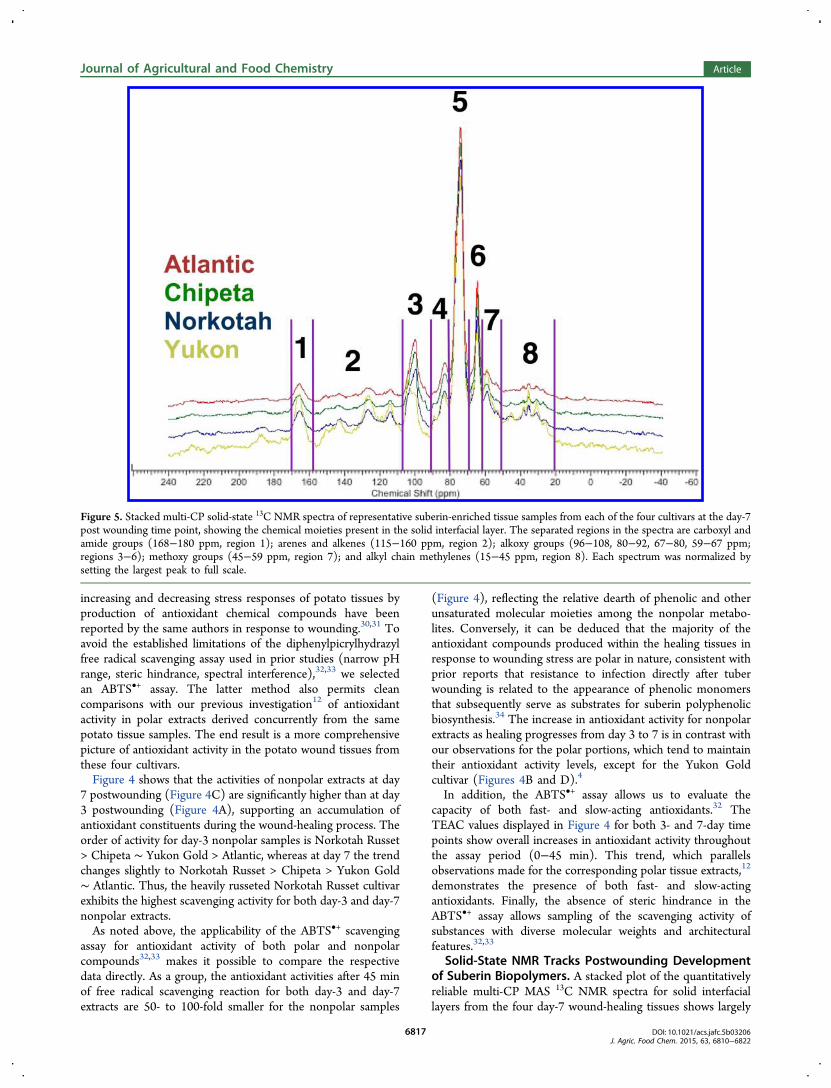
Solid-State NMR Tracks Postwounding Developmentof Suberin Biopolymers. A stacked plot of the quantitativelyreliable multi-CP MAS 13C NMR spectra for solid interfaciallayers from the four day-7 wound-healing tissues shows largely
Figure 5. Stacked multi-CP solid-state 13C NMR spectra of representative suberin-enriched tissue samples from each of the four cultivars at the day-7post wounding time point, showing the chemical moieties present in the solid interfacial layer. The separated regions in the spectra are carboxyl andamide groups (168−180 ppm, region 1); arenes and alkenes (115−160 ppm, region 2); alkoxy groups (96−108, 80−92, 67−80, 59−67 ppm;regions 3−6); methoxy groups (45−59 ppm, region 7); and alkyl chain methylenes (15−45 ppm, region 8). Each spectrum was normalized bysetting the largest peak to full scale.
Journal of Agricultural and Food Chemistry Article
DOI: 10.1021/acs.jafc.5b03206J. Agric. Food Chem. 2015, 63, 6810−6822
6817
concordant chemical shifts but clear differences in relative peakintensities (Figure 5). The spectra of the suberin samples at day7 are similar in appearance to those obtained from the day-7wound periderm in prior studies of potato tubers.19,35 (Aunique peak at 198 ppm in the Yukon day-7 sample isattributable to a carbonyl within an aldehyde group.) Becausequantitative comparisons between spectra of different plantmaterials can be skewed by inconsistencies in mass andinstrumental conditions,36 relative peak ratios within each
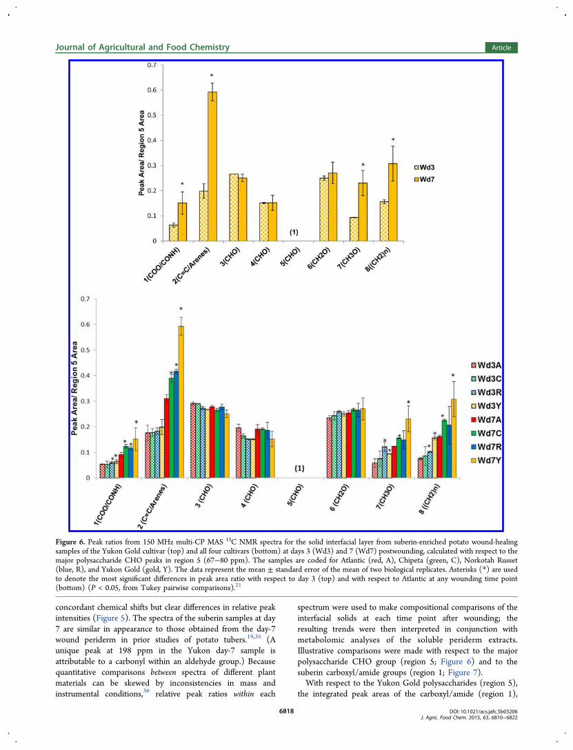
spectrum were used to make compositional comparisons of theinterfacial solids at each time point after wounding; theresulting trends were then interpreted in conjunction withmetabolomic analyses of the soluble periderm extracts.Illustrative comparisons were made with respect to the majorpolysaccharide CHO group (region 5; Figure 6) and to thesuberin carboxyl/amide groups (region 1; Figure 7).With respect to the Yukon Gold polysaccharides (region 5),
the integrated peak areas of the carboxyl/amide (region 1),
Figure 6. Peak ratios from 150 MHz multi-CP MAS 13C NMR spectra for the solid interfacial layer from suberin-enriched potato wound-healingsamples of the Yukon Gold cultivar (top) and all four cultivars (bottom) at days 3 (Wd3) and 7 (Wd7) postwounding, calculated with respect to themajor polysaccharide CHO peaks in region 5 (67−80 ppm). The samples are coded for Atlantic (red, A), Chipeta (green, C), Norkotah Russet(blue, R), and Yukon Gold (gold, Y). The data represent the mean ± standard error of the mean of two biological replicates. Asterisks (*) are usedto denote the most significant differences in peak area ratio with respect to day 3 (top) and with respect to Atlantic at any wounding time point(bottom) (P < 0.05, from Tukey pairwise comparisons).21
Journal of Agricultural and Food Chemistry Article
DOI: 10.1021/acs.jafc.5b03206J. Agric. Food Chem. 2015, 63, 6810−6822
6818
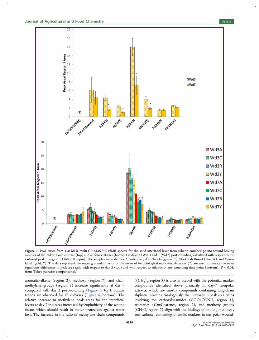
aromatic/alkene (region 2), methoxy (region 7), and chainmethylene groups (region 8) increase significantly at day 7compared with day 3 postwounding (Figure 6, top). Similartrends are observed for all cultivars (Figure 6, bottom). Therelative increase in methylene peak areas for the interfaciallayers at day 7 indicates increased hydrophobicity of the woundtissue, which should result in better protection against waterloss. The increase in the ratio of methylene chain compounds
((CH2)n, region 8) is also in accord with the potential markercompounds identified above primarily in day-7 nonpolarextracts, which are mostly compounds containing long-chainaliphatic moieties. Analogously, the increases in peak area ratiosinvolving the carboxyls/amides (COO/CONH, region 1),aromatics (CC/arenes, region 2), and methoxy groups(CH3O, region 7) align with the findings of amide-, methoxy-,and carboxyl-containing phenolic markers in our polar wound-
Figure 7. Peak ratios from 150 MHz multi-CP MAS 13C NMR spectra for the solid interfacial layer from suberin-enriched potato wound-healingsamples of the Yukon Gold cultivar (top) and all four cultivars (bottom) at days 3 (Wd3) and 7 (Wd7) postwounding, calculated with respect to thecarbonyl peak in region 1 (168−180 ppm). The samples are coded for Atlantic (red, A), Chipeta (green, C), Norkotah Russet (blue, R), and YukonGold (gold, Y). The data represent the mean ± standard error of the mean of two biological replicates. Asterisks (*) are used to denote the mostsignificant differences in peak area ratio with respect to day 3 (top) and with respect to Atlantic at any wounding time point (bottom) (P < 0.05,from Tukey pairwise comparisons).21
Journal of Agricultural and Food Chemistry Article
DOI: 10.1021/acs.jafc.5b03206J. Agric. Food Chem. 2015, 63, 6810−6822
6819
healing tissue extracts.12 These latter markers include phenolicacids and phenolic amines, including derivatives of ferulic acidthat become more abundant at day 7. Moreover, the largeraromatic-to-CHO ratios observed in day-7 Yukon Gold suberinsamples (Figures 6) fit with trends observed for the markercompounds in the polar extracts: an increased number ofaromatic markers is detected for this cultivar.12 Together, thesecorrelations suggest that both nonpolar and polar4 solublecompounds can be viewed as precursors of the suberized cellwall layer that is being formed during the healing process.At the day-7 time point, when the wound periderm layer is
forming, peak area ratios for carboxyl/amide (COO/CONH,region 1), aromatic/alkene (CC/arenes, region 2), methoxy(CH3O, region 7), and chain methylene ((CH2)n, region 8)groups are found to be the highest in Yukon Gold and lowest inAtlantic (Figure 6, bottom, and Figure S2). This observationsuggests that the suberin formed in Yukon Gold at day 7 has ahigher proportion of long-chain aliphatic compounds, whichcan provide increased hydrophobicity and lead to decreasedwater permeability. The small ratio of COO/CONH groupswith respect to polysaccharides observed for Atlantic alsosupports the hypothesis of diminished suberization that wasproposed from analysis of the nonpolar soluble markercompounds.When measured with respect to the carboxyl/amide signals
(region 1), the integrated peak areas for oxygenated aliphaticcompounds in regions 3−6 are found to decrease for allcultivars between day 3 and day 7 (Figure 7). This trendsupports and extends the ratios outlined above: whereas(region 5)/(region 1) is just the inverse of the carbonyl-to-CHO ratio, additional polysaccharide peak areas in regions 3, 4,and 6 also decrease with respect to the suberin esters andamides that contribute to region 1 of the spectrum. Thus, theselatter results reinforce the conclusion that deposition of thealiphatic-aromatic suberin polyester within the phellem cell-walltissues during development of the wound periderm diminishesthe polysaccharide-to-fatty acyl ester ratio. As noted above, therelative increase in carbonyl, aromatic, and olefinic groups canbe related to both soluble polar and nonpolar potential markercompounds, many of which contain these functional groups. Atday 7, Yukon has the lowest and Atlantic the highest peak arearatios for each of the oxygenated aliphatics in regions 3−6(Figure 7 and Figure S3).It is also notable that, among the suberin-associated groups,
the aromatic/alkene moieties (CC/arenes, region 2) showthe most dramatic increase in relative intensity with respect tothe polysaccharide carbons (Figure 6). This finding suggeststhat the majority of the compounds being deposited in thepolyphenolic domain of suberin during the 3-to-7 day timeframe originate from the polar extracts, as this is the layer that isrich in aromatic and olefinic structures (phenolic amines,flavonoids, and phenolic acids).4
Coordinated Measurements Provide a ChemicalPicture of Potato Tuber Wound Healing. GC/MSmultivariate analysis shows convergence of the nonpolarsoluble metabolite composition at the day-7 time point afterwounding of the potato tubers, suggesting induction of acommon biosynthetic pathway as a result of wounding andsuberization in all four cultivars. A similar trend was observedamong the potential polar markers.12 Analysis of the GC/MSdata leads to the identification of 28 potential cultivar-specificand wound-healing marker compounds that discriminateamong cultivar types and between wound healing time points
day 3 and 7, during closing layer and periderm development.The distribution of nonpolar soluble markers (Figure 3)dovetails with the trends observed in the solid suberincomposition of the cultivars at the same time point (Figures6 and 7). That is, at day 7, Yukon Gold has the highest andAtlantic has the lowest relative amount of solid chainmethylene-containing compounds in their respective suberinsamples. This observation parallels the trend among the solublenonpolar fatty acid markers (Figure 3B, Table 2), which at day3 display the highest number of long-chain and very-long-chainhomologues for Yukon Gold but at day 7 are diminished andputatively undergoing incorporation into the solid suberinbiopolymer. It is sensible to propose, then, that the hydro-carbon chains of the fatty acid compounds contribute to thesolid-state 13C NMR resonance corresponding to the long-chain aliphatic suberin constituents. Conversely, the nonpolarextracts from the Atlantic cultivar exhibit no day-3 markersfrom these two classes or from glyceryl esters. The paucity ofglyceryl esters, dioic acids, and hydroxyfatty acids in nonpolarAtlantic extracts, when considered in conjunction with thelowered proportions of COO/CONH groups in the interfacialsolids, suggest a diminished extent of suberization.For the solid suberized tissues (Figure 7), elevated ratios of
methylene chain-containing compounds in the wound-healingtissues of all cultivars at day 7 can increase the peridermhydrophobicity, thereby offering the potential for moreprotection against water loss. The presence of flavonoid,phenolic acid, and phenolic amine marker compounds, all ofwhich contain carboxyl or amide groups, in the polar extracts atboth day-3 and day-7 time points, could also correlate with thedeposition of such carbonyl-containing compounds in theYukon Gold suberin at both time points.12 The most vigorousdeposition of alkenes and arenes at day 7, which is observed forYukon Gold suberin samples (Figures 6 and 7), is in accordwith trends observed among the aromatic markers found in thepolar extracts of this cultivar.12 Nonetheless, we note thatrigorous validation of the metabolite profiling and solid-stateNMR results for various cultivars at different wound-healingtime points would require consideration of cultural, environ-mental, and geographic factors. A large-scale study includingadditional earlier and later wound-healing time points wouldalso provide greater insight into these dynamic changes.The antioxidant results show definitively that the scavenging
activities of the nonpolar wound tissue extracts are 50−100times smaller than those of the corresponding polar extracts, foreach time point and for all four cultivars (Figure 4). Therefore,it can be concluded that the majority of the antioxidantcompounds produced in the wound tissues are polarcompounds, such as phenolic acids and flavonoids. At bothday 3 and day 7, the nonpolar extract of Norkotah Russetdemonstrated the highest antioxidant activity, an observationthat can be attributed to the presence of unsaturated fatty acidsamong its potential markers. Among the nonpolar metabolitemarkers are compounds that may offer protection againstinvasion by plant pathogens based on their previousantimicrobial activity reported in vitro. They include unsatu-rated fatty acids, oleic acid (15, 18:1) and linoleic acid (17,18:2); saturated fatty acids, tetradecanoic acid (14, 14:0) andhexadecanoic acid (12, 16:0); and the diacid azaleic acid (5,9:0). In addition, we have previously identified markercompounds in polar extracts of the wound tissues that includephenolic amines, glycoalkaloids, flavonoids and phenolic acids,for which in vitro antimicrobial, insecticidal, and antioxidant
Journal of Agricultural and Food Chemistry Article
DOI: 10.1021/acs.jafc.5b03206J. Agric. Food Chem. 2015, 63, 6810−6822
6820
properties have been reported and which might offer protectionto the wounded tuber surface.12,37,38 Thus, the chemical pictureof the defensive armor of the potato tuber that emerges fromthe current study encompasses both polar and nonpolarmetabolites that may possess antimicrobial, antioxidant, andinsecticidal properties and also the solid suberin biopolymer,which can provide robust defensive barriers in diverse cultivars.
■ ASSOCIATED CONTENT*S Supporting InformationTable of metabolites, statistical analysis results. The SupportingInformation is available free of charge on the ACS Publicationswebsite at DOI: 10.1021/acs.jafc.5b03206.
■ AUTHOR INFORMATIONCorresponding Author*Phone: +1 212 650 8916. E-mail: [email protected] support for this work was obtained from the U.S.National Science Foundation (NSF MCB-0843627, 1411984,and 0741914 to R.E.S.), a PSC−CUNY Grant from the CityUniversity of New York (TradA-45-367 to V.C.P.), and amobility grant from the Spanish Ministry of Education(JC2010-0147 to O.S.). The NMR resources were supportedby The City College of New York (CCNY) and the CUNYInstitute of Macromolecular Assemblies, with infrastructuralassistance provided by the National Institutes of Healththrough the National Center for Research Resources(2G12RR03060) and National Institute on Minority Healthand Health Disparities (8G12 MD007603). The GC/MSinstrument was supported by NSF (CHE-0840498).NotesThe authors declare no competing financial interest.
■ ACKNOWLEDGMENTSJoe Nunez (University of California Cooperative Extension)supplied the potato tubers for analysis. The authors would liketo acknowledge Dr. Hsin Wang, Cristina Veresmortean, andBoris Kalmatsky for their valuable technical assistance with theNMR and GC/MS instruments. We thank Oseloka Chira forhis diligent work in processing the NMR data. Professor DavidJeruzalmi provided generous access to the microplate reader.
■ ABBREVIATIONS USEDABTS•+, 2,2′-azinobis (3-ethylbenzothiazoline-6-sulfonic acidammonium salt) free radical; CPMAS, cross-polarization magic-angle spinning; DPMAS, direct polarization magic-anglesp inn ing ; MSTFA, N -me thy l -N - ( t r ime thy l s i l y l ) -trifluoroacetamide; OPLS-DA, orthogonal partial least-squaresdiscriminate analysis; PCA, principal component analysis;TMCS, trimethylchlorosilane
■ REFERENCES(1) Varns, J. L.; Schaper, L. A.; Preston, D. A. Potato losses duringthe first three months of storgae for processing. Am. Potato J. 1985, 62,91−99.(2) Franke, R.; Schreiber, L. Suberin- a biopolyester formingapoplastic plant interfaces. Curr. Opin. Plant Biol. 2007, 10, 252−259.(3) Beisson, F.; Li-Beisson, Y.; Pollard, M. Solving the puzzles ofcutin and suberin polymer biosynthesis. Curr. Opin. Plant Biol. 2012,15, 329−337.(4) Bernards, M. A. Demystifying suberin. Can. J. Bot. 2002, 80, 227−240.
(5) Mattinen, M.-L.; Filpponen, I.; Jarvinen, R.; Li, B.; Kallio, H.;Lehtinen, P.; Argyropoulos, D. Structure of the polyphenoliccomponent of suberin isolated from potato (Solanum tuberosum var.Nikola). J. Agric. Food Chem. 2009, 57, 9747−9753.(6) Graca, J.; Santos, S. Suberin: A biopolyester of plant’s skin.Macromol. Biosci. 2007, 7, 128−135.(7) Jarvinen, R.; Silvestre, A. J. D.; Holopainen, U.; Kaimainen, M.;Nyyssola, A.; Gil, A. M.; Pascoal Neto, C.; Lehtinen, P.; Buchert, J.;Kallio, H. Suberin of potato (Solanum tuberosum var. Nikola):Comparison of the effect of cutinase CcCut1 with chemicaldepolymerization. J. Agric. Food Chem. 2009, 57, 9016−9027.(8) Yang, W.-L.; Bernards, M. A. Metabolite profiling of potato(Solanum tuberosum L.) tubers during wound-induced suberization.Metabolomics 2007, 3, 147−159.(9) Neubauer, J. D.; Lulai, E. C.; Thompson, A. L.; Suttle, J. C.;Bolton, M. D. Wounding coordinately induces cell wall protein, cellcycle and pectin methyl esterase genes involved in tuber closing layerand wounding periderm development. J. Plant Physiol. 2012, 169,586−595.(10) Lulai, E. C.; Corsini, D. L. Differential deposition of suberinphenolic and aliphatic domains and their roles in resistance toinfection during potato tuber (Solanum tuberosum L.) wound healing.Physiol. Mol. Plant Pathol. 1998, 53, 209−222.(11) Yang, W.-L.; Bernards, M. A. Wound-induced metabolism inpotato (Solanum tuberosum) tubers. Plant Signaling Behav. 2006, 1,59−66.(12) Dastmalchi, K.; Cai, Q.; Zhou, K.; Huang, W.; Serra, O.; Stark,R. E. Solving the Jigsaw puzzle of wound-healing potato cultivars:Metabolite profiling and antioxidant activity of polar extracts. J. Agric.Food Chem. 2014, 62, 7963−7975.(13) Choi, H.-K.; Choi, Y. H.; Verberne, M.; Lefeber, A. W. M.;Erkelens, C.; Verpoorte, R. Metabolic fingerprinting of wild type andtransgenic tobacco plants by 1H NMR and multivariate analysistechnique. Phytochemistry 2004, 65, 857−864.(14) Kim, H. K. C.; Choi, Y. H.; Verpoorte, R. NMR-basedmetabolomic analysis of plants. Nat. Protoc. 2010, 5, 536−549.(15) Katajamaa, M.; Miettinen, J.; Oresic, M. MZmine: Toolbox forprocessing and visualization of mass spectrometry based molecularprofile data. Bioinformatics 2006, 22, 634−636.(16) Worley, B.; Powers, R. Multivariate analysis in metabolomics.Curr. Metabol. 2013, 1, 92−107.(17) Wiklund, S., Multivariate Data Analysis and Modelling in“Omics”. Umetrics AB: San Jose, CA, 2008.(18) Fung, B. M.; Khitrin, A. K.; Ermolaev, K. An improvedbroadband decoupling sequence for liquid crystals and solid. J. Magn.Reson. 2000, 142, 97−101.(19) Serra, O.; Chatterjee, S.; Figueras, M.; Molinas, M.; Stark, R. E.Deconstructing a plant macromolecular assembly: Chemical archi-tecture, molecular flexibility, and mechanical performance of naturaland engineered potato suberins. Biomacromolecules 2014, 15, 799−811.(20) Johnson, R. L.; Schmidt-Rohr, K. Quantitative solid-state 13CNMR with signal enhancement by multiple cross polarization. J. Magn.Reson. 2014, 239, 44−49.(21) Jackson, S. Research methods: A modular approach; CengageLearning: Stamford, CT, 2014; p 316−317.(22) Quintanilla-Licea, R.; Morado-Castillo, R.; Gomez-Flores, R.;Laatsch, H.; Verde-Star, M. J.; Hernandez-Martínez, H.; Tamez-Guerra, P.; Tamez-Guerra, R.; Rodríguez-Padilla, C. Bioassay-guidedisolation and identification of cytotoxic compounds from Gymnosperaglutinosum leaves. Molecules 2012, 17, 11229−11241.(23) Liu, S.; Ruan, W.; Li, J.; Xu, H.; Wang, J.; Gao, Y.; Wang, J.Biological control of phytopathogenic fungi by fatty acids.Mycopathologia 2008, 166, 93−102.(24) Ginzberg, I. Wound-periderm formation. In Induced plantresistance to Herbivory, Schaller, A., Ed. Springer: Berlin, 2008.(25) Walters, D.; Raynor, L.; Mitchell, A.; Walker, R.; Walker, K.Antifungal activities of four fatty acids against plant pathogenic fungi.Mycopathologia 2004, 157, 87−90.
Journal of Agricultural and Food Chemistry Article
DOI: 10.1021/acs.jafc.5b03206J. Agric. Food Chem. 2015, 63, 6810−6822
6821
(26) Wang, W.; Tian, S.; Stark, R. E. Isolation and identification oftriglycerides and ester oligomers from partial degradation of potatosuberin. J. Agric. Food Chem. 2010, 58, 1040−1045.(27) Jung, H. W.; Tschaplinski, T. J.; Wang, L.; Glazebrook, J.;Greenberg, J. T. Priming in systemic plant immunity. Science 2009,324, 89−91.(28) Schreiber, L.; Franke, R.; Hartmann, K. Wax and suberindevelopment of native and wound periderm of potato (Solanumtuberosum) and its relation to peridermal transpiration. Planta 2005,220, 520−530.(29) Rezanka, T.; Sigler, K. Odd-numbered very-long-chain fattyacids from the microbial, animal and plant kingdoms. Prog. Lipid Res.2009, 48, 206−238.(30) Reyes, L. F.; Villarreal, J. E.; Cisneros-Zevallos, L. The increasein antioxidant capacity after wounding depends on the type of fruit orvegetable tissue. Food Chem. 2007, 101, 1254−1262.(31) Reyes, L. F.; Cisneros-Zevallos, L. Wounding stress increasesthe phenolic content and antioxidant capacity of purple-flesh potatoes(Solanum tuberosum L.). J. Agric. Food Chem. 2003, 51, 5296−5300.(32) Prior, R. L.; Wu, X.; Schaich, K. Standardized methods for thedetermination of antioxidant capacity and phenolics in foods anddietary supplements. J. Agric. Food Chem. 2005, 53, 4290−4302.(33) Magalhaes, L. M.; Segundo, M. A.; Reis, S.; Lima, J. L. F. C.Methodological aspects about in vitro evaluation of antioxidantproperties. Anal. Chim. Acta 2008, 613, 1−19.(34) Lulai, E. C., Skin-set, wound-healing, and related defects. InPotato biology and biotechnology. Advances and Perspectives, 1st ed.;Vreugdenhil, D.; Bradshaw, J.; Gebhardt, C.; Govers, F.; Mackerron,D. K. L.; Taylor, M. A.; Ross, H. A., Eds.; Elsevier: Oxford, UK, 2007;pp 471−500.(35) Yan, B.; Stark, R. E. Biosynthesis, molecular structure, anddomain architecture of potato suberin: A 13C NMR study usingisotopically labeled precursors. J. Agric. Food Chem. 2000, 48, 3298−3304.(36) Kolodziejski, W.; Klinowski, J. Kinetics of cross-polarization insolid-state NMR: A guide for chemists. Chem. Rev. 2002, 102, 613−628.(37) Back, K. Hydroxycinnamic acid amides and their possibleutilization for enhancing agronomic traits. Plant Pathology J. 2001, 17,123−127.(38) Milner, S. E.; Brunton, N. P.; Jones, P. W.; O’ Brien, N. M.;Collins, S. G.; Maguire, A. R. Bioactivities of glycoalkaloids and theiraglycones from Solanum tuberosum. J. Agric. Food Chem. 2011, 59,3454−3484.
Journal of Agricultural and Food Chemistry Article
DOI: 10.1021/acs.jafc.5b03206J. Agric. Food Chem. 2015, 63, 6810−6822
6822