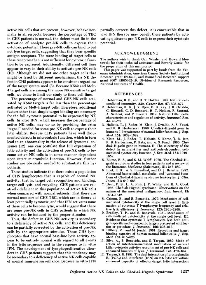
Embed Size (px)
Citation preview
Deficiency of Active Natural Killer Cellsin the Chediak-Higashi Syndrome
LOCALIZATION OF THE DEFECTUSING A SINGLE CELL
CYTOTOXICITY ASSAY
PAUL KATZ, ANNETTEM. ZAYTOUN, and ANTHONYS. FAUCI, Division of ClinicalImmunology, Department of Medicine, University of Florida College ofMedicine and the Research Service, Veterans Administration Medical Center,Gainesville, Florida 32610; Laboratory of Immunoregulation, NationalInstitute of Allergy and Infectious Diseases, National Institutes of Health,Bethesda, Maryland 20205
A B S T R A C T This study investigated the defectivenatural killer (NK) cell activity in two patients withthe Chediak-Higashi syndrome (CHS) using both astandard 51-chromium release microcytotoxicity anda single cell-in-agarose assay against K562 and Molt-4 target cells. CHSpatients were deficient in overallmaximum NK capacity, but had normal percentagesof potentially cytotoxic target binding cells. The rel-ative number of TBC that could kill bound targets (i.e.,"4active" NK cells) was significantly depressed in CHSpatients when compared with normal controls. Thediminished CHS active NK cells that were present,however, were capable of recycling and lysing mul-tiple target cells during the assay period. In vitro in-terferon (INF) treatment of normal and CHSeffectorcells did not alter target cell binding, but did increasethe maximum NK capacity, percentage of active NKcells, and the maximum recycling capacity, as well asthe rate of lysis.
These studies indicate that the depression of NKactivity in patients with CHS is secondary to a defi-ciency of active NK cells. The CHS active NK cellsthat are present, however, are capable of normal targetlysis and recycling. Potentially cytotoxic pre-NK cells,which can bind but not kill target cells, can be acti-vated by in vitro IFN to develop lytic activity. Thus,IFN treatment may be of potential benefit to the im-mune surveillance network of CHS patients by acti-
Dr. Katz is a Clinical Investigator of the Veterans Ad-ministration.
Received for publication 7 December 1981 and in revisedform 15 February 1982.
vating a population of pre-NK cells to express theircytotoxic potential.
INTRODUCTION
The natural killer (NK)l cell system is believed to bean important effector limb of the immune surveillancenetwork in animals and in man (1, 2). The in vivorelevance of this in vitro phenomenon in man has beensuggested by several lines of evidence, included inwhich is the selective impairment in NK function inthe Chediak-Higashi syndrome (CHS) (3, 4), a rareautosomal recessive disorder characterized by partialoculocutaneous albinism, severe recurrent pyogenicinfections and abnormal lysosomal granules (5, 6). Pa-tients who survive the infectious complications of thedisease often succumb to an aggressive lymphoprolif-erative disorder later in life (7). The NKdefect in thesepatients is not secondary to a lack of cells capable ofrecognizing and binding targets but appears to lie inthe subsequent lytic process itself or in the recyclingof NK cells.
To more precisely define the nature of the NKdefectin CHSpatients, our study was undertaken in whichboth a standard 51-chromium (51Cr) release micro-cytotoxicity assay and a single cell-in-agarose assaywere used. Based on the methods of Grimm and Bon-
' Abbreviations used in this paper: CHS, Chediak-Higashisyndrome; 51Cr, 51-chromium; E/T, effector/target; FCS,fetal calf serum; IFN, interferon; Km, Michaelis constant;MRC, maximum recycling capacity; NK, natural killer;TBC, target binding cell; Vmax; maximum NK capacity; V,number of killed targets.
J. Clin. Invest. The American Society for Clinical Investigation, Inc. * 0021-9738/82/06/1231/08 $1.00Volume 69 June 1982 1231-1238
1231
avida (8) and Bradley and Bonavida (9) for the mea-surement of cytolytic T cell activity, Ullberg and Jon-dal (10) have recently demonstrated that the percenttarget cell binding, the percentage of target bindingcells that go on the lysing the target (i.e., "active" NKcells), and the particular NK capacity (Vmax) of agiven heterogeneous population of effector cells canbe quantitated by simultaneously using both of theseassays. With these determinations, it is possible to thenestimate the percentage of "active" NK cells and thenumber of target cells killed by a given NK cell, thatis, the recycling capacity of a single cytotoxic cell (10).These methods have been extremely useful to date,particularly with regard to the enhancement of NKcell activity. For example, Silva et al. (11) havedemonstrated that in vitro interferon (IFN) augmentsnormal NK function by recruiting previously nonlytic"pre-NK" cells to display their cytotoxic potential aswell as by enhancing the kinetics of lysis at the singleeffector-target cell level. Similarly, it has been shownthat prostaglandin E2 (12) or moderate exercise (13)can synergistically interact with in vitro IFN to in-crease cytotoxicity by enhancing the recycling of ac-tive NK cells. Using 51Cr release microcytotoxicityand single cell assays simultaneously, we here reporta deficiency of active NK cells in CHSpatients, whichcan be partially corrected in vitro by IFN.
METHODSSubjects. Two brothers with CHS(LeR, age 29, and LaR,
age 30) were studied on five separate occasions. LeR andLaR are the products of a consanguineous marriage, andtheir detailed clinical and immunologic histories have beenextensively reported in the past (5, 6). At the time of thisstudy, the patients were free of infection and were receivingno pharmacologic agents. Neither patient had any evidenceof the progression of CHSinto the lymphoproliferative dis-order that characterizes this disease (7). Control subjects con-sisted of age- and sex-matched normal adults.
Cell suspensions. Ficcll-Hypaque-isolated peripheralblood mononuclear cells were depleted of adherent cells bypassage through nylon wool columns (14) and were used aseffector cells in all experiments. Nylon wool purificationremoved those adherent cells that might nonspecifically bindto target cells and produce a false estimate of the relativefrequency of potentially cytotoxic NK cells. Nylon wool-pu-rified lymphocytes contain <2% contaminating monocytesbased on morphology and peroxidase staining and <2%surface immunoglobulin-bearing B cells. Cells were sus-pended in RPMI 1640 supplemented with 10-15% fetal calfserum (FCS).
51Cr release assay. A described 51Cr release microcy-totoxicity assay (15) against the human erythroleukemia cellline, K562, and the human T cell line, Molt-4, was used.Briefly, 106 target cells were labeled for 1 h at 370C with300 ACi of 51Cr (ICN Nutritional Biochemicals, Cleveland,OH), washed three times, and resuspended in RPMI 1640media with 10% FCS. 104 labeled target cells (100 Al) weremixed with varying numbers of effector cells (100 Al) in V-shaped microtiter wells (Flow Laboratories, Inc., Rockville,
MD) to give final effector/target (E/T) ratios of 100:1, 50:1,20:1, and 5:1. Spontaneous release of 51Cr by target cellswas determined by placing labeled target cells in microtiterwells in the absence of effector cells. Except as noted, cul-tures were incubated at 37°C in 5% CO2 in air at 100%humidity for 4 h. Plates were then centrifuged and 100 Mlof supernatant removed and counted in a gammacounter.Percent cytotoxicity (or percent 51Cr release) was deter-mined by the formula: supernatant counts per minute minusspontaneous release counts per minute/total counts per min-ute minus spontaneous release counts per minute. In all ex-periments, spontaneous 51Cr release was <10%.
Single cell-in-agarose assay. This assay was performedby modification of the method of Ullberg and Jondal (10),based on the original descriptions of Grimm and Bonavida(8) and Bradley and Bonavida (9). Briefly, 2 X 10⁵ effectorcells and unlabeled target cells were mixed in a total volumeof 0.2 ml RPMI 1640 with 15% FCS in a 3-cm3 round bottomtube. Using these conditions, no more than one lymphocyteis bound to any target cell, and therefore a true estimate ofthe frequency of effector-target cell conjugates can be ob-tained. At higher E/T ratios, several lymphocytes may bindto a single target cell, and therefore the percentage of con-jugates cannot accurately be determined. Tubes were cen-trifuged at 500 g for 2 min and incubated at 370C for 10-20 min followed by gentle resuspension a single time witha pasteur pipette. To optimize for the number of effector-target cell conjugates formed, this gentle resuspension tech-nique was used because it has been demonstrated (9), andwe have likewise observed that less avid effector-target cellconjugates can be disrupted by vigorous resuspension. Byusing this technique, we believe that we maximize the op-portunity for these conjugates to remain intact. The cellmixture was then carefully added to 0.5 ml of 0.5% agarosein RPMI 1640 with 10 mMHepes, which was precooled atroom temperature from 470 to 390C. Cells were mixed inagarose with a pasteur pipette and then poured onto 60-mmPetri dishes (Falcon Labware, Div. Becton, Dickinson & Co.,Oxnard, CA) that had been precoated with 0.5 cm3 of 0.5%agarose. After the cell mixture in agarose had solidified, 6ml of RPMI 1640 with 15% FCS was added, and the platewas incubated as above for 4 h, except as noted. After in-cubation, the media was removed, and 2 ml of 0.1% trypanblue was added for 10 min. Plates were then washed threetimes for 5 min each with cold phosphate-buffered salineand fixed with 1% formaldehyde, which removed all extra-cellular trypan blue.
The percentage of target binding cells (TBC), -5-10%in normal subjects, was determined by counting the numberof lymphocytes binding to target cells in 200-500 countedlymphocytes. The percentage of TBC with dead targets,normally 20-25% for K562 cells and 40-60% for Molt-4 cells,was determined by counting the number of dead targets in100 effector-target conjugates. Spontaneous (or "back-ground") target cell death was determined by counting thepercentage of dead targets in the absence of effector cells.Corrections allowing for spontaneous target death are madeby applying the following formula to calculate the percent-age TBC with dead targets: (percentage of TBC with deadtargets) minus (percentage of spontaneously dead targets)multiplied by (percentage of TBCwith dead targets) (8-13).
IFN-treatment. In some experiments, effector cells werepretreated with IFN before the cytotoxicity assay. 10 X 106effector cells were incubated with 1,000 U human leukocyteIFN (generous gift of Dr. John J. Hooks obtained from Dr.K. Cantell, Karolinska Hospital, Stockholm, Sweden) orRPMI 1640 (control) for 30 min at 37°C, washed, counted,
1232 P. Katz, A. M. Zaytoun, and A. S. Fauci
and resuspended at the proper concentration. This IFN hasbeen used in vivo in clinical trials and was derived frompooled human leukocytes exposed to ultraviolet light-inac-tivated viruses. This preparation is then purified by pH treat-ment and column fraction at which point it is suitable forin vivo use. The conditions selected for IFN treatment werethose in which a maximal IFN-induced augmentation of bothnormal and CHS NK activity was observed. Using higherconcentrations of IFN or longer incubation times did notsignificantly enhance normal or CHSNKactivity to a degreegreater than that observed with the conditions reported here.
Analysis of data and statistical methods. Calculationsof cytotoxic functions were performed as outlined above andas described (10) for these assay systems. Data from the 51Crrelease assay and the single cell assay were combined as doneby Ullberg and Jondal (10) to determine particular Vmax ina given effector population. Because the dose-response curvefrom 51Cr release assays resembles Michaelis-Menten en-zyme-substrate kinetics, one can determine the Vmax andMichaelis constant (Km) values (10). Ullberg and Jondal (10)have shown that Vmax can be calculated from the Line-weaver-Burk equation:
1 Km 1 1V Vmax T Vmax'
where T is the initial number of target cells and V the num-ber of killed targets. As reported, experimental data obtainedin this manner can be approximated by determining a 5:1E/T ratio in the 51Cr release assay (5 X 10⁴ effector cellsand 104 target cells) and then using the formula:Vmax = 1.4 X 10³ + 4.2 X 10² X (percentage of cytotoxicityat 5:1). In data not shown here, for normal subjects and CHSpatients, the coefficient of correlation was >0.91 in all caseswhen this formula was compared with the more tediousLineweaver-Burk equation. Therefore, in data reported herethe more simplified formula was used.
The percentage of "active" NK cells, that is, lymphocyteswith bound and dead targets, was determined from the sin-
K562
50F
> 40
Xx00> 30
zu
120
10
gle-cell assay by multiplying the percentage of TBC by thepercentage of TBC with dead targets. This is -1-2% innormal subjects against K562 target cells and 3-5% againstMolt-4 target cells. The maximum recycling capacity (MRC),which estimates the number of targets killed by an activeNK cell in the 4-h assay, was determined by dividing Vmaxby the absolute number of active NK cells in the Vmax. TheMRCfor normal subjects is -5-6 with K562 target cells and1-3 with Molt-4 target cells.
Data were compared by the two-tailed Student's test.
RESULTS
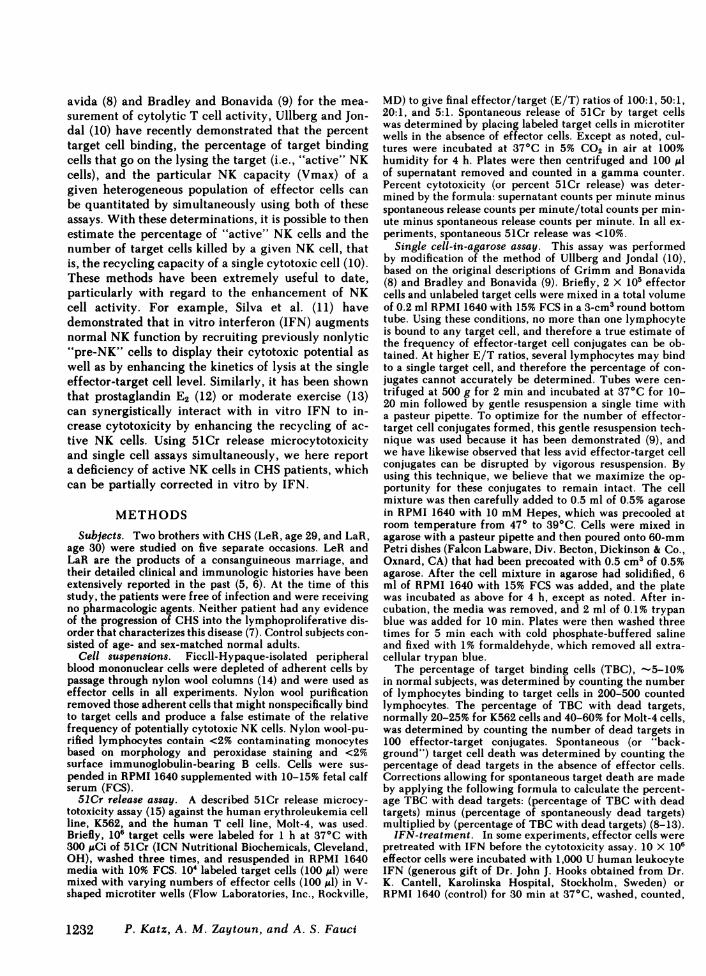
As described for these two patients (3, 4), NK activityagainst K562 and Molt-4 in a standard 51Cr releaseassay was markedly deficient at all E/T ratios whencompared with normal controls (Fig. 1). Even with theassay time extended to 18 h, the cytotoxic function ofCHS effector cells remained depressed (data notshown).
By using the 51Cr release assay and the single cell-in-agarose assay simultaneously, we were able to de-termine and compare the Vmax, percent TBC, percentTBC with dead targets, percent active NK cells, andestimated MRCfor normals and the CHSpatients. Ascalculated from 51Cr release assay data, the CHSpa-tients had significantly depres.sed values for Vmaxagainst K562 and Molt-4 targets when compared withnormal subjects (P < 0.005 for both patients for K562,P < 0.05 for both patients for Molt-4) (Tables I andII). The percentage of CHSeffector cells binding K562and Molt-4 (i.e., TBC) in the single cell assay was notstatistically different from normal individuals as hasbeen described (3). However, CHS patients LaR and
MOLT-460r
5OF
40-
30
201
10
LaR
ORLeR5:1 20:1 50:1 100:1
EFFECTOR:
RLaRLeR
5:1TARGETRATIO
20:1 50:1 100:1
FIGURE 1 NK activity of normal and CHS (LaR and LeR) effector cells against K562 andMolt-4 target cells in a standard 4-h 51Cr release assay. Data represent the mean±SEMof threeseparate experiments for K562 and four separate experiments for Molt-4.
Deficient Active NK Cells in the Chediak-Higashi Syndrome
60 r
1233
LeR were markedly deficient in the ability of theirTBC to lyse either K562 or Molt-4 (Tables I and II).While normal NK cells lysed -20% of bound K562,effector cells from both CHS patients killed only 5%of those conjugated targets (P <0.001 for both pa-tients) (Table I). Similarly, while nearly 50% of boundMolt-4 cells were lysed by normal effectors, <20% ofthese attached targets were killed by CHS NK cells(P < 0.001 for both patients) (Table II). This deficiencyin lysis was present even when the assay time wasextended to 18 h. (data not shown). Because the per-centage of active NK cells is determined by both thepercent TBC and percent TBC with dead targets, pa-tients LaR and LeR had significantly reduced per-centages of active NK cells against K562 (P < 0.005for both patients) and against Molt-4 (P < 0.001 forboth patients).
By using these data, it is possible to calculate thenumber of targets killed by a given NK cell in a 4-hassay; that is, the MRCestimates the number of timesin 4 h that a single NK cell recognizes, binds to, lysesa target, and then repeats this process. This estimateis determined from the Vmaxand percentage of activeNKcells as described in Methods (10). Although mark-edly deficient in both Vmax activity and the percent-
age of active cytotoxic cells, NK cells from LaR andLeR had normal MRCvalues against both cell lines(Tables I and II).
It has been demonstrated (3) that NK activity ofthese CHSpatients can be significantly augmented byin vitro IFN, but the level of cytotoxicity still remainssignificantly below that of non-IFN-treated normaleffector cells. Because of this, we investigated the ef-fects of in vitro IFN on the NK activity of normal andCHS lymphocytes in the currently used cytotoxicityassay systems. Weselected Molt-4 as the target becauserelatively more bound Molt-4 cells are lysed comparedwith bound K562 targets. As shown in Table III, invitro IFN increased the Vmax of normal and CHSeffector cells in the 4-h assay. The extension of theassay time to 6, 12, or 18 h did not significantly alterthe values for normal or CHSNK cells activity fromthe levels observed at 4 h. Therefore, data are reportedonly for assay times up to 4 h. The percentage of nor-mal and CHSTBC was not affected by IFN, but thepercentage of TBC with dead targets was increasedby IFN indicating that IFN induced previously non-lytic lymphocytes ("pre"-NK cells) to develop cyto-toxic activity.
To determine the effects of in vitro IFN on the ki-
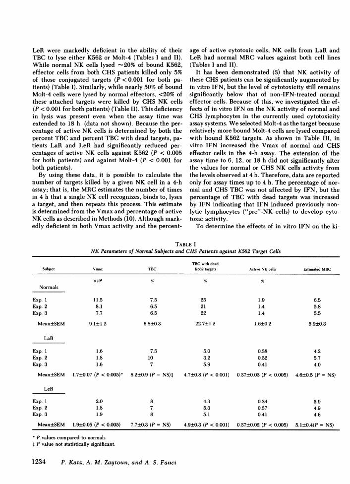
TABLE INK Parameters of Normal Subjects and CHSPatients against K562 Target Cells
TBC with deadSubject Vmax TBC K562 targets Active NK cells Estimated MRC
Xl0 % % %
Normals
Exp. 1 11.5 7.5 25 1.9 6.5Exp. 2 8.1 6.5 21 1.4 5.8Exp. 3 7.7 6.5 22 1.4 5.5
Mean±SEM 9.1±1.2 6.8±0.3 22.7±1.2 1.6±0.2 5.9±0.3
LaR
Exp. 1 1.6 7.5 5.0 0.38 4.2Exp. 2 1.8 10 3.2 0.32 5.7Exp. 3 1.6 7 5.9 0.41 4.0
Mean±SEM 1.7±0.07 (P < 0.005)' 8.2±0.9 (P = NS)I 4.7±0.8 (P < 0.001) 0.37±0.03 (P < 0.005) 4.6±0.5 (P = NS)
LeR
Exp. 1 2.0 8 4.3 0.34 5.9Exp. 2 1.8 7 5.3 0.37 4.9Exp. 3 1.9 8 5.1 0.41 4.6
Mean±SEM 1.9±0.05 (P < 0.005) 7.7±0.3 (P = NS) 4.9±0.3 (P < 0.001) 0.37±0.02 (P < 0.005) 5.1±0.4(P = NS)
P values compared to normals.P value not statistically significant.
1234 P. Katz, A. M. Zaytoun, and A. S. Fauci
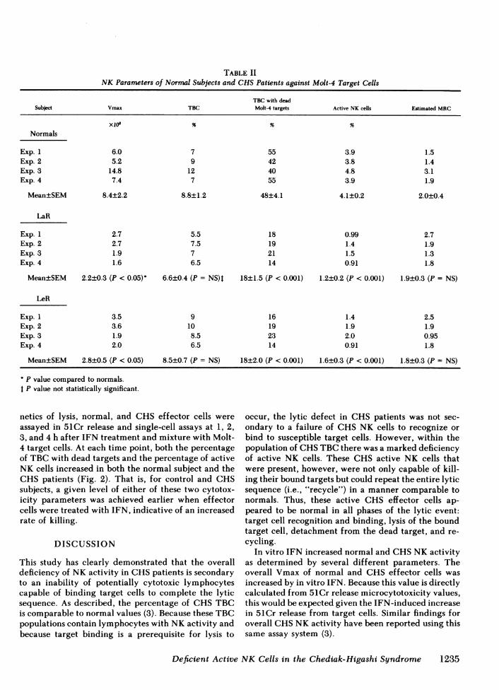
TABLE IINK Parameters of Normal Subjects and CHSPatients against Molt-4 Target Cells
TBC with deadSubject Vmax TBC Molt-4 targets Active NK cells Estimated MRC
Xl7 % % %
Normals
Exp. 1 6.0 7 55 3.9 1.5Exp. 2 5.2 9 42 3.8 1.4Exp. 3 14.8 12 40 4.8 3.1Exp. 4 7.4 7 55 3.9 1.9
Mean±SEM 8.4±2.2 8.8±1.2 48±4.1 4.1±0.2 2.0±0.4
LaR
Exp. 1 2.7 5.5 18 0.99 2.7Exp. 2 2.7 7.5 19 1.4 1.9Exp. 3 1.9 7 21 1.5 1.3Exp. 4 1.6 6.5 14 0.91 1.8
Mean±SEM 2.2±0.3 (P < 0.05) 6.6±0.4 (P = NS)I 18±1.5 (P < 0.001) 1.2±0.2 (P < 0.001) 1.9±0.3 (P = NS)
LeR
Exp. 1 3.5 9 16 1.4 2.5Exp. 2 3.6 10 19 1.9 1.9Exp. 3 1.9 8.5 23 2.0 0.95Exp. 4 2.0 6.5 14 0.91 1.8
Mean±SEM 2.8±0.5 (P < 0.05) 8.5±0.7 (P = NS) 18±2.0 (P < 0.001) 1.6±0.3 (P < 0.001) 1.8±0.3 (P NS)
Pp value compared to normals.t P value not statistically significant.
netics of lysis, normal, and CHS effector cells wereassayed in 51Cr release and single-cell assays at 1, 2,3, and 4 h after IFN treatment and mixture with Molt-4 target cells. At each time point, both the percentageof TBCwith dead targets and the percentage of activeNK cells increased in both the normal subject and theCHS patients (Fig. 2). That is, for control and CHSsubjects, a given level of either of these two cytotox-icity parameters was achieved earlier when effectorcells were treated with IFN, indicative of an increasedrate of killing.
DISCUSSION
This study has clearly demonstrated that the overalldeficiency of NK activity in CHSpatients is secondaryto an inability of potentially cytotoxic lymphocytescapable of binding target cells to complete the lyticsequence. As described, the percentage of CHSTBCis comparable to normal values (3). Because these TBCpopulations contain lymphocytes with NKactivity andbecause target binding is a prerequisite for lysis to
occur, the lytic defect in CHS patients was not sec-ondary to a failure of CHSNK cells to recognize orbind to susceptible target cells. However, within thepopulation of CHSTBC there was a marked deficiencyof active NK cells. These CHS active NK cells thatwere present, however, were not only capable of kill-ing their bound targets but could repeat the entire lyticsequence (i.e., "recycle") in a manner comparable tonormals. Thus, these active CHS effector cells ap-peared to be normal in all phases of the lytic event:target cell recognition and binding, lysis of the boundtarget cell, detachment from the dead target, and re-cycling.
In vitro IFN increased normal and CHSNKactivityas determined by several different parameters. Theoverall Vmax of normal and CHS effector cells wasincreased by in vitro IFN. Because this value is directlycalculated from 51Cr release microcytotoxicity values,this would be expected given the IFN-induced increasein 51Cr release from target cells. Similar findings foroverall CHSNKactivity have been reported using thissame assay system (3).
Deficient Active NK Cells in the Chediak-Higashi Syndrome 1235
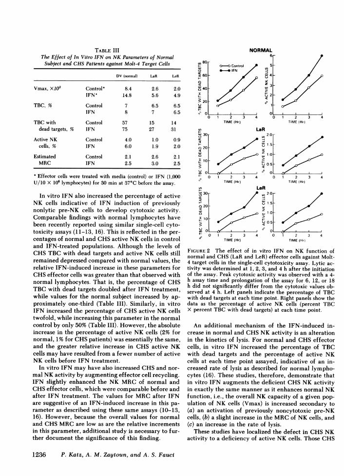
TABLE IIIThe Effect of In Vitro IFN on NK Parameters of Normal
Subject and CHSPatients against Molt-4 Target Cells
DV (normal) LaR LeR
Vmax, X103 Control' 8.4 2.6 2.0IFN° 14.8 5.6 4.9
TBC, % Control 7 6.5 6.5IFN 8 7 6.5
TBC with Control 57 15 14dead targets, % IFN 75 27 31
Active NK Control 4.0 1.0 0.9cells, % IFN 6.0 1.9 2.0
Estimated Control 2.1 2.6 2.1MRC IFN 2.5 3.0 2.5
Effector cells were treated with media (control) or IFN (1,000U/10 X 10' lymphocytes) for 30 min at 37°C before the assay.
In vitro IFN also increased the percentage of activeNK cells indicative of IFN induction of previouslynonlytic pre-NK cells to develop cytotoxic activity.Comparable findings with normal lymphocytes havebeen recently reported using similar single-cell cyto-toxicity assays (11-13, 16). This is reflected in the per-centages of normal and CHSactive NK cells in controland IFN-treated populations. Although the levels ofCHSTBC with dead targets and active NK cells stillremained depressed compared with normal values, therelative IFN-induced increase in these parameters forCHSeffector cells was greater than that observed withnormal lymphocytes. That is, the percentage of CHSTBC with dead targets doubled after IFN treatment,while values for the normal subject increased by ap-proximately one-third (Table III). Similarly, in vitroIFN increased the percentage of CHSactive NK cellstwofold, while increasing this parameter in the normalcontrol by only 50% (Table III). However, the absoluteincrease in the percentage of active NK cells (2% fornormal, 1% for CHSpatients) was essentially the same,and the greater relative increase in CHS active NKcells may have resulted from a fewer number of activeNK cells before IFN treatment.
In vitro IFN may have also increased CHSand nor-mal NKactivity by augmenting effector cell recycling.IFN slightly enhanced the NK MRCof normal andCHSeffector cells, which were comparable before andafter IFN treatment. The values for MRCafter IFNare suggestive of an IFN-induced increase in this pa-rameter as described using these same assays (10-13,16). However, because the overall values for normaland CHSMRCare low as are the relative incrementsin this parameter, additional study is necessary to fur-ther document the significance of this finding.
NORMAL6r
U) 80H
4 60a4a 40
I
20:'
H
aC
0-O Control*0* IFN
5u)
w4
z 3
U~2
601
II0 1 2 3 4
TIME (Hr)
U) u30
a 20/
0
2_ 10
O
H 0 1 2 3 4° TIME (Hr)mn Lt< 30
0 20
B iX 10
UO0 1 2 3 4&-0 TIME (Hr)
CA i0 1 2 3 4
TIME (Hr)
aR2.20
-U)
uu 1.5-
Z 1.0 _w1o
<00'
0.5
0 1 2 3 4TIME (Hr)
.eRU)2.0-
uo 1.5
O ~
z
L) 0.5
0 1 2 3 4lIME (Hr)
FIGURE 2 The effect of in vitro IFN on NK function ofnormal and CHS(LaR and LeR) effector cells against Molt-4 target cells in the single-cell cytotoxicity assay. Lytic ac-tivity was determined at 1, 2, 3, and 4 h after the initiationof the assay. Peak cytotoxic activity was observed with a 4-h assay time and prolongation of the assay for 6, 12, or 18h did not significantly differ from the cytotoxic values ob-served at 4 h. Left panels indicate the percentage of TBCwith dead targets at each time point. Right panels show thedata as the percentage of active NK cells (percent TBCX percent TBC with dead targets) at each time point.
An additional mechanism of the IFN-induced in-crease in normal and CHSNK activity is an alterationin the kinetics of lysis. For normal and CHSeffectorcells, in vitro IFN increased the percentage of TBCwith dead targets and the percentage of active NKcells at each time point assayed, indicative of an in-creased rate of lysis as described for normal lympho-cytes (16). These studies, therefore, demonstrate thatin vitro IFN augments the deficient CHSNK activityin exactly the same manner as it enhances normal NKfunction, i.e., the overall NK capacity of a given pop-ulation of NK cells (Vmax) is increased secondary to(a) an activation of previously noncytotoxic pre-NKcells, (b) a slight increase in the MRCof NK cells, and(c) an increase in the rate of lysis.
These studies have localized the defect in CHSNKactivity to a deficiency of active NK cells. Those CHS
1236 P. Katz, A. M. Zaytoun, and A. S. Fauci
active NK cells that are present, however, behave nor-mally in all respects. Because the percentage of TBCin CHSpatients is normal, the defect must lie in theactivation of nonlytic pre-NK cells to express theircytotoxic potential. These pre-NK cells can bind to butnot lyse target cells, suggesting that they bear specificantigen receptors. The mere binding of target cells tothese receptors then is not sufficient for cytotoxic func-tion to be expressed. Additionally, different cell linesappear to vary in their ability to activate pre-NK cells(10). Although we did not use other target cells thatmight be lysed by different mechanisms, the NK de-fect in CHSpatients appears to be consistent regardlessof the target system used (3). Because K562 and Molt-4 target cells are among the more NK-sensitive targetcells, we chose to limit our study to these cell lines.
The percentage of normal and CHSNK cells acti-vated by K562 targets is far less than the percentageactivated by Molt-4 target cells. Therefore, additionalfactors other than simple target binding are necessaryfor the full cytotoxic potential to be expressed by NKcells. In vitro IFN, which increases the percentage ofactive NK cells, may do so by providing the extra"signal" needed for some pre-NK cells to express theirlytic ability. Because CHS patients have well docu-mented defects in microtubule function, which in turnlead to an abnormality in the release of lysosomal en-zymes (12), one can postulate that full expression oflytic activity and the activation of pre-NK cells to be-come active NK cells is in some manner dependentupon intact microtubule function. However, furtherstudies are obviously needed to substantiate this hy-pothesis.
These studies indicate that there exists a populationof CHS lymphocytes that is capable of normal NKactivity, that is, target cell recognition and binding,target cell lysis, and recycling. CHSpatients are rel-atively deficient in this population of active NK cellswhen compared with normal subjects. That there arenormal numbers of CHSTBC, which are in theory atleast potentially cytotoxic, and that IFN activates someof these cells to become lytic, would suggest that thereare some pre-NK cells in CHSpatients in which NKactivity can be induced by the proper stimulus.
Thus, the defect in CHS NK activity is secondaryto a deficiency of active NK cells, and this deficiencycan be partially corrected by the activation of pre-NKcells by the appropriate stimulus. Those CHS lym-phocytes that have normal base-line lytic activity ap-pear to be entirely normal with regard to all eventsin the lytic sequence and in the response to in vitroIFN. The development of lymphoproliferative disor-ders in CHSpatients suggests that this tendency maybe secondary to a deficiency of active NKcells capableof normal immune surveillance. Because in vitro IFN
partially corrects this defect, it is conceivable that invivo IFN therapy may benefit these patients by acti-vating quiescent pre-NK cells to express their cytotoxicpotential.
ACKNOWLEDGMENT
The authors wish to thank Gail Whalen and Howard Mos-towski for their technical assistance and Beverly Gorski forthe preparation of this manuscript.
This paper was supported in part by funds from the Vet-erans Administration, American Cancer Society InstitutionalResearch grant IN-62-T, and Biomedical Research supportgrant S007 RR05362-19, Division of Research Resources,National Institutes of Health.
REFERENCES
1. Heberman, R. B., and H. T. Holden. 1978. Natural cell-mediated immunity. Adv. Cancer Res. 27: 305-377.
2. Heberman, R. B., J. Y. Djeu, D. H. Kay, J. R. Ortaldo,C. Riccardi, G. D. Bonnard, H. T. Holden, R. Santoni,A. Santoni, and P. Pucetti. 1979. Natural killer cells:characteristics and regulation of activity. Immunol. Rev.44: 43-70.
3. Haliotis, T., J. Roder, M. Klein, J. Ortaldo, A. S. Fauci,and R. B. Herberman. 1980. Chediak-Higashi gene inhumans. I. Impairment of natural-killer function. J. Exp.Med. 151: 1039-1048.
4. Klein, M., J. Roder, T. Haliotis, S. Korec, J. R. Jett,R. B. Herberman, P. Katz, and A. S. Fauci. 1980. Che-diak-Higashi gene in humans. II. The selectivity of thedefect in natural-killer and antibody-dependent cell-mediated cytotoxicity function. J. Exp. Med. 151: 1049-1058.
5. Blume, R. S., and S. M. Wolff. 1972. The Chediak-Hi-gashi syndrome: studies in four patients and a review ofthe literature. Medicine (Baltimore). 51: 247-280.
6. Root, R. K., A. S. Rosenthal, and D. J. Balestra. 1972.Abnormal bactericidal, metabolic, and lysosomal func-tions of Chediak-Higashi syndrome leukocytes. J. Clin.Invest. 51: 649-665.
7. Dent, P. B., L. A. Fish, J. F. White, and R. A. Good.1966. Chediak-Higashi syndrome. Observations on thenature of the associated malignancy. Lab. Invest. 15:1634-1642.
8. Grimm, E., and B. Bonavida. 1979. Mechanism of cell-mediated cytotoxicity at the single cell level. I. Esti-mation of cytotoxic T lymphocyte frequency and rela-tive lytic efficiency. J. Immunol. 123: 2861-2868.
9. Bradley, T. P., and B. Bonavida. 1981. Mechanism ofcell-mediated cytotoxicity at the single cell level. III.Evidence that cytotoxic T lymphocytes lyse both anti-gen-specific and -nonspecific targets pretreated with lec-tins or periodate. J. Immunol. 126: 208-213.
10. Ullberg, M., and M. Jondal. 1981. Recycling and targetbinding capacity of human natural killer cells. J. Exp.Med. 153: 615-628.
11. Silva, A., B. Bonavida, and S. Targan. 1980. Mode ofaction of interferon-mediated modulation of naturalkiller cytotoxic activity: recruitment of pre-NK cells andenhanced kinetics of lysis. J. Immunol. 125: 479-484.
12. Targan, S. R. 1981. The dual interaction of prostaglandinE2 (PGE2) and interferon (IFN) on NK lytic activation:enhanced capacity of effector-target lytic interactions
Deficient Active NK Cells in the Chediak-Higashi Syndrome 1237
(recycling) and blockage of pre-NK cell recruitment. J.Immunol. 127: 1424-1428.
13. Targan, S., L. Britvan, and F. Dorey. 1981. Activationof human NKCCby moderate exercise: increased fre-quency of NKcells with enhanced capability of effector-target lytic interactions. Clin. Exp. Immunol. 45: 352-360.
14. Julius, M. H., E. Simpson, and L. A. Herzenberg. 1973.A rapid method for the isolation of functional thymus-derived murine lymphocytes. Eur. J. Immunol. 3: 645-649.
15. Katz, P., C. B. Simone, P. A. Henkart, and A. S. Fauci.1980. Mechanisms of antibody-dependent cellular cy-totoxicity. The use of effector cells from chronic gran-ulomatous disease patients as investigative probes. J.Clin. Invest. 65: 55-63.
16. Targan, S., and F. Dorey. 1980. Interferon activation of"pre-spontaneous killer" (pre-SK) cells and alteration inkinetics of lysis of both "pre-SK" and active SK cells. J.Immunol. 124: 2157-2161.
17. Oliver, J. M. 1976. Impaired microtubule function cor-rectable by cyclic GMPand cholinergic agonists in theChediak-Higashi syndrome. Am. J. Pathol. 85: 395-412.
1238 P. Katz, A. M. Zaytoun, and A. S. Fauci


![Bossa Nova 2 [Almir Chediak]](https://img.pdfslide.net/doc/110x75/55cf91aa550346f57b8f6f95/bossa-nova-2-almir-chediak.jpg)

![Bossa Nova 4 [Almir Chediak]](https://img.pdfslide.net/doc/110x75/56d6be1f1a28ab301690bb78/bossa-nova-4-almir-chediak.jpg)