Embed Size (px)
Citation preview
ORIGINAL RESEARCHpublished: 13 April 2016
doi: 10.3389/fcimb.2016.00040
Frontiers in Cellular and Infection Microbiology | www.frontiersin.org 1 April 2016 | Volume 6 | Article 40
Edited by:
W. Edward Swords,
Wake Forest University Health
Sciences, USA
Reviewed by:
Janakiram Seshu,
The University of Texas at San
Antonio, USA
Stephen Peter Kidd,
University of Adelaide, Australia
*Correspondence:
Sanjay Ram
Brian J. Akerley
†These authors have contributed
equally to this work.
Received: 28 December 2015
Accepted: 21 March 2016
Published: 13 April 2016
Citation:
Wong SM, Shaughnessy J, Ram S
and Akerley BJ (2016) Defining the
Binding Region in Factor H to Develop
a Therapeutic Factor H-Fc Fusion
Protein against Non-Typeable
Haemophilus influenzae.
Front. Cell. Infect. Microbiol. 6:40.
doi: 10.3389/fcimb.2016.00040
Defining the Binding Region in FactorH to Develop a Therapeutic FactorH-Fc Fusion Protein againstNon-Typeable Haemophilusinfluenzae
Sandy M. Wong 1†, Jutamas Shaughnessy 2†, Sanjay Ram 2* and Brian J. Akerley 1*
1Department of Microbiology and Immunology, University of Mississippi Medical Center, Jackson, MS, USA, 2Division of
Infectious Diseases and Immunology, University of Massachusetts Medical School, Worcester, MA, USA
Non-typeable Haemophilus influenzae (NTHi) cause a range of illnesses including otitis
media, sinusitis, and exacerbation of chronic obstructive pulmonary disease, infections
that contribute to the problem of antibiotic resistance and are themselves often intractable
to standard antibiotic treatment regimens. We investigated a strategy to exploit binding
of the complement inhibitor Factor H (FH) to NTHi as a functional target for an
immunotherapeutic containing the NTHi binding domain of FH fused to the Fc domain
of IgG1. Chimeric proteins containing the regions that most FH-binding bacteria use to
engage human FH, domains 6 and 7 (FH6,7/Fc) and/or 18 through 20 (FH18–20/Fc),
were evaluated for binding to NTHi. FH6,7/Fc bound strongly to each of seven NTHi
clinical isolates tested and efficiently promoted complement-mediated killing by normal
human serum. FH18–20/Fc bound weakly to three of the strains but did not promote
complement dependent killing. Outer-membrane protein P5 has been implicated in FH
binding by NTHi, and FH6,7/Fc binding was greatly diminished in five of seven P5
deficient isogenic mutant strains tested, implicating an alternative FH binding protein in
some strains. Binding of FH18–20/Fc was decreased in the P5 mutant of one strain.
A murine model was used to evaluate potential therapeutic application of FH6,7/Fc.
FH6,7/Fc efficiently promoted binding of C3 to NTHi exposed to mouse serum, and
intranasal delivery of FH6,7/Fc resulted in significantly enhanced clearance of NTHi
from the lung. Moreover, a P5 deficient mutant was attenuated for survival in the lung
model, suggesting that escape mutants lacking P5 would be less likely to replace
strains susceptible to FH6,7/Fc. These results provide evidence for the potential utility
of FH6,7/Fc as a therapeutic against NTHi lung infection. FH binding is a common
property of many respiratory tract pathogens and FH/Fc chimeras may represent
promising alternative or adjunctive therapeutics against such infections, which are often
polymicrobial.
Keywords: factor H, non-typeable Haemophilus influenzae, complement, P5, outer-membrane protein, lung
infection
Wong et al. FH-Fc Immunotherapeutic against Non-Typeable Haemophilus influenza
INTRODUCTION
Non-typeable Haemophilus influenzae (NTHi), a common causeof respiratory tract infections, is associated with otitis media andsinusitis in children, and exacerbations of chronic obstructivelung disease (COPD; Murphy et al., 2009; Sethi et al., 2016).NTHi is consistently found in the lower respiratory tract in30% of COPD cases and recurrent infection by diverse NTHistrains results in exacerbation of this disease (Murphy andSethi, 2002), which afflicts greater than 6% of adults and hasbeen ranked the third leading cause of death in the U.S.(Centers for Disease Control and Prevention [CDC], 2012).Nasopharyngeal colonization with NTHi in infants predisposesto recurrent otitis media (Harabuchi et al., 1994) in whichNTHi has recently emerged as the most frequent bacterial isolate(Kaur et al., 2013). NTHi can also cause invasive infectionsincluding bacteremia, pneumonia, and meningitis, especially inneonates and individuals that are immunocompromised or havecomorbidities (Van Eldere et al., 2014; Collins et al., 2016). Otitismedia is the leading cause of pediatric antibiotic prescription,with β-lactams representing the frontline therapeutics (McCaiget al., 2002; Grijalva et al., 2009). The spread of β-lactamaseproducing NTHi as well as β-lactamase-negative ampicillinresistant strains globally has led to use of broader spectrumagents with their attendant complications (Van Eldere et al.,2014). Whereas, vaccination has been effective against type bH. influenzae with implementation of the capsular conjugatevaccine (Ladhani, 2012), it is complicated in NTHi, which lackcapsule and exhibit extensive antigenic diversity of immunogenicouter-membrane proteins among strains (Gilsdorf, 1998). Thehighly conserved NTHi protein D has been included in thepneumococcal PhiD-CV (Synflorix; GSK) vaccine, which hasshown moderate efficacy against otitis media in clinical studies.However, PhiD-CV has not been evaluated for other conditionssuch as exacerbation of COPD. Moreover, a recent study in amurine lung model was unable to demonstrate protection againstNTHi after immunization with PhiD-CV (Siggins et al., 2015).New non-antibiotic anti-infectives active against NTHi would bebeneficial as primary or adjunctive therapies.
To survive in their mammalian hosts, pathogens possessmultiple countermeasures against innate immune defenses, inwhich the complement system plays a major role (Ram et al.,2010). One strategy shared by NTHi and many medicallyimportant microbes is to bind to human complement inhibitors,including Factor H (FH), vitronectin, and C4b-binding protein,to dampen complement activation on their surfaces (Würzner,1999; Kraiczy and Würzner, 2006; Blom et al., 2009). FH inhibitsthe alternative pathway of complement by serving as a cofactorfor the factor I-mediated cleavage of C3b to the hemolyticallyinactive iC3b fragment (Pangburn et al., 1977). FH also causes“decay acceleration,” whereby it irreversibly dissociates the Bbfragment from the alternative pathway C3 convertase, C3bBb(Weiler et al., 1976; Whaley and Ruddy, 1976; Fearon andAusten, 1977). FH comprises 20 domains, also known as shortconsensus repeat domains (SCRs) or complement control proteindomains (CCPs) that are arranged in the form of a single chain(Ripoche et al., 1988). The first four N-terminal domains are
necessary and sufficient for complement inhibition (Sharma andPangburn, 1996). Pathogens bind FH regions distinct from itscomplement inhibitory domains, information that has been usedto engineer FH fusion proteins lacking complement-inhibitoryactivity to the Fc region of IgG and thereby exploit this virulenceproperty as a therapeutic target to direct Fc mediated clearanceof Neisseria meningitidis and Neisseria gonorrhoeae in animalmodels of infection (Shaughnessy et al., 2014, 2016). Thisapproach would be attractive for treatment of infections withNTHi, which frequently cause disease in the context of co-infection by other pathogens (Broides et al., 2009), primarilyinvolving Streptococcus pneumoniae, Moraxella catarrhalis, andStreptococcus pyogenes, each of which expresses FH bindingproteins (Horstmann et al., 1988; Dave et al., 2001; Bernhardet al., 2014).
Previous studies have shown that NTHi bind to FH (Hallströmet al., 2008), and that binding is mediated by the cell-surfaceouter-membrane protein P5, a member of the OmpA family ofproteins, which contributes to resistance of NTHi to killing bycomplement (Langereis et al., 2014; Rosadini et al., 2014). P5has also been described as an adhesin interacting with epithelialcells and mucosal surfaces via several potential binding partnersincluding respiratory mucin (Reddy et al., 1996), Eustachian tubemucus (Miyamoto and Bakaletz, 1996), ICAM-1 (Avadhanulaet al., 2006), and CEACAM-1 (Hill et al., 2001), however itsrole in interaction with CEACAM-1 was recently shown to beindirect (Tchoupa et al., 2015). In addition to virulence relatedphenotypes identified in vitro, P5 has been implicated in bacterialcolonization in the chinchilla (Sirakova et al., 1994) and lunginfection in mice (Wong et al., 2013; Euba et al., 2015). In thisstudy we identify the regions in FH that bind to NTHi, determinethe role of P5 in these interactions, and evaluate the ability of afusion protein that combines the NTHi-binding fragment of FHwith the Fc domain of IgG to mediate complement-dependentkilling of NTHi and facilitate clearance of bacteria in vivo in amouse lung model of NTHi infection.
MATERIALS AND METHODS
Media and Haemophilus influenzae GrowthConditionsNTHi clinical isolates NT127 (Wong et al., 2011), Hi375 (Mellet al., 2014), Hi486 (Hood et al., 1999), 86-028NP (Harrisonet al., 2005), PittGG (Buchinsky et al., 2007), R2846 (Barenkampand Leininger, 1992), and R2866 (Nizet et al., 1996; Erwinet al., 2005) were grown at 35 ± 1.5◦C in Brain Heart Infusionsupplemented with 10 µg/ml nicotinamide adenine dinucleotide(NAD) and 10 µg/ml hemin (sBHI) on agar plates or insBHI broth. DNA was transformed into naturally competentH. influenzae prepared as described (Barcak et al., 1991).Gentamicin (Gm) and 3,4-cyclohexenoesculetin-β-D-galacto-pyranoside (S-gal, Sigma-Aldrich), and D-xylose were added tosBHI at 10 µg/ml, 300 mg/L, and 1mM, respectively. FH/Fcbinding, C3 deposition and serum bactericidal assays wereperformed on NTHi grown on chocolate agar plates as describedbelow.
Frontiers in Cellular and Infection Microbiology | www.frontiersin.org 2 April 2016 | Volume 6 | Article 40
Wong et al. FH-Fc Immunotherapeutic against Non-Typeable Haemophilus influenza
Bacterial Strains and Mutants1P5 deletion mutations in Hi375, Hi486, PittGG, 86-028NP,R2846, and R2866 were created by transformation of a ∼3kb PCR product amplified with primers 5 omp1 and 3 omp2from H. influenzae Rd strain RP5G (Rosadini et al., 2014). TheNT127 P5 deletion mutant complemented with the wildtypegene from NT127 at the xyl locus, NTP5X, or carrying emptyvector sequences at the xyl locus, NTP5V, have been describedpreviously (Rosadini et al., 2014). The allelic exchange PCRproduct for deletions contains a replacement of the P5 codingregion with the aacC1 gentamicin (Gm) resistance cassetteand flanking regions for homologous recombination. GmR
transformants were selected on sBHI agar containing Gm.All strain constructions were verified by PCR amplificationacross the inserted recombinant region with primers specific forflanking sequences not contained in sequences within the P5knockout exchange delivery DNA and by PCR amplification toverify presence of the gentamicin resistance cassette with aacC1 5′
(ATGTTACGCAGCAGCAACGATGTTACGCAGCAGG) and 3′
(TTAGGTGGCGGTACTTGGGTCGAT) primers to the codingregion. Strains were additionally verified via Coomassie BrilliantBlue staining of whole-cell lysates after SDS-PAGE, and allmutants were deficient in an ∼37–39 kDa band present in thewild-type parents consistent with the predicted sizes of thecorresponding P5 proteins (Figure S1).
ComplementHuman serum was obtained from normal healthy adultvolunteers who provided informed consent. Participation wasapproved by the University ofMassachusetts Institutional ReviewBoard for the protection of human subjects. Serum was obtainedby allowing blood to clot at 25◦C for 30min followed bycentrifugation at 1500 g for 20min at 4◦C. To study the effectsof the FH/Fc proteins without confounding by natural anti-NTHiantibodies present in NHS, we depleted IgG and IgM from freshlycollected human serum, as described previously (Ray et al.,2011). Briefly, EDTA (final concentration 10mM) andNaCl (finalconcentration 1M) were added to freshly prepared human serumand treated sera was passed first over anti-human IgM agarose(Sigma), followed by passage through protein G-Sepharose; bothcolumns were equilibrated in PBS containing 10mM EDTA and1M NaCl. NaCl was added to minimize C1q depletion duringpassage of serum through the anti-human IgM column. The flow-through was collected, spin concentrated and dialyzed againstPBS/0.1mM EDTA to its original volume using a 10-kDa cutoffAmicon Ultra-15 centrifugal filter device (Millipore, Bedford,MA), sterilized by passage through a 0.22-µm filter (Millipore),aliquoted and stored at−70◦C. Hemolytic activity was confirmedusing a total complement hemolytic plate assay (The BindingSite Inc., Birmingham, U.K). Depletion of IgG and IgM wasconfirmed by dot-blot assays. In some experiments, complementactivity of serum was destroyed by heating serum at 56◦C for 1 h.Mouse complement was obtained by allowing blood obtained byterminal cardiac puncture to clot for 20min at room temperaturefollowed by incubation for 20min on ice. Serum was collectedafter centrifugation at 10,000 g for 10min at 4◦C and stored insingle-use aliquots at −80◦C. This procedure was performed in
accordance with approved IACUC protocols at the University ofMassachusetts Medical School.
FH/Fc Fusion ProteinsCloning, expression and purification of a chimeric proteincomprising human FH domains 18–20 fused to human IgG1Fc (FH18–20/Fc) and FH domains 6 and 7 fused to humanIgG1 Fc (FH6,7/Fc) have been described previously (Shaughnessyet al., 2011, 2014). Plasmids encoding the FH/Fc fusion proteinswere used to transiently transfect CHO cells using lipofectin(Life Technologies), according to themanufacturer’s instructions.Media from transfected cells was collected after 2 days andFH/Fc was purified by passage over protein A agarose. Mass wasdetermined by Coomassie Blue staining of proteins separated bySDS-PAGE and protein concentrations were determined usingthe BCA protein Assay kit (Pierce).
AntibodiesAnti-human IgG FITC (Sigma), anti-humanC3c-FITC (BioRad),and anti-mouse C3 FITC (MP Biomedicals) were used in flowcytometry assays, all at a dilution of 1:100 in Hanks BalancedSalt Solution (HBSS) containing 0.1% BSA and 1mM CaCl2 and1mMMgCl2 (HBSS++/BSA).
Flow CytometryBinding of FH/Fc fusion proteins to bacteria and humanand mouse C3 deposition on NTHi were performed by flowcytometry as described previously (Shaughnessy et al., 2011,2014; Rosadini et al., 2014). All incubations with proteins, serumand Ab were carried out in HBSS++/BSA. Data were acquired ona BD FACSCalibur flow cytometer and data were analyzed usingFlowJo software.
Serum Bactericidal AssaysNTHi harvested from chocolate agar plates following overnightgrowth were repassaged onto fresh chocolate agar plates andgrown for 5 h at 37◦C in an atmosphere containing 5%CO2. Bacteria were resuspended in HBSS++/BSA. The reactionmixture contained 20% human complement and ∼1000 CFUof NTHi and the indicated concentrations of FH/Fc in a finalvolume of 75µl. Aliquots of 12.5µl reactionmixtures were platedonto chocolate agar in duplicate at the beginning of the assay (t0)and again after incubation at 37◦C for 30min (t30). Survival wascalculated as the number of viable colonies at t30 relative to t0.
H. influenzae Competition and FHProtection Assays in the Murine LungModelH. influenzae grown to mid-log phase (OD600 0.3–0.5) in5 ml sBHI were pelleted, resuspended at the appropriateconcentrations inHank’s balance salt solution containing calciumandmagnesium chloride, and inoculated (40µl) intranasally intothe nares of 6–8 week-old female C57BL/6 mice (Charles RiverLaboratories, Wilmington, MA) anesthetized with ketamine(50mg/kg) and xylazine (5mg/kg) by intraperitoneal (IP)injection. Lungs were harvested and homogenized in 2ml of BHIat 20 h post-bacterial inoculation and plated onto sBHI agar (with
Frontiers in Cellular and Infection Microbiology | www.frontiersin.org 3 April 2016 | Volume 6 | Article 40
Wong et al. FH-Fc Immunotherapeutic against Non-Typeable Haemophilus influenza
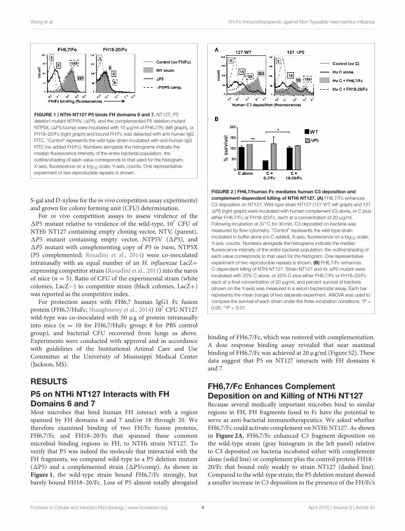
FIGURE 1 | NTHi NT127 P5 binds FH domains 6 and 7. NT127, P5
deletion mutant NTP5V, (1P5), and the complemented P5 deletion mutant
NTP5X, (1P5/comp) were incubated with 10 µg/ml of FH6,7/Fc (left graph), or
FH18–20/Fc (right graph) and bound FH/Fc was detected with anti-human IgG
FITC. “Control” represents the wild-type strain incubated with anti-human IgG
FITC (no added FH/Fc). Numbers alongside the histograms indicate the
median fluorescence intensity of the entire bacterial population; the
outline/shading of each value corresponds to that used for the histogram.
X-axis, fluorescence on a log10 scale; Y-axis, counts. One representative
experiment of two reproducible repeats is shown.
S-gal and D-xylose for the in vivo competition assay experiments)and grown for colony forming unit (CFU) determination.
For in vivo competition assays to assess virulence of the1P5 mutant relative to virulence of the wild-type, 107 CFU ofNTHi NT127 containing empty cloning vector, NTV, (parent),1P5 mutant containing empty vector, NTP5V (1P5), and1P5 mutant with complementing copy of P5 in trans, NTP5X(P5 complemented; Rosadini et al., 2014) were co-inoculatedintranasally with an equal number of an H. influenzae LacZ+expressing competitor strain (Rosadini et al., 2011) into the naresof mice (n = 5). Ratio of CFU of the experimental strain (whitecolonies, LacZ−) to competitor strain (black colonies, LacZ+)was reported as the competitive index.
For protection assays with FH6,7 human IgG1 Fc fusionprotein (FH6,7/HuFc; Shaughnessy et al., 2014) 107 CFU NT127wild-type was co-inoculated with 50 µg of protein intranasallyinto mice (n = 10 for FH6,7/HuFc group; 8 for PBS controlgroup), and bacterial CFU recovered from lungs as above.Experiments were conducted with approval and in accordancewith guidelines of the Institutional Animal Care and UseCommittee at the University of Mississippi Medical Center(Jackson, MS).
RESULTS
P5 on NTHi NT127 Interacts with FHDomains 6 and 7Most microbes that bind human FH interact with a regionspanned by FH domains 6 and 7 and/or 18 through 20. Wetherefore examined binding of two FH/Fc fusion proteins,FH6,7/Fc and FH18–20/Fc that spanned these commonmicrobial binding regions in FH, to NTHi strain NT127. Toverify that P5 was indeed the molecule that interacted with theFH fragments, we compared wild-type to a P5 deletion mutant(1P5) and a complemented strain (1P5/comp). As shown inFigure 1, the wild-type strain bound FH6,7/Fc strongly, butbarely bound FH18–20/Fc. Loss of P5 almost totally abrogated
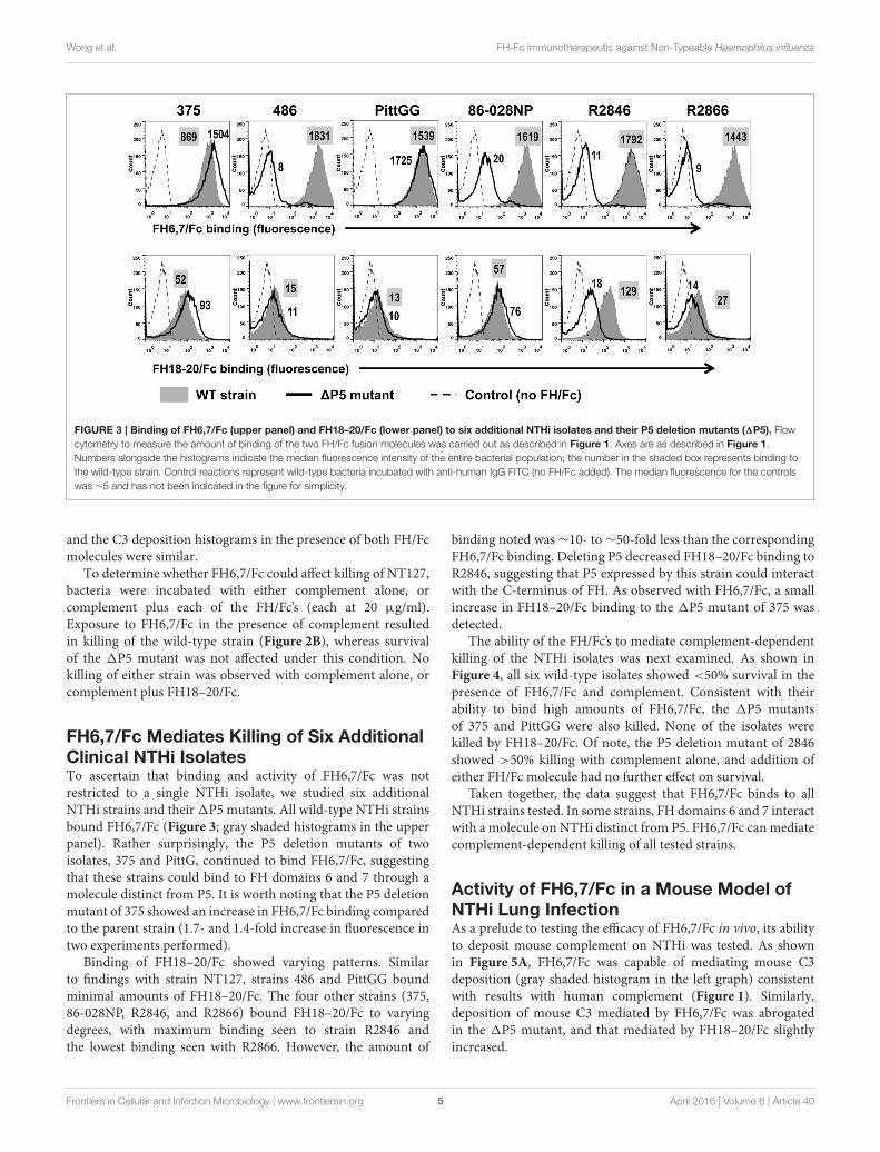
FIGURE 2 | FH6,7/human Fc mediates human C3 deposition and
complement-dependent killing of NTHi NT127. (A) FH6,7/Fc enhances
C3 deposition on NT127. Wild-type strain NT127 (127 WT; left graph) and 127
1P5 (right graph) were incubated with human complement (C) alone, or C plus
either FH6,7/Fc or FH18–20/Fc, each at a concentration of 20 µg/ml.
Following incubation at 37◦C for 30min, C3 deposited on bacteria was
measured by flow cytometry. “Control” represents the wild-type strain
incubated in buffer alone (no C added). X-axis; fluorescence on a log10 scale;
Y-axis, counts. Numbers alongside the histograms indicate the median
fluorescence intensity of the entire bacterial population; the outline/shading of
each value corresponds to that used for the histogram. One representative
experiment of two reproducible repeats is shown. (B) FH6,7/Fc enhances
C-dependent killing of NTHi NT127. Strain NT127 and its 1P5 mutant were
incubated with 20% C alone, or 20% C plus either FH6,7/Fc or FH18–20/Fc
each at a final concentration of 20 µg/ml, and percent survival of bacteria
(shown on the Y-axis) was measured in a serum bactericidal assay. Each bar
represents the mean (range) of two separate experiment. ANOVA was used to
compare the survival of each strain under the three incubation conditions. *P <
0.05; **P < 0.01.
binding of FH6,7/Fc, which was restored with complementation.A dose response binding assay revealed that near maximalbinding of FH6,7/Fc was achieved at 20 µg/ml (Figure S2). Thesedata suggest that P5 on NT127 interacts with FH domains 6and 7.
FH6,7/Fc Enhances ComplementDeposition on and Killing of NTHi NT127Because several medically important microbes bind to similarregions in FH, FH fragments fused to Fc have the potential toserve as anti-bacterial immunotherapeutics. We asked whetherFH6,7/Fc could activate complement on NTHi NT127. As shownin Figure 2A, FH6,7/Fc enhanced C3 fragment deposition onthe wild-type strain (gray histogram in the left panel) relativeto C3 deposited on bacteria incubated either with complementalone (solid line) or complement plus the control protein FH18–20/Fc that bound only weakly to strain NT127 (dashed line).Compared to the wild-type strain, the P5 deletionmutant showeda smaller increase in C3 deposition in the presence of the FH/Fc’s
Frontiers in Cellular and Infection Microbiology | www.frontiersin.org 4 April 2016 | Volume 6 | Article 40
Wong et al. FH-Fc Immunotherapeutic against Non-Typeable Haemophilus influenza
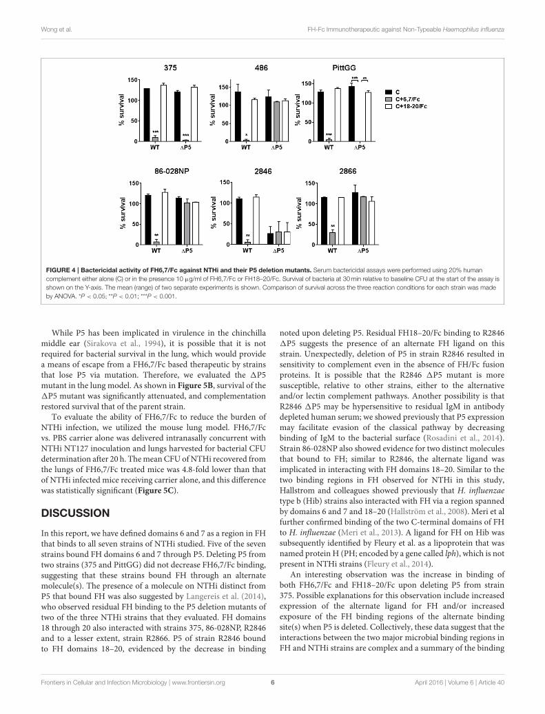
FIGURE 3 | Binding of FH6,7/Fc (upper panel) and FH18–20/Fc (lower panel) to six additional NTHi isolates and their P5 deletion mutants (1P5). Flow
cytometry to measure the amount of binding of the two FH/Fc fusion molecules was carried out as described in Figure 1. Axes are as described in Figure 1.
Numbers alongside the histograms indicate the median fluorescence intensity of the entire bacterial population; the number in the shaded box represents binding to
the wild-type strain. Control reactions represent wild-type bacteria incubated with anti-human IgG FITC (no FH/Fc added). The median fluorescence for the controls
was ∼5 and has not been indicated in the figure for simplicity.
and the C3 deposition histograms in the presence of both FH/Fcmolecules were similar.
To determine whether FH6,7/Fc could affect killing of NT127,bacteria were incubated with either complement alone, orcomplement plus each of the FH/Fc’s (each at 20 µg/ml).Exposure to FH6,7/Fc in the presence of complement resultedin killing of the wild-type strain (Figure 2B), whereas survivalof the 1P5 mutant was not affected under this condition. Nokilling of either strain was observed with complement alone, orcomplement plus FH18–20/Fc.
FH6,7/Fc Mediates Killing of Six AdditionalClinical NTHi IsolatesTo ascertain that binding and activity of FH6,7/Fc was notrestricted to a single NTHi isolate, we studied six additionalNTHi strains and their 1P5 mutants. All wild-type NTHi strainsbound FH6,7/Fc (Figure 3; gray shaded histograms in the upperpanel). Rather surprisingly, the P5 deletion mutants of twoisolates, 375 and PittG, continued to bind FH6,7/Fc, suggestingthat these strains could bind to FH domains 6 and 7 through amolecule distinct from P5. It is worth noting that the P5 deletionmutant of 375 showed an increase in FH6,7/Fc binding comparedto the parent strain (1.7- and 1.4-fold increase in fluorescence intwo experiments performed).
Binding of FH18–20/Fc showed varying patterns. Similarto findings with strain NT127, strains 486 and PittGG boundminimal amounts of FH18–20/Fc. The four other strains (375,86-028NP, R2846, and R2866) bound FH18–20/Fc to varyingdegrees, with maximum binding seen to strain R2846 andthe lowest binding seen with R2866. However, the amount of
binding noted was∼10- to∼50-fold less than the correspondingFH6,7/Fc binding. Deleting P5 decreased FH18–20/Fc binding toR2846, suggesting that P5 expressed by this strain could interactwith the C-terminus of FH. As observed with FH6,7/Fc, a smallincrease in FH18–20/Fc binding to the 1P5 mutant of 375 wasdetected.
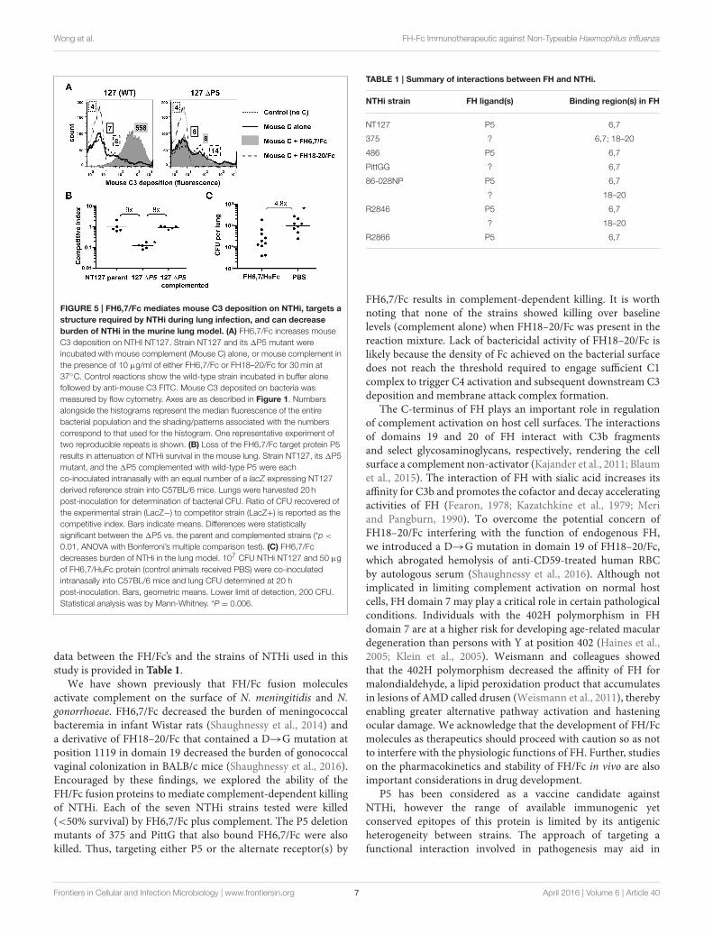
The ability of the FH/Fc’s to mediate complement-dependentkilling of the NTHi isolates was next examined. As shown inFigure 4, all six wild-type isolates showed <50% survival in thepresence of FH6,7/Fc and complement. Consistent with theirability to bind high amounts of FH6,7/Fc, the 1P5 mutantsof 375 and PittGG were also killed. None of the isolates werekilled by FH18–20/Fc. Of note, the P5 deletion mutant of 2846showed >50% killing with complement alone, and addition ofeither FH/Fc molecule had no further effect on survival.
Taken together, the data suggest that FH6,7/Fc binds to allNTHi strains tested. In some strains, FH domains 6 and 7 interactwith a molecule on NTHi distinct from P5. FH6,7/Fc canmediatecomplement-dependent killing of all tested strains.
Activity of FH6,7/Fc in a Mouse Model ofNTHi Lung InfectionAs a prelude to testing the efficacy of FH6,7/Fc in vivo, its abilityto deposit mouse complement on NTHi was tested. As shownin Figure 5A, FH6,7/Fc was capable of mediating mouse C3deposition (gray shaded histogram in the left graph) consistentwith results with human complement (Figure 1). Similarly,deposition of mouse C3 mediated by FH6,7/Fc was abrogatedin the 1P5 mutant, and that mediated by FH18–20/Fc slightlyincreased.
Frontiers in Cellular and Infection Microbiology | www.frontiersin.org 5 April 2016 | Volume 6 | Article 40
Wong et al. FH-Fc Immunotherapeutic against Non-Typeable Haemophilus influenza
FIGURE 4 | Bactericidal activity of FH6,7/Fc against NTHi and their P5 deletion mutants. Serum bactericidal assays were performed using 20% human
complement either alone (C) or in the presence 10 µg/ml of FH6,7/Fc or FH18–20/Fc. Survival of bacteria at 30min relative to baseline CFU at the start of the assay is
shown on the Y-axis. The mean (range) of two separate experiments is shown. Comparison of survival across the three reaction conditions for each strain was made
by ANOVA. *P < 0.05; **P < 0.01; ***P < 0.001.
While P5 has been implicated in virulence in the chinchillamiddle ear (Sirakova et al., 1994), it is possible that it is notrequired for bacterial survival in the lung, which would providea means of escape from a FH6,7/Fc based therapeutic by strainsthat lose P5 via mutation. Therefore, we evaluated the 1P5mutant in the lung model. As shown in Figure 5B, survival of the1P5 mutant was significantly attenuated, and complementationrestored survival that of the parent strain.
To evaluate the ability of FH6,7/Fc to reduce the burden ofNTHi infection, we utilized the mouse lung model. FH6,7/Fcvs. PBS carrier alone was delivered intranasally concurrent withNTHi NT127 inoculation and lungs harvested for bacterial CFUdetermination after 20 h. ThemeanCFU ofNTHi recovered fromthe lungs of FH6,7/Fc treated mice was 4.8-fold lower than thatof NTHi infected mice receiving carrier alone, and this differencewas statistically significant (Figure 5C).
DISCUSSION
In this report, we have defined domains 6 and 7 as a region in FHthat binds to all seven strains of NTHi studied. Five of the sevenstrains bound FH domains 6 and 7 through P5. Deleting P5 fromtwo strains (375 and PittGG) did not decrease FH6,7/Fc binding,suggesting that these strains bound FH through an alternatemolecule(s). The presence of a molecule on NTHi distinct fromP5 that bound FH was also suggested by Langereis et al. (2014),who observed residual FH binding to the P5 deletion mutants oftwo of the three NTHi strains that they evaluated. FH domains18 through 20 also interacted with strains 375, 86-028NP, R2846and to a lesser extent, strain R2866. P5 of strain R2846 boundto FH domains 18–20, evidenced by the decrease in binding
noted upon deleting P5. Residual FH18–20/Fc binding to R28461P5 suggests the presence of an alternate FH ligand on thisstrain. Unexpectedly, deletion of P5 in strain R2846 resulted insensitivity to complement even in the absence of FH/Fc fusionproteins. It is possible that the R2846 1P5 mutant is moresusceptible, relative to other strains, either to the alternativeand/or lectin complement pathways. Another possibility is thatR2846 1P5 may be hypersensitive to residual IgM in antibodydepleted human serum; we showed previously that P5 expressionmay facilitate evasion of the classical pathway by decreasingbinding of IgM to the bacterial surface (Rosadini et al., 2014).Strain 86-028NP also showed evidence for two distinct moleculesthat bound to FH; similar to R2846, the alternate ligand wasimplicated in interacting with FH domains 18–20. Similar to thetwo binding regions in FH observed for NTHi in this study,Hallstrom and colleagues showed previously that H. influenzaetype b (Hib) strains also interacted with FH via a region spannedby domains 6 and 7 and 18–20 (Hallström et al., 2008). Meri et alfurther confirmed binding of the two C-terminal domains of FHto H. influenzae (Meri et al., 2013). A ligand for FH on Hib wassubsequently identified by Fleury et al. as a lipoprotein that wasnamed protein H (PH; encoded by a gene called lph), which is notpresent in NTHi strains (Fleury et al., 2014).
An interesting observation was the increase in binding ofboth FH6,7/Fc and FH18–20/Fc upon deleting P5 from strain375. Possible explanations for this observation include increasedexpression of the alternate ligand for FH and/or increasedexposure of the FH binding regions of the alternate bindingsite(s) when P5 is deleted. Collectively, these data suggest that theinteractions between the two major microbial binding regions inFH and NTHi strains are complex and a summary of the binding
Frontiers in Cellular and Infection Microbiology | www.frontiersin.org 6 April 2016 | Volume 6 | Article 40
Wong et al. FH-Fc Immunotherapeutic against Non-Typeable Haemophilus influenza
FIGURE 5 | FH6,7/Fc mediates mouse C3 deposition on NTHi, targets a
structure required by NTHi during lung infection, and can decrease
burden of NTHi in the murine lung model. (A) FH6,7/Fc increases mouse
C3 deposition on NTHI NT127. Strain NT127 and its 1P5 mutant were
incubated with mouse complement (Mouse C) alone, or mouse complement in
the presence of 10 µg/ml of either FH6,7/Fc or FH18–20/Fc for 30min at
37◦C. Control reactions show the wild-type strain incubated in buffer alone
followed by anti-mouse C3 FITC. Mouse C3 deposited on bacteria was
measured by flow cytometry. Axes are as described in Figure 1. Numbers
alongside the histograms represent the median fluorescence of the entire
bacterial population and the shading/patterns associated with the numbers
correspond to that used for the histogram. One representative experiment of
two reproducible repeats is shown. (B) Loss of the FH6,7/Fc target protein P5
results in attenuation of NTHi survival in the mouse lung. Strain NT127, its 1P5
mutant, and the 1P5 complemented with wild-type P5 were each
co-inoculated intranasally with an equal number of a lacZ expressing NT127
derived reference strain into C57BL/6 mice. Lungs were harvested 20 h
post-inoculation for determination of bacterial CFU. Ratio of CFU recovered of
the experimental strain (LacZ−) to competitor strain (LacZ+) is reported as the
competitive index. Bars indicate means. Differences were statistically
significant between the 1P5 vs. the parent and complemented strains (*p <
0.01, ANOVA with Bonferroni’s multiple comparison test). (C) FH6,7/Fc
decreases burden of NTHi in the lung model. 107 CFU NTHi NT127 and 50 µg
of FH6,7/HuFc protein (control animals received PBS) were co-inoculated
intranasally into C57BL/6 mice and lung CFU determined at 20 h
post-inoculation. Bars, geometric means. Lower limit of detection, 200 CFU.
Statistical analysis was by Mann-Whitney. *P = 0.006.
data between the FH/Fc’s and the strains of NTHi used in thisstudy is provided in Table 1.
We have shown previously that FH/Fc fusion moleculesactivate complement on the surface of N. meningitidis and N.gonorrhoeae. FH6,7/Fc decreased the burden of meningococcalbacteremia in infant Wistar rats (Shaughnessy et al., 2014) anda derivative of FH18–20/Fc that contained a D→G mutation atposition 1119 in domain 19 decreased the burden of gonococcalvaginal colonization in BALB/c mice (Shaughnessy et al., 2016).Encouraged by these findings, we explored the ability of theFH/Fc fusion proteins to mediate complement-dependent killingof NTHi. Each of the seven NTHi strains tested were killed(<50% survival) by FH6,7/Fc plus complement. The P5 deletionmutants of 375 and PittG that also bound FH6,7/Fc were alsokilled. Thus, targeting either P5 or the alternate receptor(s) by
TABLE 1 | Summary of interactions between FH and NTHi.
NTHi strain FH ligand(s) Binding region(s) in FH
NT127 P5 6,7
375 ? 6,7; 18–20
486 P5 6,7
PittGG ? 6,7
86-028NP P5 6,7
? 18–20
R2846 P5 6,7
? 18–20
R2866 P5 6,7
FH6,7/Fc results in complement-dependent killing. It is worthnoting that none of the strains showed killing over baselinelevels (complement alone) when FH18–20/Fc was present in thereaction mixture. Lack of bactericidal activity of FH18–20/Fc islikely because the density of Fc achieved on the bacterial surfacedoes not reach the threshold required to engage sufficient C1complex to trigger C4 activation and subsequent downstream C3deposition and membrane attack complex formation.
The C-terminus of FH plays an important role in regulationof complement activation on host cell surfaces. The interactionsof domains 19 and 20 of FH interact with C3b fragmentsand select glycosaminoglycans, respectively, rendering the cellsurface a complement non-activator (Kajander et al., 2011; Blaumet al., 2015). The interaction of FH with sialic acid increases itsaffinity for C3b and promotes the cofactor and decay acceleratingactivities of FH (Fearon, 1978; Kazatchkine et al., 1979; Meriand Pangburn, 1990). To overcome the potential concern ofFH18–20/Fc interfering with the function of endogenous FH,we introduced a D→G mutation in domain 19 of FH18–20/Fc,which abrogated hemolysis of anti-CD59-treated human RBCby autologous serum (Shaughnessy et al., 2016). Although notimplicated in limiting complement activation on normal hostcells, FH domain 7 may play a critical role in certain pathologicalconditions. Individuals with the 402H polymorphism in FHdomain 7 are at a higher risk for developing age-related maculardegeneration than persons with Y at position 402 (Haines et al.,2005; Klein et al., 2005). Weismann and colleagues showedthat the 402H polymorphism decreased the affinity of FH formalondialdehyde, a lipid peroxidation product that accumulatesin lesions of AMD called drusen (Weismann et al., 2011), therebyenabling greater alternative pathway activation and hasteningocular damage. We acknowledge that the development of FH/Fcmolecules as therapeutics should proceed with caution so as notto interfere with the physiologic functions of FH. Further, studieson the pharmacokinetics and stability of FH/Fc in vivo are alsoimportant considerations in drug development.
P5 has been considered as a vaccine candidate againstNTHi, however the range of available immunogenic yetconserved epitopes of this protein is limited by its antigenicheterogeneity between strains. The approach of targeting afunctional interaction involved in pathogenesis may aid in
Frontiers in Cellular and Infection Microbiology | www.frontiersin.org 7 April 2016 | Volume 6 | Article 40
Wong et al. FH-Fc Immunotherapeutic against Non-Typeable Haemophilus influenza
bypassing this diversity. P5 may be an attractive target forsuch novel therapeutics because this molecule is ubiquitouslyexpressed, is not phase-variable, and is not subject to epigeneticregulation (Atack et al., 2015). Further, P5 deletion mutants showreduced virulence in the chinchilla otitis media model andmouselung model compared to their wild-type counterparts (Sirakovaet al., 1994), which we have substantiated in the mouse lungmodel with isogenic strains and complementation of the mutantby restoration of wildtype P5 at an ectopic chromosomal location(Figure 5B). Resistance to FH6,7/Fc, if this were to occur, wouldrequire selection of P5 deletion mutants or P5 mutants that lackthe ability to bind to human FH, both of which may place thebacterium at a fitness disadvantage as indicated by attenuationof survival of NTHi in the lung model conferred by deletion ofP5. Alternatively, it is possible that mutational loss of FH bindingby P5 may not strongly influence the course of infection if, forexample, other P5 virulence phenotypes mediated by P5 exhibitpredominant effects and do not require the same structuralregions. Langeries et al. have identified the predicted surfaceexposed loops 1 and 2 in P5 of NTHi strain R2866 as the FHinteracting regions (Langereis et al., 2014), and it is possiblethat the surface exposed loops 3 and 4 of P5 mediate alternativefunctions during infection. It will be of interest to determinewhether the role of P5 proteins in virulence vs. FH binding can bedissociated. Identification of the alternate acceptor molecule(s)for FH6,7/Fc also merits further consideration.
As an initial test of the potential therapeutic utility of FH6,7/Fcagainst NTHi we evaluated its ability to decrease survival ofNTHi in a mouse model. Murine C3 was efficiently targetedby FH6,7/Fc to the bacterial surface in the presence of mouseserum in vitro, similar to human C3 (Figure 5A), suggestingfeasibility. Encouragingly, when delivered intranasally into thelung, FH6,7/Fc was able to reduce the burden of NTHi comparedto mock treatment, producing a statistically significant 4.8-folddecrease in recovered bacteria 20 h after inoculation (Figure 5C).We note that this was a partial effect, and eradication of anongoing infection will likely require repeated doses. In addition,clinical application would likely require aerosolization of the
fusion protein to achieve similar exposure to the bacteria,however this delivery method is well-tolerated in patients treatedfor other conditions and is a clinically feasible approach.We haveprovided evidence for the efficacy of FH6,7/Fc in vivo, however,we acknowledge that further work will be needed to define itsefficacy against NTHi infection in other niches such as themiddleear, sinuses, and the bloodstream. Further, only human but notmouse FH has been reported to bind to NTHi (Langereis et al.,2014). Thus, the efficacy of FH6,7/Fc in vivo in the presence ofhuman complement inhibitors that may counteract the efficacyof the therapeutic merits study. Nevertheless, enhanced C3deposition and killing of NTHi by FH6,7/Fc in human serum thatcontains endogenous FH provides optimism for activity of thismolecule in the context of human complement.
In conclusion, FH6,7/Fc may prove a novel and promisingadjunctive therapeutic against NTHi infections, particularly ininstances of recurrent or recalcitrant infections, where multiplecourses of antibiotics have proven ineffective or only partiallyeffective and may be associated with adverse side effects.
AUTHOR CONTRIBUTIONS
SW, JS, SR, and BA designed the study, performed experiments,analyzed data, and wrote the manuscript. JS and SW contributedequally.
ACKNOWLEDGMENTS
This work was supported by grants from the National Institutesof Health/National Institutes of Allergy and Infectious Diseases,AI095740 (BA), AI111728 (to JS and SR), AI118161 (to SR) andAI114790 (to SR).
SUPPLEMENTARY MATERIAL
The Supplementary Material for this article can be foundonline at: http://journal.frontiersin.org/article/10.3389/fcimb.2016.00040
REFERENCES
Atack, J. M., Srikhanta, Y. N., Fox, K. L., Jurcisek, J. A., Brockman, K. L., Clark,
T. A., et al. (2015). A biphasic epigenetic switch controls immunoevasion,
virulence and niche adaptation in non-typeable Haemophilus influenzae. Nat.
Commun. 6, 7828. doi: 10.1038/ncomms8828
Avadhanula, V., Rodriguez, C. A., Ulett, G. C., Bakaletz, L. O., and Adderson, E. E.
(2006). Nontypeable Haemophilus influenzae adheres to intercellular adhesion
molecule 1 (ICAM-1) on respiratory epithelial cells and upregulates ICAM-
1 expression. Infect. Immun. 74, 830–838. doi: 10.1128/IAI.74.2.830-838.
2006
Barcak, G. J., Chandler, M. S., Redfield, R. J., and Tomb, J. F. (1991). Genetic
systems in Haemophilus influenzae. Methods Enzymol. 204, 321–342. doi:
10.1016/0076-6879(91)04016-H
Barenkamp, S. J., and Leininger, E. (1992). Cloning, expression, and DNA
sequence analysis of genes encoding nontypeable Haemophilus influenzae
high-molecular-weight surface- exposed proteins related to filamentous
hemagglutinin of Bordetella pertussis. Infect. Immun. 60, 1302–1313.
Bernhard, S., Fleury, C., Su, Y. C., Zipfel, P. F., Koske, I., Nordström, T., et al.
(2014). Outer membrane protein OlpA contributes to Moraxella catarrhalis
serum resistance via interaction with factor H and the alternative pathway. J.
Infect. Dis. 210, 1306–1310. doi: 10.1093/infdis/jiu241
Blaum, B. S., Hannan, J. P., Herbert, A. P., Kavanagh, D., Uhrín, D., and Stehle, T.
(2015). Structural basis for sialic acid-mediated self-recognition by complement
factor H. Nat. Chem. Biol. 11, 77–82. doi: 10.1038/nchembio.1696
Blom, A. M., Hallström, T., and Riesbeck, K. (2009). Complement evasion
strategies of pathogens-acquisition of inhibitors and beyond. Mol. Immunol.
46, 2808–2817. doi: 10.1016/j.molimm.2009.04.025
Broides, A., Dagan, R., Greenberg, D., Givon-Lavi, N., and Leibovitz, E.
(2009). Acute otitis media caused by Moraxella catarrhalis: epidemiologic
and clinical characteristics. Clin. Infect. Dis. 49, 1641–1647. doi: 10.1086/
647933
Buchinsky, F. J., Forbes, M. L., Hayes, J. D., Shen, K., Ezzo, S., Compliment, J., et al.
(2007). Virulence phenotypes of low-passage clinical isolates of nontypeable
Haemophilus influenzae assessed using the chinchilla laniger model of otitis
media. BMCMicrobiol. 7:56. doi: 10.1186/1471-2180-7-56
Frontiers in Cellular and Infection Microbiology | www.frontiersin.org 8 April 2016 | Volume 6 | Article 40
Wong et al. FH-Fc Immunotherapeutic against Non-Typeable Haemophilus influenza
Centers for Disease Control and Prevention [CDC] (2012). Chronic Obstructive
Pulmonary Disease Among Adults–United States, 2011. Morbidity and
Mortality Weekly Report Vol. 61, 938–943.
Collins, S., Vickers, A., Ladhani, S. N., Flynn, S., Platt, S., Ramsay, M. E., et al.
(2016). Clinical andmolecular epidemiology of childhood invasive nontypeable
Haemophilus influenzae disease in England andWales. Pediatr. Infect. Dis. J. 35,
e76–e84. doi: 10.1097/INF.0000000000000996
Dave, S., Brooks-Walter, A., Pangburn, M. K., and McDaniel, L. S. (2001). PspC,
a pneumococcal surface protein, binds human factor H. Infect. Immun. 69,
3435–3437. doi: 10.1128/IAI.69.5.3435-3437.2001
Erwin, A. L., Nelson, K. L., Mhlanga-Mutangadura, T., Bonthuis, P. J., Geelhood,
J. L., Morlin, G., et al. (2005). Characterization of genetic and phenotypic
diversity of invasive nontypeable Haemophilus influenzae. Infect. Immun. 73,
5853–5863. doi: 10.1128/IAI.73.9.5853-5863.2005
Euba, B., Moleres, J., Viadas, C., Ruiz de los Mozos, I., Valle, J., Bengoechea, J. A.,
et al. (2015). Relative contribution of P5 andHap surface proteins to nontypable
Haemophilus influenzae interplay with the host upper and lower airways. PLoS
ONE 10:e0123154. doi: 10.1371/journal.pone.0123154
Fearon, D. T. (1978). Regulation by membrane sialic acid of beta1H-
dependent decay-dissociation of amplification C3 convertase of the alternative
complement pathway. Proc. Natl. Acad. Sci. U.S.A. 75, 1971–1975. doi:
10.1073/pnas.75.4.1971
Fearon, D. T., and Austen, K. F. (1977). Activation of the alternative complement
pathway due to resistance of zymosan-bound amplification convertase
to endogenous regulatory mechanisms. Proc. Natl. Acad. Sci. U.S.A. 74,
1683–1687. doi: 10.1073/pnas.74.4.1683
Fleury, C., Su, Y. C., Hallström, T., Sandblad, L., Zipfel, P. F., and Riesbeck,
K. (2014). Identification of a Haemophilus influenzae factor H-Binding
lipoprotein involved in serum resistance. J. Immunol. 192, 5913–5923. doi:
10.4049/jimmunol.1303449
Gilsdorf, J. R. (1998). Antigenic diversity and gene polymorphisms inHaemophilus
influenzae. Infect. Immun. 66, 5053–5059.
Grijalva, C. G., Nuorti, J. P., and Griffin, M. R. (2009). Antibiotic prescription
rates for acute respiratory tract infections in US ambulatory settings. JAMA 302,
758–766. doi: 10.1001/jama.2009.1163
Haines, J. L., Hauser, M. A., Schmidt, S., Scott, W. K., Olson, L. M., Gallins, P., et al.
(2005). Complement factor H variant increases the risk of age-related macular
degeneration. Science 308, 419–421. doi: 10.1126/science.1110359
Hallström, T., Zipfel, P. F., Blom, A. M., Lauer, N., Forsgren, A., and Riesbeck,
K. (2008). Haemophilus influenzae interacts with the human complement
inhibitor factor H. J. Immunol. 181, 537–545. doi: 10.4049/jimmunol.181.1.537
Harabuchi, Y., Faden, H., Yamanaka, N., Duffy, L., Wolf, J., and Krystofik, D.
(1994). Nasopharyngeal colonization with nontypeableHaemophilus influenzae
and recurrent otitis media. Tonawanda/Williamsville Pediatrics. J. Infect. Dis.
170, 862–866. doi: 10.1093/infdis/170.4.862
Harrison, A., Dyer, D. W., Gillaspy, A., Ray, W. C., Mungur, R., Carson, M.
B., et al. (2005). Genomic sequence of an otitis media isolate of nontypeable
Haemophilus influenzae: comparative study with H. influenzae serotype
d, strain KW20. J. Bacteriol. 187, 4627–4636. doi: 10.1128/JB.187.13.4627-
4636.2005
Hill, D. J., Toleman, M. A., Evans, D. J., Villullas, S., Van Alphen, L., and
Virji, M. (2001). The variable P5 proteins of typeable and non-typeable
Haemophilus influenzae target humanCEACAM1.Mol.Microbiol. 39, 850–862.
doi: 10.1046/j.1365-2958.2001.02233.x
Hood, D. W., Makepeace, K., Deadman, M. E., Rest, R. F., Thibault,
P., Martin, A., et al. (1999). Sialic acid in the lipopolysaccharide of
Haemophilus influenzae: strain distribution, influence on serum resistance and
structural characterization. Mol. Microbiol. 33, 679–692. doi: 10.1046/j.1365-
2958.1999.01509.x
Horstmann, R. D., Sievertsen, H. J., Knobloch, J., and Fischetti, V. A. (1988).
Antiphagocytic activity of streptococcal M protein: selective binding of
complement control protein factor H. Proc. Natl. Acad. Sci. U.S.A. 85,
1657–1661. doi: 10.1073/pnas.85.5.1657
Kajander, T., Lehtinen, M. J., Hyvärinen, S., Bhattacharjee, A., Leung, E.,
Isenman, D. E., et al. (2011). Dual interaction of factor H with C3d
and glycosaminoglycans in host-nonhost discrimination by complement.
Proc. Natl. Acad. Sci. U.S.A. 108, 2897–2902. doi: 10.1073/pnas.10170
87108
Kaur, R., Casey, J. R., and Pichichero, M. E. (2013). Relationship with
original pathogen in recurrence of acute otitis media after completion of
amoxicillin/clavulanate: bacterial relapse or new pathogen. Pediatr. Infect. Dis.
J 32, 1159–1162. doi: 10.1097/INF.0b013e31829e3779
Kazatchkine, M. D., Fearon, D. T., and Austen, K. F. (1979). Human
alternative complement pathway: membrane-associated sialic acid regulates
the competition between B and beta1 H for cell-bound C3b. J. Immunol. 122,
75–81.
Klein, R. J., Zeiss, C., Chew, E. Y., Tsai, J. Y., Sackler, R. S., Haynes, C., et al. (2005).
Complement factor H polymorphism in age-related macular degeneration.
Science 308, 385–389. doi: 10.1126/science.1109557
Kraiczy, P., and Würzner, R. (2006). Complement escape of human pathogenic
bacteria by acquisition of complement regulators. Mol. Immunol. 43, 31–44.
doi: 10.1016/j.molimm.2005.06.016
Ladhani, S. N. (2012). Two decades of experience with theHaemophilus influenzae
serotype b conjugate vaccine in the United Kingdom. Clin. Ther. 34, 385–399.
doi: 10.1016/j.clinthera.2011.11.027
Langereis, J. D., de Jonge, M. I., and Weiser, J. N. (2014). Binding of human
factor H to outer membrane protein P5 of non-typeable Haemophilus
influenzae contributes to complement resistance. Mol. Microbiol. 94, 89–106.
doi: 10.1111/mmi.12741
McCaig, L. F., Besser, R. E., and Hughes, J. M. (2002). Trends in antimicrobial
prescribing rates for children and adolescents. JAMA 287, 3096–3102. doi:
10.1001/jama.287.23.3096
Mell, J. C., Sinha, S., Balashov, S., Viadas, C., Grassa, C. J., Ehrlich, G. D., et al.
(2014). Complete genome sequence of Haemophilus influenzae strain 375 from
the middle ear of a pediatric patient with otitis media. Genome Announc.
2:e01245–14. doi: 10.1128/genomeA.01245-14
Meri, S., and Pangburn, M. K. (1990). Discrimination between activators and
nonactivators of the alternative pathway of complement: regulation via a sialic
acid/polyanion binding site on factor H. Proc. Natl. Acad. Sci. U.S.A. 87,
3982–3986. doi: 10.1073/pnas.87.10.3982
Meri, T., Amdahl, H., Lehtinen, M. J., Hyvarinen, S., McDowell, J. V.,
Bhattacharjee, A., et al. (2013). Microbes bind complement inhibitor factor
H via a common site. PLoS Pathog. 9:e1003308. doi: 10.1371/journal.ppat.10
03308
Miyamoto, N., and Bakaletz, L. O. (1996). Selective adherence of non-
typeable Haemophilus influenzae (NTHi) to mucus or epithelial cells in the
chinchilla eustachian tube and middle ear. Microb. Pathog. 21, 343–356. doi:
10.1006/mpat.1996.0067
Murphy, T. F., Faden, H., Bakaletz, L. O., Kyd, J. M., Forsgren, A., Campos, J., et al.
(2009). NontypeableHaemophilus influenzae as a pathogen in children. Pediatr.
Infect. Dis. J. 28, 43–48. doi: 10.1097/INF.0b013e318184dba2
Murphy, T. F., and Sethi, S. (2002). Chronic obstructive pulmonary disease: role
of bacteria and guide to antibacterial selection in the older patient. Drugs Aging
19, 761–775. doi: 10.2165/00002512-200219100-00005
Nizet, V., Colina, K. F., Almquist, J. R., Rubens, C. E., and Smith, A. L. (1996). A
virulent nonencapsulated Haemophilus influenzae. J. Infect. Dis. 173, 180–186.
doi: 10.1093/infdis/173.1.180
Pangburn, M. K., Schreiber, R. D., and Müller-Eberhard, H. J. (1977). Human
complement C3b inactivator: isolation, characterization, and demonstration of
an absolute requirement for the serum protein beta1H for cleavage of C3b and
C4b in solution. J. Exp. Med. 146, 257–270. doi: 10.1084/jem.146.1.257
Ram, S., Lewis, L. A., and Rice, P. A. (2010). Infections of people with complement
deficiencies and patients who have undergone splenectomy. Clin. Microbiol.
Rev. 23, 740–780. doi: 10.1128/CMR.00048-09
Ray, T. D., Lewis, L. A., Gulati, S., Rice, P. A., and Ram, S. (2011). Novel blocking
human IgG directed against the pentapeptide repeat motifs of Neisseria
meningitidis Lip/H.8 and Laz lipoproteins. J. Immunol. 186, 4881–4894. doi:
10.4049/jimmunol.1003623
Reddy, M. S., Bernstein, J. M., Murphy, T. F., and Faden, H. S. (1996). Binding
between outer membrane proteins of nontypeable Haemophilus influenzae and
human nasopharyngeal mucin. Infect. Immun. 64, 1477–1479.
Ripoche, J., Day, A. J., Harris, T. J., and Sim, R. B. (1988). The complete amino
acid sequence of human complement factor H. Biochem. J. 249, 593–602. doi:
10.1042/bj2490593
Rosadini, C. V., Gawronski, J. D., Raimunda, D., Argüello, J. M., and Akerley,
B. J. (2011). A novel zinc binding system, ZevAB, is critical for survival of
Frontiers in Cellular and Infection Microbiology | www.frontiersin.org 9 April 2016 | Volume 6 | Article 40
Wong et al. FH-Fc Immunotherapeutic against Non-Typeable Haemophilus influenza
nontypeable Haemophilus influenzae in a murine lung infection model. Infect.
Immun. 79, 3366–3376. doi: 10.1128/IAI.05135-11
Rosadini, C. V., Ram, S., and Akerley, B. J. (2014). Outer membrane protein P5
is required for resistance of nontypeable Haemophilus influenzae to both the
classical and alternative complement pathways. Infect. Immun. 82, 640–649.
doi: 10.1128/IAI.01224-13
Sethi, S., Anzueto, A., Miravitlles, M., Arvis, P., Alder, J., Haverstock, D., et al.
(2016). Determinants of bacteriological outcomes in exacerbations of chronic
obstructive pulmonary disease. Infection 44, 65–76. doi: 10.1007/s15010-015-
0833-3
Sharma, A. K., and Pangburn, M. K. (1996). Identification of three physically and
functionally distinct binding sites for C3b in human complement factor H
by deletion mutagenesis. Proc. Natl. Acad. Sci. U.S.A. 93, 10996–11001. doi:
10.1073/pnas.93.20.10996
Shaughnessy, J., Gulati, S., Agarwal, S., Unemo, M., Ohnishi, M., Su, X.
H., et al. (2016). A novel factor H-Fc chimeric immunotherapeutic
molecule against Neisseria gonorrhoeae. J. Immunol. 196, 1732–1740. doi:
10.4049/jimmunol.1500292
Shaughnessy, J., Ram, S., Bhattacharjee, A., Pedrosa, J., Tran, C., Horvath, G.,
et al. (2011). Molecular characterization of the interaction between sialylated
Neisseria gonorrhoeae and factor H. J. Biol. Chem. 286, 22235–22242. doi:
10.1074/jbc.M111.225516
Shaughnessy, J., Vu, D. M., Punjabi, R., Serra-Pladevall, J., DeOliveira, R. B.,
Granoff, D. M., et al. (2014). Fusion protein comprising factor H domains 6
and 7 and human IgG1 Fc as an antibacterial immunotherapeutic. Clin. Vaccine
Immunol. 21, 1452–1459. doi: 10.1128/CVI.00444-14
Siggins, M. K., Gill, S. K., Langford, P. R., Li, Y., Ladhani, S. N., and Tregoning, J.
S. (2015). PHiD-CV induces anti-Protein D antibodies but does not augment
pulmonary clearance of nontypeable Haemophilus influenzae in mice. Vaccine
33, 4954–4961. doi: 10.1016/j.vaccine.2015.07.034
Sirakova, T., Kolattukudy, P. E., Murwin, D., Billy, J., Leake, E., Lim, D., et al.
(1994). Role of fimbriae expressed by nontypeable Haemophilus influenzae
in pathogenesis of and protection against otitis media and relatedness of the
fimbrin subunit to outer membrane protein A. Infect. Immun. 62, 2002–2020.
Tchoupa, A. K., Lichtenegger, S., Reidl, J., and Hauck, C. R. (2015). Outer
membrane protein P1 is the CEACAM-binding adhesin of Haemophilus
influenzae.Mol. Microbiol. 98, 440–455. doi: 10.1111/mmi.13134
Van Eldere, J., Slack, M. P., Ladhani, S., and Cripps, A. W. (2014). Non-typeable
Haemophilus influenzae, an under-recognised pathogen. Lancet Infect. Dis. 14,
1281–1292. doi: 10.1016/S1473-3099(14)70734-0
Weiler, J. M., Daha, M. R., Austen, K. F., and Fearon, D. T. (1976). Control
of the amplification convertase of complement by the plasma protein
beta1H. Proc. Natl. Acad. Sci. U.S.A. 73, 3268–3272. doi: 10.1073/pnas.73.
9.3268
Weismann, D., Hartvigsen, K., Lauer, N., Bennett, K. L., Scholl, H. P.,
Charbel Issa, P., et al. (2011). Complement factor H binds malondialdehyde
epitopes and protects from oxidative stress. Nature 478, 76–81. doi: 10.1038/
nature10449
Whaley, K., and Ruddy, S. (1976). Modulation of the alternative complement
pathways by beta 1 H globulin. J. Exp. Med. 144, 1147–1163. doi:
10.1084/jem.144.5.1147
Wong, S. M., Bernui, M., Shen, H., and Akerley, B. J. (2013). Genome-wide
fitness profiling reveals adaptations required by Haemophilus in coinfection
with influenza A virus in the murine lung. Proc. Natl. Acad. Sci. U.S.A. 110,
15413–15418. doi: 10.1073/pnas.1311217110
Wong, S. M., St Michael, F., Cox, A., Ram, S., and Akerley, B. J. (2011).
ArcA-regulated glycosyltransferase lic2B promotes complement evasion and
pathogenesis of nontypeable Haemophilus influenzae. Infect. Immun. 79,
1971–1983. doi: 10.1128/IAI.01269-10
Würzner, R. (1999). Evasion of pathogens by avoiding recognition or eradication
by complement, in part via molecular mimicry. Mol. Immunol. 36, 249–260.
doi: 10.1016/S0161-5890(99)00049-8
Conflict of Interest Statement: The authors declare that the research was
conducted in the absence of any commercial or financial relationships that could
be construed as a potential conflict of interest.
Copyright © 2016 Wong, Shaughnessy, Ram and Akerley. This is an open-access
article distributed under the terms of the Creative Commons Attribution License
(CC BY). The use, distribution or reproduction in other forums is permitted,
provided the original author(s) or licensor are credited and that the original
publication in this journal is cited, in accordance with accepted academic practice.
No use, distribution or reproduction is permitted which does not comply with these
terms.
Frontiers in Cellular and Infection Microbiology | www.frontiersin.org 10 April 2016 | Volume 6 | Article 40


![Kumar et al., 1:6 Open Access Scientific Reports · Bacteriocin preparation was then filter sterilized using Millipore 0.45 mm filters [12]. Pediocin activity aasay Bacteriocin activity](https://img.pdfslide.net/doc/110x75/5e495a1b23b22c0e2470af9d/kumar-et-al-16-open-access-scientific-reports-bacteriocin-preparation-was-then.jpg)