Embed Size (px)
Citation preview
CLINICAL IMPORTANCEUrolithiasis is a common disorder of the urinary tract in dogs.However, the incidence (annual rate of appearance of new casesamong the entire population at risk for the disease) of canineurolithiasis has not been established. Urolithiasis was diagnosedin 3,628 of 676,668 dogs (0.53%) admitted to veterinary teach-ing hospitals in North America between 1980 and 1993. Theproportion of dogs with urolithiasis admitted to veterinary hos-pitals in Germany was similar (Lulich et al, 1995).
Clinical signs of urolithiasis may be the first indication ofunderlying systemic disorders, or defects in the structure orfunction of the urinary tract (Table 38-1). Uroliths may passthrough various parts of the excretory pathway of the urinarytract, they may dissolve, they may become inactive or they maycontinue to form and grow. If uroliths associated with clinicalsigns are allowed to remain untreated, they may result in seri-ous sequelae. Despite urolith removal by voiding, dissolutionprotocols, or surgery, uroliths frequently recur if risk factorsassociated with their formation are not suppressed or corrected.
Urolithiasis should not be viewed as a single disease, butrather as a sequela of one or more underlying abnormalities.
The fact that urolith formation is often erratic and unpre-dictable indicates that several interrelated complex physiologicand pathologic factors are involved. Therefore, detection ofuroliths is only the beginning of the diagnostic process. De-termination of urolith composition narrows etiologic possibili-ties. Knowledge of the patient’s food and how it is fed andserum and urine concentrations of lithogenic minerals, crystal-lization promoters, crystallization inhibitors and their interac-tions aids in the diagnosis, treatment and prevention ofurolithiasis (Box 38-1).
FORMATION OF UROLITHS
Initiation and GrowthUrolith formation is associated with two complementary butseparate phases: initiation and growth. It appears that initiatingevents are not the same for all types of uroliths. In addition, fac-tors that initiate urolith formation may be different from thosethat allow urolith growth.
The initial step in urolith formation is formation of a crystalnidus (or crystal embryo). This initiation phase of urolith for-
Chapter
38Canine Urolithiasis:
Definitions, Pathophysiologyand Clinical Manifestations
Carl A. Osborne
Jody P. Lulich
Lisa K. Ulrich
“If the patient you treat is harmed more than helped, then best leave the stones alone. But by taking a look at the thoughts in this book,
ways to treat stones by how patients eat you’ll be shown.” Carl A. Osborne, 1999
mation, called nucleation, is dependent on supersaturation ofurine with lithogenic crystalloids. The inciting factor and theprecise sequence of events that lead to the formation of mosttypes of stones are still unknown. The degree of urine supersat-uration may be influenced by the magnitude of renal excretionof crystalloids, urinary pH and/or crystallization inhibitors orpromoters in urine. Noncrystalline proteinaceous matrix sub-stances may also play a role in nucleation in some instances.
Three theories have been proposed to explain initiation oflithogenesis: 1) supersaturation-crystallization theory, 2) ma-trix-nucleation theory and 3) crystallization-inhibition theory(Osborne and Kruger, 1984). Each theory emphasizes a singlefactor. The supersaturation-crystallization theory incriminatesexcessive supersaturation of urine with urolith-forming crystal-loids as the primary event in lithogenesis. In this hypothesis,crystal nucleation is considered to be a physiochemical processinvolving precipitation of crystalloids from a supersaturatedsolution. Urolith formation is thought to occur independentlyof preformed matrix or crystallization inhibitors.
The matrix-nucleation theory incriminates preformedorganic matrix (thought to be a mucoprotein with mineral-
binding properties) as the primary determinant in lithogenesis.This theory is based on the assumption that preformed organ-ic matrix forms an initial nucleus that subsequently permitsurolith formation by precipitation of crystalloids. The role oforganic matrix in lithogenesis has not been defined with cer-tainty; however, the similarity of the overall composition ofmatrix from human uroliths of various mineral compositionsupports this hypothesis.
The crystallization-inhibition theory proposes that reductionor absence of organic and inorganic inhibitors of crystallizationis the primary determinant of calcium oxalate and calciumphosphate lithogenesis. This theory is based on the fact thatseveral lithogenic substances in urine are maintained in solutionat concentrations significantly higher than is possible in water(i.e., driving forces for crystal precipitation of normally saturat-ed urine are minimized by crystallization inhibitors). Similarly,inhibitors are important in minimizing crystal growth andaggregation. These three theories are not mutually exclusive. Infact, supersaturation of urine with the crystal’s components is aprerequisite for each theory of nucleation.
Further growth of the crystal nidus depends on: 1) whetheror not it remains in the lumen of the excretory pathway of theurinary system, 2) the degree and duration of supersaturation ofurine with crystalloids identical or different from those in thenidus and 3) physical characteristics of the crystal nidus.Crystals that are compatible with other crystalloids may alignthemselves and grow on the surface of other crystals. This iscalled epitaxial growth. Epitaxy may represent a heterogeneousform of nucleation, and may account for some mixed and com-pound uroliths. For example, in people, the structural similari-ties of uric acid and calcium oxalate permit urolith growth byepitaxy (Coe, 1977).
NucleationNucleation refers to the initial event in the formation (or pre-cipitation) of uroliths and is characterized by the appearance ofsubmicroscopic molecular aggregates of crystalloids. Initially,the aggregates are approximately 100 molecules in size and rep-resent potential crystal embryos (or a nidus). Crystals representan orderly arrangement of atoms in a periodic pattern or lattice.To become a urolith, crystal embryos must have a latticearrangement that allows continued growth. They must also belarge enough to prevent dispersion back into the dissolvedphase (Pak, 1976).
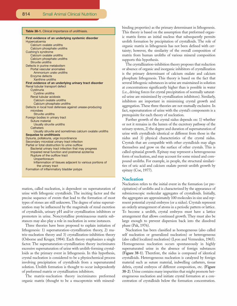
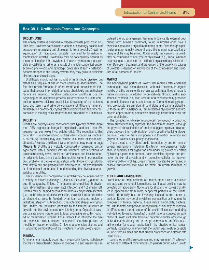
Nucleation has been classified as homogeneous (also calledself nucleation or generalized nucleation) or heterogeneous(also called localized nucleation) (Lyon and Vermeulen, 1965).Homogeneous nucleation occurs spontaneously in highlysupersaturated urine in the absence of foreign substances(Figure 38-1). Therefore, the nidus is composed of identicalcrystalloids. Heterogeneous nucleation is catalyzed by foreignmaterial such as suture material, indwelling catheters, tissuedebris, crystal embryos of different composition, etc. (Figure38-2). Urine contains many impurities that might promote het-erogeneous nucleation and initiate crystal formation at a con-centration of crystalloids below the formation concentration.
Small Animal Clinical Nutrition814
Table 38-1. Clinical importance of urolithiasis.
First evidence of an underlying systemic disorderHypercalcemia
Calcium oxalate urolithsCalcium phosphate uroliths
Cushing’s syndromeCalcium oxalate urolithsCalcium phosphate urolithsStruvite uroliths
Defects in purine metabolismPortal vascular anomalies
Ammonium urate urolithsEnzyme defects
Xanthine urolithsFirst evidence of an underlying urinary tract disorderRenal tubular transport defect
CystinuriaCystine uroliths
Renal tubular acidosisCalcium oxalate urolithsCalcium phosphate uroliths
Defects in local host defenses against urease-producing microbes
Struvite urolithsForeign bodies in urinary tract
Suture materialUsually struvite uroliths
CathetersUsually struvite and sometimes calcium oxalate uroliths
Sequelae to urolithiasisDysuria, pollakiuria, urge incontinenceSecondary microbial urinary tract infectionPartial or total obstruction to urine outflow
Bacterial urinary tract infection that may progressImpaired renal function and postrenal azotemiaRupture of the outflow tract
UroperitoneumInflammation of tissues adjacent to various portions of
the urinary tractFormation of inflammatory bladder polyps
815Introduction to Canine Urolithiasis
UROLITHIASISThe urinary system is designed to dispose of waste products in sol-uble form. However, some waste products are sparingly soluble andoccasionally precipitate out of solution to form crystals. Growth oraggregation of microscopic crystals may lead to formation ofmacroscopic uroliths. Urolithiasis may be conceptually defined asthe formation of uroliths anywhere in the urinary tract from less sol-uble crystalloids of urine as a result of multiple congenital and/oracquired physiologic and pathologic processes. If such crystalloidsbecome trapped in the urinary system, they may grow to sufficientsize to cause clinical signs.
Urolithiasis should not be thought of as a single disease, butrather as a sequela of one or more underlying abnormalities. Thefact that urolith formation is often erratic and unpredictable indi-cates that several interrelated complex physiologic and pathologicfactors are involved. Therefore, detection of uroliths is only thebeginning of the diagnostic process. Determination of urolith com-position narrows etiologic possibilities. Knowledge of the patient’sfood, and serum and urine concentrations of lithogenic minerals,crystallization promoters, crystallization inhibitors and their interac-tions aids in the diagnosis, treatment and prevention of urolithiasis.
UROLITHSUroliths are polycrystalline concretions that typically contain morethan 95% organic or inorganic crystalloids, and less than 5%organic matrices (weight vs. weight ratio). (The exception to thisgenerality is infection-induced uroliths which contain as much as50% matrix). Uroliths may also contain a number of minor con-stituents. A variety of different types of uroliths may occur in dogs(Figure 1). Uroliths are typically composed of organized crystalaggregates with a complex internal structure. Cross sections ofuroliths frequently reveal nuclei and laminations, and less frequent-ly radial striations. Urine that bathes uroliths varies in composition(and probably in degree of saturation with lithogenic crystalloids)from day to day and perhaps from hour to hour. This phenomenonis of conceptual importance in understanding the physical charac-teristics of uroliths.
The incidence and composition of uroliths may be influenced bya variety of factors including: 1) species, 2) breed, 3) gender, 4)age, 5) geography, 6) food, 7) anatomic abnormalities, 8) physio-logic abnormalities, 9) urinary tract infection and 10) urinary pH.Uroliths may be named according to mineral composition, location(i.e., nephroliths, ureteroliths, cystoliths, vesical calculi, urethroliths)or shape (i.e., smooth, faceted, pyramidal, laminated, mulberry,jackstone, staghorn or branched). Characteristic shapes of crystalsand uroliths are influenced primarily by the internal structure ofcrystals and the environment in which they form. Crystals of calci-um oxalate monohydrate tend to fuse, producing smoothly round-ed or mammillated uroliths. Local factors that influence the sizeand shape of uroliths include: 1) number of uroliths present, 2)mobility or fixation of uroliths, 3) flow characteristics of urine and4) anatomic configuration of the structure in which uroliths grow.
MINERALA mineral is a naturally occurring, inorganically formed substancethat has a characteristic chemical composition and usually has an
ordered atomic arrangement that may influence its external geo-metric form. Minerals commonly found in uroliths often have achemical name and a crystal (or mineral) name. Even though a par-ticular mineral usually predominates, the mineral composition ofmany uroliths may be mixed. Occasionally, the center of a urolithmay be composed of one type of crystalloid (e.g., silica), whereasouter layers are composed of a different crystalloid (especially stru-vite). Detection, treatment and prevention of the underlying causesof urolithiasis depend on knowledge of the composition and struc-ture of all portions of uroliths.
MATRIXThe nondialyzable portion of uroliths that remains after crystallinecomponents have been dissolved with mild solvents is organicmatrix. Uroliths consistently contain variable quantities of organicmatrix substances in addition to crystalloids. Organic matrix sub-stances identified in human uroliths and experimentally producedin animals include matrix substances A, Tamm-Horsfall glycopro-tein, uromucoid, serum albumin and alpha and gamma globulins.Of these, matrix substance A, Tamm-Horsfall glycoprotein and uro-mucoid appear to be quantitatively more significant than alpha andgamma globulins.
The complex of diverse mucoprotein compounds composingmatrix substances may represent the skeleton of uroliths. Althoughthe physical characteristics of uroliths suggest organized relation-ships between the matrix skeleton and crystalline building blocks,the role of each of these components in formation, retention andgrowth of uroliths is still poorly understood.
Organic matrix may affect urolith formation by one or more ofseveral mechanisms including: 1) sites of heterogeneous nucle-ation, 2) templates for organizing and modifying growth of crystals3) binding agents that cement urolith particles together and pro-mote retention of crystals and 4) protective colloids that preventfurther growth of uroliths. Organic matrix may also be composed ofpassive substances that have no effect on urolith formation orgrowth.
NUCLEI AND LAMINATIONSExamination of cross sections of uroliths often reveals a nucleusand adjacent peripheral laminations. Laminated uroliths may bedetected by radiography. Nuclei are focal points (or cores) that dif-fer in appearance from more peripheral portions of the urolith.Nuclei are usually but not invariably located in the center ofuroliths. Nuclei may be of crystalline composition or they may becomposed of foreign material, tissue debris, blood clots, bacteria,etc. The mineral composition of crystalline nuclei may be identicalor different from the remainder of the urolith. Nuclei surrounded bywell-defined layers (or lamellae) of solid material suggest an earlyphase of urolith evolution. However, crystalline nuclei large enoughto be detected visually are too large to represent an initial crys-talline nidus for crystal nucleation in the physiochemical sense.Centrally located nuclei imply that the urolith was freely accessibleto urine from all sides and that growth proceeded at a similar rateon all sides.
Laminated uroliths are common and may represent: 1) alternat-ing bands of different mineral types, 2) periods during which urolith
Box 38-1. Urolithiasis Terms and Concepts.

These substances may be thought of as facilitators or potentia-tors of crystallization. Any crystal type may be a potential nidusfor nucleation of another crystal type. A greater degree ofsupersaturation (i.e., a higher formation product) is required forhomogeneous nucleation than for heterogeneous nucleation.Once nucleation has occurred, however, crystal growth canoccur at any degree of supersaturation (even at metastability).
Undersaturated SolutionsAn undersaturated solution contains a sufficiently low concen-tration of a crystalloid to permit dissolution of additional quan-tities of the crystalloid. Urine is undersaturated when the soluteconcentration (or activity product) is less than the solubility ofthe solute in question. Formation of urine that is undersaturat-ed with lithogenic crystalloids may permit varying degrees ofurolith dissolution.
Saturated SolutionsSaturated solutions are in equilibrium with undissolved solute
at a given temperature. Saturated solutions contain so muchdissolved substances that no more can be dissolved at a giventemperature. With respect to urine, the saturation concentra-tion is that concentration of a crystalloid that remains un-changed when the urine is mixed with uroliths (or the solidphase) containing that crystalloid. The saturation of salts inurine is influenced by several variables including pH, ionicstrength and temperature.
Supersaturated SolutionsA supersaturated solution is more saturated with a substance ata given temperature than would be normally expected (i.e., it isany concentration greater than the saturation concentration).Supersaturated urine contains a greater concentration of a crys-talloid (cystine, phosphate, calcium, ammonium, etc.) than theassociated solvent (water) would be predicted to be able to nor-mally hold in solution. Supersaturation can vary in degree.Urine is metastable at lower levels of supersaturation. At high-er levels of supersaturation, however, urine becomes unstable
Small Animal Clinical Nutrition816
growth occurred without interruption or3) alternating periods of precipitation ofminerals and gel. Although a difference inappearance between two consecutivelayers should prompt suspicion of differ-ences in composition, this is not alwaysthe case.
MATRIX CONCRETIONSBy definition, a urolith must contain someminerals. However, concretions com-posed primarily (more than 65%) ofmatrix may occur. These concretions,commonly called matrix stones, oftenoccur in the urethra of male cats andsheep, and sometimes occur in dogs andpeople. They may form a cast of that por-tion of the excretory pathway in whichthey are formed (e.g., urethral plugs),implying a rapid rate of formation. Indogs, matrix concretions usually occursecondary to bacterial infections.
COMPOUND UROLITHSCompound uroliths have one or more lay-ers of mineral composition (e.g., struvite)different from minerals identified in thenucleus (e.g., calcium oxalate).
MIXED UROLITHSMixed uroliths contain more than onemineral, neither of which composes atleast 70% of the urolith, but without anucleus or well-defined laminations.
The Bibliography for Box 38-1 can be found at www.markmorris.org.
Figure 1. Different mineral types of canine uroliths illustrating common sizes, shapes andsurface characteristics. 1) Calcium oxalate dihydrate; 2) Calcium oxalate dihydrate; 3)Calcium oxalate monohydrate; 4) Calcium oxalate monohydrate; 5) Calcium oxalate mono-hydrate; 6) Calcium oxalate dihydrate; 7) Cystine; 8) Cystine; 9) Ammonium urate (left urolithhas been bisected to illustrate laminations); 10) Ammonium urate; 11) Ammonium urate; 12)Struvite; 13) Struvite; 14) Compound urolith with a nidus of calcium oxalate monohydratesurrounded by a shell of struvite and calcium carbonate apatite; 15) Compound urolith with anidus of silica surrounded by shells containing a mixture of calcium oxalate, silica andammonium urate; 16) Silica; 17) Silica; 18) Silica; 19) Struvite that has the shape of the uri-nary bladder and proximal urethra; 20) Struvite that has the shape of the renal pelvis andproximal ureter.
Box 38-1 continued
1 2
3
4
5
6
10
11
12
13
14
15
7 8 9 16 17
18
19
20
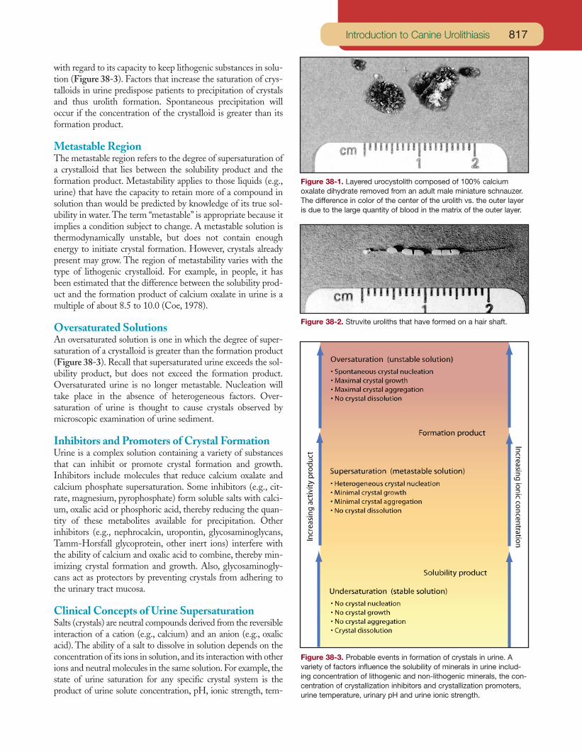
with regard to its capacity to keep lithogenic substances in solu-tion (Figure 38-3). Factors that increase the saturation of crys-talloids in urine predispose patients to precipitation of crystalsand thus urolith formation. Spontaneous precipitation willoccur if the concentration of the crystalloid is greater than itsformation product.
Metastable RegionThe metastable region refers to the degree of supersaturation ofa crystalloid that lies between the solubility product and theformation product. Metastability applies to those liquids (e.g.,urine) that have the capacity to retain more of a compound insolution than would be predicted by knowledge of its true sol-ubility in water. The term “metastable” is appropriate because itimplies a condition subject to change. A metastable solution isthermodynamically unstable, but does not contain enoughenergy to initiate crystal formation. However, crystals alreadypresent may grow. The region of metastability varies with thetype of lithogenic crystalloid. For example, in people, it hasbeen estimated that the difference between the solubility prod-uct and the formation product of calcium oxalate in urine is amultiple of about 8.5 to 10.0 (Coe, 1978).
Oversaturated SolutionsAn oversaturated solution is one in which the degree of super-saturation of a crystalloid is greater than the formation product(Figure 38-3). Recall that supersaturated urine exceeds the sol-ubility product, but does not exceed the formation product.Oversaturated urine is no longer metastable. Nucleation willtake place in the absence of heterogeneous factors. Over-saturation of urine is thought to cause crystals observed bymicroscopic examination of urine sediment.
Inhibitors and Promoters of Crystal FormationUrine is a complex solution containing a variety of substancesthat can inhibit or promote crystal formation and growth.Inhibitors include molecules that reduce calcium oxalate andcalcium phosphate supersaturation. Some inhibitors (e.g., cit-rate, magnesium, pyrophosphate) form soluble salts with calci-um, oxalic acid or phosphoric acid, thereby reducing the quan-tity of these metabolites available for precipitation. Otherinhibitors (e.g., nephrocalcin, uropontin, glycosaminoglycans,Tamm-Horsfall glycoprotein, other inert ions) interfere withthe ability of calcium and oxalic acid to combine, thereby min-imizing crystal formation and growth. Also, glycosaminogly-cans act as protectors by preventing crystals from adhering tothe urinary tract mucosa.
Clinical Concepts of Urine SupersaturationSalts (crystals) are neutral compounds derived from the reversibleinteraction of a cation (e.g., calcium) and an anion (e.g., oxalicacid). The ability of a salt to dissolve in solution depends on theconcentration of its ions in solution, and its interaction with otherions and neutral molecules in the same solution. For example, thestate of urine saturation for any specific crystal system is theproduct of urine solute concentration, pH, ionic strength, tem-
817Introduction to Canine Urolithiasis
Figure 38-1. Layered urocystolith composed of 100% calciumoxalate dihydrate removed from an adult male miniature schnauzer.The difference in color of the center of the urolith vs. the outer layeris due to the large quantity of blood in the matrix of the outer layer.
Figure 38-2. Struvite uroliths that have formed on a hair shaft.
Figure 38-3. Probable events in formation of crystals in urine. Avariety of factors influence the solubility of minerals in urine includ-ing concentration of lithogenic and non-lithogenic minerals, the con-centration of crystallization inhibitors and crystallization promoters,urine temperature, urinary pH and urine ionic strength.
Small Animal Clinical Nutrition818
perature and preformed chemical complexes.To illustrate these principles, consider pure water as a solu-
tion and calcium oxalate as a salt. Small amounts of calciumoxalate added to water dissolve completely because water isundersaturated with calcium and oxalic acid ions. As more cal-cium oxalate is added, the water’s capacity to dissolve addition-al calcium oxalate is decreased until the solution becomes satu-rated. In this context, saturation of the solution with calciumand oxalic acid ions occurs when no additional calcium oxalatecan be dissolved at a given pH and temperature of the solution.If additional calcium oxalate is added, it will appear as a solid.
As in water, calcium oxalate can also be dissolved in under-saturated urine. However, unlike water, urine is a complex solu-tion containing a unique combination of ionic and nonionicmolecules that may increase the solubility of calcium oxalate.Therefore, calcium oxalate added beyond the point of satura-tion will remain in solution. Thus, the solution becomes super-saturated with calcium and oxalic acid ions. Supersaturation isconceptually significant because the solution contains enoughenergy to form solids from dissolved ions (i.e., it is thermody-namically unstable). When supersaturated, the solution must“struggle” to maintain the homogeneous nature between theions it contains. One method by which the solution returns tothermodynamic stability is by concentrating excess calcium andoxalic acid ions as solids or crystals on pre-existing surfaces ortemplates (e.g., other crystals or foreign material). This phe-nomenon is called heterogeneous nucleation. However, if thesolution becomes oversaturated by addition of more calciumand oxalic acid ions, calcium oxalate crystals will form withoutan existing template (so-called homogeneous nucleation). Aftercrystals have formed, available thermodynamic energy favorscrystal growth whereby free ions become incorporated into thecrystals. Crystal growth continues until ions in solution becomedepleted, allowing the solution to return to thermodynamic sta-bility (or saturation). Crystals retained in the urinary tract maygrow (the second phase of urolith formation).
Urine is a complex solution containing “inert” ions (i.e., sul-fate, sodium, potassium, magnesium) unlikely to chemicallybond with calcium and oxalic acid. In this way, they increasecalcium oxalate solubility. The negative ions (e.g., sulfate) sur-round positive calcium ions, and the positive ions (e.g., sodium,potassium, magnesium) surround negative oxalic acid ions. Thenet effect is a decrease in attraction between calcium and oxal-ic acid ions. Because calcium and oxalic acid ion interaction isrequired for crystal formation, the solubility of calcium oxalateincreases as the concentration of “inert” ions increases.
Supersaturation of urine with certain lithogenic ions also de-pends on another group of substances called “crystallizationinhibitors.” These include citric acid and pyrophosphates thatchelate calcium but remain dissolved in solution. Likewise, certainmucoproteins, glycosaminoglycans, glycoproteins (e.g., nephro-calcin) and other poorly identified substances may interact withcalcium. The result is a decrease in the amount of calcium avail-able to bind with oxalic acid (and phosphoric acid). It is of signif-icance that these inhibitors have been found to be deficient orabnormal in some calcium oxalate urolith-forming patients.
Activity ProductThe product of the chemical activities of two ionic materials iscalled the activity product. It is a mathematical expression usedto estimate the degrees of saturation (i.e., undersaturation,supersaturation or oversaturation) of a dog’s urine with litho-genic minerals (Figure 38-3). In addition to concentration ofminerals, it encompasses other variables including urinary pHand ionic strength of the solution (Pak et al, 1977). Activityproduct encompasses solubility product and formation product.Activity products are calculated by measuring total concentra-tions of major ionizable solutes in urine. For efficiency, comput-er programs are commonly used to aid calculation of ion con-centrations and activity products (Brown et al, 1994).
Solubility ProductThe solubility product is a type of activity product reflecting theurine’s ability to dissolve a known concentration of lithogenicions at variable but known pH and temperature. It is constantfor each mineral component at a given temperature and pH.Urine is saturated when the solubility product value is reached.Below this value, urine is undersaturated with lithogenic ions;above this value urine is supersaturated. When devising dietaryand medical protocols to dissolve or prevent urolith formation,the goal is to achieve an activity product less than the solubili-ty product (or a state of undersaturation) (Figure 38-3).
Formation ProductThe formation product is a type of activity product reflectingthe concentration of ions at which precipitation of solute(homogeneous nucleation and eventually crystal formation)occurs at a given pH and temperature. It is the upper limit ofmetastability. Urinary pH may affect the ionization of someurine constituents and thus their solubility. If urinary pH variesduring the day, urine may be intermittently supersaturated oroversaturated. Ion activities above the formation product areassociated with an unstable state of oversaturation resulting inspontaneous crystal formation and rapid crystal growth.Because this condition may be influenced by the product ofseveral factors (including the time of incubation, a crystallizablematrix and inhibitors of nucleation) in addition to the concen-tration of lithogenic crystalloids, it is commonly called the for-mation product. In people, as mentioned above, the formationproduct for calcium oxalate is approximately 8.5 to 10 timesgreater than its solubility product (Coe, 1978). This indicatesthat urine, because of the addition of a variety of crystallizationinhibitors, must be saturated at least eight times above the sol-ubility product before crystals will form. In general, urine ofurolith formers is more supersaturated with respect to the con-stituents of their uroliths than is the urine of normal subjects.
PATIENT ASSESSMENT
History and Physical ExaminationThe history of dogs with urolithiasis depends on: 1) anatomiclocation(s) of uroliths, 2) duration of uroliths in specific loca-
tion(s), 3) physical characteristics of uroliths (size, shape, num-ber), 4) secondary urinary tract infection (UTI) and virulenceof infecting organism(s) and 5) presence of concomitant dis-eases in the urinary tract and other body systems. After a diag-nosis of urolithiasis has been confirmed, the history and physi-cal examination should focus on detection of any underlying ill-ness that may predispose the dog to urolith formation.
A dietary history should also be obtained for all patients withurolithiasis, with the objective of identifying risk factors thatpredispose the patient to specific mineral types. Likewise, own-ers should be questioned about vitamin-mineral supplements,previous illnesses and medications that may predispose thepatient to various types of uroliths.
Signs typical of lower urinary tract disease include dysuria,pollakiuria, hematuria, urge incontinence, paradoxical inconti-nence and voiding small uroliths during micturition. Signs ofuremia may occur if urine flow has been obstructed for a suffi-cient period, or if there is extravasation of urine into the peri-toneal cavity due to rupture of the excretory pathways.
Signs of upper tract disease include painless hematuria andpolyuria if sufficient nephrons have impaired function. Ab-dominal pain may occur if there is overdistention of the renalpelvis with urine due to outflow obstruction (Table 38-2).Many patients with uroliths have no clinical signs. Absence ofsigns is especially common in patients with nephroliths.
If gross hematuria is present, determining when during theprocess of micturition it is most severe may be of value in local-izing its source. If hematuria occurs throughout micturition,lesions (including uroliths) may be present in the kidneys,ureters, urinary bladder, prostate gland and/or urethra. If hema-turia occurs primarily at the end of micturition, lesions of theventral bladder wall or intermittent renal hematuria should besuspected. If hematuria occurs at the beginning or is independ-ent of micturition, lesions in the urethra or genital tract shouldbe suspected.
Digital palpation of the entire urethra, including evaluationby rectal examination, may reveal urethroliths or urolithslodged in the bladder neck. A firm, non-yielding mass may bepalpated in the urinary bladder if a solitary urolith is present; agrating sensation confined to the bladder may be detected ifmultiple uroliths are present. It may be impossible to palpatesmall or solitary urocystoliths if the bladder wall is contractedand/or thickened due to inflammation. Likewise, it may beimpossible to palpate uroliths in a distended or overdistendedbladder. In this situation, the bladder should be repalpated afterurine has been eliminated by voiding, manual compression ofthe bladder, cystocentesis or catheterization. One should sus-pect urethroliths when urethral catheters cannot be advancedinto the bladder. However, inability to advance a catheterthrough the urethra may also be associated with urethral stric-tures or space occupying lesions that partially or totally occludethe urethral lumen.
In the absence of infection or outflow obstruction, abnormal-ities are usually not associated with nephroliths unless bilateralnephroliths are associated with sufficient renal damage to causeuremia. If infection or obstruction is present, there may be pain
in the area of the kidneys and/or palpable enlargement of theaffected kidney(s). Concomitant bacterial pyelonephritis maybe associated with polysystemic signs due to sepsis.
Diagnostic StudiesUrinalysisResults of urinalysis are usually characterized by abnormalitiestypical of inflammation (pyuria, proteinuria, hematuria andincreased numbers of epithelial cells), which may or may not beassociated with infection. Whereas urease-producing microbes(staphylococci, Proteus spp., ureaplasmas) may cause infection-induced struvite (magnesium ammonium phosphate) urolithsto form, opportunistic bacteria that are not lithogenic (e.g.,Escherichia coli and streptococci) may colonize the urinary tractas a result of urolith-induced alterations in local host defenses.Quantitative urine culture of all patients with uroliths is recom-
819Introduction to Canine Urolithiasis
Table 38-2. Clinical signs of uroliths that may be associatedwith urinary system dysfunction.
UrethrolithsAsymptomaticDysuria, pollakiuria, urge incontinence and/or periuriaGross hematuriaPalpable urethral urolithsSpontaneous voiding of small urolithsPartial or complete urine outflow obstruction
Overflow incontinenceAnuriaPalpation of an overdistended and painful urinary bladderUrinary bladder rupture, abdominal distention and
abdominal painSigns of postrenal azotemia (anorexia, depression, vomiting
and diarrhea)Signs associated with concurrent urocystoliths, ureteroliths
and/or renolithsUrocystolithsAsymptomaticDysuria, pollakiuria and urge incontinenceGross hematuriaPalpable bladder urolithsPalpably thickened urinary bladder wallPartial or complete urine outflow obstruction of bladder neck
(See Urethroliths.)Other signs associated with concurrent urethroliths, ureteroliths
and/or renolithsUreterolithsAsymptomaticGross hematuriaConstant abdominal painUnilateral or bilateral urine outflow obstruction
Palpably enlarged kidney(s)Signs of postrenal azotemia (See Urethroliths.)
May have other signs associated with concurrent urethroliths, urocystoliths and/or nephroliths
NephrolithsAsymptomaticGross hematuriaConstant abdominal painSigns of systemic illness if generalized renal infection is present
(anorexia, depression, fever and polyuria)Palpably enlarged kidney(s)Signs of postrenal azotemia (See Urethroliths.)Other signs associated with concurrent urethroliths, urocys-
toliths and/or ureteroliths
mended because knowledge of bacterial type is important inpredicting the mineral composition of uroliths, and in selectingan appropriate antimicrobial agent for treatment.
The pH of urine obtained from patients with uroliths is vari-able; however, it may become persistently alkaline if secondaryinfection with urease-producing bacteria occurs. The signifi-cance of a single urinary pH measurement should be interpret-ed cautiously because there are significant fluctuations through-out the day, especially with respect to the time, amount andtypes of food consumption. In general, magnesium ammoniumphosphate and calcium phosphate uroliths are associated withalkaline urine, whereas ammonium urate, sodium urate, uricacid, calcium oxalate, cystine and silica uroliths tend to be asso-ciated with acidic urine.
The advent of effective dietary and medical protocols to dis-solve and prevent uroliths in dogs and cats has resulted inrenewed interest in detection and interpretation of crystalluria.Evaluation of urine crystals may aid in: 1) detection of disorderspredisposing animals to urolith formation, 2) estimation of themineral composition of uroliths and 3) evaluation of the effec-tiveness of dietary and medical protocols initiated to dissolve orprevent uroliths.
Crystals form only in urine that is or recently has been super-saturated with lithogenic substances. Therefore, crystalluriarepresents a risk factor for urolithiasis. However, detection ofurine crystals is not synonymous with urolithiasis and clinicalsigns associated with uroliths. Nor are urine crystals irrefutableevidence of a urolith-forming tendency. For example, crystal-luria that occurs in individuals with anatomically and function-ally normal urinary tracts is usually harmless because the crys-
tals are eliminated before they aggregate or grow to sufficientsize to interfere with normal urinary function. In addition, crys-tals that form after elimination or removal of urine from thepatient often are of no clinical importance. Identification ofcrystals that have formed in vitro does not justify therapy.
Detection of some types of crystals (e.g., cystine and ammo-nium urate) in clinically asymptomatic patients, frequent detec-tion of large aggregates of crystals (e.g., calcium oxalate or mag-nesium ammonium phosphate) in apparently normal individu-als, or detection of any form of crystals in fresh urine collectedfrom patients with confirmed urolithiasis may be of diagnostic,prognostic and therapeutic importance. Large crystals andaggregates of crystals are more likely to be retained in the uri-nary tract, and therefore may be of greater clinical significancethan small or single crystals.
Although there is not a direct relationship between crystal-luria and urolithiasis, detection of crystals in urine is proof thatthe urine sample is oversaturated with lithogenic substances.However, oversaturation may occur as a result of in vitro eventsin addition to or instead of in vivo events. Therefore, care mustbe used not to overinterpret the significance of crystalluria. Invivo variables that influence crystalluria include: 1) the concen-tration of lithogenic substances in urine (which in turn is influ-enced by their rate of excretion and the volume of water inwhich they are excreted), 2) urinary pH (Table 38-3), 3) thesolubility of lithogenic substances and 4) excretion of diagnos-tic agents (e.g., radiopaque contrast media) and medications(e.g., sulfonamides).
In vitro variables that influence crystalluria include: 1) tem-perature, 2) evaporation, 3) urinary pH and 4) the technique of
Small Animal Clinical Nutrition820
Table 38-3. Common characteristics of selected urine crystals.
Urinary pH at which crystals commonly form
Crystal types Appearances Acidic Neutral AlkalineAmmonium urate Yellow-brown spherulites, thorn apples + + +Amorphous urates Amorphous or spheroidal yellow-brown structures + ± -Bilirubin Reddish-brown needles or granules + - -Calcium carbonate Large yellow-brown spheroids with radial striations, or - ± +
small crystals with spheroidal or dumbbell shapesCalcium oxalate dihydrate Small colorless envelopes (octahedral form) + + ±Calcium oxalate monohydrate Small spindles “hempseed” or dumbbells + + ±Calcium phosphate Amorphous or long thin prisms ± + +Cholesterol Flat colorless plates with corner notch + + -Cystine Flat colorless hexagonal plates + + ±Hippuric acid Four- to six-sided colorless elongated plates or prisms + + ±
with rounded cornersLeucine Yellow-brown spheroids with radial and concentric laminations + + -Magnesium ammonium phosphate Three- to six-sided colorless prisms ± + +Sodium urate Colorless or yellow-brown needles or slender prisms, + ± -
sometimes in clusters or sheavesSulfa metabolites Sheaves of needles with central or eccentric binding, + ± -
sometimes fan-shaped clustersTyrosine Fine colorless or yellow needles arranged in sheaves or rosettes + - -Uric acid Diamond or rhombic rosettes, or oval plates, structures + - -
with pointed ends, occasionally six-sided platesXanthine Yellow-brown amorphous, spheroidal or ovoid structures + ± -Key: + = crystals commonly occur at this pH, ± = crystals may occur at this pH, but are more common at the other pH, - = crystals areuncommon at this pH.
specimen preparation (e.g., centrifugation vs. noncentrifugationand volume of urine examined) and preservation. As men-tioned above, in vitro changes that occur after urine collectionmay enhance formation or dissolution of crystals. Although invitro changes may be used to enhance detection of certain typesof crystals (e.g., acidification to cause precipitation of cystine),in vitro crystal formation may have no clinical relevance to invivo formation of crystals in urine. When knowledge of in vivourine crystal type is especially important, fresh, warm speci-mens should be serially examined. The number, size and struc-
ture of crystals should be evaluated, as well as their tendency toaggregate.
Urinary pH influences the formation and persistence of sev-eral types of crystals.Therefore, it is often useful to consider pHwhen interpreting crystalluria (Table 38-3). Different crystalstend to form and persist in certain urinary pH ranges, althoughthere are exceptions. Exceptions may be related to large con-centrations of lithogenic substances in urine or recent in vivo orin vitro changes in urinary pH.
Refrigeration is an excellent method to preserve many phys-
821Introduction to Canine Urolithiasis
Figure 38-4. Photomicrographs of common crystals found in urine sediment. Calcium oxalate monohydrate (dumbbell form, large arrow) andcalcium oxalate dihydrate (octahedral form, small arrows) (Top, Left). Calcium oxalate dihydrate; octahedral form (Top, Right). Magnesiumammonium phosphate (struvite); prisms (Middle, Left). Cystine; flat, colorless hexagonal plates (Middle, Right). Ammonium urate; thorn appleform (Bottom, Left). Amorphous xanthine; spheroids (Bottom, Right).

ical, chemical and morphologic properties of urine sediment.However, refrigeration must be used with caution when evalu-ating crystalluria from qualitative and quantitative standpoints.Although refrigeration of urine samples is likely to enhanceformation of various types of crystals, this phenomenon mayhave no relationship to events occurring in the patient’s body.
Crystalluria may also be influenced by food, including waterintake. Dietary influence on crystalluria is of diagnostic impor-tance because urine crystal formation that occurs while patientsare consuming hospital foods may be dissimilar to urine crystalformation that occurs when patients are consuming foods fedat home.
Microscopic evaluation of urine crystals should not be usedas the sole criterion to predict the mineral composition ofmacroliths in patients with confirmed urolithiasis (Table 38-3and Figure 38-4). Only quantitative analysis can provide defin-itive information about the mineral composition of the entireurolith. However, interpretation of crystalluria in light of other
clinical findings often allows the clinician to tentatively identi-fy the mineral composition of uroliths, especially their outer-most layers. Subsequent reduction or elimination of crystals bytherapy provides a useful index of the efficacy of medical anddietary protocols designed to dissolve or prevent uroliths.
Radiography and UltrasonographyThe primary objective of radiographic or ultrasonographic eval-uation of patients suspected of having uroliths is to determinethe site(s), number, density and shape of uroliths. However, thesize and number of uroliths are not a reliable index of the prob-able efficacy of therapy. After urolithiasis has been confirmed,radiographic or ultrasonographic evaluation also aids in detec-tion of predisposing abnormalities (Table 38-4).
The size, number, location and mineral composition ofuroliths influence their radiographic and ultrasonographicappearance. Most uroliths greater than 3 mm in diameterhave varying degrees of radiodensity, and therefore can bedetected by survey abdominal radiography or ultrasonography(Osborne et al, 1995). Very small uroliths (<3 mm in diame-ter) may not be visualized by survey radiography or ultra-sonography. Uroliths greater than 1 mm in diameter can usu-ally be detected by double-contrast cystography, providedexcessive contrast medium is not used (Feeney et al, 1999).Table 38-5 lists relative densities of common uroliths basedon survey radiography (Feeney et al, 1999). Because of signif-icant variation, the radiodensity of uroliths is not by itself areliable index of mineral composition.
Uroliths greater than 3 mm in diameter are not commonlyradiolucent. An exception to this generality is uroliths com-posed of 100% ammonium or sodium urate or uric acid.However, in our experience many ammonium urate uroliths ofdogs are marginally radiodense. This finding may be related toa variable quantity of phosphates and other minerals in urateuroliths of dogs.
Matrix uroliths may be radiolucent or have some radiodensi-
Small Animal Clinical Nutrition822
Table 38-4. Advantages and disadvantages of survey radiography, double-contrast radiography and ultrasonography in assessing uroliths.
Survey Double-contrast Parameters radiography radiography UltrasonographyAssessment of urethroliths Yes, if radiodense Indirectly* PoorAssessment of radiolucent urocystoliths Unreliable Yes YesDistinguishing blood clots from urocystoliths No Probably YesAssessment of laminated urocystoliths Best of the three methods Probably NoAssessment of other bladder disorders Unreliable Yes SometimesAssessment of urocystolith number Yes (>3 mm) Yes (>1 mm) Equipment and observer dependentAssessment of urocystolith size Yes (>3 mm) Yes (>1 mm) Equipment and observer dependentAssessment of urocystolith density Yes (>3 mm) No NoAssessment of urocystolith shape Yes (>3 mm) Yes (>1 mm) NoImmediate postsurgical assessment for uroliths Yes Not recommended** No (air artifacts) Risk of air artifact in bladder No Yes NoRisk of iatrogenic bacterial urinary tract infection No Yes NoExposure to ionizing radiation Yes Yes NoNecessary to remove hair No No OftenAuthors’ overall choice Screening Investigation Third choice*During transurethral catheterization. **Due to risk of iatrogenic bacterial urinary tract infection.
Table 38-5. Comparison of relative densities of commonuroliths detected by survey radiography.*
Mineral types Relative atomic number**Water 7.7Urate 6.9-7.7Struvite 9.81Cystine 10Silica 11.6Calcium oxalate dihydrate 13Calcium oxalate monohydrate 13.6Cortical bone 15Calcium phosphate 15.9*Adapted from Feeney DA, Weichselbaum RC, Jessen CR, etal. Imaging canine urocystoliths: Detection and prediction ofmineral content. Veterinary Clinics of North America: SmallAnimal Practice 1999; 29: 59-72.**Effective atomic numbers (Zeff), which is the sum of differentelements in the urocystolith and is related to its mass.
ty. Blood clots are radiolucent and may be mistaken for radiolu-cent uroliths. Radiolucent uroliths may be readily distinguishedfrom blood clots when evaluated by two-dimensional, gray-scale ultrasonography. Uroliths are usually in the dependentportion of the bladder lumen, produce sharply marginatedshadows containing few echoes and are associated with acousticshadowing. Blood clots may be located anywhere in the blad-der lumen, typically have an irregular outline and indistinctmargins and are not associated with acoustic shadowing.
Uroliths that are radiodense on survey radiographs mayappear to be radiolucent when evaluated by positive-contrastradiography. This finding is related to the fact that manyuroliths are more radiodense than body tissue, but less radio-dense than the contrast material. A diagnosis of radiolucenturoliths should be based on their radiodensity compared withsoft tissues, and not their radiodensity compared with positive-contrast medium.
A urolith may be larger than that depicted by its radiodensi-ty if only a portion of it contains radiodense minerals.This phe-nomenon is most likely to occur with rapidly growing struviteuroliths that contain large quantities of matrix.
Hematology and Serum ChemistryHemograms of dogs with uroliths are usually normal unlessthere is concomitant generalized infection of the kidneys orprostate gland associated with leukocytosis. Microcytosis, ane-mia, target cells and leukocytosis have occasionally been associ-ated with portal vascular anomalies in dogs with and withouturate uroliths (Cornelius et al, 1975; Ewing et al, 1974;Griffiths et al, 1981; Rothuizen and van den Ingh, 1980).
Serum chemistry values are usually normal in patients withinfection-induced magnesium ammonium phosphate, cys-tine and silica uroliths unless obstruction of urine outflow orgeneralized renal infection leads to changes characteristic ofrenal failure. Although most patients with calcium oxalateand calcium phosphate uroliths are normocalcemic, some arehypercalcemic.
Calcium phosphate and sterile struvite uroliths may be asso-ciated with distal renal tubular acidosis characterized by hyper-chloremic (normal anion gap) metabolic acidosis, urinary pHvalues consistently greater than approximately 6 andhypokalemia.
A variety of biochemical alterations may exist in patientswith urate urolithiasis. The following changes may be observedin patients with urate uroliths due to congenital or acquiredhepatic disorders (Rothuizen and van den Ingh, 1980; Barrettet al, 1976; Marretta et al, 1981): 1) decreased urea nitrogenconcentrations, 2) decreased total protein and albumin concen-trations, 3) abnormal bile acid concentrations, 4) increased con-centrations of total bilirubin and fasting blood ammonia and 5)increased serum alanine aminotransferase and serum alkalinephosphatase enzyme activities. Dogs with portal vascularanomalies typically have reduced hepatic functional mass andaltered portal blood flow evidenced by abnormally elevated bileacid concentrations, prolonged sulfobromophthalein retentiontimes and abnormal ammonia tolerance tests (Griffiths et al,
1981; Rothuizen and van den Ingh, 1980; Barrett et al, 1976;Marretta et al, 1981; Center et al, 1985).
Urine ChemistryDetection of the underlying causes of specific types of urolithi-asis is often linked to evaluation of the biochemical composi-tion of urine. For best results, at least one and preferably twoconsecutive 24-hour urine samples should be collected becausedetermination of fractional excretion of many metabolites in“spot” urine samples does not accurately reflect 24-hourmetabolite excretion (Table 38-6).
Water consumption and hydration status must be consideredwhen interpreting laboratory results. Decreased water con-sumption and dehydration are associated with several alter-ations, including decreased renal clearance of metabolites andincreased urine specific gravity and urine solute concentrations(Taburu et al, 1993). Caution must be used in interpreting 24-hour excretion of solutes in the diagnosis and therapy of uro-lithiasis if hospitalized animals consume less water than in thehome environment.
Urine concentrations of potentially lithogenic metabolitesare also influenced by the amount and composition of foodconsumed, and whether urine was collected during conditionsof fasting or food consumption (Lulich et al, 1991, 1991a).Aldosterone secretion increases following food deprivation.Increased aldosterone secretion promotes renal tubular sodiumreabsorption and potassium excretion. As a consequence, plas-ma potassium concentration decreases, urinary potassium ex-cretion increases and urinary sodium and chloride excretiondecrease (Lulich et al, 1991a). Urinary calcium, magnesium anduric acid excretions are reduced during fasting. However, uri-nary excretion of phosphorus, oxalate and citrate are apparent-ly unaffected by fasting (Lulich et al, 1991a). In dogs, urinaryammonia, titratable acid and hydrogen ion excretion decreaseand urinary pH values increase when food is withheld (Lulichet al, 1991a; Lemieux and Plante, 1968). Therefore, values for24-hour urinary solute excretion may differ when measured fol-lowing food consumption vs. values obtained when food iswithheld.
Consumption of food stimulates gastric secretion of hydro-chloric acid. As a result, concentrations of chloride decrease andbicarbonate increase in venous blood draining the stomach.Total serum concentration of carbon dioxide increases. Theresulting metabolic alkalosis is commonly called the postpran-dial alkaline tide. Urinary pH will increase unless acidifyingsubstances are contained in the food. In a study of healthy bea-gles, eating was associated with increased urinary excretion ofhydrogen ions, ammonia, sodium, potassium, calcium, magne-sium and uric acid (Lulich et al, 1991a).
Laboratory results may be markedly affected by changes infoods fed in a home environment vs. different foods fed in ahospital environment. For example, urinary excretion of poten-tially lithogenic metabolites while animals consume foods fedin the hospital may be different from those excreted by animalseating at home. To determine the influence of home-fed foodson laboratory test results, consider asking clients to bring
823Introduction to Canine Urolithiasis

Small Animal Clinical Nutrition824
Table 38-6. Protocol for measuring 24-hour urinary excretion of various substances associated with urolithiasis.
Technique1. To allow for food acclimation, feed the patient either the food it was consuming just before urolith formation or a standard food at
home for 10 to 14 days. We commonly use Prescription Diet k/d Canine* as the standard food.2. If possible, house and feed the dog in the urine collection cage for at least one day before urine collection. As dogs become accli-
mated to their new environment, they are more likely to consume quantities of food and water similar to that consumed in their homeenvironment.
3. Begin each 24-hour urine collection period by removing urine from the urinary bladder by transurethral catheterization. This urine isdiscarded. Record the actual time that urine collection is initiated.
4. Weigh the dog.5. Then feed the dog its food as if at home. Water should be continuously available for consumption.6. Begin administering a broad-spectrum antibiotic that achieves high concentrations in urine to prevent catheter-induced urinary tract
infection. The dosage, dosing interval and route of administration should be based on manufacturer recommendations.7. Keep the patient in the collection cage during urine collection. When using metabolism cages designed for urine collection, catheteri-
zation of the urinary tract is unnecessary except at the end of the 24 hours. House-trained dogs may not voluntarily void in theircage. Bladder catheterization may be necessary to obtain urine from these dogs. Dogs may be catheterized as often as necessary tokeep them comfortable (usually every six to eight hours).
8. Catheterize the urinary bladder at the end of 24 hours to remove all urine. Save this urine.9. Record the exact time of collection termination.
10. Pool all urine collected during the 24-hour period in a single container and measure its volume.11. Thoroughly mix the pooled urine before removing aliquots for analysis.
Preservation1. Preservatives have different roles, but are often used to minimize bacterial growth, reduce chemical decomposition, solubilize con-
stituents that might otherwise precipitate out of solution, or decrease atmospheric oxidation of unstable compounds.2. The method of preservation may vary depending on the substances being measured and the tests used to measure them. Consult
the laboratory to determine the recommended method of preservation.3. Preservatives should not be added to some specimens because of possible interference with analytical methods.4. Refrigeration is a common method for preserving urine collected for analysis. Urine removed by intermittent catheterization can be
stored in a refrigerator in clean containers with screw top lids. Containers used for continuous collection beneath metabolism cagescan be surrounded by ice packs and then insulated. Refrigeration causes some minerals to precipitate out of solution.
5. Specimens can be acidified (add 10 ml of 1 N hydrochloric acid per liter to achieve a pH of 3 or less) to preserve oxalate and calciumfor analysis. However, acidified urine is unsuitable for measuring uric acid because it precipitates in acidic solutions.
Storage of selected analytes in urine1. No single preservative is ideal if multiple substances in urine are to be analyzed. To minimize degradation, we routinely collect urine
under conditions of refrigeration. Immediately following urine collection, preservatives are added to appropriate aliquots of urine forstorage until analysis.
2. Uric acid and xanthine: Aliquots of urine should be diluted (1 ml of urine with 19 ml of distilled water) to preserve uric acid and xan-thine. This mixture can then be frozen.
3. Ammonia: Aliquots of urine (3 to 5 ml) may be frozen for up to 30 days.4. Oxalate: Aliquots of urine (2 ml) are diluted with 1 N hydrochloric acid (1.66 ml) and then frozen.
Calculations1. Calculating 24-hour urine volume
a. Although 24-hour urine specimens are recommended to minimize the effects of short-term biologic variations in mineral excretion,collecting perfectly timed 24-hour samples may be difficult. The following formula can be used to adjust actual urine volume to a24-hour period: 1,440 ÷ actual time interval (minutes) x urine volume (1,440 = number of minutes in 24 hours).
b. Example: A 24-hour urine collection was started at 9:30 a.m. and ended the following day at 8:30 a.m. A total of 350 ml of urinewere collected during this period. What is the 24-hour urine volume? 1,440 ÷ 1,380 x 350 = 356.2 ml.
2. Converting mmol/l to mg/dla. Scientists are striving to adopt a uniform system of measurement termed the System International d ‘Unites to standardize meas-
urements. In this system, concentration is often expressed as moles, millimoles or micromoles of a substance per liter of fluid.Most normal values in the United States are expressed as mg/dl. The following formula can be used to convert mmol/l to mg/dl:mmol/l x atomic weight of substance ÷ 10. The atomic weights of elements can be found in the periodic tables of general chem-istry books.
b. Example: The concentration of calcium from a 24-hour urine sample was 1.35 mmol/l. Convert this value to mg/dl for comparisonwith normal values. The atomic weight of calcium is 40.08. 1.35 mmol/l x 40.08 ÷ 10 = 5.4 mg/dl.
3. Calculating mg/kg/24-hour or mEq/kg/24-hour excretiona. Excretion of metabolites is often expressed on a per kg basis to standardize excretion for dogs of different weights. The following
formula can be used to standardize excretion rates: Concentration of substance x 24-hour urine volume ÷ body weight in kg. Theunits used to express the volume of urine and the concentration of the substance evaluated must be the same.
b. Example: The concentration of calcium in a 24-hour urine sample was 5.4 mg/dl. A total of 356.2 ml of urine was collected. Thedog weighed 10 kg. What is the daily calcium excretion on a per kg basis? First, express the volume of urine collected in the sameunits as the concentration of the substance measured. The 356.2 ml = 3.562 dl; therefore, 5.4 mg/dl x 3.562 dl ÷ 10 kg = 1.92mg/kg/24 hours.
Additional considerations1. Midpoint blood samples. Evaluation of blood during the midpoint of a 24-hour urine collection may help determine if changes in urine
concentration reflect changes in serum or plasma concentration of analytes. This information can help detect underlying causes andmechanisms of abnormal mineral excretion. Likewise, evaluation of blood concentrations of some hormones (i.e., parathyroid hor-mone, calcitriol, etc.) may be helpful in determining the role of hormones in the regulation of mineral excretion.
home-fed foods for use during periods of diagnostic hospital-ization (Osborne et al, 1990).
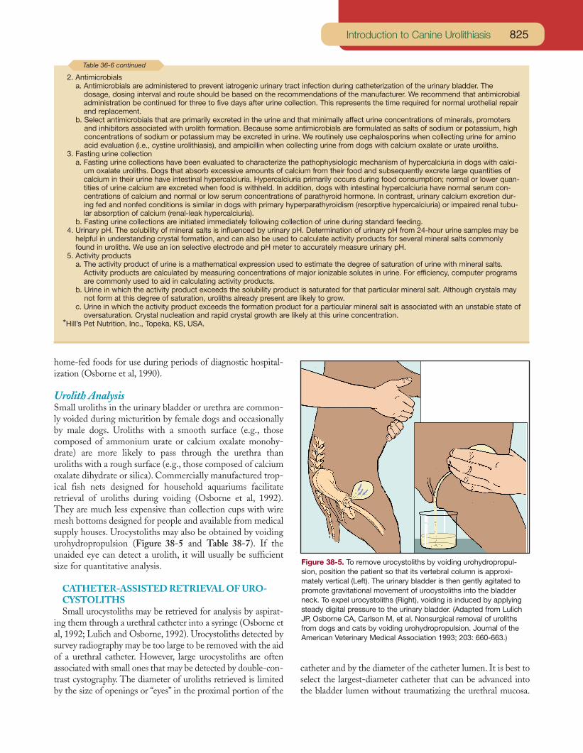
Urolith AnalysisSmall uroliths in the urinary bladder or urethra are common-ly voided during micturition by female dogs and occasionallyby male dogs. Uroliths with a smooth surface (e.g., thosecomposed of ammonium urate or calcium oxalate monohy-drate) are more likely to pass through the urethra thanuroliths with a rough surface (e.g., those composed of calciumoxalate dihydrate or silica). Commercially manufactured trop-ical fish nets designed for household aquariums facilitateretrieval of uroliths during voiding (Osborne et al, 1992).They are much less expensive than collection cups with wiremesh bottoms designed for people and available from medicalsupply houses. Urocystoliths may also be obtained by voidingurohydropropulsion (Figure 38-5 and Table 38-7). If theunaided eye can detect a urolith, it will usually be sufficientsize for quantitative analysis.
CATHETER-ASSISTED RETRIEVAL OF URO-CYSTOLITHSSmall urocystoliths may be retrieved for analysis by aspirat-
ing them through a urethral catheter into a syringe (Osborne etal, 1992; Lulich and Osborne, 1992). Urocystoliths detected bysurvey radiography may be too large to be removed with the aidof a urethral catheter. However, large urocystoliths are oftenassociated with small ones that may be detected by double-con-trast cystography. The diameter of uroliths retrieved is limitedby the size of openings or “eyes” in the proximal portion of the
catheter and by the diameter of the catheter lumen. It is best toselect the largest-diameter catheter that can be advanced intothe bladder lumen without traumatizing the urethral mucosa.
825Introduction to Canine Urolithiasis
2. Antimicrobialsa. Antimicrobials are administered to prevent iatrogenic urinary tract infection during catheterization of the urinary bladder. The
dosage, dosing interval and route should be based on the recommendations of the manufacturer. We recommend that antimicrobialadministration be continued for three to five days after urine collection. This represents the time required for normal urothelial repairand replacement.
b. Select antimicrobials that are primarily excreted in the urine and that minimally affect urine concentrations of minerals, promotersand inhibitors associated with urolith formation. Because some antimicrobials are formulated as salts of sodium or potassium, highconcentrations of sodium or potassium may be excreted in urine. We routinely use cephalosporins when collecting urine for aminoacid evaluation (i.e., cystine urolithiasis), and ampicillin when collecting urine from dogs with calcium oxalate or urate uroliths.
3. Fasting urine collectiona. Fasting urine collections have been evaluated to characterize the pathophysiologic mechanism of hypercalciuria in dogs with calci-
um oxalate uroliths. Dogs that absorb excessive amounts of calcium from their food and subsequently excrete large quantities ofcalcium in their urine have intestinal hypercalciuria. Hypercalciuria primarily occurs during food consumption; normal or lower quan-tities of urine calcium are excreted when food is withheld. In addition, dogs with intestinal hypercalciuria have normal serum con-centrations of calcium and normal or low serum concentrations of parathyroid hormone. In contrast, urinary calcium excretion dur-ing fed and nonfed conditions is similar in dogs with primary hyperparathyroidism (resorptive hypercalciuria) or impaired renal tubu-lar absorption of calcium (renal-leak hypercalciuria).
b. Fasting urine collections are initiated immediately following collection of urine during standard feeding.4. Urinary pH. The solubility of mineral salts is influenced by urinary pH. Determination of urinary pH from 24-hour urine samples may be
helpful in understanding crystal formation, and can also be used to calculate activity products for several mineral salts commonlyfound in uroliths. We use an ion selective electrode and pH meter to accurately measure urinary pH.
5. Activity productsa. The activity product of urine is a mathematical expression used to estimate the degree of saturation of urine with mineral salts.
Activity products are calculated by measuring concentrations of major ionizable solutes in urine. For efficiency, computer programsare commonly used to aid in calculating activity products.
b. Urine in which the activity product exceeds the solubility product is saturated for that particular mineral salt. Although crystals maynot form at this degree of saturation, uroliths already present are likely to grow.
c. Urine in which the activity product exceeds the formation product for a particular mineral salt is associated with an unstable state ofoversaturation. Crystal nucleation and rapid crystal growth are likely at this urine concentration.
*Hill’s Pet Nutrition, Inc., Topeka, KS, USA.
Table 36-6 continued
Figure 38-5. To remove urocystoliths by voiding urohydropropul-sion, position the patient so that its vertebral column is approxi-mately vertical (Left). The urinary bladder is then gently agitated topromote gravitational movement of urocystoliths into the bladderneck. To expel urocystoliths (Right), voiding is induced by applyingsteady digital pressure to the urinary bladder. (Adapted from LulichJP, Osborne CA, Carlson M, et al. Nonsurgical removal of urolithsfrom dogs and cats by voiding urohydropropulsion. Journal of theAmerican Veterinary Medical Association 1993; 203: 660-663.)

Well-lubricated, soft, flexible catheters are preferable to lessflexible ones. The size of openings in the proximal portion ofthe catheter may be enlarged with a scalpel, razor blade or scis-sors to facilitate retrieval of urocystoliths. However, care mustbe used not to weaken the catheter to the point where it couldbreak while being inserted into or removed from the urethraand urinary bladder.
Uroliths may be retrieved by catheter aspiration as follows(Figure 38-6). With the patient in lateral recumbency, a well-lubricated catheter should be advanced through the urethrainto the bladder lumen. The tip of the catheter should be posi-tioned so that it will not interfere with movement of the blad-der wall as fluid is aspirated from the bladder lumen. If the uri-nary bladder is not distended with urine, it should be partiallydistended with physiologic (0.9%) saline solution. As a rule ofthumb, a normal, empty canine or feline urinary bladder can bepartially distended by injecting 3 to 4 ml of fluid per kg bodyweight. However, the urinary bladder should be palpated perabdomen during the time it is distended with saline solution toensure that it is not overdistended.
The next step is crucial to successful retrieval of urocystoliths.While urine (and saline solution) is aspirated into the syringe,
Small Animal Clinical Nutrition826
Table 38-7. Voiding urohydropropulsion: A nonsurgical technique for removing small urocystoliths.
1. Perform appropriate diagnostic studies, including completeurinalysis, quantitative urine culture and diagnostic radiog-raphy. Determine the location, size, surface contour andnumber of urocystoliths.
2. Anesthetize the patient, if needed.3. If the urinary bladder is not distended with urine, moderate-
ly distend it with a physiologic solution (e.g., saline,Ringer’s, etc.) injected through a transurethral catheter. Toprevent overdistention, palpate the bladder per abdomenduring infusion. Remove the catheter.
4. Position the patient such that the vertebral spine is approxi-mately vertical.
5. Gently agitate the urinary bladder, with the objective of pro-moting gravitational movement of urocystoliths into thebladder neck.
6. Induce voiding by manually expressing the urinary bladder.Use steady digital pressure rather than an intermittentsqueezing motion.
7. Collect urine and uroliths in a cup. Compare urolith numberand size to those detected by radiography and submit themfor quantitative analysis.
8. If needed, repeat Steps 3 through 7 until the number ofuroliths detected by radiography are removed or untiluroliths are no longer voided.
9. Perform double-contrast cystography to ensure that nouroliths remain in the urinary bladder. Repeat voiding urohy-dropropulsion if small urocystoliths remain.
10. Administer prophylactic antimicrobials for three to five days,or longer if needed.
11. Monitor the patient for adverse complications (i.e., hema-turia, dysuria, bacterial urinary tract infection and urethralobstruction with uroliths).
12. Formulate appropriate recommendations to minimize urolithrecurrence or to manage uroliths remaining in the urinarytract on the basis of quantitative mineral analysis of voidedurocystoliths.
Figure 38-6. Illustration of catheter-assisted retrieval of urocys-toliths. With the patient in lateral recumbency, uroliths have gravitat-ed to the dependent portion of the urinary bladder (Top). The blad-der lumen has been distended by injection of 0.9% saline solution.Vigorous movement of the abdomen in an up-and-down motion dis-perses uroliths throughout fluid in the bladder lumen (Middle).Aspiration of fluid from the urinary bladder during movement of theabdominal wall (Bottom) may result in movement of one or moresmall uroliths into the catheter and syringe. (Adapted from Lulich JP,Osborne CA. Catheter assisted retrieval of canine and feline urocys-toliths. Journal of the American Veterinary Medical Association 1992;201: 111-113.)

an assistant should vigorously and repeatedly move the patient’sabdomen in an up-and-down motion. This maneuver dispers-es uroliths located in the dependent portion of the bladderthroughout fluid in the bladder lumen. Small uroliths in thevicinity of the catheter tip may then be aspirated into thecatheter along with the urine-saline mixture. It may be neces-sary to repeat this sequence of steps several times before a suf-ficient number of uroliths are retrieved. The bladder lumenshould be redistended with saline solution each time. Difficultyin aspirating urine and saline solution into the syringe may becaused by poor positioning of the catheter tip or by partialocclusion of the catheter lumen with one or more uroliths.Flushing saline solution through the catheter after it has beenremoved from the patient often results in the retrieval ofuroliths that occlude the catheter lumen.
Care must be used not to overdistend the urinary bladderwith saline solution because this will increase the space inwhich the uroliths are suspended. Because patients withuroliths are predisposed to catheter-induced bacterial UTIs,antimicrobial therapy should be considered immediately beforethis procedure and for an appropriate period afterward. Properselection, insertion and positioning of urethral catheters mini-mize iatrogenic trauma to the lower urinary tract.
COLLECTION AND QUANTITATIVE ANALYSISOF URINE CRYSTALSIf available data do not indicate the probable mineral compo-
sition of uroliths and if uroliths cannot be retrieved with the aidof a urethral catheter, consider preparing a large pellet of urinecrystals by centrifugation of urine in a conical-tip centrifugetube (Osborne et al, 1992, 1995). The quantity of crystallinesediment available for analysis may be increased by repeatedlyremoving the supernatant after centrifugation, adding addi-tional noncentrifuged urine to the tube containing sedimentand again centrifuging the preparation. If the conditions thatcaused urolith formation are still present, evaluation of the pel-let formed from crystalline sediment by quantitative methodsdesigned for urolith analysis may provide meaningful informa-tion about the mineral composition of a patient’s uroliths.However, crystals identified by this method may only reflect theouter portions of compound uroliths. Therefore, results ofquantitative urine crystal analysis should be interpreted in con-junction with other pertinent clinical data.
QUANTITATIVE ANALYSIS OF UROLITHSThe location, number, size, shape, color and consistency of
uroliths removed from the urinary tract should be recorded. Alluroliths should be saved in a container (preferably a sterile one)and submitted for analysis. Do not give uroliths to ownersbefore analysis. If multiple uroliths are present, one may beplaced into a container of 10% buffered formalin for deminer-alization and microscopic examination. However, formalinshould not be used to preserve uroliths for mineral analysisbecause formalin may alter the results. Because many urolithscontain two or more mineral components, it is important toexamine representative portions. The mineral composition of
crystalline nuclei may be identical or different from outer layersof uroliths (Figure 38-7). The nuclei of uroliths should be ana-lyzed separately from outer layers because knowledge of themineral composition of the nuclei may suggest the initiatingcause of the urolith. Uroliths should not be broken before sub-mission because the central core may be distorted or lost.
Routine analysis of uroliths by qualitative methods of chem-ical analysis is not recommended. The major disadvantage ofthis procedure is that only some of the chemical radicals andions can be detected. In addition, the proportion of the differ-ent chemical constituents in the urolith cannot be quantified.In contrast to chemical methods of analysis, physical methodshave proved to be far superior in identification of crystallinesubstances. Physical methods also permit detection of silica anddrugs and drug metabolites. They also permit differentiation ofvarious subgroups of minerals (e.g., calcium oxalate monohy-drate and calcium oxalate dihydrate, or uric acid, ammoniumacid urate and xanthine) and allow semiquantitative determina-tion of various mineral components. Physical methods com-monly used by laboratories that specialize in quantitativeurolith analysis include a combination of polarizing light mi-croscopy, x-ray diffractometry and infrared spectroscopy(Osborne et al, 1983; Zinn et al, 1986; Ulrich et al, 1996). Somelaboratories also are equipped to perform elemental analysiswith an energy dispersive x-ray microanalyzer or by neutronactivation. Occasionally, chemical methods of analysis andpaper chromatography may be used to supplement informationprovided by physical methods. Chapter 46 lists selected labora-tories that perform quantitative urolith analysis.
UROLITH CULTUREBacterial culture of the interior of uroliths is indicated if: 1)
urine obtained from the patient has not been previously cul-tured, 2) culture of urine obtained from patients suspected ofhaving struvite uroliths yields no growth or 3) the patient has a
827Introduction to Canine Urolithiasis
Figure 38-7. Schematic demonstrating the different componentsthat may be observed on the cut surface of a bisected urolith.(Adapted from Osborne CA, Lulich JP, Polzin DJ, et al. Analysis of77,000 canine uroliths: Perspectives from the Minnesota UrolithCenter. Veterinary Clinics of North America: Small Animal Practice1999; 29: 23.)
UTI with bacteria that do not produce urease. Bacteria har-bored inside uroliths are not always the same as those presentin urine. Bacteria detected within uroliths probably representthose present at the time they were formed, and may serve as asource of recurrent UTI. Bacteria may remain viable withinuroliths for long periods. In a pilot study, we cultured viablestaphylococci from struvite uroliths removed from a miniatureschnauzer up to three months following surgery. If all theuroliths have not been removed from the patient, knowledge ofthe type and antimicrobic susceptibility of bacteria inside
uroliths that have been voided or removed may be of therapeu-tic significance. Procedures for culture of microbes from theinner portions of uroliths have been developed and can be per-formed by most veterinary diagnostic laboratories (Osborne etal, 1983; Ruby and Ling, 1986).
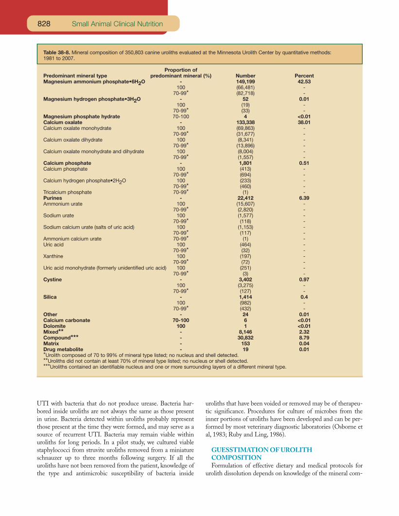
GUESSTIMATION OF UROLITH COMPOSITIONFormulation of effective dietary and medical protocols for
urolith dissolution depends on knowledge of the mineral com-
Small Animal Clinical Nutrition828
Table 38-8. Mineral composition of 350,803 canine uroliths evaluated at the Minnesota Urolith Center by quantitative methods: 1981 to 2007.
Proportion of Predominant mineral type predominant mineral (%) Number PercentMagnesium ammonium phosphate•6H2O - 149,199 42.53
100 (66,481) -70-99* (82,718) -
Magnesium hydrogen phosphate•3H2O - 52 0.01100 (19) -
70-99* (33) -Magnesium phosphate hydrate 70-100 4 <0.01Calcium oxalate - 133,338 38.01Calcium oxalate monohydrate 100 (69,863) -
70-99* (31,677) -Calcium oxalate dihydrate 100 (8,341) -
70-99* (13,896) -Calcium oxalate monohydrate and dihydrate 100 (8,004) -
70-99* (1,557) -Calcium phosphate - 1,801 0.51Calcium phosphate 100 (413) -
70-99* (694) -Calcium hydrogen phosphate•2H2O 100 (233) -
70-99* (460) -Tricalcium phosphate 70-99* (1) -Purines - 22,412 6.39Ammonium urate 100 (15,607) -
70-99* (2,820) -Sodium urate 100 (1,577) -
70-99* (118) -Sodium calcium urate (salts of uric acid) 100 (1,153) -
70-99* (117) -Ammonium calcium urate 70-99* (1) -Uric acid 100 (464) -
70-99* (32) -Xanthine 100 (197) -
70-99* (72) -Uric acid monohydrate (formerly unidentified uric acid) 100 (251) -
70-99* (3) -Cystine - 3,402 0.97
100 (3,275) -70-99* (127) -
Silica - 1,414 0.4100 (982) -
70-99* (432) -Other - 24 0.01Calcium carbonate 70-100 6 <0.01Dolomite 100 1 <0.01Mixed** - 8,146 2.32Compound*** - 30,832 8.79Matrix - 153 0.04Drug metabolite - 19 0.01*Urolith composed of 70 to 99% of mineral type listed; no nucleus and shell detected.**Uroliths did not contain at least 70% of mineral type listed; no nucleus or shell detected.***Uroliths contained an identifiable nucleus and one or more surrounding layers of a different mineral type.
829Introduction to Canine Urolithiasis
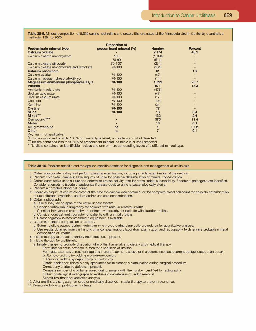
Table 38-9. Mineral composition of 5,050 canine nephroliths and ureteroliths evaluated at the Minnesota Urolith Center by quantitativemethods: 1991 to 2006.
Proportion of Predominate mineral type predominant mineral (%) Number PercentCalcium oxalate - 2,174 43.1Calcium oxalate monohydrate 100 (1,168) -
70-99 (511) -Calcium oxalate dihydrate 70-100* (234) -Calcium oxalate monohydrate and dihydrate 70-100 (161) -Calcium phosphate - 81 1.6Calcium apatite 70-100 (67) -Calcium hydrogen phosphate•2H2O 70-100 (14) -Magnesium ammonium phosphate•6H2O 70-100 1,299 25.7Purines - 671 13.3Ammonium acid urate 70-100 (476) -Sodium acid urate 70-100 (47) -Sodium calcium urate 70-100 (17) -Uric acid 70-100 104 -Xanthine 70-100 (24) -Cystine 70-100 77 -Silica 70-100 18 0.4Mixed** - 132 2.6Compound*** - 575 11.4Matrix - 13 0.3Drug metabolite na 1 0.02Other na 7 0.1Key: na = not applicable.*Uroliths composed of 70 to 100% of mineral type listed; no nucleus and shell detected.**Uroliths contained less than 70% of predominant mineral; no nucleus or shell detected.***Uroliths contained an identifiable nucleus and one or more surrounding layers of a different mineral type.
Table 38-10. Problem-specific and therapeutic-specific database for diagnosis and management of urolithiasis.
1. Obtain appropriate history and perform physical examination, including a rectal examination of the urethra.2. Perform complete urinalysis; save aliquots of urine for possible determination of mineral concentration.3. Obtain quantitative urine culture and determine urease activity; test for antimicrobial susceptibility if bacterial pathogens are identified.
Consider attempts to isolate ureaplasmas if urease-positive urine is bacteriologically sterile.4. Perform a complete blood cell count.5. Freeze an aliquot of serum collected at the time the sample was obtained for the complete blood cell count for possible determination
of urea nitrogen, creatinine, calcium and/or uric acid concentrations.6. Obtain radiographs.
a. Take survey radiographs of the entire urinary system.b. Consider intravenous urography for patients with renal or ureteral uroliths.c. Consider intravenous urography or contrast cystography for patients with bladder uroliths.d. Consider contrast urethrography for patients with urethral uroliths.e. Ultrasonography is recommended if equipment is available.
7. Determine mineral composition of uroliths.a. Submit uroliths passed during micturition or retrieved during diagnostic procedures for quantitative analysis.b. Use results obtained from the history, physical examination, laboratory examination and radiography to determine probable mineral
composition of uroliths.8. Initiate therapy to eradicate urinary tract infection, if present.9. Initiate therapy for urolithiasis.
a. Initiate therapy to promote dissolution of uroliths if amenable to dietary and medical therapy.Formulate followup protocol to monitor dissolution of uroliths.Formulate alternative treatment options if uroliths do not dissolve or if problems such as recurrent outflow obstruction occur.b. Remove uroliths by voiding urohydropropulsion.c. Remove uroliths by nephrotomy or cystotomy.Obtain bladder or kidney biopsy specimens for microscopic examination during surgical procedure.Correct any anatomic defects, if present.Compare number of uroliths removed during surgery with the number identified by radiography.Obtain postsurgical radiographs to evaluate completeness of urolith removal.Submit uroliths for quantitative analysis.
10. After uroliths are surgically removed or medically dissolved, initiate therapy to prevent recurrence.11. Formulate followup protocol with clients.
Small Animal Clinical Nutrition830
position of uroliths. Because a variety of different types ofuroliths and nephroliths occur in dogs (Tables 38-8 and 38-9),veterinarians should use a protocol that facilitates determina-tion of the mineral composition of uroliths based on probabili-ty (Tables 38-10 and 38-11). Formulation of dietary and med-ical therapy based on the mineral composition of uroliths deter-mined by this protocol is usually associated with a high degreeof success in dissolving uroliths or arresting their growth.
Attempts to induce dissolution of uroliths may be hamperedif the uroliths are heterogeneous in composition. This has notbeen a significant problem in dogs with uroliths composed pri-marily of magnesium ammonium phosphate with small quan-tities of calcium apatite because the solubility characteristics ofthe two minerals are similar. Veterinarians have encountereddifficulty in dissolving uroliths composed primarily of struvite
with an outer shell composed primarily of calcium apatite. Dif-ficulty will also be encountered in attempting to induce com-plete dissolution of a urolith with a nucleus of calcium oxalate,calcium phosphate, ammonium urate or silica and a shell ofstruvite because the solubility characteristics of this combina-tion of minerals are dissimilar. These phenomena should beconsidered if dietary and medical therapy seems to be ineffec-tive after initially reducing the size of uroliths (Box 38-2).
REFERENCES
The references for Chapter 38 can be found atwww.markmorris.org.
OVERVIEWSurgery has been the time-honored method of managing all types ofurolithiasis in dogs. Although surgery has been an effective methodthat provides immediate elimination of uroliths, it is associated withseveral limitations, including: 1) persistence of underlying causesand a high rate of recurrence of uroliths despite surgery, 2) patientfactors that enhance adverse consequences of general anesthesiaor surgery and 3) inability to remove all uroliths or fragments ofuroliths during surgery. In addition, situations occasionally arise inwhich owners of companion animals will not consent to surgicaltherapy but will consider dietary and medical therapy. Dietary andmedical dissolution of uroliths may be considered for these andother reasons (i.e., the urolith is asymptomatic).
Results of several experimental and clinical investigations haveconfirmed the efficacy of dietary and medical dissolution of canineuroliths. Despite the feasibility of dissolution of uroliths, however, thisform of therapy is associated with potential hazards. Uroliths alwayspredispose patients to urinary tract infection (UTI) and obstructiveuropathy. Risks and benefits of dietary and medical vs. surgical anddietary and medical therapy must be considered for each patient.
Detailed descriptions of nonsurgical and surgical methods forreestablishing urine outflow are beyond the scope of the discussionbut are available elsewhere. Likewise, details pertaining to surgicalremoval of uroliths, endoscopic and percutaneous manipulation ofuroliths, chemolysis via nephrostomy, disintegration of renal andureteral uroliths via ultrasound and shockwave lithotripsy have beenreviewed.
DIETARY AND MEDICAL DISSOLUTION OF UROLITHSTherapy should not be initiated before appropriate samples havebeen collected for diagnosis. The objectives of dietary and medicalmanagement of uroliths are to arrest further growth and to promoteurolith dissolution by correcting or controlling underlying abnormali-ties. For therapy to be effective, it must induce undersaturation ofurine with lithogenic crystalloids by: 1) increasing the solubility ofcrystalloids in urine, 2) increasing the volume of urine in which crys-
talloids are dissolved or suspended and 3) reducing the quantity oflithogenic crystalloids in urine. For example, attempts to increase thesolubility of crystalloids in urine often include administration of med-ications designed to change urinary pH to create a less favorableenvironment for crystallization. Likewise, diuresis is commonlyinduced to increase the volume of urine in which crystalloids are dis-solved or suspended. A dietary change is an example of a method toreduce the quantity of lithogenic crystalloids in urine.
In general, dietary and medical treatment should be formulated instepwise fashion, with the initial goal of reducing the urine concen-tration of lithogenic substances. Medications that have the potentialto induce a sustained alteration in body composition of metabolites,in addition to urine concentration of metabolites, should be reservedfor patients with active or frequently recurrent uroliths. Caution mustbe used so that the side effects of treatment are not more detrimen-tal than the effects of the uroliths.
Results of experimental and clinical studies have revealed that thesize and number of uroliths do not dictate the likelihood of responseto therapy. We have had success in dissolving uroliths that are smalland large, single and multiple. However the rate of dissolution isrelated to size and surface area of the urolith exposed to urine. Justas one large ice cube dissolves more slowly than an equal volume ofcrushed ice, one large urolith will dissolve more slowly than an equalvolume of many smaller uroliths. Rate of dissolution is influenced bysurface area of the urolith exposed to undersaturated urine.
Difficulty in inducing complete dissolution of uroliths by creatingurine that is undersaturated with the suspected lithogenic crystalloidshould prompt consideration that: 1) the wrong mineral componentwas identified, 2) the nucleus of the urolith is of different mineralcomposition than outer portions and/or 3) the owner or the patient isnot complying with therapy.
SURGICAL REMOVAL OF UROLITHSDetection of uroliths is not in itself an indication for surgery.However, along with dietary and medical management, surgicalintervention may play an important role in therapy of urolithiasis.
Box 38-2. Principles of Urolith Treatment and Prevention.
831Introduction to Canine Urolithiasis
Surgical candidates include patients: 1) with urolith-inducedobstruction to urine outflow that cannot be corrected by nonsurgicaltechniques, especially in patients with concomitant UTI, 2) withuroliths that are refractory to current methods of dietary and med-ical dissolution (e.g., silica, calcium oxalate and calcium phosphateuroliths), 3) with uroliths that are increasing in size or numberdespite dietary and medical therapy designed to inhibit their growthor cause their dissolution (especially if they are obstructing urineoutflow or causing progressive deterioration in renal function), 4)with nephroliths and renal dysfunction of such a nature that the timerequired to induce dietary and medical dissolution is likely to beassociated with more renal dysfunction than that associated withsurgical procedures, 5) with anatomic defects of the urogenital tractthat predispose patients to recurrent UTI and urolithiasis and areamenable to surgical correction at the time uroliths are removed and6) unable to respond to dietary and medical management becauseof poor client compliance with therapeutic recommendations.
Complete obstruction to urine outflow caused by uroliths inpatients with concomitant UTI should be regarded as a surgicalemergency. In this situation, rapid spread of infection and associat-ed damage to the urinary tract, especially the kidneys, are likely toinduce septicemia and peracute renal failure caused by a combina-tion of obstruction and pyelonephritis.
Unilateral nephroliths and ureteroliths that cause outflow obstruc-tion and markedly impair function of the associated kidney should bemanaged by surgical intervention or (if possible) percutaneousnephropyelolithotomy. Dietary and medical therapy designed toinduce urolith dissolution over several weeks in patients with poorlydraining kidneys is unlikely to be effective because the urolith(s) willnot be continuously bathed with newly formed urine modified toinduce litholysis. The same concept applies to urethroliths that can-not be removed by nonsurgical methods.
Surgical removal of uroliths followed by dietary and medicallitholytic protocols may be of value in some patients. Examplesinclude patients in which uroliths or fragments of uroliths remainafter surgery, and patients with crystalluria of a character and mag-
nitude that indicate rapid recurrence is likely. Surgical incisionsshould be repaired using meticulous technique if protein-restrictedcanine litholytic foods are used.
PREVENTION OF UROLITH RECURRENCEUroliths tend to recur. Prevention of recurrent uroliths, whichreduces the need for dietary and medical therapy and surgery, istherefore cost effective. In general, preventive strategies aredesigned to eliminate or control the underlying causes of varioustypes of uroliths. When causes cannot be identified, preventivestrategies encompass efforts to minimize risk factors associatedwith lithogenesis. These strategies commonly include dietary modi-fication.
The Bibliography for Box 38-2 can be found atwww.markmorris.com
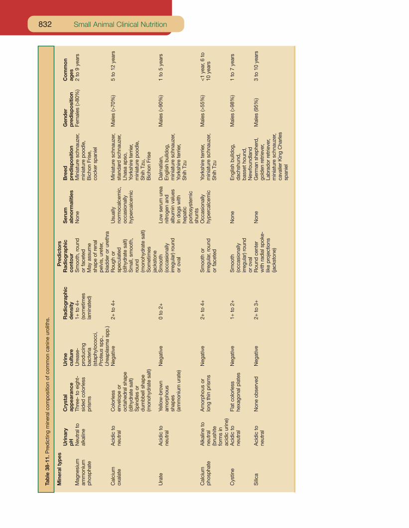
Small Animal Clinical Nutrition832
Tab
le38
-11.
Pre
dict
ing
min
eral
com
posi
tion
ofco
mm
onca
nine
urol
iths.
Min
eral
type
sP
redi
ctor
sU
rina
ryC
ryst
alU
rine
Rad
iogr
aphi
cR
adio
grap
hic
Ser
umB
reed
Gen
der
Com
mon
pHap
pear
ance
cultu
rede
nsity
cont
our
abno
rmal
ities
pred
ispo
sitio
npr
edis
posi
tion
ages
Mag
nesi
umN
eutr
alto
Thre
e-to
eigh
t-U
reas
e-1+
to4+
Sm
ooth
,rou
ndN
one
Min
iatu
resc
hnau
zer,
Fem
ales
(>80
%)
2to
9ye
ars
amm
oniu
mal
kalin
esi
ded
colo
rless
prod
ucin
g(s
omet
imes
orfa
cete
dm
inia
ture
pood
le,
phos
phat
epr
ism
sba
cter
iala
min
ated
)M
ayas
sum
eB
icho
nFr
ise,
(sta
phyl
ococ
ci,
shap
eof
rena
lco
cker
span
iel
Pro
teus
spp.
,pe
lvis
,ure
ter,
Ure
apla
sma
spp.
)bl
adde
ror
uret
hra
Cal
cium
Aci
dic
toC
olor
less
Neg
ativ
e2+
to4+
Rou
ghor
Usu
ally
Min
iatu
resc
hnau
zer,
Mal
es(>
70%
)5
to12
year
sox
alat
ene
utra
len
velo
peor
spec
ulat
edno
rmoc
alce
mic
,st
anda
rdsc
hnau
zer,
octa
hedr
alsh
ape
(dih
ydra
tesa
lt)oc
casi
onal
lyLh
asa
apso
,(d
ihyd
rate
salt)
Sm
all,
smoo
th,
hype
rcal
cem
icYo
rksh
irete
rrie
r,S
pind
les
orro
und
min
iatu
repo
odle
,du
mbb
ells
hape
(mon
ohyd
rate
salt)
Shi
hTz
u,(m
onoh
ydra
tesa
lt)S
omet
imes
Bic
hon
Fris
eja
ckst
one
Ura
teA
cidi
cto
Yello
w-b
row
nN
egat
ive
0to
2+S
moo
thLo
wse
rum
urea
Dal
mat
ian,
Mal
es(>
90%
)1
to5
year
sne
utra
lam
orph
ous
(occ
asio
nally
nitr
ogen
and
Eng
lish
bulld
og,
shap
esirr
egul
ar)r
ound
albu
min
valu
esm
inia
ture
schn
auze
r,(a
mm
oniu
mur
ate)
orov
alIn
dogs
with
York
shire
terr
ier,
hepa
ticS
hih
Tzu
port
osys
tem
icsh
unts
Cal
cium
Alk
alin
eto
Am
orph
ous
orN
egat
ive
2+to
4+S
moo
thor
Occ
asio
nally
York
shire
terr
ier,
Mal
es(>
55%
)<
1ye
ar,6
toph
osph
ate
neut
ral
long
thin
pris
ms
irreg
ular
,rou
ndhy
perc
alce
mic
min
iatu
resc
hnau
zer,
10ye
ars
(bru
shite
orfa
cete
dS
hih
Tzu
form
sin
acid
icur
ine)
Cys
tine
Aci
dic
toFl
atco
lorle
ssN
egat
ive
1+to
2+S
moo
thN
one
Eng
lish
bulld
og,
Mal
es(>
98%
)1
to7
year
sne
utra
lhe
xago
nalp
late
s(o
ccas
iona
llyda
chsh
und,
irreg
ular
)rou
ndba
sset
houn
d,or
oval
New
foun
dlan
dS
ilica
Aci
dic
toN
one
obse
rved
Neg
ativ
e2+
to3+
Rou
ndce
nter
Non
eG
erm
ansh
ephe
rd,
Mal
es(9
5%)
3to
10ye
ars
neut
ral
with
radi
alsp
oke-
gold
enre
trie
ver,
like
proj
ectio
nsLa
brad
orre
trie
ver,
(jack
ston
e)m
inia
ture
schn
auze
r,ca
valie
rK
ing
Cha
rles
span
iel