Embed Size (px)
Citation preview
DEGENERATIVE INSTABILITY
OF THE LUMBAR SPINE
IMAGING AND CLINICAL FEATURES
F. Postacchini, M. Paolino Clinica Ortopedica Università Sapienza, Roma
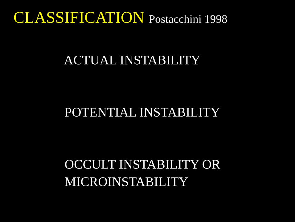
CLASSIFICATION Postacchini 1998
ACTUAL INSTABILITY
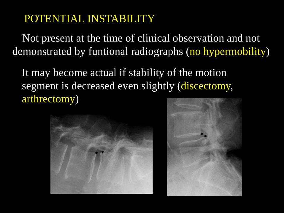
POTENTIAL INSTABILITY
OCCULT INSTABILITY OR
MICROINSTABILITY
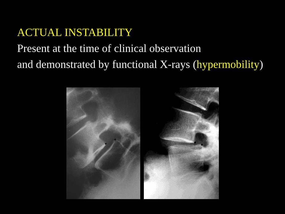
ACTUAL INSTABILITY
Present at the time of clinical observation
and demonstrated by functional X-rays (hypermobility)
. . . .
Not present at the time of clinical observation and not
demonstrated by funtional radiographs (no hypermobility)
It may become actual if stability of the motion
segment is decreased even slightly (discectomy,
arthrectomy)
POTENTIAL INSTABILITY
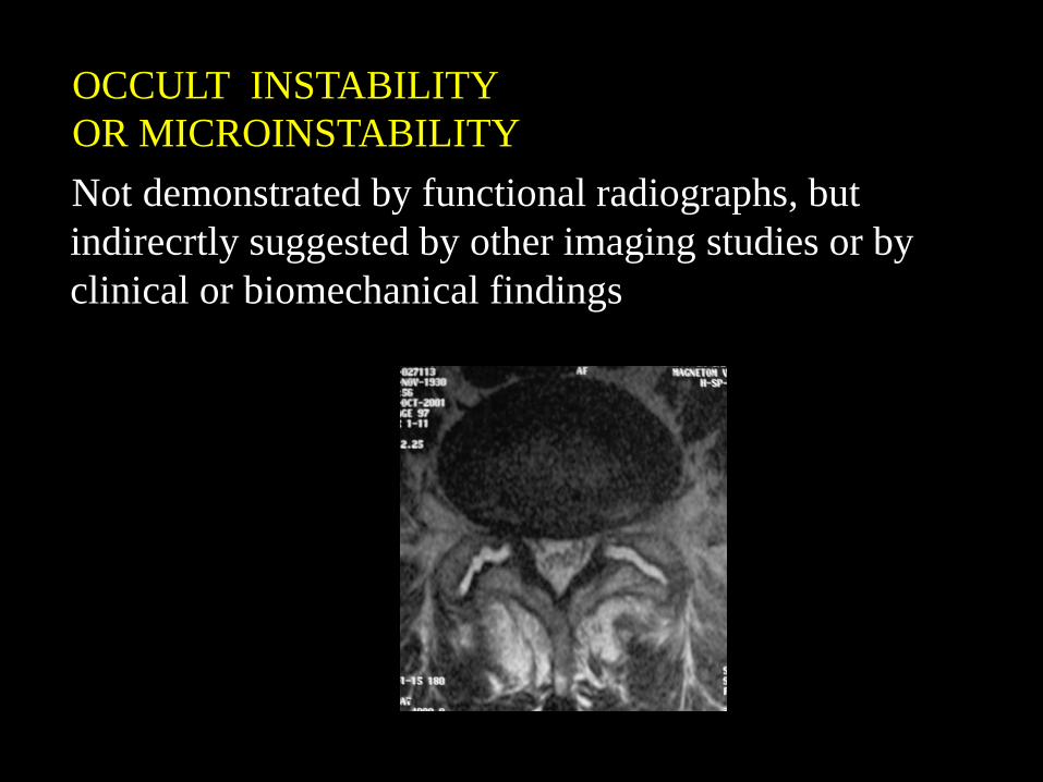
Not demonstrated by functional radiographs, but
indirecrtly suggested by other imaging studies or by
clinical or biomechanical findings
OCCULT INSTABILITY
OR MICROINSTABILITY
IMAGING
&
BIOMECHANICS
INSTABILITY
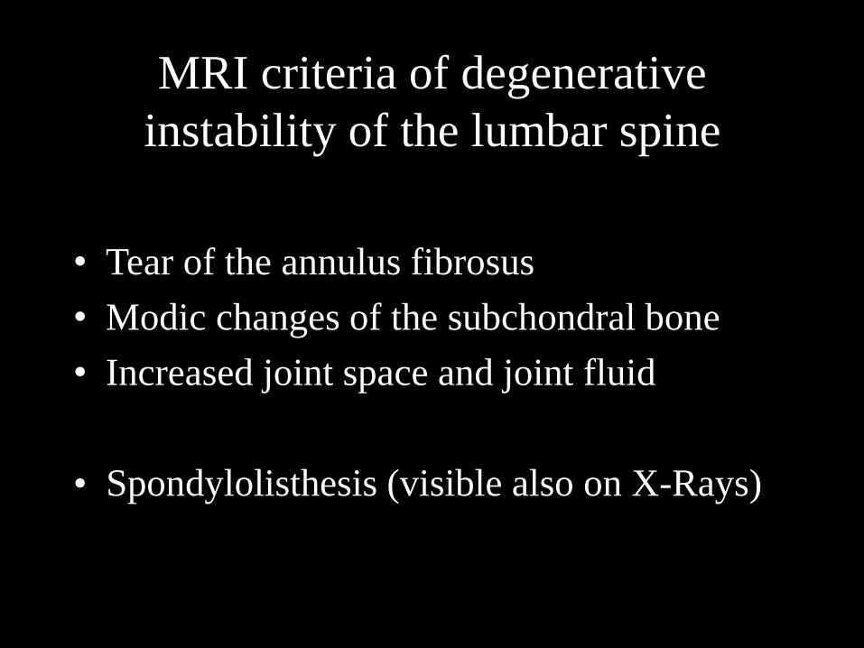
MRI criteria of degenerative
instability of the lumbar spine
• Tear of the annulus fibrosus
• Modic changes of the subchondral bone
• Increased joint space and joint fluid
• Spondylolisthesis (visible also on X-Rays)
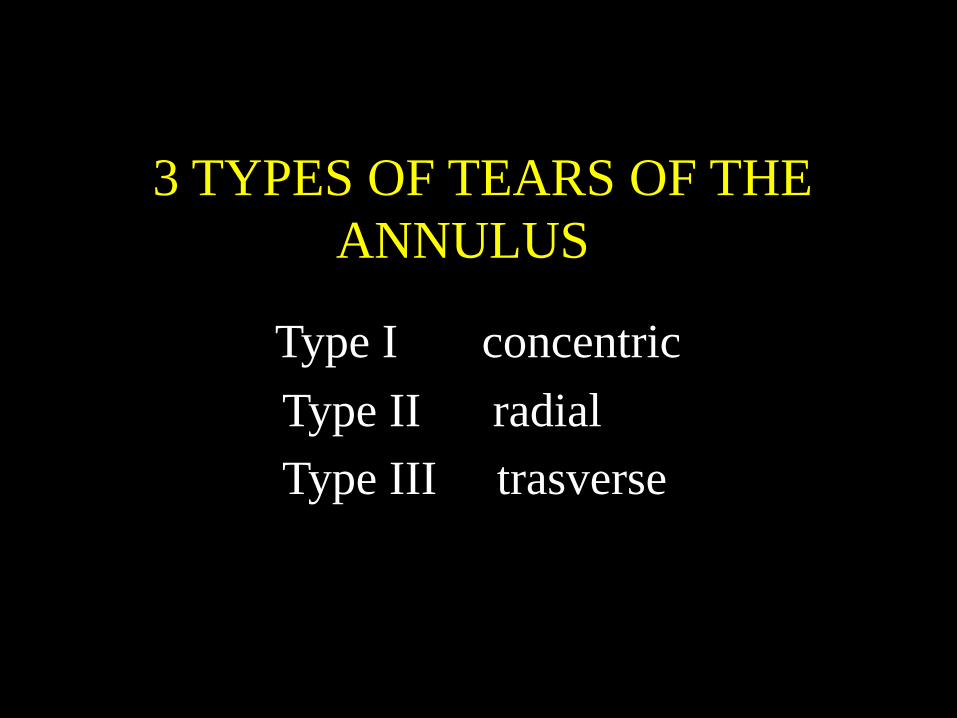
3 TYPES OF TEARS OF THE
ANNULUS
Type I concentric
Type II radial
Type III trasverse
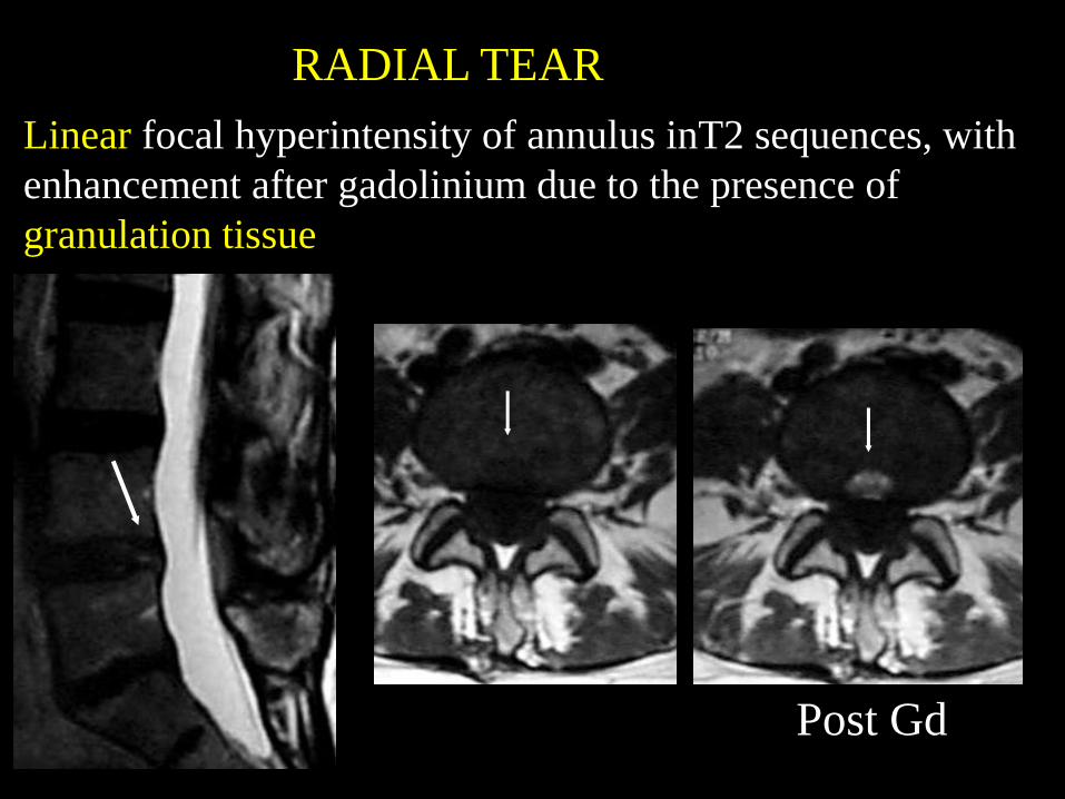
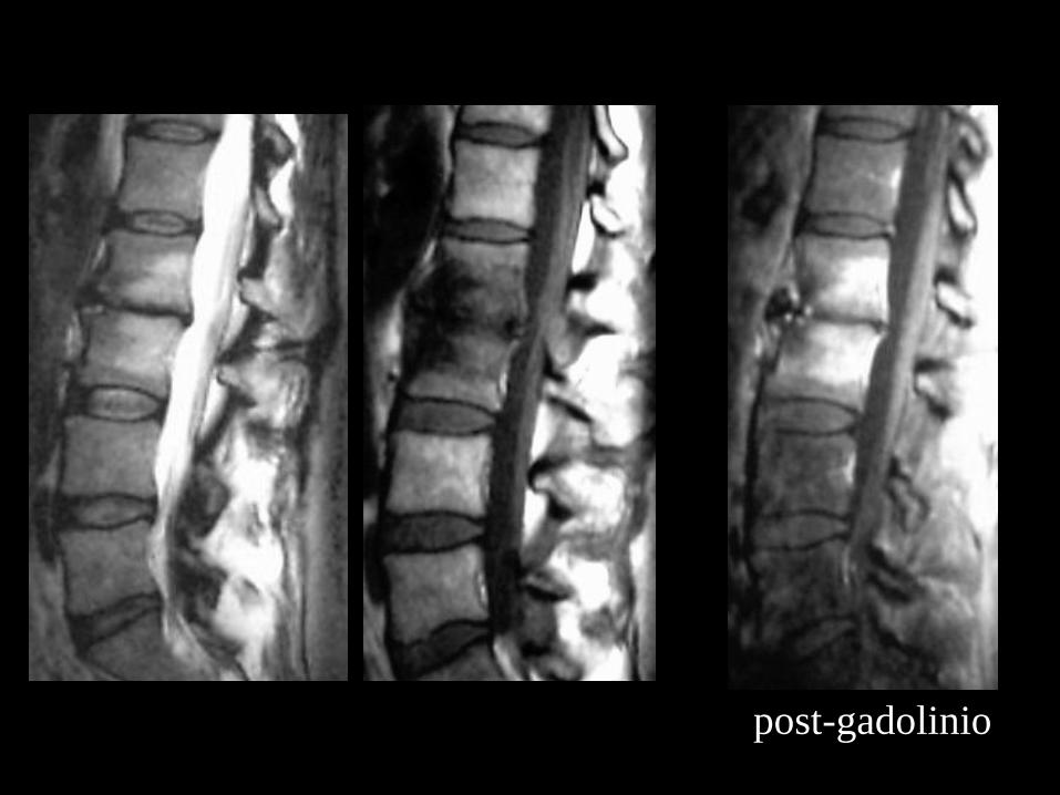
Linear focal hyperintensity of annulus inT2 sequences, with
enhancement after gadolinium due to the presence of
granulation tissue
Post Gd
RADIAL TEAR
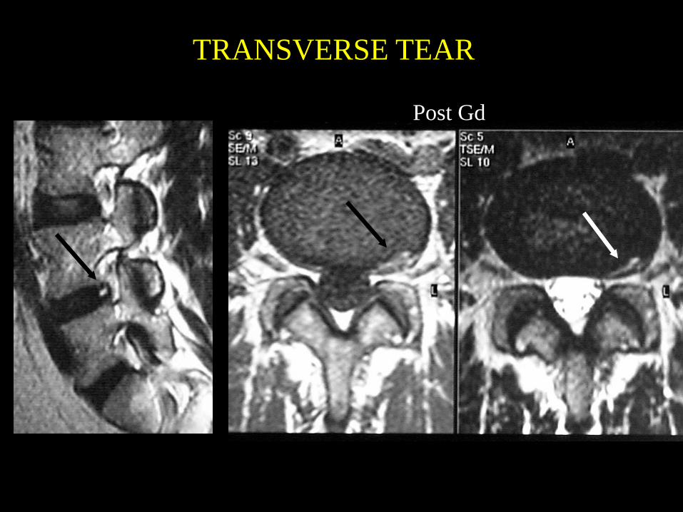
TRANSVERSE TEAR
Post Gd
Intervertebral disk appearance correlated with
stiffness of lumbar spinal motion segments
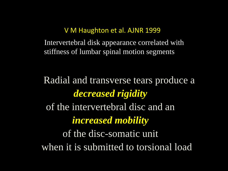
Radial and transverse tears produce a
decreased rigidity
of the intervertebral disc and an
increased mobility
of the disc-somatic unit
when it is submitted to torsional load
V M Haughton et al. AJNR 1999
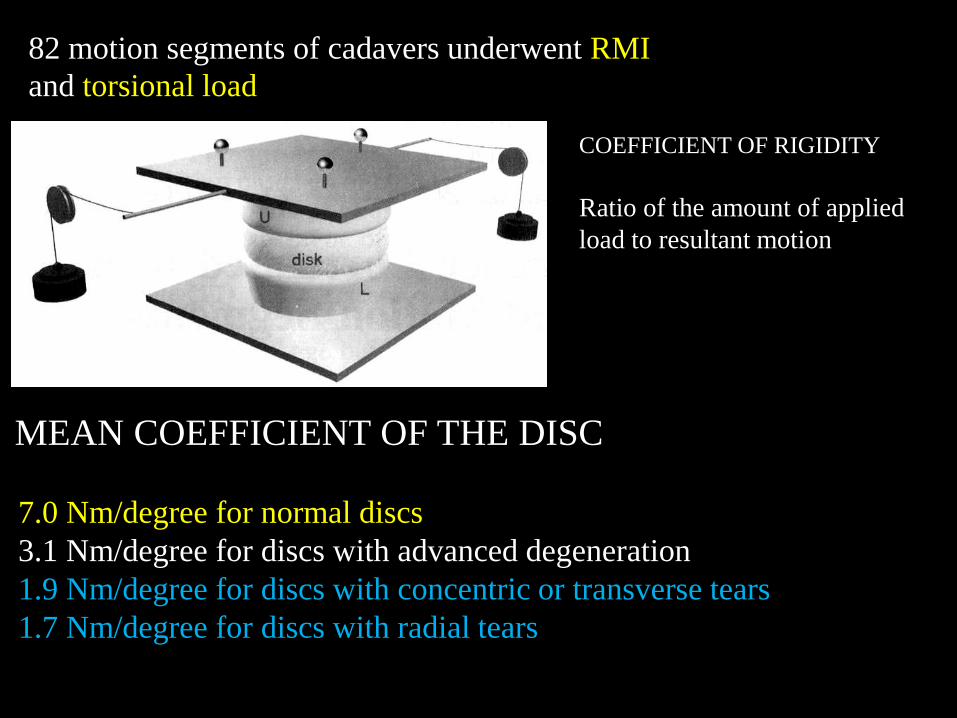
7.0 Nm/degree for normal discs
3.1 Nm/degree for discs with advanced degeneration
1.9 Nm/degree for discs with concentric or transverse tears
1.7 Nm/degree for discs with radial tears
MEAN COEFFICIENT OF THE DISC
COEFFICIENT OF RIGIDITY
Ratio of the amount of applied
load to resultant motion
82 motion segments of cadavers underwent RMI
and torsional load
Radial and transverse tears of the annulus, i.e. those
of the posterior outermost part of the disc,
may be associated to discogenic
pain
In 67% of patients with radial tears on RMI
Yu et al. AJNR 2002
discography elicits pain
MODIC CHANGES
Type I: hypo in T1 and hyper in T2, replacement
of yellow marrow with fibrovascular tissue
Typo I associated to LBP
Toyone et al. JBJS 1994
Type III: hypo in T1 and T2
sclerosis of subcondral bone
Type II: hyper in T1 and hyper in
T2, chronic change
Unclear association with
instability
Bram et al. 2005
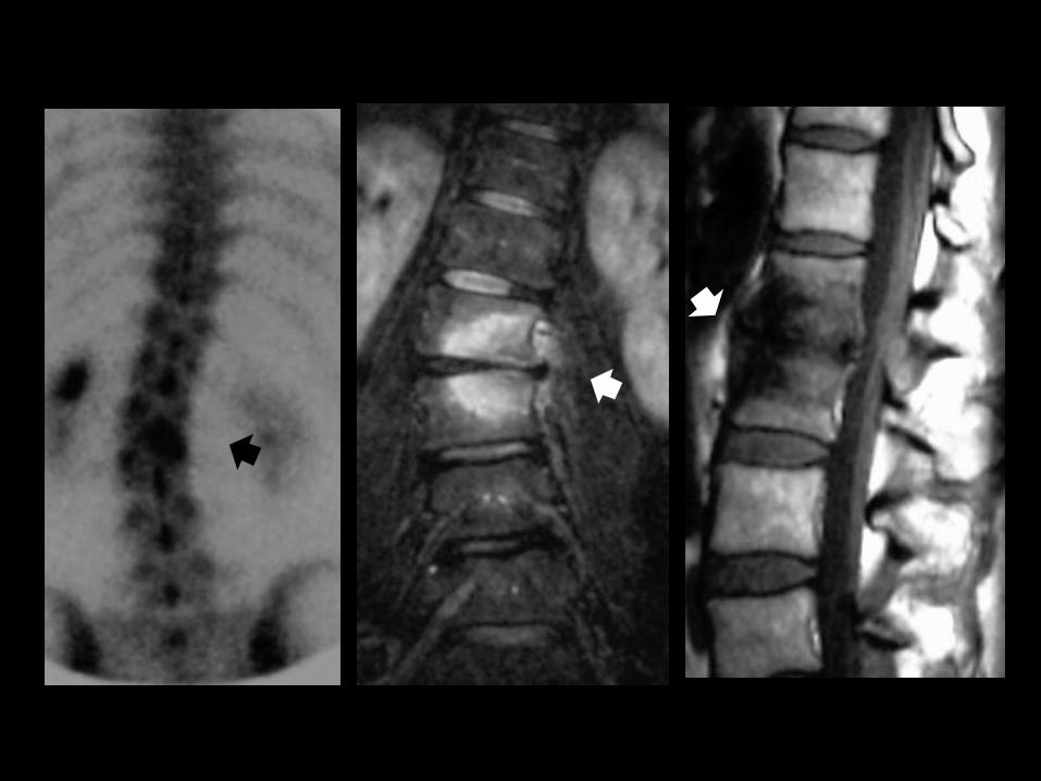
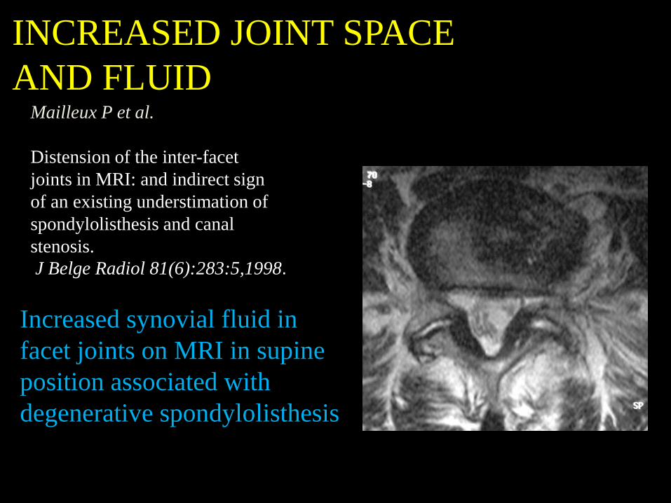
INCREASED JOINT SPACE
AND FLUID Mailleux P et al.
Distension of the inter-facet
joints in MRI: and indirect sign
of an existing understimation of
spondylolisthesis and canal
stenosis.
J Belge Radiol 81(6):283:5,1998.
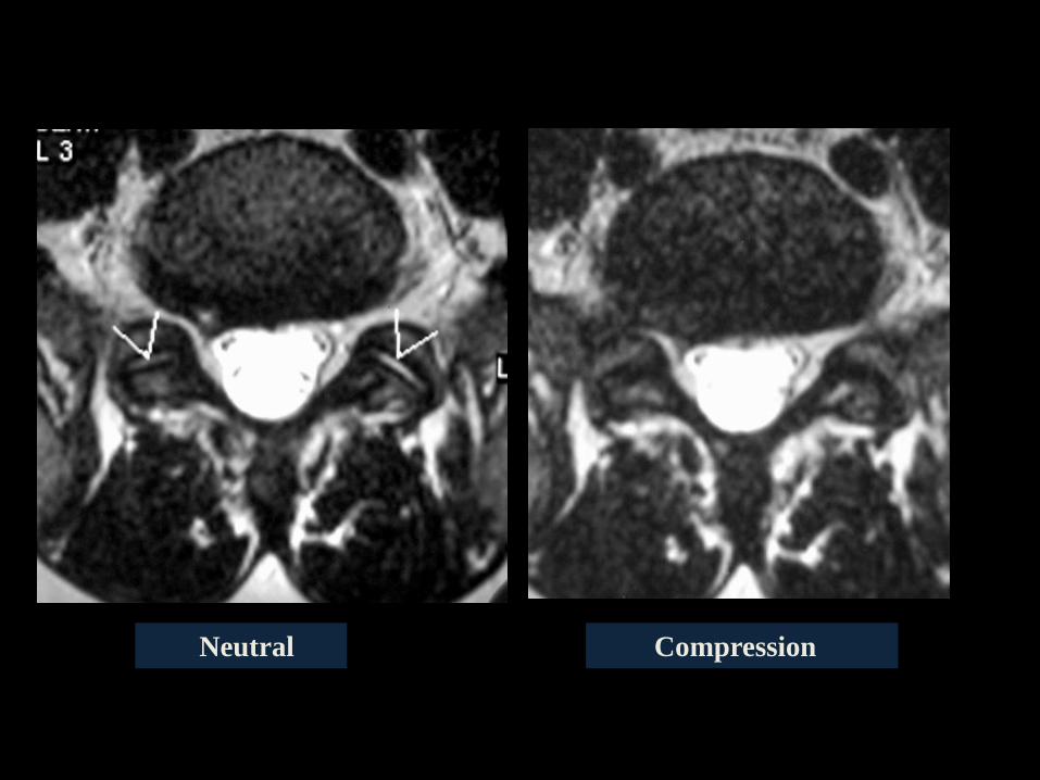
Increased synovial fluid in
facet joints on MRI in supine
position associated with
degenerative spondylolisthesis
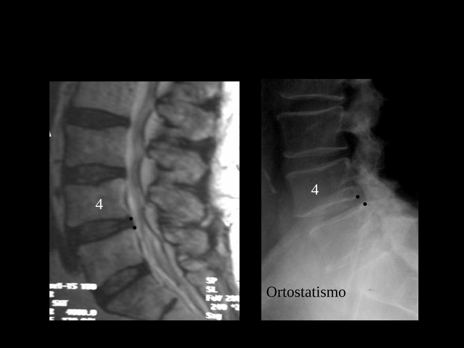
4
Ortostatismo
4 4
. . . .
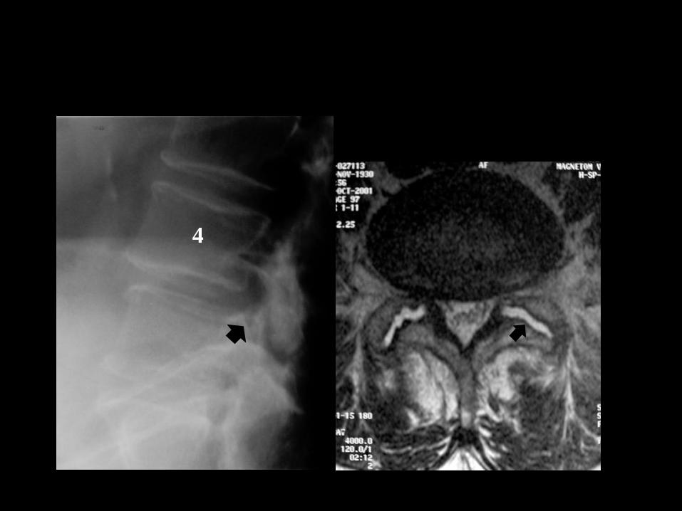
4
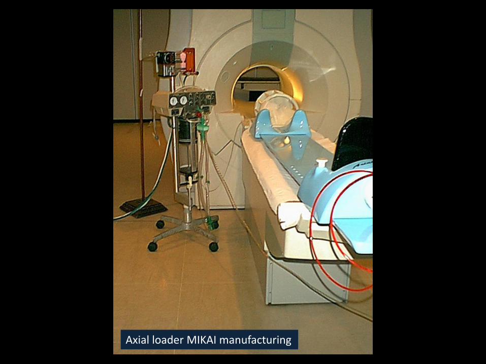
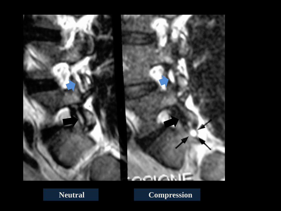
Axial loader MIKAI manufacturing
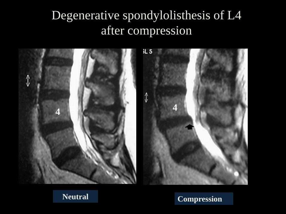
Compression Neutral
Degenerative spondylolisthesis of L4
after compression
4 4
Compression Neutral
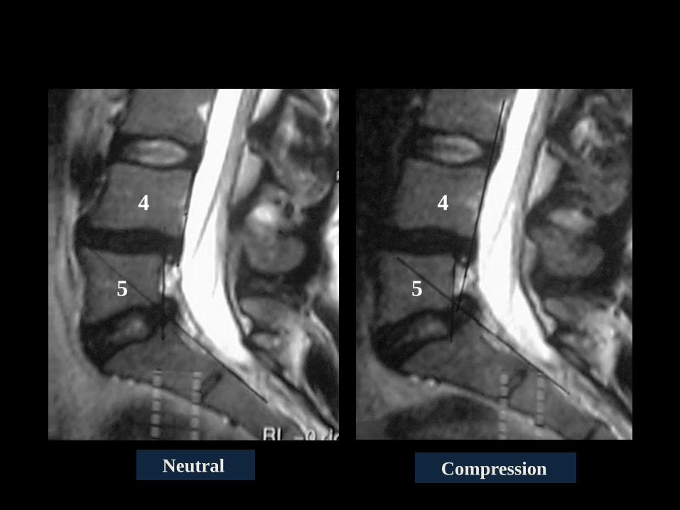
4 4 4
5 5
Neutral Compression
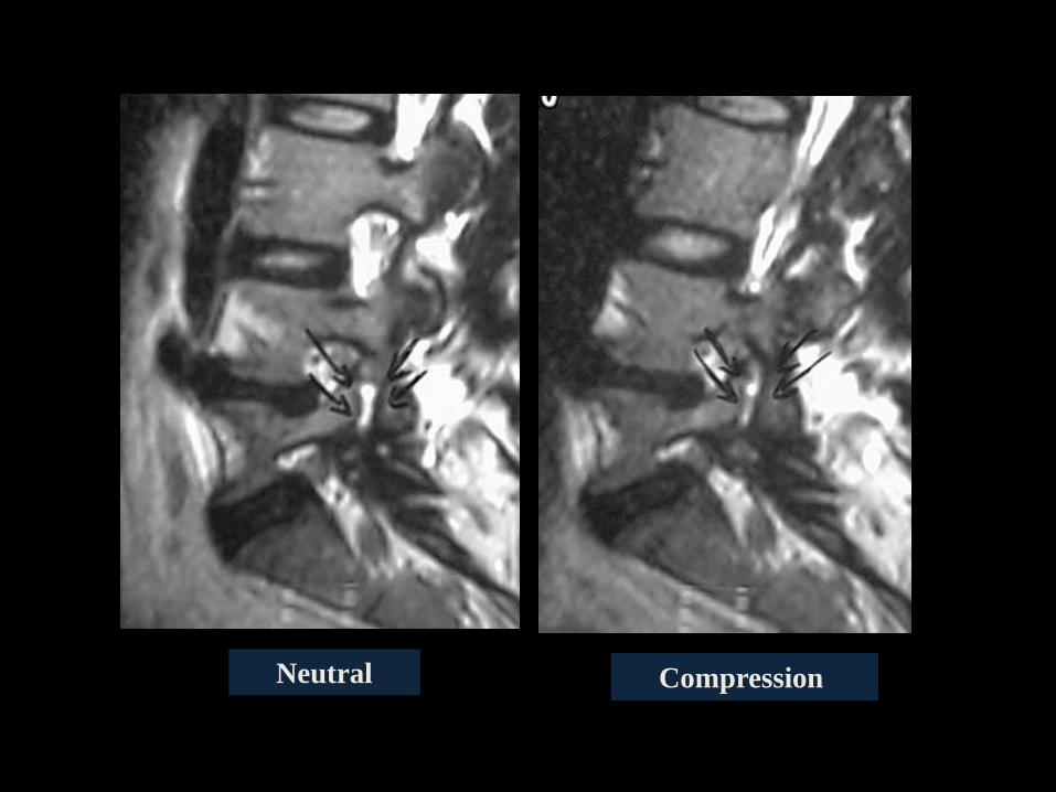
Compression Neutral
Compression Neutral
The decrease-redistribution of
synovial fluid of facet joints:
Probably the most interesting observation
as regard to the MRI in compression
Sign of microinstability ?
CONCLUSIONS IMAGING
• Some MRI findings may lead to suspect an
instability
• They are the tears of the annulus fibrosus,
Modic I changes of subcondral bone and
particularly the increase in synovial fluid of
facet joints
CLINICAL FEATURES
Low back and/or radicular pain
Pain in the prolonged standing position (post office) and
improvement in the supine position (frequent)
Pain in prolonged walking (fairly frequent)
Pain in physical efforts (frequent, aspecific)
DEGENERATIVE SPONDYLOLISTHESIS
SYMPTOMS
SIGNS
Pain on flexion-extension of the spine (fairly frequent, aspecific.)
Pain on local pressure (frequent, aspecific)
LOW BACK PAIN
BUT INSTABILITY, EVEN
ACTUAL, IS ALWAYS
ASSOCIATED WITH LOW BACK
PAIN ?
NO
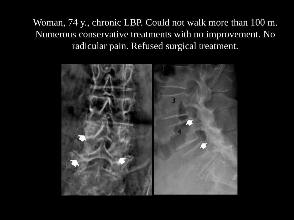
Woman, 74 y., chronic LBP. Could not walk more than 100 m.
Numerous conservative treatments with no improvement. No
radicular pain. Refused surgical treatment.
3
4
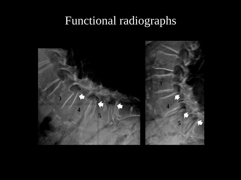
Functional radiographs
3
4 5
3
4
5
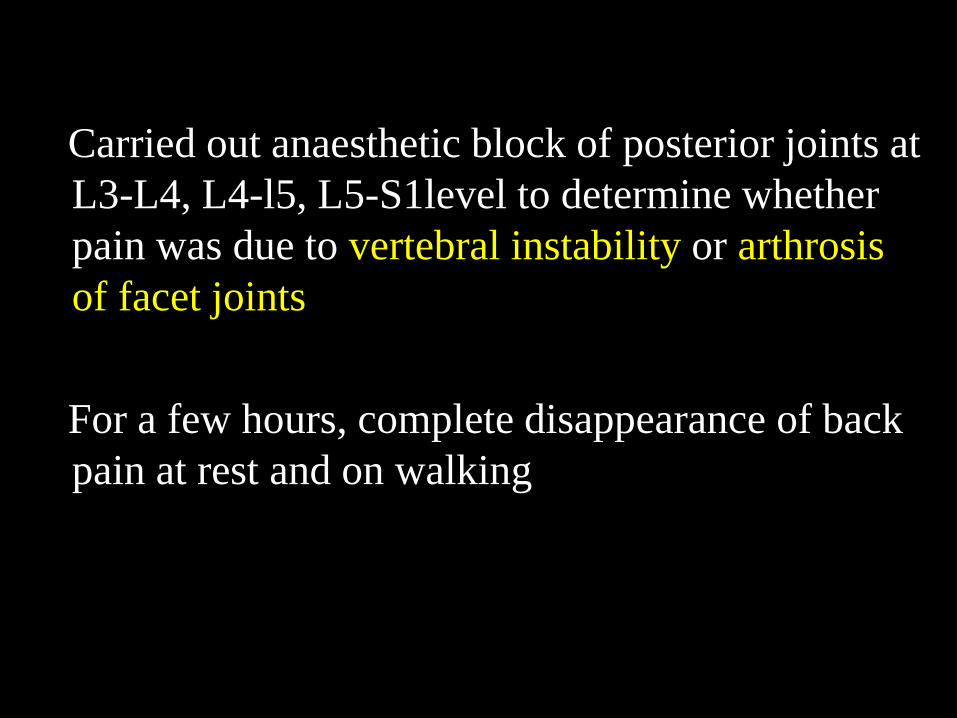
Carried out anaesthetic block of posterior joints at
L3-L4, L4-l5, L5-S1level to determine whether
pain was due to vertebral instability or arthrosis
of facet joints
For a few hours, complete disappearance of back
pain at rest and on walking
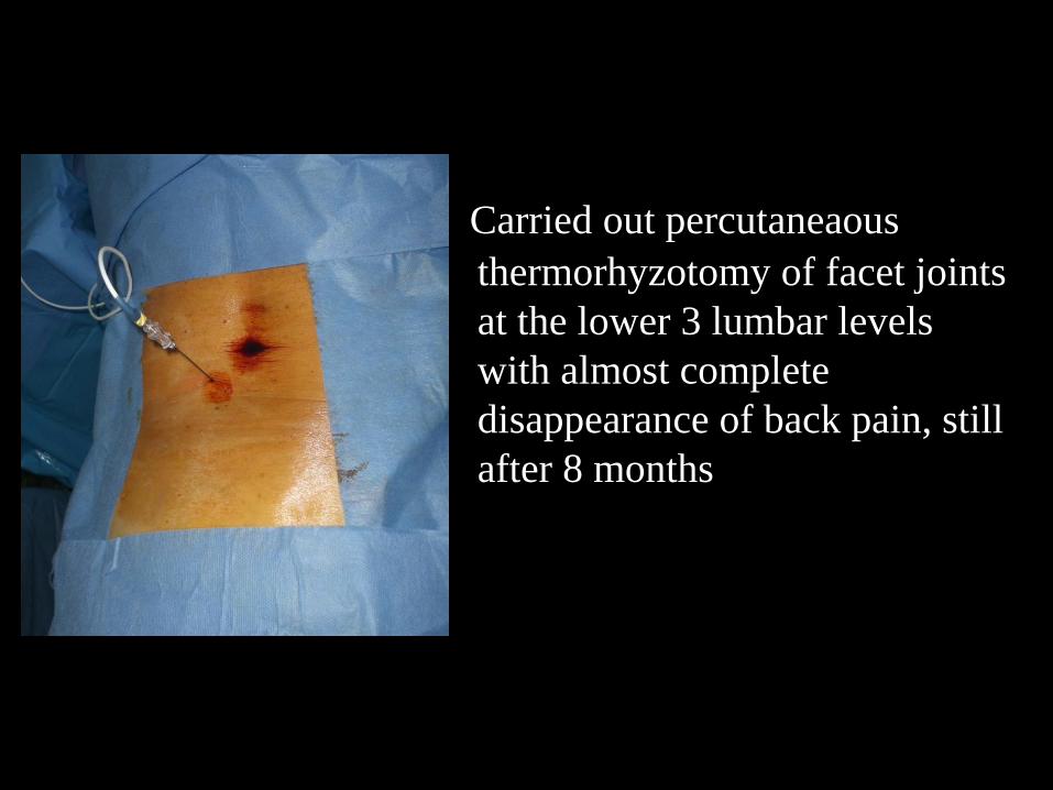
Carried out percutaneaous
thermorhyzotomy of facet joints
at the lower 3 lumbar levels
with almost complete
disappearance of back pain, still
after 8 months
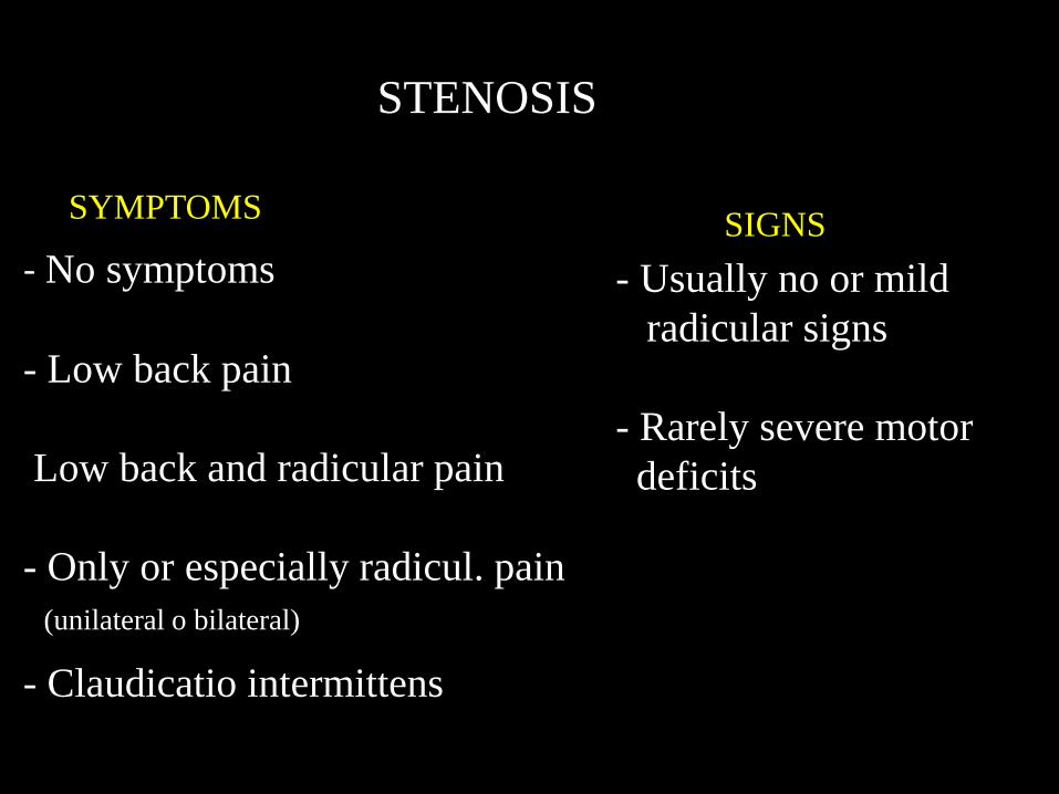
- No symptoms
- Low back pain
Low back and radicular pain
- Only or especially radicul. pain
(unilateral o bilateral)
- Claudicatio intermittens
SIGNS
- Usually no or mild
radicular signs
- Rarely severe motor
deficits
SYMPTOMS
STENOSIS
DIAGNOSIS
- Clinical signs (+ -)
- Imaging : RMI (+++), CT (++)
- EMG (+ -)
- History (+++)
- anamnesi (+++)
DISCOGENIC INSTABILITY
Continuous LBP or intermittent LBP with acute episodes
(frequent, aspecific)
SYMPTOMS
LBP during heavy physical activity (frequent, aspecific)
LBP on trunk flexion and torsion
(frequent, fairly aspecific)
SIGNS
Pain on trunk flexion (frequent, aspecific)
Pain on torsion, lateral inclination (frequente)
Pain on local pressure (frequent, aspecific)
NO RADICOLARI SIGNS
MODIC 1
No LBP
SYMPTOMS
SIGNS
Occasional LBP
Intermittent LBP or continuous pain on physical activities
(exercises), efforts, trunk flexion (frequent)
Pain on trunk flexion (frequent, aspecific)
Pain on pressure (aspecific)
NO RADICULAR SIGNS
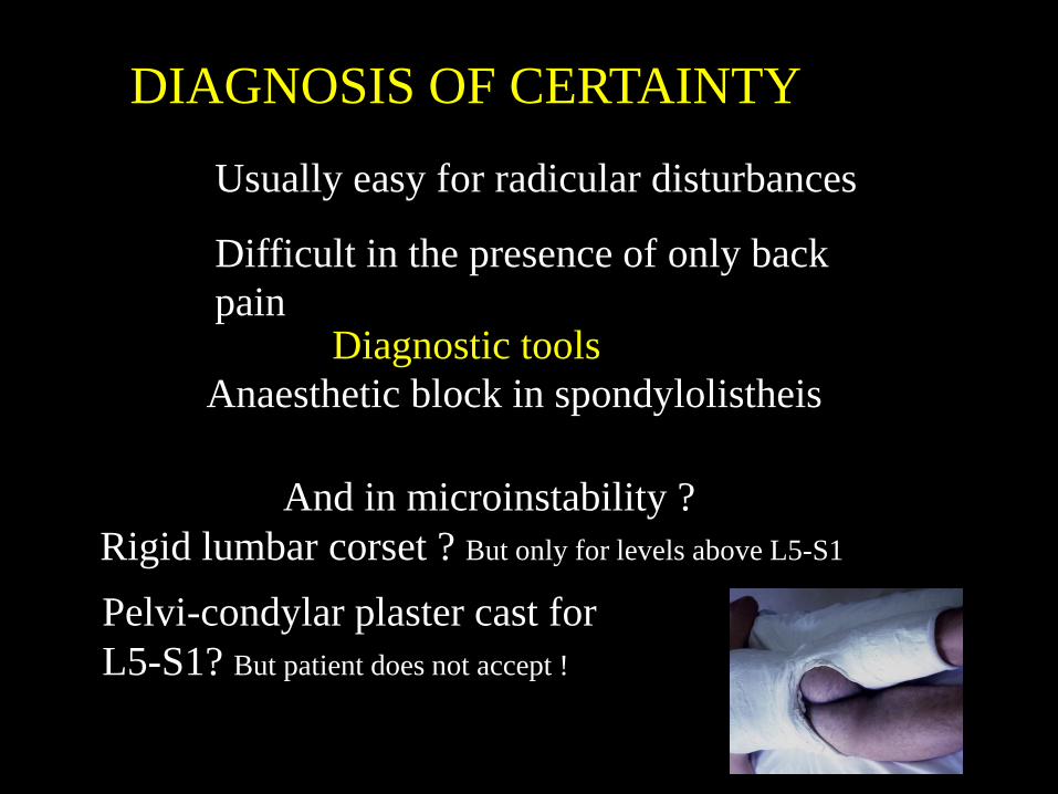
DIAGNOSIS OF CERTAINTY
Usually easy for radicular disturbances
Difficult in the presence of only back
pain Diagnostic tools
Anaesthetic block in spondylolistheis
And in microinstability ?
Rigid lumbar corset ? But only for levels above L5-S1
Pelvi-condylar plaster cast for
L5-S1? But patient does not accept !
CONCLUSIONS 1
There are certain instabilities: actual instability
There are possible instabilities, difficult to demonstrate
and based on indirect biomechanical, imaging and
clinical data: microinstability
There are probable instabilities: potential instabilities
Attention: even the actual instabilities can be
simple hypermobilities
CONCLUSIONS 2
The vertebral clinical features are very variables and are
mostly aspecific
Diagnosgtic tests may at times be useful
Microinstability is still a condition that is scarcely
known to a large extent
The most reliable symptoms are: pain in prolonmged
standing and walking

GRAZIE
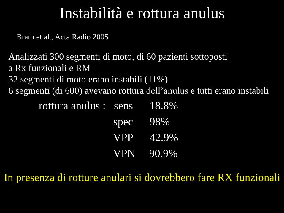
Instabilità e rottura anulus
rottura anulus : sens 18.8%
spec 98%
VPP 42.9%
VPN 90.9%
Bram et al., Acta Radio 2005
In presenza di rotture anulari si dovrebbero fare RX funzionali
Analizzati 300 segmenti di moto, di 60 pazienti sottoposti
a Rx funzionali e RM
32 segmenti di moto erano instabili (11%)
6 segmenti (di 600) avevano rottura dell’anulus e tutti erano instabili
post-gadolinio

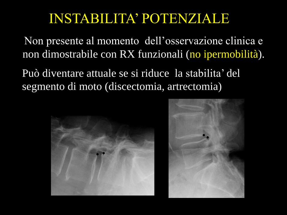
. . . .
Non presente al momento dell’osservazione clinica e
non dimostrabile con RX funzionali (no ipermobilità).
Può diventare attuale se si riduce la stabilita’ del
segmento di moto (discectomia, artrectomia)
INSTABILITA’ POTENZIALE
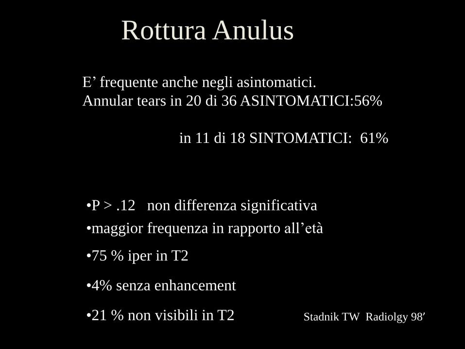
Rottura Anulus
E’ frequente anche negli asintomatici.
Annular tears in 20 di 36 ASINTOMATICI:56%
in 11 di 18 SINTOMATICI: 61%
Stadnik TW Radiolgy 98’
•P > .12 non differenza significativa
•maggior frequenza in rapporto all’età
•75 % iper in T2
•4% senza enhancement
•21 % non visibili in T2
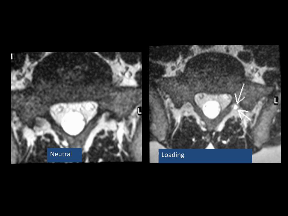
Loading Neutral
4
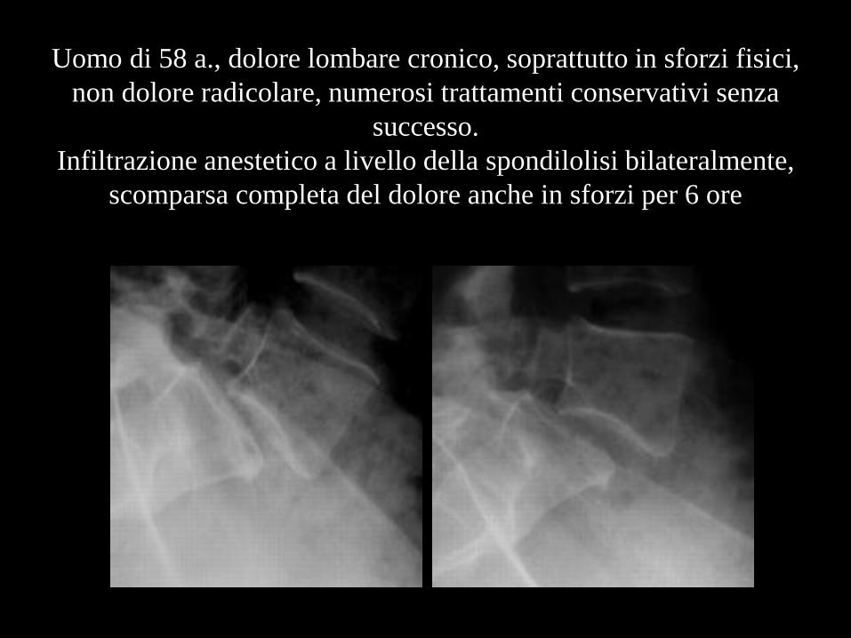
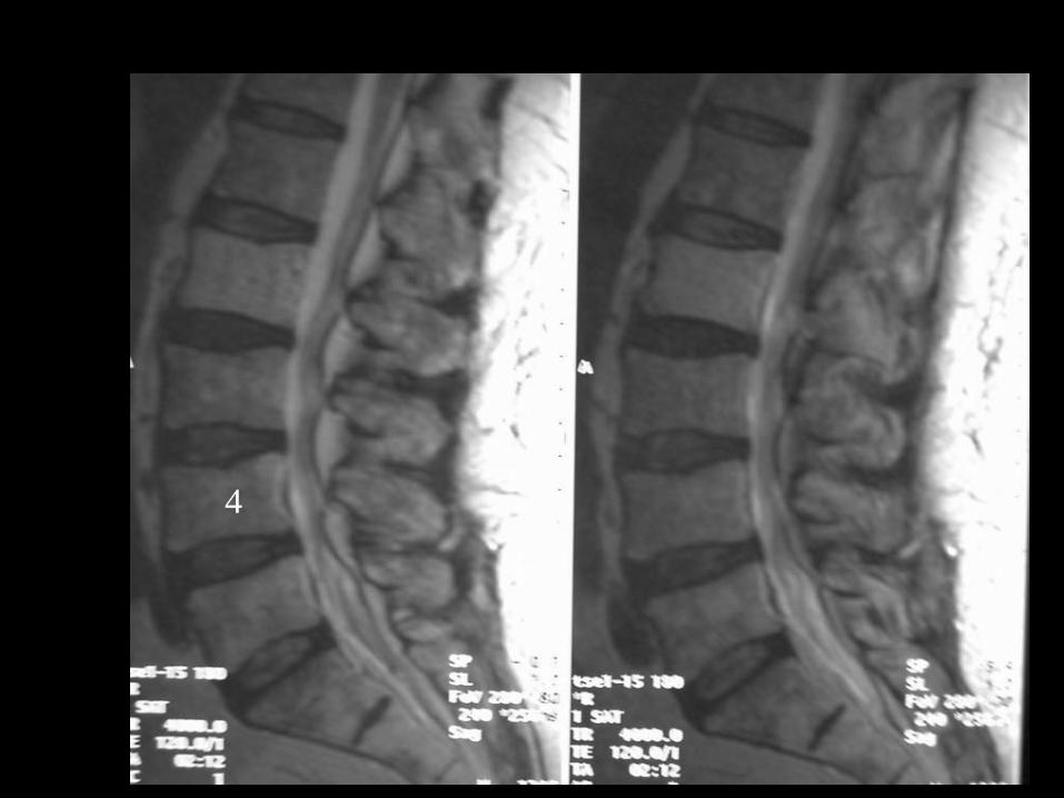
Uomo di 58 a., dolore lombare cronico, soprattutto in sforzi fisici,
non dolore radicolare, numerosi trattamenti conservativi senza
successo.
Infiltrazione anestetico a livello della spondilolisi bilateralmente,
scomparsa completa del dolore anche in sforzi per 6 ore
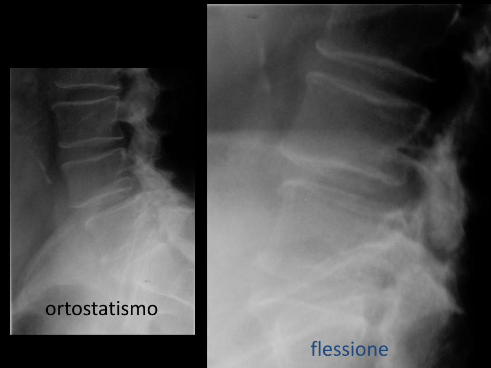
ortostatismo
flessione
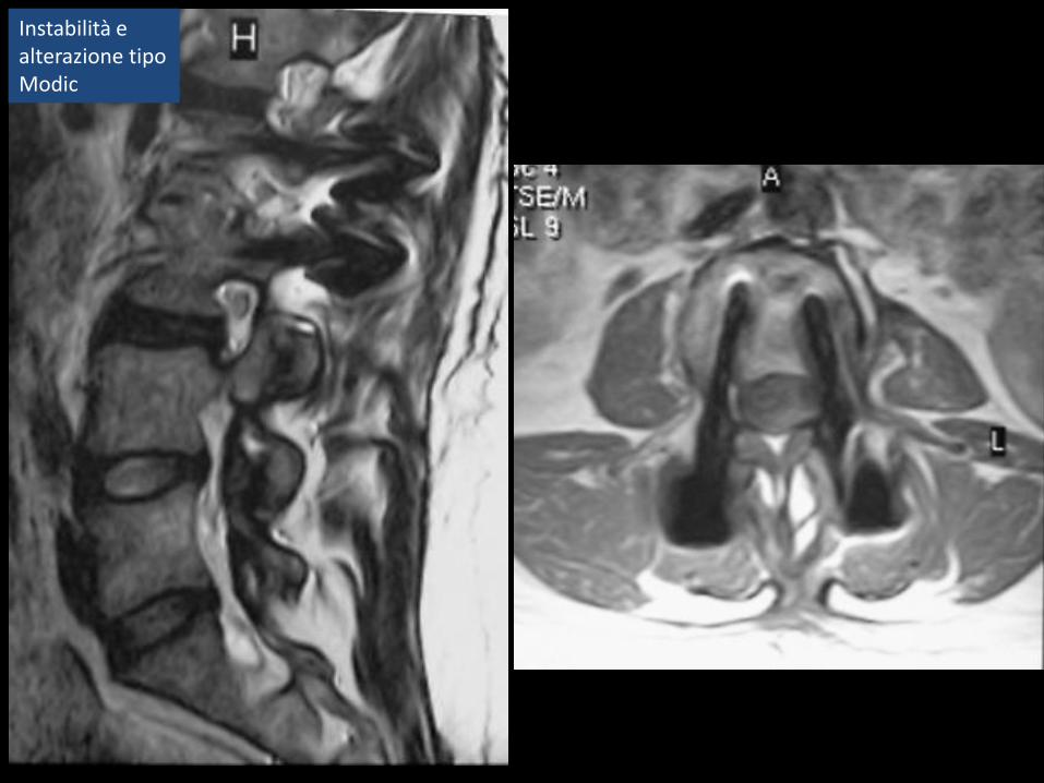
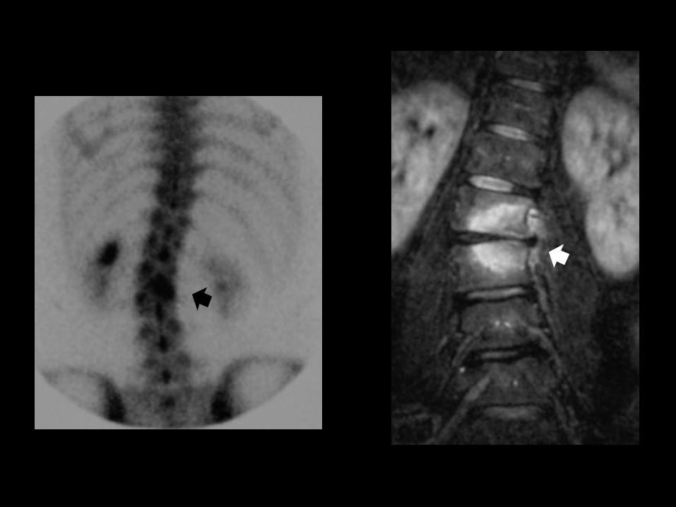
Instabilità e alterazione tipo Modic

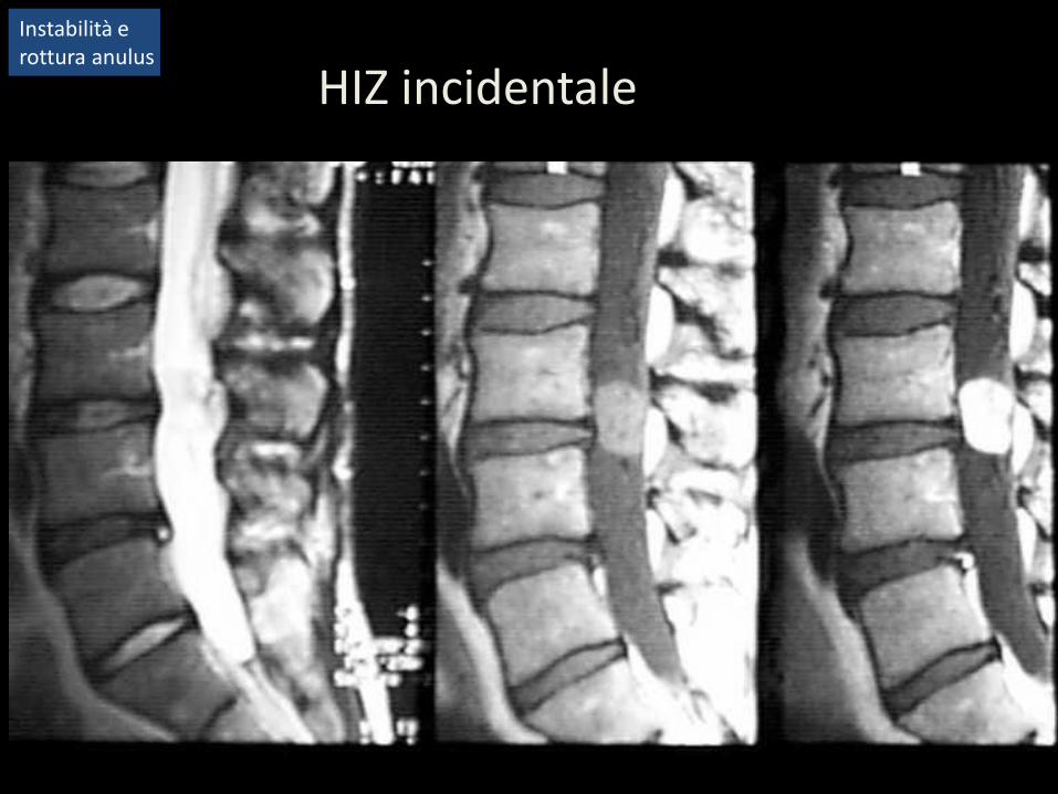
HIZ incidentale
Instabilità e rottura anulus
HIZ, esperienza personale
In 124 Pz con lombalgia rilevo di HIZ nel 32% (40 Pz.)
6 Pz presentavano anche alterazione degenerativa dell’osso subcondrale
Instabilità e rottura anulus

La perdita di capacità del rachide, sotto carichi
fisiologici, di mantenere i rapporti tra le vertebre
Pope, Panjabi 1985: A loss of motion segment stiffness, such that force application to that motion
segment produces abnormally great motion compared to that of a normal spine.
Una perdita di rigidità del segmento di moto, tale che la forza applicata
a quel segmento produce un movimento abnormemente maggiore che
in un rachide normale
Frymoyer, Selby 1985: An abnormal response to applied loads characterized kinematically by
abnormal movement in the motion segment beyond normal constraints
Un’ anomala risposta ai carichi applicati caratterizzata cinematicamente
da un abnorme movimento nel segmento di moto oltre i normali limiti
White III, Panjabi 1978: The loss of the ability of the spine under physiologic loads to
maintain relationships between vertebrae
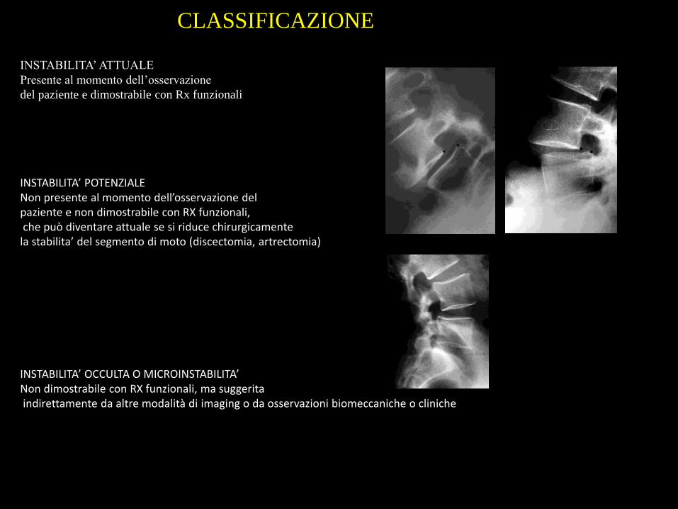
INSTABILITA’ ATTUALE
Presente al momento dell’osservazione
del paziente e dimostrabile con Rx funzionali
INSTABILITA’ POTENZIALE Non presente al momento dell’osservazione del paziente e non dimostrabile con RX funzionali, che può diventare attuale se si riduce chirurgicamente la stabilita’ del segmento di moto (discectomia, artrectomia) INSTABILITA’ OCCULTA O MICROINSTABILITA’ Non dimostrabile con RX funzionali, ma suggerita indirettamente da altre modalità di imaging o da osservazioni biomeccaniche o cliniche
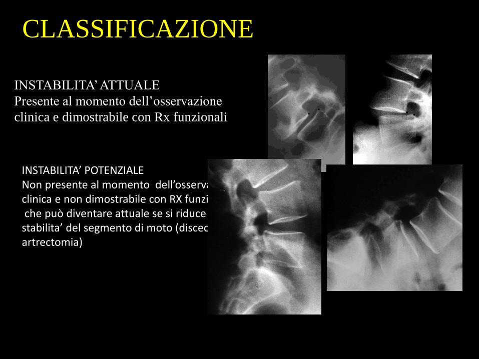
CLASSIFICAZIONE
CLASSIFICAZIONE
INSTABILITA’ ATTUALE
Presente al momento dell’osservazione
clinica e dimostrabile con Rx funzionali
INSTABILITA’ POTENZIALE Non presente al momento dell’osservazione clinica e non dimostrabile con RX funzionali, che può diventare attuale se si riduce la stabilita’ del segmento di moto (discectomia, artrectomia)

CLASSIFICAZIONE
INSTABILITA’ ATTUALE
INSTABILITA’ POTENZIALE
INSTABILITA’ OCCULTA O
MICROINSTABILITA’
INSTABILITA’ BIOMECCANICA (MATEMATICA-
SPERIMENTALE)
INSTABILITA’ “RADIOLOGICA”
INSTABILITA’ CLINICA
DIRETTA INDIRETTA
• DIAGNOSI
• DIAGNOSI
IDENTIFICAZIONE DELLA INSTABILITA’

CLASSIFICAZIONE
TIPI DI PATOLOGIE
• SPONDILO ISTMICA
• SPONDILO DEGENERATIVA
• DISCOPATIA DEGENERATIVA
• SCOLIOSI
QUADRI CLINICI
IMAGING
TRACTION SPUR
A 74-year old woman complained of chronic back pain, which worsened on walking. In the last few months, pain had become more severe and prevent her walking more than 100 metres. She had no leg pain.
She had undergone several conservative
treatments (anti-inflammatory medication,
phisical therapy and mild exercises) with
no significant improvement
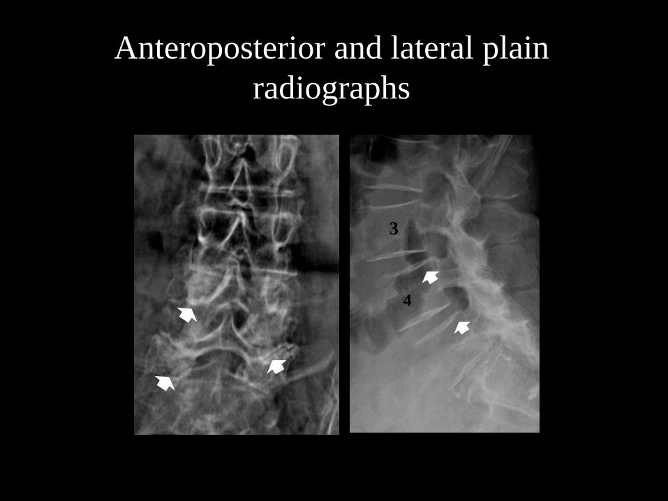
Anteroposterior and lateral plain
radiographs
3
4

Flexion-extension radiographs
3
4 5
3
4
5
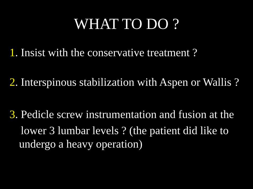
WHAT TO DO ?
1. Insist with the conservative treatment ?
2. Interspinous stabilization with Aspen or Wallis ?
3. Pedicle screw instrumentation and fusion at the
lower 3 lumbar levels ? (the patient did like to
undergo a heavy operation)
We carried out an injection of a local anhaestetic at L5-S1, L4-L5 and L3-L4 on both sides to determine whether low back pain was due to vertebral instability or arthrotic changes of the facet joints
For a few hours after the injection the patient had a complete disappearance of back pain also on prolonged walking
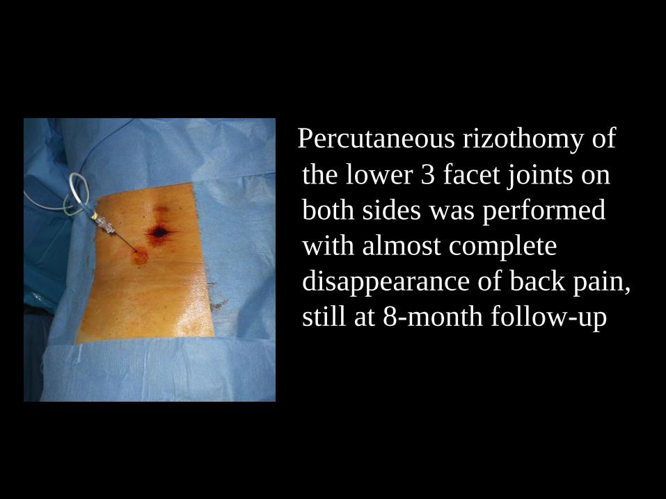
Percutaneous rizothomy of
the lower 3 facet joints on
both sides was performed
with almost complete
disappearance of back pain,
still at 8-month follow-up