Embed Size (px)
Citation preview
The PDF of the article you requested follows this cover page.
This is an enhanced PDF from The Journal of Bone and Joint Surgery
73:802-808, 1991. J. Bone Joint Surg. Am.HN Herkowitz and LT Kurz
intertransverse process arthrodesisprospective study comparing decompression with decompression and Degenerative lumbar spondylolisthesis with spinal stenosis. A
This information is current as of September 27, 2006
Reprints and Permissions
Permissions] link. and click on the [Reprints andjbjs.orgarticle, or locate the article citation on
to use material from thisorder reprints or request permissionClick here to
Publisher Information
www.jbjs.org20 Pickering Street, Needham, MA 02492-3157The Journal of Bone and Joint Surgery
on September 27, 2006 www.ejbjs.orgDownloaded from
Copyright 1991 by The Journal ofBone and Joint Surgery, Incorporated
802 ThE JOURNAL OF BONE AND JOINT SURGERY
Degenerative Lumbar Spondylolisthesis with Spinal Stenosis
A PROSPECTIVE STUDY COMPARING DECOMPRESSION
WITH DECOMPRESSION AND INTERTRANSVERSE PROCESS ARTHRODESIS*t
BY HARRY N. HERKOWITZ, M.D4, AND LAWRENCE T. KURZ, M.D4, ROYAL OAK, MICHIGAN
From the Section of Spine Surgery, Department of Orthopaedic Surgery, William Beaumont Hospital, Royal Oak
ABSTRACT: Fifty patients who had spinal stenosisassociated with degenerative lumbar spondylolisthesiswere prospectively studied clinically and radiographi-cally to determine ifconcomitant intertransverse-processarthrodesis provided better results than decompressivelaminectomy alone. There were thirty-six women andfourteen men. The mean age of the twenty-five patientswho had had an arthrodesis was 63.5 years and that ofthe twenty-five patients who had not had an arthrodesis,sixty-five years. The level of the operation was betweenthe fourth and fifth lumbar vertebrae in forty-one pa-tients and between the third and fourth lumbar verte-brae in nine patients. The patients were followed for amean of three years (range, 2.4 to four years). In thepatients who had had a concomitant arthrodesis, theresults were significantly better with respect to relief ofpain in the back and lower limbs.
Degenerative lumbar spondylolisthesis was apparentlyfirst described in the German literature, as pseudospondy-
lolisthesis, by Junghanns23 in 193 1 , and in the English-language literature by Macnab29 in 1950. The modern con-cept of degenerative spondylolisthesis was described byNewman� in 1955; however, the operative management ofthis disorder when accompanied by spinal stenosis has re-mained controversial, despite a clearer understanding of itspathogenesis and pathology’ ,6,1417,24,26,43#{149}
Some authors have reported satisfactory results withdecompressive laminectomy alone5’79”2”9’�, while others
have advocated that a spinal arthrodesis be done concom-itantly with the decompression2’4”2”6’242839’49. It is difficult
to compare these series because of differences in the patientpopulations, operative procedures, surgeons, postoperativemanagement, grading of results, and levels of the spine atwhich the operation was done. Therefore, the indications
for concomitant arthrodesis with decompressive laminec-tomy in the operative management of patients who havedegenerative lumbar spondylolisthesis and spinal stenosishave remained unclear.
To determine these indications, a prospective study wasperformed to compare the results of decompression alone
* No benefits in any form have been received or will be received froma commercial party related directly or indirectly to the subject ofthis article.No funds were received in support of this study.
t Read in part at the Annual Meeting of the International Society forthe Study of the Lumbar Spine, Boston, Massachusetts, June 17, 1990.
t Suite 100, 16800 West Twelve Mile Road, Southfield, Michigan48076-2176.
with those of concomitant intertransverse-process arthro-desis at the level of the decompression for the managementof degenerative spondylolisthesis at a single level associatedwith lumbar spinal stenosis.
Materials and Methods
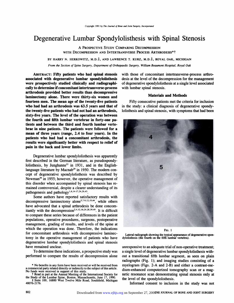
Fifty consecutive patients met the criteria for inclusionin the study: a clinical diagnosis of degenerative spondy-lolisthesis and spinal stenosis, with symptoms that had been
FIG. 1
Lateral radiograph showing the typical appearance ofdegenerative spon-
dylolisthesis (the fourth on the fifth lumbar vertebra).
unresponsive to an adequate trial of non-operative treatment;a single level of degenerative lumbar spondylolisthesis with-
out a transitional fifth lumbar segment, as seen on plainradiographs (Fig. 1); and imaging studies consisting of amyelogram (Figs. 2-A and 2-B) and either a contrast-me-
dium-enhanced computerized tomography scan or a mag-netic resonance scan demonstrating spinal stenosis only atthe level of the spondylolisthesis.
Informed consent to inclusion in the study was not
on September 27, 2006 www.ejbjs.orgDownloaded from
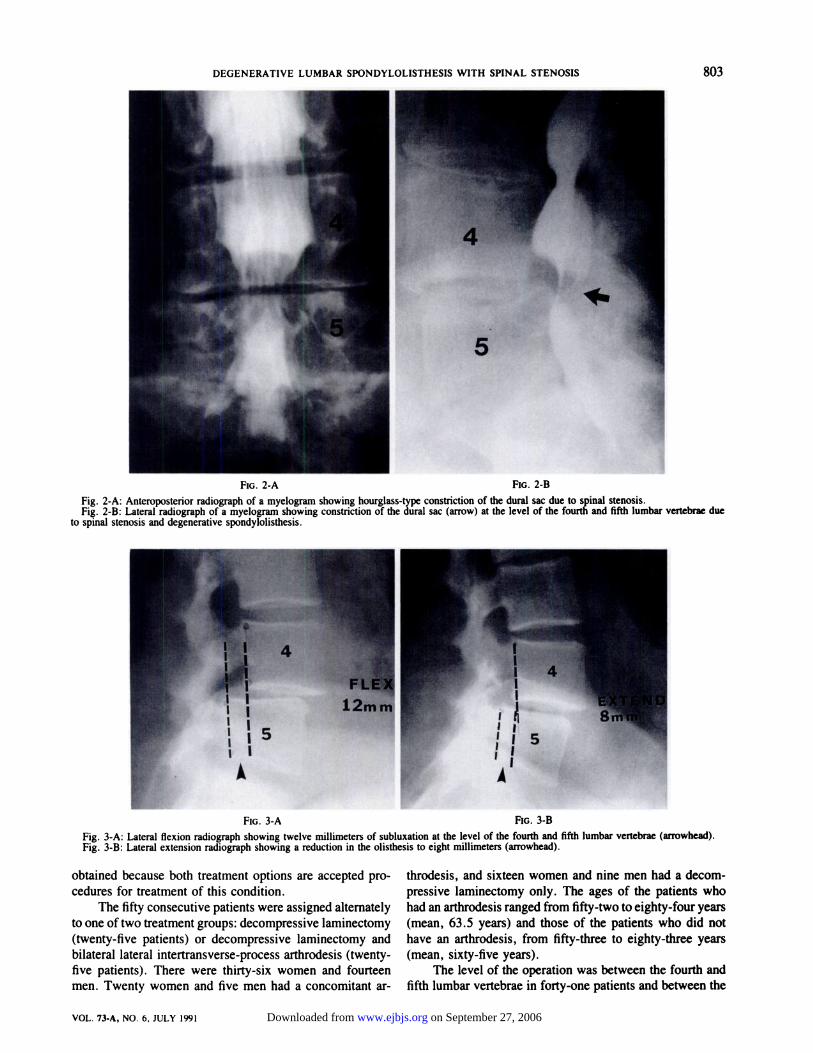
FIG. 2-A FIG. 2-B
Fig. 2-A: Anteroposterior radiograph of a myelogram showing hourglass-type constriction of the dural sac due to spinal stenosis.Fig. 2-B: Lateral radiograph of a myelogram showing constriction of the dural sac (arrow) at the level of the fourth and fifth lumbar vertebrae due
to spinal stenosis and degenerative spondylolisthesis.
FIG. 3-A FIG. 3-B
Fig. 3-A: Lateral flexion radiograph showing twelve millimeters of subluxation at the level of the fourth and fifth lumbar vertebrae (arrowhead).Fig. 3-B: Lateral extension radiograph showing a reduction in the olisthesis to eight millimeters (arrowhead).
DEGENERATIVE LUMBAR SPONDYLOLISTHESIS WITH SPINAL STENOSIS 803
VOL. 73-A, NO. 6, JULY 1991
obtained because both treatment options are accepted pro-
cedures for treatment of this condition.The fifty consecutive patients were assigned alternately
to one of two treatment groups: decompressive laminectomy(twenty-five patients) or decompressive laminectomy andbilateral lateral intertransverse-process arthrodesis (twenty-five patients). There were thirty-six women and fourteenmen. Twenty women and five men had a concomitant ar-
throdesis, and sixteen women and nine men had a decom-pressive laminectomy only. The ages of the patients whohad an arthrodesis ranged from fifty-two to eighty-four years(mean, 63 .5 years) and those of the patients who did nothave an arthrodesis, from fifty-three to eighty-three years
(mean, sixty-five years).The level of the operation was between the fourth and
fifth lumbar vertebrae in forty-one patients and between the
on September 27, 2006 www.ejbjs.orgDownloaded from
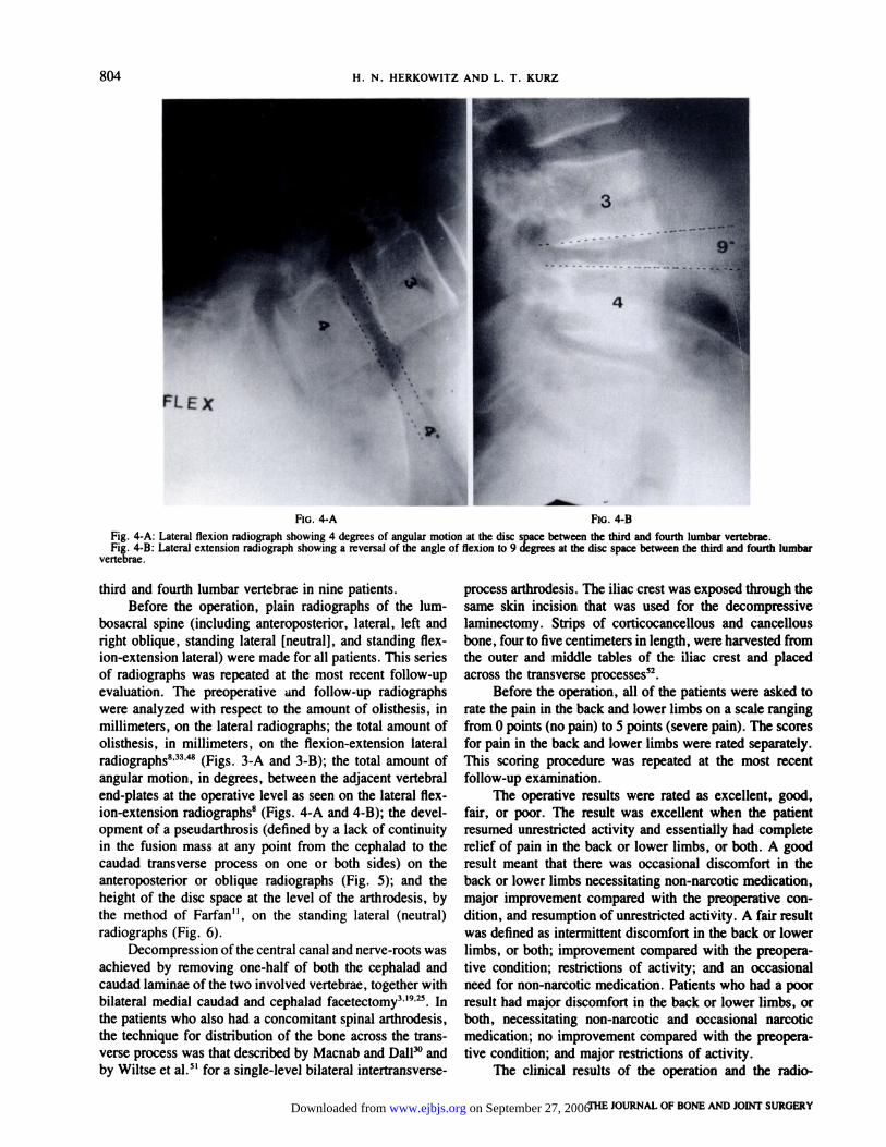
FIG. 4-A FiG. 4-B
804 H. N. HERKOWITZ AND L. T. KURZ
THE JOURNAL OF BONE AND JOINT SURGERY
Fig. 4-A: Lateral flexion radiograph showing 4 degrees of angular motion at the disc space between the third and fourth lumbar vertebrae.Fig. 4-B: Lateral extension radiograph showing a reversal of the angle of flexion to 9 degrees at the disc space between the third and fourth lumbar
vertebrae.
third and fourth lumbar vertebrae in nine patients.Before the operation, plain radiographs of the lum-
bosacral spine (including anteropostenor, lateral, left andright oblique, standing lateral [neutral], and standing flex-ion-extension lateral) were made for all patients. This series
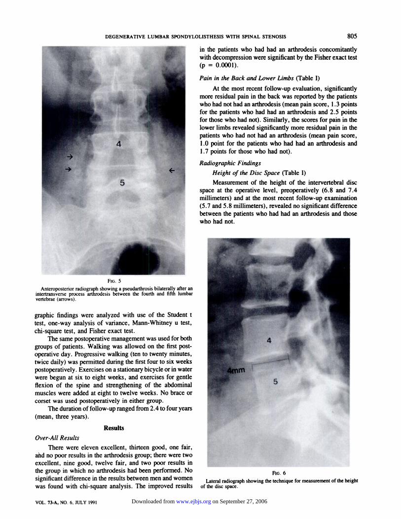
of radiographs was repeated at the most recent follow-upevaluation. The preoperative and follow-up radiographswere analyzed with respect to the amount of olisthesis, inmillimeters, on the lateral radiographs; the total amount ofolisthesis, in millimeters, on the flexion-extension lateralradiographs8’33’� (Figs. 3-A and 3-B); the total amount ofangular motion, in degrees, between the adjacent vertebralend-plates at the operative level as seen on the lateral flex-ion-extension radiographs8 (Figs. 4-A and 4-B); the devel-opment of a pseudarthrosis (defined by a lack of continuityin the fusion mass at any point from the cephalad to the
caudad transverse process on one or both sides) on theanteroposterior or oblique radiographs (Fig. 5); and the
height of the disc space at the level of the arthrodesis, bythe method of Farfan’ ‘ , on the standing lateral (neutral)radiographs (Fig. 6).
Decompression of the central canal and nerve-roots was
achieved by removing one-half of both the cephalad and
caudad laminae of the two involved vertebrae, together withbilateral medial caudad and cephalad facetectomy3”9’�. Inthe patients who also had a concomitant spinal arthrodesis,the technique for distribution of the bone across the trans-verse process was that described by Macnab and DalP#{176}and
by Wiltse et al.5’ for a single-level bilateral intertransverse-
process arthrodesis. The iliac crest was exposed through thesame skin incision that was used for the decompressivelaminectomy. Strips of corticocancellous and cancellous
bone, four to five centimeters in length, were harvested from
the outer and middle tables of the iliac crest and placedacross the transverse processes52.
Before the operation, all of the patients were asked torate the pain in the back and lower limbs on a scale rangingfrom 0 points (no pain) to 5 points (severe pain). The scoresfor pain in the back and lower limbs were rated separately.This scoring procedure was repeated at the most recent
follow-up examination.The operative results were rated as excellent, good,
fair, or poor. The result was excellent when the patientresumed unrestricted activity and essentially had completerelief of pain in the back or lower limbs, or both. A goodresult meant that there was occasional discomfort in theback or lower limbs necessitating non-narcotic medication,major improvement compared with the preoperative con-
dition, and resumption of unrestricted activity. A fair resultwas defined as intermittent discomfort in the back or lowerlimbs, or both; improvement compared with the preopera-
tive condition; restrictions of activity; and an occasional
need for non-narcotic medication. Patients who had a poorresult had major discomfort in the back or lower limbs, orboth, necessitating non-narcotic and occasional narcoticmedication; no improvement compared with the preopera-tive condition; and major restrictions of activity.
The clinical results of the operation and the radio-
on September 27, 2006 www.ejbjs.orgDownloaded from
I
I1�.,.,V
4
5..�,
FIG. 5
DEGENERATIVE LUMBAR SPONDYLOLISTHESIS WITH SPINAL STENOSIS 805
VOL. 73.A, NO. 6, JULY 1991
c-i in the patients who had had an arthrodesis concomitantly
.� I with decompression were significant by the Fisher exact test
� (p = 0.0001).
Pain in the Back and Lower Limbs (Table I)
At the most recent follow-up evaluation, significantlymore residual pain in the back was reported by the patientswho had not had an arthrodesis (mean pain score, 1 .3 points
for the patients who had had an arthrodesis and 2.5 pointsfor those who had not). Similarly, the scores for pain in the
lower limbs revealed significantly more residual pain in the
patients who had not had an arthrodesis (mean pain score,1.0 point for the patients who had had an arthrodesis and
1 .7 points for those who had not).
Radiographic Findings
Height of the Disc Space (Table I)
Measurement of the height of the intervertebral discspace at the operative level, preoperatively (6.8 and 7.4millimeters) and at the most recent follow-up examination(5.7 and 5.8 millimeters), revealed no significant difference
between the patients who had had an arthrodesis and those
who had not.
Anteroposterior radiograph showing a pseudarthrosis bilaterally after anintertransverse process arthrodesis between the fourth and fifth lumbarvertebrae (arrows).
graphic findings were analyzed with use of the Student t
test, one-way analysis of variance, Mann-Whitney u test,chi-square test, and Fisher exact test.
The same postoperative management was used for bothgroups of patients. Walking was allowed on the first post-operative day. Progressive walking (ten to twenty minutes,
twice daily) was permitted during the first four to six weekspostoperatively. Exercises on a stationary bicycle or in waterwere begun at six to eight weeks, and exercises for gentle
flexion of the spine and strengthening of the abdominalmuscles were added at eight to twelve weeks. No brace orcorset was used postoperatively in either group.
The duration of follow-up ranged from 2.4 to four years
(mean, three years).
Results
Over-All Results
There were eleven excellent, thirteen good, one fair,ahd no poor results in the arthrodesis group; there were twoexcellent, nine good, twelve fair, and two poor results inthe group in which no arthrodesis had been performed. No �G 6
significant difference in the results between men and women �teral radiograph showing the technique for measurement of the height
was found with chi-square analysis. The improved results of the disc space.
on September 27, 2006 www.ejbjs.orgDownloaded from
806 H. N. HERKOWITZ AND L. T. KURZ
ThE JOURNAL OF BONE AND JOINT SURGERY
Olisthesis (Table I)
The patients who had not had an arthrodesis had asignificant increase in olisthesis (5.3 to 7.9 millimeters)compared with the preoperative value, including the changeoccurring on flexion and extension (3.4 to 5.8 millimeters)compared with normal values, as seen on the preoperativeand most recent follow-up radiographs. Seven (28 per cent)of the twenty-five patients who had had an arthrodesis and
twenty-four (96 per cent) of the twenty-five patients whohad not had an arthrodesis had increased olisthesis after theoperation.
All seven patients who had had an arthrodesis and hadincreased olisthesis postoperatively had an excellent or good
result, compared with eleven of the twenty-four patientswho had not had an arthrodesis and had increased olisthesis.None of the seven patients who had had an arthrodesis andhad increased olisthesis had a fair or poor result, comparedwith thirteen of the twenty-four patients who had not hadan arthrodesis.
Vertebral Angulation (Table I)
There was a significant increase in angular motion on
lateral bending compared with the preoperative values (9.6compared with 12. 8 degrees) in the patients who had hadan arthrodesis (p = 0.002). One-way analysis of variance
demonstrated that the postoperative radiographic findings
of olisthesis that were noted on flexion and extension andof angular motion that were seen on lateral bending weresignificant contributors to the poorer clinical results in thepatients who had not had a concomitant arthrodesis.
Pseudarthrosis
Pseudarthrosis of the fusion mass was observed in nine(36 per cent) of the twenty-five patients who had had anarthrodesis. In two of these patients, the pseudarthrosis was
noted on both sides of the fusion mass, and in the otherseven, it was confined to one side. However, the clinicalresult was excellent in five and good in four patients.
Discussion
The literature has been unclear with respect to the roleof arthrodesis when a decompressive laminectomy is per-
formed for spinal stenosis associated with degenerative lum-bar spondylolisthesis2’7”22#{176}22’28’41’45’47. Satisfactory resultsafter decompressive laminectomy alone have been reportedin 60 to 96 per cent of patients47�#{176}22’28.Residual or recurrentpain in the back or lower limbs, or both, has been noted,however, in as many as 73 per cent of such patients, with
residual low-back pain reported more frequently�#{176}.This maybe related, to some degree, to residual instability, or it maybe related to the development of increased olisthesis post-operatively47’2’2t2227’2843’45’47. In some series, the olisthesishas been found to increase significantly more often in pa-
tients who have a poor result7’22.In reports on the results of decompressive laminectomy
alone, concomitant arthrodesis was recommended for all
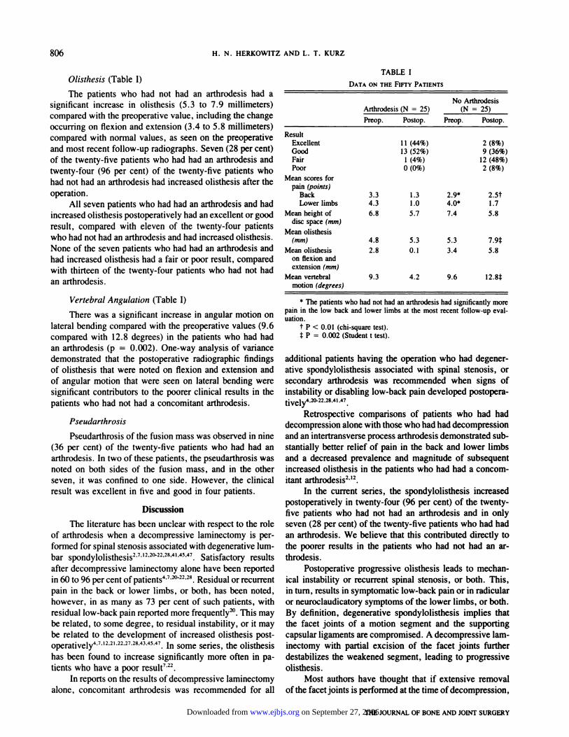
TABLE I
DATA ON THE Fivr� PATIENTS
Arthrode sis (N = 25)No Arthrodesis
(N = 25)
Preop. Postop. Preop. Postop.
ResultExcellent 1 1 (44%) 2 (8%)Good 13 (52%) 9 (36%)Fair 1 (4%) 12 (48%)Poor 0 (0%) 2 (8%)
Mean scores forpain (points)
Back 3.3 1.3 2.9* 2.5tLowerlimbs 4.3 1.0 4.0* 1.7
Mean height of 6.8 5.7 7.4 5.8disc space (mm)
Mean olisthesis(nun) 4.8 5.3 5.3 7.9�
Meanolisthesis 2.8 0.1 3.4 5.8on flexion andextension (mm)
Mean vertebral 9.3 4.2 9.6 12.8�motion (degrees)
* The patients who had not had an arthrodesis had significantly more
pain in the low back and lower limbs at the most recent follow-up eval-uation.
t P < 0.01 (chi-square test).
t P = 0.002 (Student t test).
additional patients having the operation who had degener-ative spondylolisthesis associated with spinal stenosis, or
secondary arthrodesis was recommended when signs ofinstability or disabling low-back pain developed postopera-
44,47.
Retrospective comparisons of patients who had had
decompression alone with those who had had decompression
and an intertransverse process arthrodesis demonstrated sub-stantially better relief of pain in the back and lower limbsand a decreased prevalence and magnitude of subsequentincreased olisthesis in the patients who had had a concom-itant arthrodesis2”2.
In the current series, the spondylolisthesis increasedpostoperatively in twenty-four (96 per cent) of the twenty-five patients who had not had an arthrodesis and in onlyseven (28 per cent) of the twenty-five patients who had hadan arthrodesis. We believe that this contributed directly tothe poorer results in the patients who had not had an ar-throdesis.
Postoperative progressive olisthesis leads to mechan-
ical instability or recurrent spinal stenosis, or both. This,
in turn, results in symptomatic low-back pain or in radicularor neuroclaudicatory symptoms of the lower limbs, or both.
By definition, degenerative spondylolisthesis implies thatthe facet joints of a motion segment and the supportingcapsular ligaments are compromised. A decompressive lam-inectomy with partial excision of the facet joints further
destabilizes the weakened segment, leading to progressiveolisthesis.
Most authors have thought that if extensive removalofthe facetjoints is performed at the time of decompression,
on September 27, 2006 www.ejbjs.orgDownloaded from
DEGENERATIVE LUMBAR SPONDYLOLISTHESIS WITH SPINAL STENOSIS 807
VOL. 73-A, NO. 6, JULY 1991
an arthrodesis should be added. The criteria of Wiltse et
al.�#{176}for inclusion of a spinal arthrodesis after decompressivelaminectomy in patients who have degenerative spondylo-listhesis were an age of less than sixty years for patients
who have had total removal of the facet joints and of less
than fifty-five years for patients who have had central de-
compression and in whom the facet joints are intact. Al-though these criteria have served as guidelines in the past,it must be recognized that the patient’s chronological andphysiological ages often do not coincide, especially as we
enter an era when the elderly are living longer, are healthier,and lead more active lives. Therefore, age restraints on theperformance of operative procedures have less validity thanin the past. In the current series, age and sex were notsignificant predictors of the outcome of the operation.
The preoperative and postoperative heights of the discspace were calculated to determine if a normal disc space,without major degenerative narrowing, would predispose to
an increase in postoperative olisthesis or affect the outcome,and whether the height of the disc space could be used asa determinant for recommendation of a concomitant spinalarthrodesis. Lombardi et al. noted that disc spaces that were
more than six millimeters in height tended to slip the mostafter decompression’-�. Although these authors did not spe-
cifically state that this was a criterion for performance ofan arthrodesis after decompression, that was implied by theirfindings. Johnsson et al. noted no significant difference inthe height of the disc space between patients who had good
and poor results after decompressive laminectomy, but theydid observe a decrease in the height of the disc space afterthe operation22. Although the technique of measurement thatwas used in the current series differed from that of Johnsson
et al. , our results demonstrated a general tendency for theheight of the disc space to decrease in both the patients whohad had an arthrodesis and in those who had not, and thismeasurement was not useful for prediction of the necessityof an arthrodesis.
Despite the fact that a pseudarthrosis occurred in nine(36 per cent) of the twenty-five patients who had had anarthrodesis, the result was excellent in five and good in fourpatients. It should be noted that seven patients had a pseu-darthrosis on only one side of the arthrodesis. Although two
patients had a bilateral non-union, attainment of a solidfusion on one side of the arthrodesis or the development of
a fibrous union appeared to provide sufficient structural sup-port to prevent progressive olisthesis.
As can be seen from the literature, the criteria for theperformance of a spinal arthrodesis after decompression for
spinal stenosis secondary to degenerative lumbar spondy-
lolisthesis have been based on retrospective analysis andanecdotal information3133’3538. The preoperative factors thathave often been cited as influencing the results are age, sex,the severity of preoperative pain in the back, the amount ofpreoperative olisthesis, and the amounts of olisthesis andvertebral motion seen on postoperative flexion-extension
radiographs�#{176}22’�#{176}.These factors were evaluated in thepresent study. The severity of preoperative pain in the backand lower limbs, the age and sex of the patient, and theamount of olisthesis preoperatively and on flexion-extensionradiographs postoperatively were not significantly different
between the two groups. There was, however, a significantdifference postoperatively for all of these parameters exceptage and sex, and they also were found to be directly relatedto the outcome.
The indications for an arthrodesis have been linked toexcessive removal of the facet joints and disruption of thepars interarticularis during operative decompression�#{176}’42.Some surgeons also have recommended the so-called towel-clip maneuver to distract adjacent spinous processes. If ex-cessive movement is noted, an arthrodesis is performed. Inthe current series, both groups had the same procedure foroperative decompression. Despite this limited decompres-sion, there were significantly more postoperative symptomsof pain in the back and lower limbs and vertebral motionin the patients who had had decompression without spinal
arthrodesis.In summary, the determination of which patients
should have a spinal arthrodesis concomitant with de-
compression cannot be based on the preoperative or intra-operative factors that have been discussed. The results ofthis prospective study clearly demonstrate that decompres-sive lumbar laminectomy with intertransverse process ar-throdesis is the operative procedure of choice for patients
who have spinal stenosis associated with degenerative lum-bar spondylolisthesis at a single level. The age and sex ofthe patient and the height of the disc space did not influencethe outcome of the operation. A significant postoperativeincrease in olisthesis, olisthesis on flexion-extension radio-
graphs, and vertebral motion at the operative level occurredmore often in the patients who had not had an arthrodesisand was associated with a poorer clinical result. The de-velopment of a pseudarthrosis did not preclude a successfulresult.
NoTE: The authors thank Daniel Barth Jones, Ph.D. , of Beaumont Research Institute, for thestatistical analysis.
References
1. BOLESTA, M. J. , and BOHLMAN, H. H.: Degenerative Spondylolisthesis. In Instructional Course Lectures, The American Academy of OrthopaedicSurgeons. Vol. 38, pp. 157-165. Park Ridge, Illinois, The American Academy of Orthopaedic Surgeons, 1989.
2. BOLESTA, M. J. , and BOHLMAN, H. H.: Degenerative Spondylolisthesis: The Role of Arthrodesis. Orthop. Trans. , 13: 564, 1989.3. BOOTH, R. E. , JR.: Spinal Stenosis. In Instructional Course Lectures, The American Academy of Orthopaedic Surgeons. Vol. 35, pp. 420-435.
St. Louis, C. V. Mosby, 1986.4. BROWN, M. D. , and LOCKWOOD, J. M.: Degenerative Spondylolisthesis. In Instructional Course Lectures, The American Academy of Orthopaedic
Surgeons. Vol. 32, pp. 162-169. St. Louis, C. V. Mosby, 1983.5. CAUCHOLX, J.; BEN0IST, M.; and CHASSAING, V.: Degenerative Spondylolisthesis. Clin. Orthop. . 115: 122-129, 1976.6. CHANG, K. W. , and MCAFEE, P. C.: Degenerative Spondylolisthesis and Degenerative Scoliosis Treated with a Combination Segmental Rod-
Plate and Transpedicular Screw Instrumentation System: A Preliminary Report. J. Spinal Dis. , 1: 247-251, 1989.
on September 27, 2006 www.ejbjs.orgDownloaded from
808 H. N. HERKOWITZ AND L. T. KURZ
7. DALL, B. E. , and RowE, D. E.: Degenerative Spondylolisthesis. Its Surgical Management. Spine, 10: 668-672, 1985.8. Dupuis, P. R.; YONG-HING, KEN; CASSIDY, J. D.; and KIRKALDY-WILLIS, W. H.: Radiologic Diagnosis of Degenerative Lumbar Spinal Instability.
Spine, 10: 262-276, 1985.9. EPSTEIN, J. A.; EPSTEIN, B. S.; and LAVINE, LEROY: Nerve Root Compression Associated with Narrowing of the Lumbar Spinal Canal. J. Neurol.,
Neurosurg. and Psychiat. , 25: 165-176, 1962.10. EPSTEIN, J. A. ; EPSTEIN, B. S. ; LAVINE, L. S. ; CARRAS, ROBERT; and ROSENTHAL, A. D. : Degenerative Lumbar Spondylolisthesis with an Intact
Neural Arch (Pseudospondylolisthesis). J. Neurosurg. , 44: 139-147, 1976.11. FARFAN, H. F.: Mechanical Disorders of the Low Back. Philadelphia, Lea and Febiger, 1973.12. FErrER, H. L.; WIESEL, S. W.; CUCKLER, J. M.; and ROTHMAN, R. H.: Degenerative Spondylolisthesis. To Fuse or Not to Fuse. Spine, 10: 287-
289, 1985.13. FITZGERALD, J. A. W. , and NEWMAN, P. H.: Degenerative Spondylolisthesis. J. Bone and Joint Surg. , 58-B(2): 184-192, 1976.14. GARFIN, S. ; GLOVER, M. ; BooTH, R. ; SIME0NE, F. ; and ROTHMAN, R. H. : Laminectomy: A Review of the Pennsylvania Hospital Experience.
J. Spinal Dis., 1: 133-161, 1989.15. GRABIAS, STANLEY: Current Concepts Review. The Treatment of Spinal Stenosis. J. Bone and Joint Surg., 62-A: 308-313, March 1980.16. HANLEY, E. N., JR.: Decompression and Distraction-Derotation Arthrodesis for Degenerative Spondylolisthesis. Spine, 11: 269-276, 1986.17. HANRAETS, P. R.: The Degenerative Back and Its Differential Diagnosis. Amsterdam, Elsevier, 1959.18. HERKOWITZ, H. N.: The Role of Fusion in Decompressive Surgery of the Lumbar Spine. Read at the Sixth Symposium of the Spine Study Group,
Palm Springs, California, Nov. 6, 1988.19. HERKOWITZ, H. N. , and GARFIN, S. R.: Decompressive Surgery for Spinal Stenosis. Scm. Spine Surg. , 1: 163-167, 1989.20. HERRON, L. D., and TRIPPI, A. C.: LA-S Degenerative Spondylolisthesis. The Results of Treatment by Decompressive Laminectomy without
Fusion. Spine, 14: 534-538, 1989.21. JOHNSSON, K.-E.; WILLNER, STIG; and JOHNSSON, KJELL: Postoperative Instability After Decompression for Lumbar Spinal Stenosis. Spine, 11:
107-110, 1986.22. JOHNSSON, K.-E.; REDLUND-JOHNELL, INGA; UDEN, ALF; and WILLNER, STIG: Preoperative and Postoperative Instability in Lumbar Spinal Stenosis.
Spine, 14: 591-593, 1989.23. JUNGHANNS, HERBERT: Spondylolisthesen ohne Spalt im ZwischengelenkstUck (“Pseudospondylolisthesen”). Arch. Orthop. Unfall-Chir., 29: 118-
127, 1931.24. KANEDA, KIYOSHI; KAZAMA, HISASHI; SATOH, SHIGEN0Bu; and FUJIYA, MASANORI: Follow-up Study of Medial Facetectomies and Posterolateral
Fusion with Instrumentation in Unstable Degenerative Spondylolisthesis. Clin. Orthop. , 203: 159-167, 1986.25. KIRKALDY-WILLIS, W. H.; PAINE, K. W. E.; CAUCHOIX, JEAN; and MCIVOR, GRAEME: Lumbar Spinal Stenosis. Clin. Orthop., 99: 30-50, 1974.26. KIRKALDY-WILLIS, W. H.; WEDGE, J. H.; YONG-HING, K.; and REILLY, J.: Pathology and Pathogenesis of Lumbar Spondylosis and Stenosis.
Spine, 3: 320-328, 1978.27. LEE, C. K.: Lumbar Spinal Instability (Olisthesis) After Extensive Posterior Spinal Decompression. Spine, 8: 429-433, 1983.28. LOMBARDI, J. S. ; WILTSE, L. L.; REYNOLDS, JAMES; WIDELL, E. H.; and SPENCER, CURTIS, ifi: Treatment of Degenerative Spondylolisthesis.
Spine, 10: 821-827, 1985.29. MACNAB, IAN: Spondylolisthesis with an Intact Neural Arch - The So-Called Pseudo-Spondylolisthesis. J. Bone and Joint Surg., 32-B(3): 325-
333, 1950.30. MACNAB, IAN, and DALL, DESMOND: The Blood Supply of the Lumbar Spine and Its Application to the Technique of Intertransverse Lumbar
Fusion. J. Bone and Joint Surg. , 53-B(4): 628-638, 1971.31. NACHEMSON, ALF: The Role of Spine Fusion. Question 8. Spine, 6: 306-307, 1981.32. NACHEMSON, ALF: Lumbar Spine Instability. A Critical Update and Symposium Summary. Spine, 10: 290-291, 1985.33. NASCA, R. J.: Surgical Management of Lumbar Spinal Stenosis. Spine, 12: 809-816, 1987.34. NEWMAN, P. H.: Spondylolisthesis, Its Cause and Effect. Ann. Roy. Coll. Surg. , 16: 305-323, 1955.35. NEWMAN, P. H.: Stenosis of the Lumbar Spine in Spondylolisthesis. Clin. Orthop., 115: 116-121, 1976.36. NEWMAN, P. H.: Surgical Treatment for Spondylolisthesis in the Adult. din. Orthop., 117: 106-1 1 1, 1976.37. PAINE, K. W. E.: Results of Decompression for Lumbar Spinal Stenosis. Clin. Orthop. , 115: 96-100, 1976.38. POSNER, iRA; WHITE, A. A., Ill; EDWARDS, W. 1.; and HAYES, W. C.: A Biomechanical Analysis of the Clinical Stability of the Lumbar and
Lumbosacral Spine. Spine, 7: 374-389, 1982.39. REYNOLDS, J. B. , and WILTSE, L. L.: Surgical Treatment of Degenerative Spondylolisthesis. An Abstract. Spine, 4: 148-149, 1979.40. ROMBOLD, CHARLES: Treatment of Spondylolisthesis by Posterolateral Fusion, Resection of the Pars Interarticularis, and Prompt Mobilization of
the Patient. J. Bone and Joint Surg. , 48-A: 1282-1300, Oct. 1966.41. ROSENBERG, N. J.: Degenerative Spondylolisthesis. Predisposing Factors. I. Bone and Joint Surg., 57-A: 467474, June 1975.42. SHENKIN, H. A. , and HASH, C. J.: Spondylolisthesis After Multiple Bilateral Laminectomies and Facetectomies for Lumbar Spondylosis. Follow-
up Review. J. Neurosurg. , 50: 45-47, 1979.43. SLENJUEWICZ, P. J. , and FLATLEY, T. J.: Postoperative Spondylolisthesis. Clin. Orthop. , 221: 172-180, 1987.44. SPENGLER, D. M.: Current Concepts Review. Degenerative Stenosis of the Lumbar Spine. J. Bone and Joint Surg., 69-A: 305-308, Feb. 1987.45. TILE, MARVIN; MCNEiL, S. R.; ZARINS, R. K.; PENNAL, G. F.; and GARSIDE, S. H.: Spinal Stenosis. Results of Treatment. Clin. Orthop., 115:
104-108, 1976.46. VERBIEST, HENK: Results of Surgical Treatment of Idiopathic Developmental Stenosis of the Lumbar Vertebral Canal. A Review of Twenty-seven
Years’ Experience. J. Bone and Joint Surg., 59-B(2): 181-188, 1977.47. WHITE, A. H. , and WILTSE, L. L.: Postoperative Spondylolisthesis. in Lumbar Spondylosis: Diagnosis, Management, and Surgical Treatment,
pp. 184-194. Chicago, Year Book Medical, 1977.48. WHITE, A. A. , III; PANJABI, M. M.; POSNER, IRA; EDWARDS, W. T.; and HAYES, W. C.: Spinal Stability: Evaluation and Treatment. in Instructional
Course Lectures, The American Academy of Orthopaedic Surgeons. Vol. 30, pp. 457-483. St. Louis, C. V. Mosby, 1981.49. WILISE, L. L.: Common Problems of the Lumbar Spine. Degenerative Spondylolisthesis and Spinal Stenosis. J. Contin. Ed. Orthop., 7: 17-30,
50. WILTSE, L. L.; KIRKALDY-WILLIS, W. H.; and MCIVOR, G. W. D.: The Treatment of Spinal Stenosis. Clin. Orthop., 115: 83-91, 1976.51. WILTSE, L. L.; BATEMAN, J. G.; HUTCHINSON, R. H.; and NELSON, W. E.: The Paraspinal Sacrospinalis-Splitting Approach to the Lumbar Spine.
J. Bone and Joint Surg. , 50-A: 919-926, July 1968.52. WISNESKI, R. J. , and ROTHMAN, R. H. : Posterior Intertransverse Process Fusion: Indications, Pathomechanics and Results. in Lumbar Spine
Surgery: Techniques and Complications, p. 2721. Edited by A. H. White, R. H. Rothman, and C. D. Ray. St. Louis, C. V. Mosby, 1987.
THE JOURNAL OF BONE AND JOINT SURGERY on September 27, 2006 www.ejbjs.orgDownloaded from
![Spondylolisthesis mimicking the progression of dissection ...Degenerative spondylolisthesis (DS) is common in el-derly patients.[2] It is characterized by the displacement, usually](https://img.pdfslide.net/doc/110x75/60803130ef7e7377cd0eda00/spondylolisthesis-mimicking-the-progression-of-dissection-degenerative-spondylolisthesis.jpg)