Embed Size (px)
Citation preview

1
Degradation of cyclin A is regulated by acetylation Francesca Mateo1, Miriam Vidal-Laliena1, Nuria Canela1, Luca Busino2, Marian A. Martinez-Balbas3, Michele Pagano2, Neus Agell1 and Oriol Bachs1+. 1Department of Cell Biology and Pathology, Faculty of Medicine, University of
Barcelona, Casanova 143, 08036-Barcelona, Spain, 2Department of Pathology and
NYU Cancer Institute, New York University School of Medicine, 550 First Ave.,
MSB 599 New York, NY 10016,USA and 3Instituto de Biología Molecular de
Barcelona, Consejo Superior de Investigaciones Científicas (CSIC). Josep Samitier
1,5. Parc Científic de Barcelona. 08028-Barcelona. Spain.
+ Corresponding author. Tel: 34 93 403 52 86, Fax: 34 93 402 19 07;
E-mail: [email protected]
Running title: acetylation and cyclin A degradation
Key words: acetylation, cyclin A, degradation, P/CAF, ubiquitylation,
Exact character count: 4360
brought to you by COREView metadata, citation and similar papers at core.ac.uk
provided by Digital.CSIC

2
Abstract
Cyclin A accumulates at the onset of S phase, remains high during G2 and
early mitosis and is degraded at prometaphase. Here, we report that cyclin A
directly interacts with the acetyltransferase P/CAF and is acetylated at
lysines 54, 68, 95 and 112. Maximal acetylation occurs simultaneously to
ubiquitylation at mitosis, indicating a role of acetylation on cyclin A
stability. A non-acetylatable mutant in which these lysines were substituted
by arginines (cycA 4R) cannot be ubiquitylated, is more stable than cycA
WT and arrests cell cycle at mitosis. Despite of that, it interacts with the
proteins needed for its degradation (cdks, Cks, Cdc20, Cdh1 and APC/C).
CycA 4R shows higher affinity for cdks than cycA WT and cycA 4R-cdk
complexes display higher kinase activity than control complexes. All these
results indicate that cyclin A acetylation at specific lysines is crucial for
cyclin A stability and also plays a role in the regulation of cyclin A-cdk
activity.

3
Introduction
Cell cycle progression is governed by the family of cyclin dependent kinases
(cdks) (Morgan, 1997). Their activities are regulated by binding to
regulatory subunits called cyclins, phosphorylation and binding to inhibitory
proteins (Sherr and Roberts, 1999). During cell cycle, specific pairs of
cyclin-cdks are formed and activated. Cdk1 together with cyclins A and B
governs G2/M transition. G1 progression is under the control of cyclin D-
cdk4/6. Cyclin E-cdk2 triggers DNA synthesis and cyclin A-cdk2 drives S
phase progression (Malumbres and Barbacid, 2005). Whereas the levels of
most cdks are relatively constant during cell cycle those of cyclins fluctuate,
and in that way, they bind to and activate specific cdks.
Cyclin A levels are low during G1 but they increase at the onset of S
phase, when it contributes to the stimulation of DNA synthesis (Rosenberg
et al., 1995; Resnitzky et al., 1995). The amount of cyclin A remains high
after S phase and in early mitosis when, by associating with cdk1, it drives
the initiation of chromosome condensation and possibly nuclear envelope
breakdown (Furuno et al., 1999; Pagano and Draetta, 1991; Gong et al.,
2007). It is destroyed during prometaphase by the Anaphase Promoting
Complex/Cyclosome (APC/C) via proteasome (den Elzen and Pines, 2001).
Cyclin B levels rise during G2 and then it binds to cdk1. This complex
promotes the completion of chromosome condensation and spindle
assembly, thus driving cell cycle progression until metaphase. Cyclin B is
degraded during metaphase, significantly later than cyclin A (Hagting et al.,
2002). Because each cyclin is responsible for the phosphorylation of a
specific subset of cdk substrates, it is expected that after their degradation
their specific substrates would be dephosphorylated. Thus, the ordered
destruction of the different cyclins helps to order the sequence of events in

4
late mitosis (Bloom and Cross, 2007). In fact, on time degradation of cyclins
A and B is a key event for mitosis progression and non-degradable mutants
of cyclin A cause cell arrest in metaphase, whereas those of cyclin B block
cells during anaphase (Parry and O'Farrell, 2001; Sullivan and Morgan,
2007).
The signals that trigger cyclin A degradation at prometaphase are still
a matter of controversy. Degradation is induced by APC/C bound to the
targeting subunit Cdc20 (APC/CCdc20) that is activated by phosphorylation
by cyclin B-cdk1. Cyclin A degradation is spindle-checkpoint independent
and thus, it starts as soon as APC/CCdc20 is activated (den Elzen and Pines,
2001; Geley et al., 2001). In contrast, cyclin B1 degradation by APC/CCdc20
is sensitive to the spindle assembly checkpoint. Therefore, at prometaphase
unattached sister chromatids generate signals that allow inhibitory
components of the spindle-assembly checkpoint, such as Mad2, to bind to
Cdc20 and block its ability to interact with cyclin B1 (Fang et al., 1998;
Sudakin et al., 2001). Moreover, a recent report indicates that to maintain the
cell cycle arrest induced by the spindle assembly checkpoint, Cdc20 must be
ubiquitylated and degraded(Nilsson et al., 2008). Only when all chromatids
are attached to the mitotic spindle at metaphase, the spindle-assembly
checkpoint becomes inactivated and then cyclin B1 can be degraded. This
different behaviour of cyclin A and cyclin B degradation by the same
APC/C complex indicates that distinct signals participate in targeting these
cyclins for ubiquitylation and the subsequent degradation during mitosis
(Geley et al., 2001).
In general, cyclins have a “destruction box” which is a sequence that
is recognized by the ubiquitylation machinery in order to degrade these
proteins (Glotzer et al., 1991). Cyclin A also has an extended “destruction

5
box” that includes aa 47-72 (Klotzbucher et al., 1996). However, in order to
totally avoid cyclin A ubiquitylation and degradation the first 171 aa of
cyclin A must be eliminated, revealing that in addition to the extended
“destruction box” more sequences from the amino terminus are needed for
cyclin A degradation (Fung et al., 2005). Moreover, the association of cyclin
A to its cdk partner is needed for its degradation, suggesting that Cdc20
binds to cyclin A through an extended motif that includes its amino terminus
but also its cdk partner (Wolthuis et al., 2008).
Here we report that cyclin A can be acetylated in vivo and in vitro by
the acetyltransferase P/CAF at four specific lysine residues located in its
amino terminus. When these residues are substituted by arginines, cyclin A
cannot be ubiquitylated, is much more stable and causes cell cycle arrest at
G2/M. Therefore, our results indicate that acetylation is a critical signal in
the regulation of cyclin A degradation.

6
Results
The putative in vivo acetylation of cyclin A was analyzed in HCT116 cells
transfected with HA-cyclin A. By immunoprecipitation (IP) with anti-HA
antibodies followed by western blotting (WB) with anti-acetylated lysines
(acetylK), we observed that cyclin A was acetylated in the cells (Figure 1a).
We subsequently aimed to identify the acetyltransferases that could be
responsible for this acetylation. Thus, in vitro assays, using GST-cyclin A as
a substrate and different acetyltransferases as enzymes were performed. As
shown in Figure 1b, cyclin A was acetylated by P/CAF but not by CBP or
Tip60. To further determine whether P/CAF was also involved in the in vivo
acetylation of cyclin A, cells were transfected with Flag-P/CAF and HA-
cyclin A and treated with control or P/CAF-specific siRNAs. We then
performed IP on cell extracts with anti-HA and the levels of cyclin A
acetylation were determined by WB. Results revealed that the decrease of
P/CAF strongly reduced cyclin A acetylation (Figure 1c), indicating a role of
P/CAF in the in vivo acetylation of cyclin A.
Among other regulatory functions, acetylation of lysine residues
might affect protein stability in different ways. Thus, we decided to
investigate whether acetylation could participate in the regulation of cyclin
A stability. Cyclin A degradation largely depends on its N-terminal region
and the lysine residues involved in cyclin A ubiquitylation and degradation
are located in the first 171 aa of its sequence (Figure 2a) (den Elzen and
Pines, 2001; Geley et al., 2001). To study whether the acetylation sites were
located in this cyclin A region that contains 12 lysines, in vitro spot mapping
assays were performed. Thus, 13 peptides (each one containing one or two
consecutive lysines) belonging to this N-terminal region of cyclin A were
synthesized and spotted on a membrane that was subjected to acetylation

7
assays with P/CAF (supplementary Figure S1). An acetylatable peptide from
histone H3 was used as a control. Results indicated that peptides containing
K54, K68, K95 and K112 were acetylated (Figure 2b). To analyze whether
these lysines were the major acetylation sites in the full length protein, we
generated a cyclin A mutant in which lysines 54, 68, 95 and 112 were
substituted by arginines (cycA 4R). This mutant was used for in vitro
acetylation assays with P/CAF. Results indicated that differently from cyclin
A WT (cycA WT) that was clearly acetylated by P/CAF, cycA 4R was not
(Figure 2c). Finally, to further determine whether these four lysines were
the major in vivo acetylation sites, cells were tranfected with Flag-cycA WT
or Flag-cycA 4R, subsequently subjected to IP with anti-Flag and then
cyclin A acetylation was determined by WB. Results revealed that cycA WT
was acetylated whereas the mutant cycA 4R was not, indicating that these
four lysines are the major in vivo acetylation sites (Figure 2d).
The putative in vivo interaction between cyclin A and P/CAF was first
analyzed by fluorescence microscopy in CFP-cyclin A and YFP-P/CAF
transfected cells. Results showed that both proteins co-localized in the
nucleus (Figure 3a). The interaction between both proteins was further
determined by IP experiments with anti-HA in cells co-transfected with
YFP-P/CAF and HA-cyclin A. Subsequent WB analysis demonstrated the
co-immunoprecipitation of cyclin A, P/CAF and cdk2 (Figure 3b). Finally,
Surface Plasmon Resonance analyses demonstrated the direct association
between cyclin A and P/CAF (Figure 3c). We were also interested in
determining the interaction between cyclin A and P/CAF during the cell
cycle. For that purpose we first analyzed the levels of cyclin A and P/CAF in
cells synchronized at different phases of the cell cycle. We observed that the
levels of P/CAF were high during S phase and G2/M, decreased at

8
metaphase and remained low during G1. This behaviour was similar to that
observed for cyclin A (Figure 3d, left panel). Cdk2 was detected overall the
cell cycle although the levels slightly varied depending on the cell cycle
phase. To determine the interaction between cyclin A and P/CAF, IP
experiments with anti-HA were performed in cells transfected with YFP-
P/CAF and HA-cyclin A. WB analysis of the immunoprecipitates indicated
that cyclin A and P/CAF mostly interacted during S phase and G2/M, when
both proteins were most abundant. At these specific points of the cell cycle
cyclin A also interacted with cdk2. We also aimed to determine the
acetylation status of cyclin A. Interestingly, maximal acetylation of cyclin A
was observed during G2/M although the protein was also acetylated during S
phase (Figure 3d, right panel).
To study the role of acetylation on cyclin A stability, we determined
the half-life of cycA WT and cycA 4R in transfected cells. Figure 4a shows
that the half-life of cycA WT is of around 6 h whereas that of cycA 4R is
much longer. As it has been reported that non-degradable forms of cyclin A
(lacking a part of the N-terminus) cause arrest of cells in metaphase (Geley
et al., 2001; Fung et al., 2005), we aimed to study the effect of the cycA 4R
mutant on cell cycle progression. FACS analysis revealed a substantial block
in G2/M in cells transfected with the cycA 4R mutant, whereas only a slight
effect was observed in cells transfected with cycA WT (Figure 4b).
We further explored the possibility that the increased stability of cycA
4R could be due to defects in its ubiquitylation. Thus, in vivo ubiquitylation
assays were performed. Cells were transfected with HA-ubiquitin plus Flag-
cycA WT or Flag-cycA 4R, then subjected to IP with anti-Flag, and finally
the ubiquitylation levels were analyzed by WB with anti-Flag and anti-HA.
Results indicated that cycA WT was clearly ubiquitylated whereas

9
ubiquitylation of cycA 4R was strongly reduced (Figure 5a). The low levels
of ubiquitylation and degradation shown by cycA 4R could be due to two
different possibilities: 1) these four lysines are actually ubiquitylation sites
or 2) these four lysines are acetylation sites needed for the subsequent
ubiquitylation of other lysines. To discriminate between these two
possibilities we studied the in vivo ubiquitylation of Flag-cycA 4Q, a mutant
in which lysines 54, 68, 95 and 112 of cyclin A were substituted by
glutamines. This form is considered to be a pseudoacetylated mutant because
of the structure similarity between glutamine (Q) and the acetylated-lysine
residue (Hecht et al., 1995; Li et al., 2002). Results indicated that differently
from cycA 4R, the mutant cycA 4Q was ubiquitylated similarly to cycA WT
(Figure 5a). Interestingly, the half-life of cycA 4Q is shorter than that of
cycA 4R but not as short as that of cycA WT, indicating that cycA 4Q can
be degraded although not so efficiently as cycA WT (supplementary Figure
S2b). Therefore, these results indicate that K54, K68, K95 and K112 are not
ubiquitylation sites but acetylation sites needed for the ubiquitylation of
other lysines and for the subsequent degradation of cyclin A.
Next we analyzed the acetylation and ubiquitylation of cyclin A
during cell cycle. Thus, Flag-cycA WT-transfected cells were synchronized
by a double thymidine block and subjected to IP with anti-Flag at different
times after the release of the blockade. When WB with anti-acetylK was
performed, a peak of cyclin A acetylation was observed at 4-6 h after the
release (Figure 5b, bottom panel). Interestingly, during the same period of
time a peak of cyclin A ubiquitylation was also observed (Figure 5b, upper
panel). The simultaneous acetylation and ubiquitylation of cyclin A supports
that acetylation is involved in cyclin A ubiquitylation and degradation.
FACS analysis revealed that under our experimental conditions at 4-6 h after

10
the release of the double-thymidine block cells are in mitosis (supplementary
Figure S3).
We further aimed to analyze the mechanisms underlying the
decreased ubiquitylation of cycA 4R. It is known that to be degraded cyclin
A has to form a cyclin A-Cdk-Cks complex that is recruited to the
phosphorylated APC/C by its Cks protein. Cdc20 attached to this complex
causes cyclin A to be degraded (Wolthuis et al., 2008). Thus, we analyzed
the interactions of Flag-cycA WT and Flag-cycA 4R with Cdc20, Cdh1,
APC3, Cks1/2, cdk1 and cdk2 in asynchronously growing cells. Results
showed that the interaction with Cdc20 and APC3 was similar in both cases
but cycA 4R showed an increased interaction with cdk1, cdk2, Cks and
Cdh1 (Figure 6a). The interaction between the mutant cycA 4R and Cdc20
was further confirmed by IP experiments with anti-Flag in cells transfected
with Flag-Cdc20 plus CFP-cycA WT or CFP-cycA 4R (Figure 6b, upper
panel). Moreover, the increased interaction of Cdh1 with cycA 4R was also
confirmed by IP experiments with anti-HA in cells transfected with HA-
Cdh1 plus Flag-cycA WT or Flag-cycA 4R (Figure 6b, bottom panel).
Experiments carried out in cells synchronized in S phase or G2/M revealed
that the interactions of cycA WT or cycA 4R with these proteins at these
specific points are similar to those observed in asynchronously growing cells
(supplementary Figure S4).
The increased interaction of cycA 4R with cdk1 and cdk2 is of
particular interest because it might affect their kinase activity. To analyze
this possibility, cells transfected with Flag-cycA WT or Flag-cycA 4R were
subjected to IP with anti-Flag and the associated cdk activity was determined
in the immunoprecipitates. We observed that cycA 4R-cdk complexes
display higher kinase activity than that of cycA WT-cdk complexes (Figure

11
6c). When cells were subjected to IP with anti-cdk2 we also observed an
increased association of cycA 4R with cdk2 respect to that shown by cycA
WT, and also a higher kinase activity in the cycA 4R-cdk2 complexes
(Figure 6d). So, in addition to the role in cyclin A stability, lysines 54, 68,
95 and 112 also play a role in the regulation of cyclin A interaction with cdk
and its associated kinase activity.
12
Discussion
The different timing of cyclin A and cyclin B degradation at mitosis and the
diverse sensitivity of these cyclins to the spindle assembly checkpoint
indicates that specific mechanisms target each one of these cyclins for
degradation (van Leuken et al., 2008). We report here that cyclin A
acetylation by the acetyltranferase P/CAF participates in the signalling
pathway that targets cyclin A for degradation at early mitosis. Acetylation is
a post-translational modification occurring at the Nε-amino-group of lysines
that might regulate protein functions in many different ways as for instance,
protein-protein and protein-DNA interactions and protein stability. Lately, a
number of reports have revealed that lysine acetylation might act as a direct
signal enhancing protein degradation for proteins such as E2F1 (Galbiati et
al., 2005), HIF-1α (Jeong et al., 2002), SV40 T antigen (Shimazu et al.,
2006), and pRB (Leduc et al., 2006). Moreover, the interplay between lysine
acetylation and ubiquitylation has been reinforced by the evidence that at
least four acetyltransferases (p300, CBP, P/CAF and TAF1) possess intrinsic
ubiquitin activating/conjugating or ligase activities (Sadoul et al., 2008).
We report here that P/CAF acetylates cyclin A at lysines K54, K68,
K95 and K112 and that these lysines are the major acetylation sites both in
vivo and in vitro. These specific residues are located in the amino terminal
domain of cyclin A that has been involved in the stability of the protein
(Wolthuis et al., 2008). In fact, two of these lysines, K54 and K68 were
already described as important residues for the ubiquitylation and
degradation of cyclin A. Specifically, the authors reported that substitution
of K37, K54 and K68 by arginines generates a more stable cyclin A but this
mutant was still ubiquitylated (Fung et al., 2005).

13
Our results indicate that lysines K54, K68, K95 and K112 are critical
residues for ubiquitylation and degradation of cyclin A. A possible role of
these lysines in these processes is to act as acetylation sites needed for the
subsequent ubiquitylation of other lysine residues. This is supported by the
evidence that the ubiquitylation of the non-acetylatable mutant cycA 4R is
strongly reduced whereas the pseudoacetylated mutant cycA 4Q was
ubiquitylated similarly to cycA WT. As glutamines cannot be ubiquitylated,
the ubiquitylation sites in the cycA 4Q mutant must be other lysines
probably located in the amino terminus of cyclin A. Thus, these results
suggest that likely cyclin A acetylation at these specific lysines signals this
protein for the subsequent ubiquitylation and degradation.
According to our observations, P/CAF is the principal histone
acetyltransferase involved in cyclin A acetylation. In addition to acetylate
histones, P/CAF also participates in the reversible acetylation of various
transcriptional regulators as the general transcription factors TFIIEβ and
TFIIF (Imhof et al., 1997) and the sequence-specific transcription factors
E2F1 (Martinez-Balbas et al., 2000), c-myc (Patel et al., 2004), myo D
(Patel et al., 2004) and p53 (Gu and Roeder, 1997; Sakaguchi et al., 1998). It
has been implicated in many important cellular processes such as
transcription, differentiation, proliferation and apoptosis (Schiltz and
Nakatani, 2000). In the cell P/CAF is a subunit of multiprotein complexes
that posses global histone acetylation activity and locus-specific co-activator
functions together with acetyl transferase activity on non-histone susbtrates
(Nagy and Tora, 2007). Recently, it has been described that in addition to
acetylate p53, the intrinsic ubiquitylation activity of P/CAF controls the
stability of the oncoprotein Hdm2, indicating an important role of this
acetylase in the DNA damage checkpoint (Linares et al., 2007).

14
Interestingly, the levels of P/CAF oscillate during cell cycle similarly
to those of cyclin A. They are low at G1, increase during S phase and remain
high during G2 and early mitosis to finally decrease before metaphase. As
the decrease of cyclin A is produced by degradation by the APC/CCdc20
complex, the similar behaviour of P/CAF suggests that it could also be an
APC/CCdc20 substrate. However, this is something that needs to be
investigated.
Cyclin A associates with P/CAF during S phase and this interaction is
maintained until early mitosis, then before metaphase this complex is
disrupted. Concomitant to its association with P/CAF, cyclin A becomes
acetylated. A more detailed time-course analysis indicates that cyclin A
acetylation increases at early mitosis simultaneously to cyclin A
ubiquitylation. All these data support that acetylation by P/CAF targets
cyclin A for its ubiquitylation.
To be degraded cyclin A has to be bound to a cdk subunit which in
turn has to be associated with a Cks protein. This cyclin A-cdk-Cks complex
is then recruited to the phosphorylated APC/C by its Cks subunit. The
Cdc20 attached to cyclin A causes cyclin A to be degraded regardless of the
spindle checkpoint being active or not (Wolthuis et al., 2008). Thus, a
possibility is that cyclin A acetylation at these specific lysines might be
required for the interaction with some of these proteins of the ubiquitylation
machinery. However, the analysis of the interactions of the non-acetylatable
mutant cycA 4R with the proteins of the APC/CCdc20 ubiquitylation complex
ruled out this possibility because this mutant retains the ability to interact
with all the proteins of the complex (Cdc20, Cdh1, APC3, cdks and Cks).
Thus, the lack of ubiquitylation is not due to a reduced ability to form
ubiquitylation complexes. A possible interpretation of these results is that
15
acetylation of lysines 54, 68, 95 and 112 is needed for the correct
incorporation of ubiquitin molecules on specific sites of cyclin A.
An unexpected result was the observation that the levels of cdk1, cdk2
and Cks associated with cycA 4R are much higher than those bound to cycA
WT. These results might be interpreted in the sense that cycA 4R displays a
much higher affinity for these proteins than cycA WT. As Cks does not
directly interact with cyclin A but associates with the cdk subunits, probably
the higher affinity of cycA 4R for this protein is a consequence of its
increased affinity for the cdks. The increased affinity of cycA 4R for the
cdks is accompanied by a higher activity of these complexes compared to
that displayed by cycA WT-cdks. Likely, this increased kinase activity is
due to the fact that cycA 4R is able to generate more complexes with cdks
than cycA WT does. The putative role of the augmented affinity of cycA 4R
for cdks and the subsequent elevated kinase activity of these complexes on
the ubiquitylation block still remains unknown.
Likely, this increased kinase activity is due to the fact that cycA 4R is
able to generate more complexes with cdks than cycA WT does.
As a summary, results presented here reveal that acetylation at
specific lysines is a new mechanism that targets cyclin A for degradation at
early mitosis. In addition to that, our results also revealed an unexpected
new mechanism for the regulation of cdk activity that depends on the
integrity of four specific lysines of cyclin A.

16
Materials and methods
Plasmids
cDNA of wild type cyclin A was cloned into pGEX6P1, pEFHA, pcDNA3-
Flag and pECFP-C1 vectors. CycA 4R and 4Q mutants were generated by
site-directed mutagenesis. pCX-Flag-P/CAF, pGEX2TKP-HATP/CAF (352-
658), pGEX4T2-P/CAF (full length), pGEX2T-CBP and pGEX2T-Tip60
were a kind gift from MA. Martínez-Balbás. pcDNA3.1-HA-Cdh1 and Flag-
Cdc20 were a kind gift from M. Pagano. P/CAF shRNA and control shRNA
were purchased from Sigma.
Antibodies and reagents
Antibodies against cyclin A (H-432), cdk2 (M-2), Cdc20 (H175) and Cks1/2
(FL-79) were purchased from Santa Cruz Biotechnology. Anti-Cdh1 (34-
2000) was purchased from Zymed. Anti-acetylK (#9441) and anti-phospho-
histone H3 (Ser 28) (#9713) were from Cell Signaling. Antibodies against
Flag (F7425), HA (H6908) and P/CAF (P7493) were purchased from Sigma,
and APC3/Cdc27 (ab10538) from Abcam. For IPs we used monoclonal anti-
HA agarose and monoclonal anti-Flag M2 affinity gel from Sigma.
[14C]acetylCoA was purchased from Perkin Elmer. Thymidine, Nocodazol
and Cycloheximide were from Sigma. ALLN was from Calbiochem.
Protein purification and in vitro acetylation
Protein expression and purification was performed as described (Canela et
al., 2006). Acetylase assays were performed as described (Martinez-Balbas
et al., 2000). For cyclin A acetylation assays, 1-10 μl of the different
acetylases (5,000-10,000 cpm activity on histones) were incubated with 6

17
μM of purified GST or GST-cyclin A and 0.02 μCi [14C]acetylCoA. For the
spot-mapping experiment, the membrane was incubated in 3 ml of HAT
buffer (50 mM Tris-HCl pH 8, 500 mM NaCl, 0.1 mM EDTA, 5% glycerol,
0.1% NP-40) in the presence of GST-HATP/CAF and [14C]acetylCoA, for 30
min at 30ºC. Then, the membrane was washed, dried and subjected to
autoradiography.
Immunoprecipitation
Cells were lysed in RIPA buffer (50 mM Tris-HCl pH 7.5, 150 mM NaCl,
1% NP-40, 0.5% Sodium deoxycholate, 0.1%SDS, 1 mM EDTA, 1 mM
DTT, 1 mM PMSF, 0.1 mM Na3VO4, 0.5 μg/μl aprotinin, 10 μg/μl
leupeptin) for 30 min on ice. After centrifugation, lysates (0.2-2 mg of
protein) were incubated with Flag or HA agarose beads for 2 h at 4ºC. After
3 washes with RIPA buffer, Laemmli buffer was added to the samples and
they were electrophoresed.
Surface Plasmon Resonance experiments
The Surface Plasmon Resonance analysis was performed at room
temperature using a Biacore T100 (Biacore International AB). P/CAF
purified protein was immobilized on a carboxymethylated dextran sensor
chip (CM5) using the amine coupling method as described by the
manufacturer. A blank immobilization was performed using the same
method and was used as the reference surface. Purified full-length cyclin A
was diluted in HBS-EP buffer (Biacore International AB) and was injected
over the flow cells at a flow rate of 30 μl/min for 60 s. Following a
dissociation time of 120 s, final regeneration of the surface was performed

18
with a short pulse of 0.05% (w/v) SDS. The interaction between P/CAF and
cyclin A was detected and presented as a sensorgram by plotting resonance
units against time.
Cell culture, synchronization and treatments
Cells were grown in Dulbecco's modified Eagle's medium supplemented
with 10% fetal calf serum. Transfected synchronized cells were obtained as
described (Donzelli et al., 2002). Specific experiments were performed with
cells that were cultured in the presence of 10 μg/ml cycloheximide.
Flow citometry analyses
Cells were fixed with 70% cold ethanol for 2 h at 4ºC, washed with PBS,
and finally incubated with 50 μg/ml of propidium iodide (Sigma) and 200
μg/ml RNase for 30 min at room temperature. Analysis of DNA content was
carried out in a BD Biosciences FACS Canto II. Data was analysed with
WinMDI 2.9 software.
In vivo ubiquitylation assays
Cells were transfected with indicated plasmids. 24 h after transfection, cells
were replated and treated with 100 μM ALLN for 16 h. Then, cells were
harvested and subjected to IP.
Immunoprecipitation and kinase assays
After three washes in RIPA buffer and two in kinase buffer (50 mM Hepes
pH 7.4, 2.5 mM EGTA, 10 mM MgCl2) immunoprecipitates were
resuspended in a final volume of 30 μl of kinase buffer containing 12.5 μM

19
ATP, 1 μCi of [32P]ATP, 2 mM dithiothreitol, and 2 μg of histone H1 for 30
min at 30°C. Reactions were stopped by the addition of Laemmli buffer. The
samples were then electrophoresed on 12% SDS-polyacrylamide gels and
then stained with Coomassie Blue and dried. The radioactivity associated to
the gels was detected with a PhosphorImager.
Supplementary information is available at the oncogene’s web site
ACKNOWLEDGEMENTS
The research was supported by grants SAF2006-05212 and SAF2007-60491
from the Ministerio de Educación y Ciencia of Spain and RETICS
RD06/0020/0010 from the Instituto de Salud Carlos III.

20
Legends to the Figures
Figure 1 Cyclin A is acetylated by P/CAF in vivo and in vitro. (a) HCT116
cells transfected with HA-cyclin A were subjected to IP with anti-HA or IgG
as a control, followed by WB with anti-HA or anti-acetylK. (b) Purified
GST-cyclin A was subjected to in vitro acetylation assays using the catalytic
domain of P/CAF (GST-HATP/CAF), GST-CBP or GST-Tip60 in the
presence of [14C]acetylCoA. Purified GST was used as a negative control
substrate. In the assays with P/CAF or Tip60 their autoacetylation was used
as a positive control, whereas in the case of CBP, histones were used as a
positive control substrate. Acetylated proteins were visualized by
autoradiography (upper panel). A loading control gel was stained with
coomassie blue (bottom panel). (c) HCT-116 cells were transfected with
Flag-P/CAF, HA-cyclin A and control or P/CAF siRNA. Extracts were
prepared and expression of P/CAF was analysed by WB (left panel). IPs
against HA or IgG as a control were performed with part of the extracts
followed by western blot with anti-HA or anti-acetylK (right panel).
Figure 2 Cyclin A is acetylated at lysines 54, 68, 95 and 112. (a) Schematic
representation of cyclin A lysine residues and domains. (b) Thirteen peptides
including one or two consecutive lysines from the cyclin A fragment
including aa 1-171, were spotted on a membrane (see also Fig S1). As a
positive control a peptide from histone H3 was added. The membrane was
subjected to in vitro acetylation assays with P/CAF and [14C]acetylCoA.
Acetylation was visualized by autoradiography. (c) Purified GST-cycA WT
and GST-cycA 4R were used in in vitro acetylation assays with P/CAF.
Acetylation was visualized by autoradiography (left panel). A loading

21
control gel was stained with coomassie blue (right panel). (d) HeLa cells
were transfected with Flag-cycA WT or Flag-cycA 4R. Cell extracts were
subjected to IP with anti-Flag followed by WB with anti-Flag and anti-
acetylK.
Figure 3 Cyclin A is acetylated and interacts with P/CAF at S and G2/M
phases of the cell cycle. (a) COS cells were transfected with CFP-cyclin A
and YFP-P/CAF and co-localization of both proteins was studied by
fluorescence microscopy. (b) HeLa cells were transfected with HA-cyclin A
and YFP-P/CAF. Cell extracts were subjected to IP using HA or IgG as a
control followed by WB with antibodies against HA, P/CAF and cdk2. A
sample of cell lysate (input) was used as a control. (c) The putative direct
interaction between P/CAF and cyclin A was studied by Surface Plasmon
Resonance as described in methods section. P/CAF was fixed on the matrix
and cyclin A was left to circulate on the chip. The interaction was
represented in the sensorgram. (d) HeLa cells were transfected with HA-
cyclin A and YFP-P/CAF and synchronized by a double-thymidine block or
nocodazol as described in methods section. Then, the levels of P/CAF,
cyclin A and cdk2 were determined by WB (left panel). To confirm the time
of mitosis a western blot with antibodies against phosphorylated histone H3
was performed (bottom, left panel). Cell extracts were subjected to IP with
anti-HA and the amount of P/CAF, cyclin A, acetylated cyclin A and cdk2
was analyzed by WB (right panel).
Figure 4 CycA 4R is much more stable than cycA WT and induces cell
cycle arrest at G2/M. (a) HeLa cells were transfected with Flag-cycA WT or
Flag-cycA 4R, treated with cyclohexymide (CHX) and collected at different

22
times of treatment. Cyclin A levels were analyzed by western blot. Cdk2
levels were used as a loading control. The amount of Flag-cyclin A was
quantitated and represented in the graph. Results are the mean ± SE of 8
independent experiments. (b) HeLa cells were mock-transfected as a control
or transfected with CFP-cycA WT or CFP-cycA 4R. FACS analysis of the
population of transfected cells was performed (bottom panel) and
represented in a graph (upper panel). Control of expression of CFP-cycA
WT and 4R is shown in Figure S2a.
Figure 5 Cyclin A acetylation is simultaneous to its ubiquitylation. (a) HeLa
cells were transfected with HA-ubiquitin plus Flag-cycA WT, Flag-cycA 4R
or Flag-cycA 4Q and treated with the proteasome inhibitor ALLN. Then,
they were lysed and subjected to IP with anti-Flag or IgG as a control. The
levels of ubiquitylated cyclin A were determined by WB with anti-Flag and
anti-HA. (b) Flag-cycA WT-transfected cells were treated with the
proteasome inhibitor ALLN, collected at different times after the release of a
double-thymidine block and subjected to IP with anti-Flag. Cyclin A
ubiquitylation was determined by western blot with anti-Flag and cyclin A
acetylation with anti-acetylK. A shorter exposure of WB anti-Flag is shown
in the middle panel.
Figure 6 Differential interaction of cycA WT and 4R with components of
the ubiquitylation machinery and cdk1 and cdk2. (a) 293T cells were
transfected with Flag-cycA WT or Flag-cycA 4R, then lysed, and IPs with
anti-Flag were performed. The presence of APC3, Cdh1, Cdc20, Cks1/2,
cdk1 and cdk2 in the immunoprecipitates was determined by WB. (b) As
described in (a), but co-transfecting Flag-Cdc20 with CFP-cycA WT or 4R,

23
or co-transfecting HA-Cdh1 with Flag-cycA WT or 4R. (c, d) 293T cells
were transfected with Flag-cycA WT or 4R, lysed and immunoprecipitated
with anti-Flag (c) or anti-CDK2 (d). Kinase assays of the
immunoprecipitates were performed and phosphorylation of histone H1 was
detected by PhosphorImager. Kinase activity was normalized to the amount
of immunoprecipitated cdk2 and represented in the graphs. Results shown
are the mean ± SE of 3 independent experiments. Asterisk indicates an
unspecific band detected by anti-Flag antibody.

24
References
Bloom J , Cross FR. (2007). Multiple levels of cyclin specificity in cell-cycle control. Nat. Rev. Mol. Cell Biol. 8: 149-160
Canela N, Orzaez M, Fucho R, Mateo F, Gutierrez R, Pineda-Lucena A, Bachs O, Perez-Paya E. (2006). Identification of an Hexapeptide That Binds to a Surface Pocket in Cyclin A and Inhibits the Catalytic Activity of the Complex Cyclin-dependent Kinase 2-Cyclin A. Journal of Biological Chemistry 281: 35942-35953
den Elzen N , Pines J. (2001). Cyclin A is destroyed in prometaphase and can delay chromosome alignment and anaphase. J. Cell Biol. 153: 121-136
Donzelli M, Squatrito M, Ganoth D, Hershko A, Pagano M, Draetta GF. (2002). Dual mode of degradation of Cdc25 A phosphatase. EMBO J. 21: 4875-4884
Fang G, Yu H, Kirschner MW. (1998). Direct binding of CDC20 protein family members activates the anaphase-promoting complex in mitosis and G1. Mol. Cell 2: 163-171
Fung TK, Yam CH, Poon RY. (2005). The N-terminal regulatory domain of cyclin A contains redundant ubiquitination targeting sequences and acceptor sites. Cell Cycle. 4: 1411-1420
Furuno N, den Elzen N, Pines J. (1999). Human cyclin A is required for mitosis until mid prophase. J. Cell Biol. 147: 295-306
Galbiati L, Mendoza-Maldonado R, Gutierrez MI, Giacca M. (2005). Regulation of E2F-1 after DNA damage by p300-mediated acetylation and ubiquitination. Cell Cycle 4: 930-939
Geley S, Kramer E, Gieffers C, Gannon J, Peters JM, Hunt T. (2001). Anaphase-promoting complex/cyclosome-dependent proteolysis of human cyclin A starts at the beginning of mitosis and is not subject to the spindle assembly checkpoint. J. Cell Biol. 153: 137-148
Glotzer M, Murray AW, Kirschner MW. (1991). Cyclin is degraded by the ubiquitin pathway. Nature 349: 132-138
Gong D, Pomerening JR, Myers JW, Gustavsson C, Jones JT, Hahn AT, Meyer T, Ferrell JE, Jr. (2007). Cyclin A2 regulates nuclear-envelope breakdown and the nuclear accumulation of cyclin B1. Curr. Biol. 17: 85-91
Gu W , Roeder RG. (1997). Activation of p53 sequence-specific DNA binding by acetylation of the p53 C-terminal domain. Cell 90: 595-606

25
Hagting A, den Elzen N, Vodermaier HC, Waizenegger IC, Peters JM, Pines J. (2002). Human securin proteolysis is controlled by the spindle checkpoint and reveals when the APC/C switches from activation by Cdc20 to Cdh1. J. Cell Biol. 157: 1125-1137
Hecht A, Laroche T, Strahl-Bolsinger S, Gasser SM, Grunstein M. (1995). Histone H3 and H4 N-termini interact with SIR3 and SIR4 proteins: a molecular model for the formation of heterochromatin in yeast. Cell 80: 583-592
Imhof A, Yang XJ, Ogryzko VV, Nakatani Y, Wolffe AP, Ge H. (1997). Acetylation of general transcription factors by histone acetyltransferases. Curr. Biol. 7: 689-692
Jeong JW, Bae MK, Ahn MY, Kim SH, Sohn TK, Bae MH, Yoo MA, Song EJ, Lee KJ, Kim KW. (2002). Regulation and destabilization of HIF-1alpha by ARD1-mediated acetylation. Cell 111: 709-720
Klotzbucher A, Stewart E, Harrison D, Hunt T. (1996). The 'destruction box' of cyclin A allows B-type cyclins to be ubiquitinated, but not efficiently destroyed. EMBO J. 15: 3053-3064
Leduc C, Claverie P, Eymin B, Col E, Khochbin S, Brambilla E, Gazzeri S. (2006). p14ARF promotes RB accumulation through inhibition of its Tip60-dependent acetylation. Oncogene 25: 4147-4154
Li M, Luo J, Brooks CL, Gu W. (2002). Acetylation of p53 inhibits its ubiquitination by Mdm2. J. Biol. Chem. 277: 50607-50611
Linares LK, Kiernan R, Triboulet R, Chable-Bessia C, Latreille D, Cuvier O, Lacroix M, Le Cam L, Coux O, Benkirane M. (2007). Intrinsic ubiquitination activity of PCAF controls the stability of the oncoprotein Hdm2. Nat. Cell Biol. 9: 331-338
Malumbres M , Barbacid M. (2005). Mammalian cyclin-dependent kinases. Trends in Biochemical Sciences 30: 630-641
Martinez-Balbas MA, Bauer UM, Nielsen SJ, Brehm A, Kouzarides T. (2000). Regulation of E2F1 activity by acetylation. EMBO J. 19: 662-671
Morgan DO. (1997). Cyclin-dependent kinases: engines, clocks, and microprocessors. Annu. Rev. Cell Dev. Biol. 13: 261-291
Nagy Z , Tora L. (2007). Distinct GCN5/PCAF-containing complexes function as co-activators and are involved in transcription factor and global histone acetylation. Oncogene 26: 5341-5357
Nilsson J, Yekezare M, Minshull J, Pines J. (2008). The APC/C maintains the spindle assembly checkpoint by targeting Cdc20 for destruction. Nat. Cell Biol.
Pagano M , Draetta G. (1991). Cyclin A, cell cycle control and oncogenesis. Prog. Growth Factor Res. 3: 267-277

26
Parry DH , O'Farrell PH. (2001). The schedule of destruction of three mitotic cyclins can dictate the timing of events during exit from mitosis. Curr. Biol. 11: 671-683
Patel JH, Du Y, Ard PG, Phillips C, Carella B, Chen CJ, Rakowski C, Chatterjee C, Lieberman PM, Lane WS, Blobel GA, McMahon SB. (2004). The c-MYC oncoprotein is a substrate of the acetyltransferases hGCN5/PCAF and TIP60. Mol. Cell Biol. 24: 10826-10834
Resnitzky D, Hengst L, Reed SI. (1995). Cyclin A-associated kinase activity is rate limiting for entrance into S phase and is negatively regulated in G1 by p27Kip1. Mol. Cell Biol. 15: 4347-4352
Rosenberg AR, Zindy F, Le Deist F, Mouly H, Metezeau P, Brechot C, Lamas E. (1995). Overexpression of human cyclin A advances entry into S phase. Oncogene 10: 1501-1509
Sadoul K, Boyault C, Pabion M, Khochbin S. (2008). Regulation of protein turnover by acetyltransferases and deacetylases. Biochimie 90: 306-312
Sakaguchi K, Herrera JE, Saito S, Miki T, Bustin M, Vassilev A, Anderson CW, Appella E. (1998). DNA damage activates p53 through a phosphorylation-acetylation cascade. Genes Dev. 12: 2831-2841
Schiltz RL , Nakatani Y. (2000). The PCAF acetylase complex as a potential tumor suppressor. Biochim. Biophys. Acta 1470: M37-M53
Sherr CJ , Roberts JM. (1999). CDK inhibitors: positive and negative regulators of G1-phase progression. Genes Dev. 13: 1501-1512
Shimazu T, Komatsu Y, Nakayama KI, Fukazawa H, Horinouchi S, Yoshida M. (2006). Regulation of SV40 large T-antigen stability by reversible acetylation. Oncogene 25: 7391-7400
Sudakin V, Chan GK, Yen TJ. (2001). Checkpoint inhibition of the APC/C in HeLa cells is mediated by a complex of BUBR1, BUB3, CDC20, and MAD2. J. Cell Biol. 154: 925-936
Sullivan M , Morgan DO. (2007). Finishing mitosis, one step at a time. Nat. Rev. Mol. Cell Biol. 8: 894-903
van Leuken R, Clijsters L, Wolthuis R. (2008). To cell cycle, swing the APC/C. Biochim. Biophys. Acta 1786: 49-59
Wolthuis R, Clay-Farrace L, van Zon W, Yekezare M, Koop L, Ogink J, Medema R, Pines J. (2008). Cdc20 and Cks direct the spindle checkpoint-independent destruction of cyclin A. Mol. Cell 30: 290-302
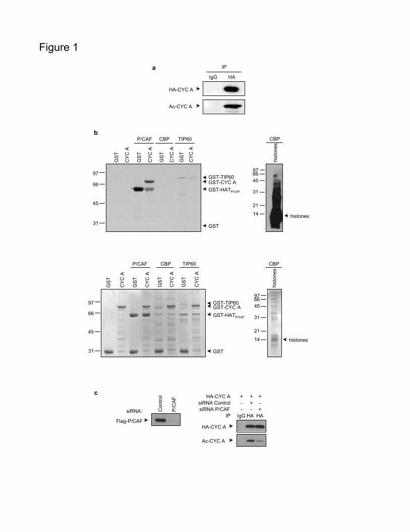
Figure 1
aIgG HA
HA-CYC A
Ac-CYC A
IP
b
GS
T
CY
C A
GS
T
CY
C A
GS
T
CY
C A
GS
T
CY
C A
P/CAF CBP TIP60
31
45
66
97 GST-TIP60GST-CYC AGST-HATP/CAF
GST
GS
T
CY
C A
GS
T
CY
C A
GS
T
CY
C A
GS
T
CY
C A
P/CAF CBP TIP60
GST-TIP60GST-CYC AGST-HATP/CAF
GST31
45
66
97
hist
ones
CBP
histones
31
456697
21
14
hist
ones
CBP
31
456697
21
14 histones
siRNA: Con
trol
P/C
AF
Flag-P/CAFHA-CYC A
Ac-CYC A
siRNA ControlsiRNA P/CAF
IP
- + -- - +
IgG HA HA
HA-CYC A + + +c

Figure 2
CYCLIN A
Lysine residuesD-boxExtended D-boxCyclin box
b
H3 3 4 7 9 11
3. K544. K687. K959. K112 and K113
11. K112
c d
IgG WT 4R
IP FLAG
Ac-CYC A
Flag-CYC A
a
1 171
1 432
GS
T
CY
C A
WT
CY
C A
4R
31
45
66
97GST-CYC A
GST-HATP/CAF
GST
GS
T
CY
C A
WT
CY
C A
4R
31
45
66
97GST-CYC A
GST-HATP/CAF
GST

Figure 3
ba
Input IgG HA
IP
YFP-P/CAF
HA-CYC A
CDK2
cyclin A P/CAF merge
c
RU
50
200
100
50 150 2500
Time (s)
Res
pons
e (0
=bas
elin
e)
0
-50
150
200100
_ CYC A WT _ Control
d Input
G1
S G2/
M
met
apha
se
IP HA
IgG
HA-CYC A
CDK2
YFP-P/CAF
H3-P
G1
S G2/
M
met
apha
se
Ac-CYC A
HA-CYC A
CDK2
YFP-P/CAF
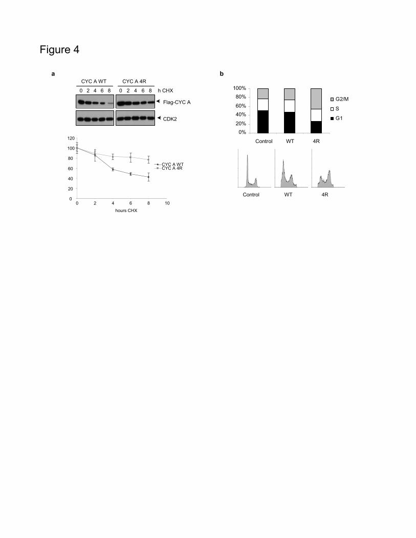
Figure 4
a
0 2 4 6 8 0 2 4 6 8
CYC A WT CYC A 4R
h CHX
Flag-CYC A
CDK2
0%20%
40%60%
80%100%
Control WT 4R
G2/M
S
G1
Control WT 4R
b
0
20
40
60
80
100
120
0 2 4 6 8 10hours CHX
CYC A WTCYC A 4R

Figure 5
0 1 2 3 4 5 6 7AsIgG
+ + + + + + + + +- ALLN
h post-thymidine
Flag-CYC A
Ub-Flag-CYC A
Ac-CYC A
IP FLAGb
a
IgG WT WT 4R 4QALLN: + - + + +
IP FLAG
IgG WT WT 4R 4Q+ - + + +
IP FLAG
WT WT 4R 4Q- + + +
Input
Ub-CYC A
Flag-CYC A
WB FLAG WB HAWB FLAG
50
75100
150250
50
75
100
150
250
Flag-CYC A

Figure 6
c d
IgG WT 4R
IP CDK2
CDK2
Flag-CYC A
H1-P
CDK2
Flag-CYC A
H1-P
IgG WT 4R
IP FLAG
0
20
40
60
80
100
CYC A WT CYC A 4R
rela
tive
units
0
20
40
60
80
100
CYC A WT CYC A 4R
rela
tive
units
b
IgG WT 4R
IP HA
HA-Cdh1
Flag-CYC A
WT 4R
Input
a
Flag-CYC A
Cdc20
Cdh1
IgG WT 4R
IP FLAG
CDK2
CDK1
WT 4R
Input
Cks1/2
APC3
Flag-Cdc20
CFP-CYC A
IgG WT 4R
IP FLAG
WT 4R
Input
*