Embed Size (px)
Citation preview
DEGRADATION OF ISOLATED DEOXYRIBONUCLEIC ACID MEDIATED BY NITROSO-CHLORAMPHENICOL
POSSIBLE ROLE IN CHLORAMPHENICOL-INDUCED APLASTIC
ANEMIA
THOWXS MURRAY, KATHLEEN M. DOWNEV and ADEI. A. YUNIS*+
Departments of Medicine and Biochemistry. University of Miami School of Medicine. Miami. FL 33101. and the Howard Hughes Medical Institute. Miami. FL 33101. U.S.A.
Abstract-Reduction of the nitro group of chloramphenicol (CAP) gives rise to more highly reactive intermediates which may be involved in the aplastic anemia associated with CAP use. One such intermediate. nitroso-chloramphcnicol (NO-CAP). has been found to be a potent agent for mediating degradation of isolated DNA. In a reaction mixture containing 100 PM NO-CAP, 100 FM CuC12. and 5 mM NADH. 7 ~1.g of Escherichin co/i [‘HIDNA was completely degraded to acid-soluble fragments in 30 min. Damage to DNA was in the form of single-stranded scissions. The requirement for copper was specific. andcopper chelating rcagcnts blocked the degradation. The need ?or a reducing agknt could be met equallv well bv NADH or NADPH. but not bv sulfhvdrvl reagents such as glutathione, dithiothreitol and 2:mercapioethanol. Oxygen was also necessary’fo; the NO-CAP-mediated DNA damage, with reduced forms of oxygen participating in the reaction. A role for H20~ was indicated by the inhibition of the degradation seen when catalase was included in the mixture. Hydroxyl radicals are known to be produced in the reaction of HJO: with certain transition metals. Scavangers of hydroxyl radicals also inhibited strand-scission. suggesting rhat the radicals may be the primary agents m DNA degradation. The importance of the nitroso moiety of NO-CAP was evidenced by the lack of DNA damage seen when NO-CAP was replaced by CAP under the conditions tested.
Use of the antimicrobial agent, chloramphenicol (CAP)* [o-(-)-threo-I-p-nitrophenyl-2-dichloro- acetamido-1,3-propanediol]. is sometimes associated with the rare but, often, fatal complication of aplastic anemia [l-3]. It has been postulated that the anemia might result from the interaction of more highly reactive metabolic reduction products of CAP with bone marrow stem cells rather than being a direct consequence of the CAP molecule [4. S]. In this regard, nitroso-chloramphenicol (NO-CAP). which contains a nitroso group in place of thep-NO? moiety of CAP, is suspected to be a potential mediator of the drug-induced aplasia [4]. The recent demonstra- tion that extracts from human liver preparations are capable of reducing the p-NO? group of CAP is consistent with this hypothesis 161.
Although there is no direct e\ idence to date linking NO-CAP to the generation of aplastic anemia, a number of studies from our laboratory have dem- onstrated that it is more reactive and more cytotoxic than the parent compound, CAP. A high degree of
* Howard Hughes Investigator. t Send all correspondence to: Adel A. Yunis, M.D.,
Department of Medicine (R-38). Universitv of Miami School of Medicine, P.O. Box 016960. Miamj, FL 33101. U.S.A.
$ Abbreviations: CAP. chloramphenicol; TAP. thiam- phenicol; NO-CAP, nitroso-chloramphenicol; .OH, hydroxyl radical; and EGTA, ethyleneglycol-bis (amino- ethylether) tetra-acetate.
2291
biochemical reactivity is typical of aromatic nitroso compounds, and covalent binding of NO-CAP to human lymphocytes in culture is Ifi-fold greater than that of CAP [7]. Similarly, toxic effects of nitroso aromatics have been reported [S] and nitroso metab- olites have been implicated as the mediators of the mutagenic potential of some nitroaromatics [9, lo]. NO-CAP has been shown previously to be much more cytotoxic to human bone marrow cells in cul- ture than CAP, with 5 x lO-‘M NO-CAP causing 50% cell death in 48 hr. Additional experiments suggested that the cytotoxicity seen with NO-CAP might involve interaction of this compound with DNA. The nitroso derivative inhibited DNA syn- thesis in marrow cells at much lower concentrations than CAP [4]. Further, only proliferating cells in culture are susceptible to NO-CAP toxicity, and the irreversible effects are greatest as cells proceed into and through the DNA synthesis portion (S-phase) of the cell cycle [II].
Prompted by these observations, studies were undertaken to examine the interaction of NO-CAP with DNA. Data presented here clearly demonstrate that NO-CAP (or a compound in equilibrium with it in the reaction mixture) can cause single-strand scissions in isolated DNA. Metal ion and reducing agent requirements for the reaction are discussed as well as the involvement of oxygen and its reduction products. The significance of NO-CAP-mediated DNA damage as a mechanism of drug toxicity is considered, in addition to the potential relevance of this to the problem of drug-induced aplastic anemia.

2292 T. MURRAY. K. M. DOWNEY and A. A. Y~NI~
MATERIALS AND METHODS
Assay for DNA cleavage based on formation of acid-soluble products. Degradation of Escherichia coli [3H]DNA (label in thymidine bases) was assessed by following the formation of acid-soluble products. The reaction mixture contained 100 mM potassium phosphate buffer, pH 8.0. 100pM NO- CAP, 5 mM NADH, 7 pg sonicated E. co/i ]jH]DNA (average molecular weight of DNA fragments, 60,000), and 50 pM CUCIZ in a final volume of 0.1 ml. Mixtures were incubated at 37” for 30 min before the reaction was halted by the addition of 0.5 ml of a 1 M perchloric acid solution containing 1 mM EDTA. A O.l-ml aliquot of salmon sperm DNA (2 mgiml) was then added, and solutions were chilled at 0” for 10 min. The mixtures were centrifuged at 6700g for 10 min, and radiolabeled, acid-soluble products in the supernatant fractions were deter- mined in a liquid scintillation spectrometer.
Isolation of form I DNA. Superhelical covalently closed circular DNA (form I) was isolated from a commercial preparation (Boehringer) of bacterio- phage PMZDNA by a cesium chlorideiethidium bromide centrifugation step. The DNA (0.5 A?(,,,unit) was layered on top of a cesium chloride solution (d = 1.56giml) containing 150 mM NaCI, 15 mM sodium citrate, pH 7.0. and 1 .O mg of ethidium bro- mide in a final volume of 7.5 ml. Solutions were placed in Beckman 50Ti polyallomer tubes which had been prewashed in a 10mM EDTA solution. Mineral oil was added to fill the tubes to 0.6 cm from the top, and tubes were centrifuged at 35.OOOrpm for 48 hr at 4” in a Spinco 50 Ti fixed angle rotor. Solutions were drained dropwise from a hole punc- tured in the bottom of the tubes, and the band of form I DNA was collected. Ethidium bromide was removed by passing the DNA solution through a Dowex 50 column (0.7 x 3 cm) and the DNA was dialyzed for 24 hr against three changes of 150 mM NaCl, 15 mM sodium citrate, pH 7.0, to eliminate the cesium chloride.
Agarose gel electrophoresis. The horizontal gel electrophoresis equipment used was purchased from Aquebogue Machine Shops, Aquebogue, NY. Aga- rose gels (la, w/w) were prepared in buffer (TAE) containing 40 mM Tris, 5 mM sodium acetate and 1 mM EDTA. pH 7.8. Gel dimensions were approx- imately 22 x 13 x 0.4cm. Reaction mixtures were adjusted to 4.3% Ficoll70 and 0.02% bromophenol blue before 50-,ul aliquots were applied to wells. Electrophoresis was performed in TAE buffer at a potential of 30V using a Schlumberger (Benton Harbor, MI) power supply (SP-2717). DNA bands were visualized by immersing the gels in ethidium bromide solutions in TAE buffer for 30 min.
Materials. NO-CAP was prepared from CAP as previously described by Dr. M. Corbett, University of Miami, Rosenstiel School of Marine Atmospheric Sciences [12]. Sonicated E. coli [‘H]DNA (0.18 pCi/AzM, unit) was purchased from Miles Bio- chemicals, Elkhart, IN. Neocuproine. L-arginine. t.-cysteine, p-aminobenzoate, imidazole. 2-mer- captoethanol. glutathione, 8-hydroxyquinoline, nitrobenzene, nitrosobenzene. ethidium bromide. salmon sperm DNA. Tris. and catalase (H:O2:H~O~
oxidoreductase, EC 1.11.1.6) were obtained from the Sigma Chemical Co., St. Louis, MO. Thiourea, triethylenetetraamide tetrahydrochloride. sodium diethyldithiocarbamate, and 2,2’-bipyridine were from Eastman Kodak Chemicals, Rochester, NY. EDTA, EGTA. ammonium formate. potassium phosphate, CdCI:. NiCI-. MnCI?, MgCl:. Fe(NH4)2(SOd)2.6H20, FeCh, CaC1:.2H?O, CuC12.2H20. ZnSOJ.7H:O. and sodium acetate were purchased from Mallinckrodt . Jersey City, NJ, NADPH and bacteriophage PM2-DNA were from Boehringer Mannheim Biochemicals. Indianapolis. IN; dithiothreitol, glycylglycine and NADH were obtained from Calbiochem, San Diego, CA: agarose was purchased from Pharmacia Fine Chemicals, Pis- cataway, NJ; BeCI: was from the Aldrich Chemical Co., Milwaukee, WI; COCIZ was from the J. T. Baker Co., Phillipsburg. NJ; Dowex 50 was purchased from Biorad Laboratories, Richmond, CA; and sodium benzoate was from MCB Manufacturing Chemists Inc., Cincinnati, OH. CsClz was from Schwartz Bio- research, Inc., Orangeburg, NY and CAP was pro- vided by the Parke-Davis Co., Detroit. MI.
RESULTS
Requirements for degradation of DNA by NO- CAP. Reduction of the p-NO2 group of CAP to the nitroso derivative conferred upon the molecule the ability to cause breaks in double-stranded DNA. A time course of this activity against E. coli ]“H]DNA indicated that NO-CAP rapidly produced extensive damage (Fig. 1). Nearly all of the DNA (7 pg) pres- ent in the reaction mixture was converted into acid-soluble fragments within 30min by 100pM NO-CAP. The reaction required copper and a reducing agent (e.g. NADH) and was strongly inhibited (90%) if oxygen was removed and the incubation was carried out under a Nz atmosphere (Table I). DNA degradation was temperature sen- sitive, being completely blocked at 0” and reduced by 80% at 22”.
The nitroso moiety of NO-CAP was implicated as the structural feature of the molecule involved in this reaction, as equal concentrations of C.4P were inactive with regard to DNA damage. The concen- tration dependence of the DNA nicking activity of
0 10 20 30 IO 50 60
TIM6 (lin)
Fig. 1. NO-CAP-mediated degradation of DNA as a func- tion of time. The release of acid-soluble DNA fragments in the presence of 100 PM NO-CAP, 100 PM CuCI: and S mM NADH was determined as described in Materials
and Methods.

Degradation of DNA by CAP reduction products
Table 1. Requirements for degradation of DNA by NO-CAP*
Components c/C Acid soluble
Complete Minus Cu(II) Minus NADH Minus NO-CAP Minus oxygen
Plus CAP, minus NO-CAP, NADH and
100 <l <I <I 10
Cu(II) plus CAP, minus NO-CAP
<I <l
* All samples were 1OOmM in potassium phosphate, pH 8.0, and contained 7 pg of E. co/i [jH]DNA. In addition. the complete reaction mixture contained, in a final volume of 0.1 ml. 50 FM CuCl:, 5 mM NADH, and 100 PM NO-CAP. When NO-CAP was replaced by CAP. it was also 100pM. The minus oxygen experiment was carried out in sealed tubes under a NZ atmosphere. Acid-soluble radioactivity after a 30-min incubation at 37” was determined as described in Materials and Methods.
NO-CAP is shown in Fig. 2. NO-CAP levels of 5 PM and above readily produced demonstrable levels of acid-soluble fragments in 30 min under the condi- tions employed here. Further evidence linking the p-NO group to the DNA damage came from parallel studies with nitrosobenzene. It was found to produce a very similar dose-response pattern (87% acid-solu- ble DNA with 100pM nitrosobenzene), while the activity of nitrobenzene was comparable to that of CAP (cl% release of acid-soluble DNA with 100 PM nitrobenzene).
Nature of the DNA damage produced by NO-CAP. The assay system used to assess the type of damage to DNA mediated by NO-CAP employed superhel- ical covalently closed circular PM2-DNA as sub- strate. Introduction of a single-stranded scission into the superhelical DNA (form I) converts it into a relaxed open circle conformation (form II) [13]. Direct production of linear duplex DNA (form III) requires a double-stranded break in the supercoil (although linear duplex DNA is also produced if two single-stranded scissions are generated in close prox- imity along the DNA molecule). The three forms
-/ , .1 1 10 100
NO-CIP (PM)
Fig. 3. Agarose gel electrophoresis of PM2-DNA following incubation with CAP and NO-CAP. Supercoiled PM2- DNA (form I, 80 ng) was incubated for 30 min in 100 mM potassium phosphate buffer, pH 8.0, and (a) 1 PM NO- CAP. NADH (500 PM) and CuClz (2 PM); (b) 5 PM NO- CAP. NADH (500pM) and CuClz (2 PM); (c) 1OyM NO-CAP, NADH (500,uM) and CUCIZ (2 yM). (d) no additions, (e) NADH (500pM) and CuClz (2pM); (f) 100 PM CAP; and (g) 100 ,uM CAP, NADH (500 PM) and CuClz (2pM). Reaction mixtures were then adjusted to 4.3% Ficoll 70 and 0.02% bromophenol blue. Aliquots were electrophoresed for 15 hr at 30 V on horizontal gels. DNA bands corresponding to open circle (form II). linear duplex (form III) and supercoiled (form I) DNA were
Fig. 2. Effect of NO-CAP concentration on DNA degra- dation by NO-CAP. Various amounts of NO-CAP were added to reaction mixtures containing 50pM CuCIz and 5 mM NADH, and acid-soluble counts were determined after a 30-min incubation at 37” as described in Materials
and Methods. visualized with ethidium bromide.
2293
are readily distinguished electrophoretically on aga- rose gels under appropriate conditions [14]. This type of analysis applied to NO-CAP actions on DNA showed that the drug induced single-stranded rather than double-stranded scissions (Fig. 3). Further, since the introduction of as little as one single- stranded nick causes a difference in electrophoretic mobility, it is possible to detect DNA damage at drug levels lower than would be required to generate
abc de fg
-

2294 T. MIJRUY. K. M. DowNI-I’~~~ A. A. YIINI\
Table 2. Reducing agent specificity for degradation of DNA by NO-CAP’
Concn $4’ Acid soluble Reducing agent (mM) -NO-CAP +NO-CAP
NADH 0.5 <l 19.2 1 .o <I 4X.5 5.0 Cl 100
NADPH 0.5 <I 23.5 1.0 <I 4X.9 5.0 3.1 IO0
* DNA degradation was assessed as described in Materials and Methods following incubation in the presence of 100 PM NO-CAP and 50 PM CuCll.
acid-soluble DNA fragments. In a 30-min incuba- tion, NO-CAP at 1 ,uM produced at least one scission and possibly several in nearly every molecule of the PM2-DNA (80 ng) present in the assay. Higher con- centrations of NO-CAP (5 or 1OpM) or longer incubation times (data not shown) resulted in more extensive DNA damage. In contrast, no strand scis- sion was detected with CAP at concentrations as high as 100 PM.
Reducing agent specificity. The requirement for a reducing agent in the reaction can be fulfulled equally well by NADH or NADPH (Table 2). The concen- tration dependence for both was similar. with 5 mM being sufficient for complete degradation of the DNA in 30 min. Higher levels of NADH were some- what less effective under these conditions (81% acid-soluble counts at 8mM NADH). In contrast. sulfhydryl compounds such as dithiothreitol, gluta- thione and 2-mercaptoethanol were ineffective at comparable concentrations (Cl% acid-soluble counts under all conditions tested). Rather than act- ing as co-factors in the reaction, these thiol agents inhibited the strand scission. Addition of 1 mM thiol to reactions containing 5 mM NADH completely blocked the production of acid-soluble fragments (data not shown), possibly by direct interaction of the thiol with the p-NO, since aromatic nitroso groups are known to react with glutathione to yield reduction products of the nitroso group [ 151.
Metal ion requirernmt. The results listed in Table 1 show that. in addition to a reducing agent. the degradation of DNA by NO-CAP was dependent upon the presence of a metal ion. In tests designed to determine the speciticitv of this requirement. Cu(l1) was the only transition metal found to be effective [Cd(II). Co(I1). Ni(l1). Zn(I1). Mn(I1). Mg(II), Fe(I1). Fe(lI1). Be(Il) and &(I) all yielded ~2% acid-soluble fragments under similar condi- tions]. Assays containing Cu(I1) concentrations below 10 !IM showed greatly reduced strand-scission (18% acid-soluble DNA at 5 I’M) as did Cu( II) levels of 200uM and above (30% acid soluble-DNA at 200,~M). Iron( the metal ion which i!, needed for bleomycin-mediated strand-scission of DNA [IO, 171. could not replace Cu(I1) in the reaction involving NO-CAP. Also. none of the metal ions above caused detectable DNA degradation at the concentrations tested in the absence of NO-CAP.
The need for Cu(I1) in the reaction was further substantiated by the inhibition of DNA degradation observed when metal chelators were added to the complete reaction mixture (Table 3). At SO~IM (a 2.5fold excess). all chelators tested except imidazole and 2,2’-bipyridine completely prevented the break- down of DNA. The effectiveness of these two com- pounds was proportionately increased as the con- centrations were doubled. Since imidazole and 2.2’-bipyridine have lower stability constants [1X] fol the Cu(II)-chelator complex than do other chelators listed in Table 3. the chelator data are consistent with a specificitv for c‘u(lI1) in the reaction.
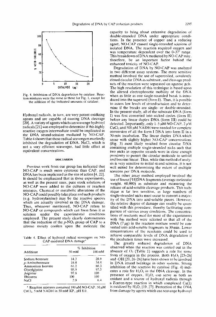
Effect of cutaiusr und h~,dro.ryl mdicczl .>caorngrrs on the degrudutim of DNA hv NO-CAP. Since the extent of DNA breakdown is-greatly reduced when the reaction is conducted anerobically. a possible role for reduction products of oxygen was con- sidered. An involvement of 11~0~ (or other species in rapid equilibrium with it) in the reaction was indicated by the inhibition of DNA degradation seen when catalase was included in the assay mixture (Fig. 4). It is unlikely that catalase was exerting its effect by chelating copper, since heat-denatured catalase has no effect.
Chelated transition metals have been shown pre- viously to reduce 1 I:O: to hydroxyl radicals [ 191.
Table 3. Inhibition of DNA degradation by metal chelating agcntx -___-
Concn ’ c Acid-\olublr
Metal chelator (uM) -NO-CAP t NO-CAP
None so I. t 100 EDTA so I I EGTA 50 I. I I L-Cysteine 50 ‘Z 1 2.7 Triethylenetetraamine 50 I c I 8-Hydroxyquinoline so <I I c I Neocuproine SO I I Sodium diethyldithiocarbamate so <’ .I I Imidazole 50 c I ‘)2.1 2,2’-Bipyridine SO J. I X5. I
Imidazole 100 ~ I 75.1 2.2’-Bipyridine 100 s:. I 70. I
* Reaction mixtures contained 100 ,uM NO-CAP. 10 !tM Tuc‘i,. and 7 mhl NADH.
Degradation of DNA by CAP reduction products 2295
CATAldSt (yg)
Fig. 4. Inhibition of DNA degradation by catalase. Reac- tion mixtures were the same as those for Fig. 1, except for
the addition of the indicated amounts of catalase.
Hydroxyl radicals, in turn, are very potent oxidizing agents and are capable of causing DNA cleavage [20]. A variety of agents which can scavenge hydroxyl radicals [21] was employed to determine if this highly reactive oxygen intermediate could be implicated in the DNA strand-scission mediated by NO-CAP. Table 4 shows that these radical scavengers markedly inhibited the degradation of DNA. NaCl, which is not a very efficient scavenger, had little effect at equivalent concentrations.
DISCUSSION
Previous work from our group has indicated that NO-CAP is much more cytotoxic than CAP, and DNA has been implicated as the site of action [4,22]. It should be emphasized that in those experiments, as well as the present ones, pure stock solutions of NO-CAP were added to the cultures or reaction mixtures. Chemical or metabolic alterations of the NO-CAP could possibly take place and such products (e.g. hydroxylamine) may be the reactive species which are actually involved in the DNA damage. Thus, whenever mentioned, NO-CAP refers to NO-CAP or compounds which can form from it in solution under the experimental conditions employed. The present study clearly demonstrates that the reduction of the p-NO2 group of CAP to a nitroso moiety confers upon the molecule the
Table 4. Effect of hydroxyl radical scavengers on NO- CAP-mediated DNA damage*
% Inhibition Additions 50 mM 100 mM
Sodium benzoate 14.3 28.9 p-Aminobenzoate 16.8 34.0 Ammonium formate 61.2 86.3 Glycylglycine 95.9 97.5 Arginine 97.8 100 Thiourea 100 100 NaCI 2.9 6.4
* Reaction mixtures contained 100 PM NO-CAP, 50 PM CuCl*, 5 mM NADH in 50 mM KP,, pH 8.0.
capacity to bring about extensive degradation of double-stranded DNA under appropriate condi- tions. In the presence of copper and a reducing agent, NO-CAP caused single-stranded scissions of isolated DNA. The reaction required oxygen and was temperature dependent over the C-37” range. This breakdown of DNA mediated by NO-CAP may, therefore, be an important factor behind the enhanced toxicity of NO-CAP.
Degradation of DNA by NO-CAP was analyzed in two different assay systems. The more sensitive method involved the use of supercoiled, covalently closed circular DNA as substrate, and cleavage prod- ucts of the reaction were separated on agarose gels. The high resolution of this technique is based upon the altered electrophoretic mobility of the DNA when as little as one single-stranded break is intro- duced into the supercoil (form I). Thus, it is possible to assess low levels of strand-scission and to deter- mine if the breaks are single- or double-stranded. In the present study, all of the substrate DNA (form I) was first converted into nicked circles (form II) before any linear duplex DNA (form III) could be detected. Importantly, only 1-5 PM NO-CAP, 2 PM CuC12 and 500 PM NADH were needed for complete conversion of all the form I DNA into form II in a 30-min incubation. The linear duplex DNA which arose with slightly higher NO-CAP concentrations (Fig. 3) most likely resulted from circular DNA containing multiple single-stranded nicks such that two nicks in opposite strands were in close enough proximity to permit the circular molecule to unfold and become linear. Thus, while this method of analy- sis is very sensitive to initial strand scission, it is not well suited for determining the extent of multiple scissions per DNA molecule.
The other assay method employed involved the use of linear [3H]DNA fragments (average molecular weight, 60,000) as substrate and measuring the release of acid-soluble cleavage products. This tech- nique is far less sensitive, as large numbers of single-stranded nicks must occur to convert a major- ity of the DNA into acid-soluble pieces. However, the relative degree of damage can readily be quan- tified with this procedure, thereby facilitating com- parison of various assay conditions. The concentra- tions of reactants used for most of the experiments with this method were selected so that all of the DNA (7,~g) in the reaction mixture would be con- verted into acid-soluble fragments in 30 min. Lower concentrations of the reactants could be used to achieve comparable levels of DNA degradation if the incubation times were increased.
The greatly reduced degradation of DNA observed when the reaction was carried out in the absence of 02 (Table 1) suggests a role for some form of oxygen in the process. Both H202 [23-261 and .OH [20,2+28] have been shown to be involved in DNA strand breakage in other systems. Strong inhibition of the reaction by catalase (Fig. 4) indi- cates a role for Hz01 in the DNA cleavage. In the presence of copper, Hz02 can serve as both an oxidant and a source of hydroxyl radicals through a Fenton-type reaction in which complexed Cu(1) is oxidized by H20? 119,271. Protection of the DNA from damage by agents which can scavenge hydroxyl
2296 T. M~JRKA’I, K. M. Dow~tj
radicals (Table 4) suggests that the latter may also 2. be involved in the strand scission.
and A. A. YLJNIX
There are a number of similarities between the degradation of DNA involving the nitroso analog of CAP and that mediated by I.lO-phenanthroline. Both of these reactions require copper and a reducing agent [29,30] and both can be inhibited by catalase [26,30] and hydroxyl radical scavengers [26]. DNA damage in the latter system is believed to result from hydroxyl radicals generated in the reduction of HzOl by a l,lO-phenanthroline-Cu(I) complex, with the reducing agent serving to convert the complexed Cu(I1) to Cu(1). It has further been suggested that 1 ,lO-phenanthroline promotes the DNA degradation
3 4,
5,
6.
7.
8. (),
D. M. Williams, R. E. Lynch and G. E. Carwright. Semin. Hemat. 10. 195 (1973). A. A. Yunis and G. R. Bloomberg. Prog. I-femur. 4. 13X (1964). A. A. Yhis. A. M. Miller. 2. Salem and Ci. K. Arimura. .I. Lab. clin. Med. 96. 36 (19X0). J. H. Weisburger. Y. Shirasu. P. H. Grantham and E. K. Weisburger. J. hiol. Chem. 242. 371 (1967). Z. Salem. T. Murray and A. A. Yunis. J. Luh. cl;rl. Med. 97. XXI (IYXl). T. Murray and A. A. Yunis. J. Lab. c/in. Med. 98. 396 (1981). M. Kiese and K. Taeger, Riartrt?,l-Schmie~febe~g’,s Arc/r. Pharmac.292. 59 (lY76).
by bringing the site of hydroxyl radical production
underway in our laboratoiy ;o determine if DNA close to liable sites in DNA 1261. Studies are currentlv
damage mediated by NO-CAP involves any of these factors.
10. C. W. Chiu. L. H. Lee. C. Y. Wang and G. T. Bryan. _
B. W. Ames. E. G. Gurney. J. A. Miller and H. Bartsch. Proc. natn. Acad. Sci. C/.S.A. 69, 3128 (1971).
11
12.
13.
14.
Mutation Res. 58. I I (197X). A. M. Miller and A. A. Yunis. Phurmacology, 24. hl (1982).
It is apparent from the data presented here that NO-CAP can act as a potent mediator of DNA strand scission. This property appears to be due to the aromatic nitroso moiety of the molecule. since equal molar concentrations of nitrosobenzene can replace NO-CAP in the reaction, while CAP and nitrobenzene will not. Several nitroheterocyclic drugs (including metronidazole) [31, 321 and CAP [33] have also recently been linked to DNA strand scission following electrolytic reduction of the nitro groups, although for these compounds the identities of the reduced species have not been established. Thus, the reaction of NO-CAP with DNA discussed herein may be just one example of a more general class of reactions.
In conclusion, this study demonstrates that a potential intermediate of CAP could interact with other cellular components to cause breaks in DNA. It is, therefore of interest as a potential mechanism of drug toxicity, especially in light of the previously cited cytotoxic effects of NO-CAP on human lym- phocytes and marrow cells in culture [4]. In addition, the present data also raise the intriguing possibility that DNA damage mediated by NO-CAP could be involved in one of the early events leading to drug-induced aplastic anemia.
Acknowledgements-This work was supported in part h) USPHS Grants AM 05472, AM 26218 and CA 30081. The authors express their gratitude to Dr. Walter Scott for the use of his facilities in the electrophoresis experiments. We also thank Dr. Benito Que for his helpful discussions and Mrs. Yolanda Capo for the technical preparation of this manuscript.
REFERENCES
1. A. A. Yunis, in The Year in Hematology (Eds. R. Silber, J. LoBue and A. S. Gordon), p. 143. Plenum. New York (1978).
1.5 16.
17.
1x.
19.
20.
21.
22.
23.
24.
25.
26.
27.
28.
29.
30.
31.
32.
33.
M. D. Corbett and B. R. Chipko. Antimicroh. Agertt~ Chemother. 13, 193 (lY78). J. Vinograd. J. Lebowitz and R. Watson, J. moles. Biol. 33. 173 (1967). J. E. Strong and S. T. Crooke. in Bleornycin: Chemrcal. Biochemical, and Biolo&alAspect.s (Ed. S. M. Hecht), p. 244. Springer, New York (i979). P. Ever. Chem. Biol. Interact. 24. 227 (1979) E. A. Sausville. J. Peisach and S. B. Hor&tz. Hio- chemisrry 17, 2740 (1978). E. A. Sausville. R. W. Stein, J. Peisach and S. B. Horwitz. Biochemistry 17, 2746 (197X). The Stabilitv Consrants of Metal Ion C‘omplexes (Com- piled by L. G. Sillen and A. E. Martell). The Chemical Society, London (1964). K. L. Fang. P. B. McCoy, J. L. Poyer and B. B. Keclc. Chem. Biol. interact. 15. 77 (1976). P. R. Armel. G. F. Strniste and S. S. Wallace, Radrar. Res. 69, 328 (lY77). M. Anbar and P. Neta. Int. J. uppl. Rudiat. 1.totope.s 18. 493 (1067). A. A. Yunis. A. M. Miller. Z. Salem and G. K. Arimura. Clin. Toxtc. 17. 359 ( 1980). L. E. Marshall. D. R. Graham, K. A. Reich and D. S. Sigman, Biochemisfry 20. 234 (1981). H. J. Rhaese and E. Freese. Biochim. biophy~. Acrcr 155, 476 (1968). H. Massie, H. Samis and M. Baird, Biochim. hlophys. Acta 272, 539 (1972). B. G. Que. K. M. Downey and A. G. So, Biochemistr~~ 19. 5987 (1980). S. A. Lesko. R. J. Lorentzen and P. 0. P. Ts’o, Biochemistry 19. 3023 (1980). J. W. Lown and S-K. Sim, Biochem. hionhvs. Re.s. , Commun. 77. 1150 (1977). K. M. Downey. B. G. Que and A. G. So. Hiochcw7.
biophys. Res. Commun. 93, 264 (1980). D. S. Sigman. D. R. Graham, V. D. D’Aurora and A. M. Stein. J. hiol. Chem. 254, 1226a (1979). R. C. Knight. I. M. Skolimowski and D. 1. Edwards. Biochem. Pharmuc. 27. 2089 (1978). D. A. Rowley. R. C. Knight. I. M. Skolimowskl and D. I. Edwards. Biochern. Pharmac. 28. 3009 (1979). I. M. Skolimowski, D. A. Rowley. R. C. Knight and D. I. Edwards. J. Antimicroh. C‘hemother. 7. S93 (1981).