Embed Size (px)
Citation preview

Case report
Delayed calyceal cutaneous fistula after renal transplantation
Luay P. Susan, M.D. James D. Daughtry, M.D. Department of Urology
William E. Braun, M.D.
Department of Hypertension and Nephrology
Lynn H. Banowsky, M.D. Ralph A. Straffon, M.D.
Department of Urology
Rafael Valenzuela, M.D.
Department of Immunopathology
Calyceal cutaneous fistula is a serious complica-tion of renal transplantation. It is usually caused by segmental renal infarction resulting from an unrec-ognized ligated or severed accessory renal vessel during donor nephrectomy. Unless associated with trauma, a calyceal fistula usually develops within the first 3 months of transplantation. This is the first reported case of a calyceal cutaneous fistula in a renal transplant patient manifested 2 years after renal transplantation and associated with acute pyelonephritis and apparently newly developed amyloidosis. Prompt surgical intervention with pri-mary closure of the fistula achieved a successful outcome.
Urinary extravasation is a perplexing problem of renal transplantation; its incidence varies between 8% and 17%.''2 Although oliguria is a common finding in most of the cases, the initial diagnosis is usually unrecognized because of concomitant rejec-tion or infection or both.3 We report a case of calyceal cutaneous fistula in a renal transplant patient with presenting symptoms of acute pyelo-nephritis.
Case report A 51-year-old Caucas ian m a n whose original renal
disease was focal sclerosing glomerulonephr i t i s received a second renal al lograft . A Terasaki " C " ma tched renal allograft dona ted by a sibling had been rejected a f te r 41
287
uses require permission. on March 18, 2022. For personal use only. All otherwww.ccjm.orgDownloaded from
288 Cleveland Clinic Quarterly Vol. 45, No. 1
months of good funct ion . T h e second renal a l lograf t , f rom a cadaver donor , ma tched for only H L A ant igen func t ioned immediate ly . By the 15th day the serum creat in ine was 1.8 m g / d l .
T h e pat ient did well unt i l 23 months later when he was admi t t ed because of fever, chills, and pain over the allograft . Both ur ine and blood cultures grew Escherichia coli a n d his serum creat inine level increased to 4.4 m g / d l . Excretory urography, cystography, and re t rograde pyelography were normal . Renal t ransplant angiography revealed pat-ent renal vessels with a clearance t ime of 2 seconds a n d no evidence of infarct ion {Fig. 1). H e was treated with a course of cepha-lothin intravenously for 2 weeks. At the t ime of discharge the ur ine was sterile a n d the serum creat inine level was 1.6 m g / d l .
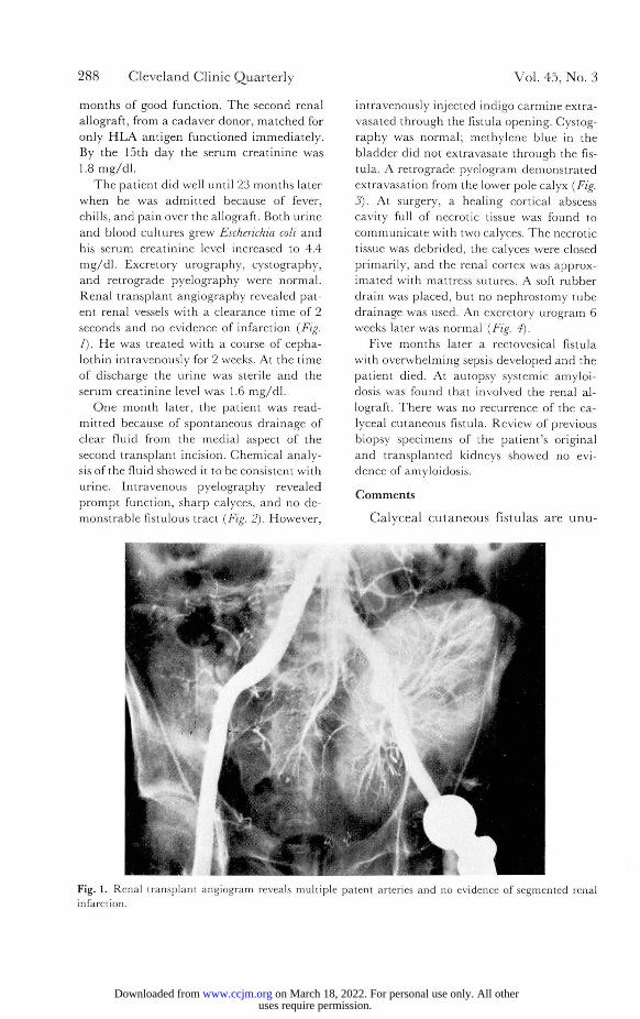
O n e mon th later, the pat ient was read-mi t ted because of spontaneous dra inage of clear fluid from the medial aspect of the second t ransplant incision. Chemical analy-sis of the fluid showed it to be consistent wi th urine. In t ravenous pyelography revealed p rompt funct ion, sharp calyces, and no de-mons t rab le fistulous tract (Fig. 2). However ,
intravenously injected indigo ca rmine extra-vasated through the fistula opening. Cystog-raphy was normal ; methylene blue in the b ladder did not extravasate through the fis-tula. A re t rograde pyelogram demons t ra ted extravasat ion f rom the lower pole calyx (Fig. 3). At surgery, a healing cortical abscess cavity full of necrotic tissue was found to communica t e with two calyces. T h e necrotic tissue was debr ided, the calyces were closed primari ly, and the renal cortex was approx-imated with mattress sutures. A soft rubber drain was placed, but no nephros tomy tube dra inage was used. An excretory u rogram 6 weeks later was normal (Fig. 4).
Five mon ths later a rectovesical fistula with overwhelming sepsis developed and the pat ient died. At autopsy systemic amyloi-dosis was found that involved the renal al-lograft . T h e r e was no recurrence of the ca-lyceal cu taneous fistula. Review of previous biopsy specimens of the pat ient ' s original and t ransplanted kidneys showed no evi-dence of amyloidosis.
C o m m e n t s
Calyceal cutaneous fistulas are unu-
Fig. 1. Renal transplant angiogram reveals multiple patent arteries and no evidence of segmented renal infarction.
uses require permission. on March 18, 2022. For personal use only. All otherwww.ccjm.orgDownloaded from
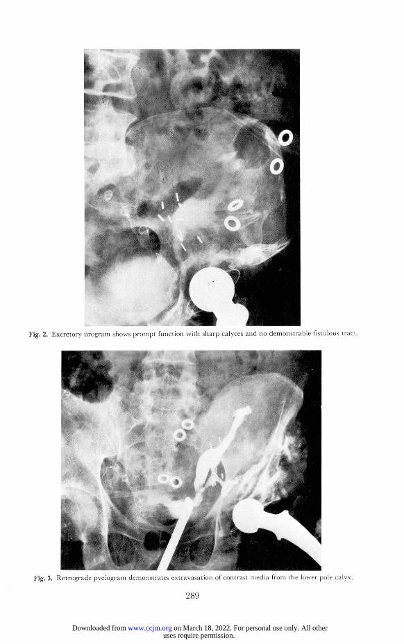
Fig. 3. Retrograde pyelogram demonstrates extravasation of contrast media from the lower pole calyx.
289
uses require permission. on March 18, 2022. For personal use only. All otherwww.ccjm.orgDownloaded from

290 Cleveland Clinic Quarterly Vol. 45, No. 1
Fig. 4. Normal excretory urogram 6 weeks following primary closure of the fistula.
sual complications of renal transplan-tation. Williams et al3 reported two cases in 170 transplants; Anderson et al,4 one case in 125 transplants; and Schiff et al,5 three cases in 134 trans-plants. The overall incidence of calyceal fistulas is 1.3%.
The most important contributing fac-tor is segmental renal infarction result-ing from severing an unrecognized ac-cessory renal artery or inadvertently li-gating a small polar vessel. Since caly-ceal fistulas occur more commonly in patients receiving renal allografts sup-plied by two or more renal arteries, failure to revascularize successfully the renal segment supplied by the vessel leads to renal infarction and production of the fistula. Although our patient had
two renal arteries supplying the allo-graft, both were proved to be patent by angiography and no infarction was demonstrated. Localized spasm of the renal artery during perfusion has been suspected to cause segmental renal is-chemia and subsequent fistula forma-tion,6 but with the use of systemic phe-noxybenzamine and local procaine in-fusion, vascular spasm has been amelio-ra t ed . 7 , 8 ^ essence, calyceal fistulas typ-ically are early complications of renal transplantation and are most likely to occur during the first 3 months of sur-g e r y . A delayed calyceal fistula of 1 year's duration developed after an au-tomobile accident that had caused blunt trauma to the allograft.9
Although in humans, renal cortical
uses require permission. on March 18, 2022. For personal use only. All otherwww.ccjm.orgDownloaded from

Fall 1978 Delayed calyceal cutaneous fistula 291
necrosis has been observed after hyper-acute rejection,1 neither that early form of rejection nor chronic rejection was evident in this transplanted kidney. The infusion of certain bacterial toxins has also been demonstrated to cause cortical necrosis similar to that seen in the rejec-tion graft.11 It is likely that cortical is-chemia in our patient was due to severe acute pyelonephritis that occurred 2 years following transplantation and caused cortical necrosis and fistula for-mation. Amyloidosis also may have con-tributed to development of the fistula.
It is important to be alerted to this complication when recurrent upper uri-nary tract infection occurs in renal transplant patients. Prompt surgical in-tervention is mandatory. Although ne-phrostomy tubes have been used by oth-ers,3"6'9 debridement and primary clo-sure of the calyces appear to be more feasible because foreign bodies are elim-inated, the incidence of urinary leakage is reduced, and early recovery is pro-moted.
References 1. Palmer JM, Kountz SL, Swenson RS, et al:
Urinary tract morbidity in renal transplan-
tation. Arch Surg 98: 352-356, 1969. 2. Prout GR Jr, Hume DM, Lee HM, et al:
Some urological aspects in 93 consecutive renal homotransplants in modified recipients. J Urol 97: 409-425, 1967.
3. Williams G, Birtch AG, Wilson RE, et al: Urological complications of renal transplan-tation. Br J Urol 42: 21-28, 1970.
4. Anderson EE, Glenn JF, Seigier HF, et al: Urologie complications in renal transplanta-tion. J Urol 107: 187-192, 1972.
5. Schiff M Jr, McGuire EJ, Weiss RM, et al: Management of urinary fistulas after renal transplantation. J Urol 115: 251-256, 1976.
6. Fox M, Tottenham RC: Urinary fistula from segmental infarction in a transplanted kid-ney; recovery following surgical repair. Br J Urol 44: 336-338, 1972.
7. Beizer FO, Kountz SL: Preservation and transplantation of human cadaver kidneys; a two-year experience. Ann Surg 172: 394-404, 1970.
8. Beizer FO, Reed TW, Pryor JP, et al: Cause of renal injury in kidneys obtained from ca-daver donors. Surg Gynecol Obstet 130: 467-477, 1970.
9. Schiff M Jr, McGuire EJ, Webster J: Success-ful management of caliceal fistulas following renal transplantation. Arch Surg 110: 1129-1132, 1975.
10. Williams GM, Hume DM, Hudson RP Jr, et al: "Hyperacute" renal-homograft rejection in man. N Engl J Med 279: 611-618, 1968.
11. Klassen J, Milgrom F: Studies on cortical necrosis in renal grafts. Transplant Proc 3:598-601, 1971.
uses require permission. on March 18, 2022. For personal use only. All otherwww.ccjm.orgDownloaded from