Embed Size (px)
Citation preview
Contents lists available at ScienceDirect
Bone Reports
journal homepage: www.elsevier.com/locate/bonr
Delayed tooth movement in Runx2+/− mice associated with mTORC2 instretch-induced bone formation
Tomo Aonumaa, Nagato Tamamurab,1, Tomohiro Fukunagaa, Yuichi Sakaia, Nobuo Takeshitaa,Shohei Shigemia, Takashi Yamashiroc, Irma Thesleffd, Teruko Takano-Yamamotoa,e,⁎
a Division of Orthodontics and Dentofacial Orthopedics, Tohoku University Graduate School of Dentistry, 4-1, Seiryo-machi, Aoba-ku, Sendai, Miyagi 980-8575, JapanbDepartment of Orthodontics and Dentofacial Orthopedics, Okayama University Graduate School of Medicine, Dentistry and Pharmaceutical Sciences, 2-5-1 Shikata-cho,Okayama City, Okayama 700-8558, Japanc Department of Orthodontics and Dentofacial Orthopedics, Osaka University Graduate School of Dentistry, 1-8 Yamada-Oka, Suita, Osaka 565-0871, Japand Research Program in Developmental Biology, Institute of Biotechnology, POB56, University of Helsinki, 00014 Helsinki, Finlande Department of Biomaterials and Bioengineering, Faculty of Dental Medicine, Hokkaido University, Kita 13, Nishi 7, Kita-ku, Sapporo, Hokkaido 060-8586, Japan
A R T I C L E I N F O
Keywords:Runx2+/− miceBMSCsOsteoblastMechanical stressmTORC2Experimental tooth movement
A B S T R A C T
Runt-related transcription factor 2 (Runx2) is an essential transcription factor for osteoblast differentiation, andis activated by mechanical stress to promote osteoblast function. Cleidocranial dysplasia (CCD) is caused bymutations of RUNX2, and CCD patients exhibit malocclusion and often need orthodontic treatment. However,treatment is difficult because of impaired tooth movement, the reason of which has not been clarified. Weexamined the amount of experimental tooth movement in Runx2+/− mice, the animal model of CCD, andinvestigated bone formation on the tension side of experimental tooth movement in vivo. Continuous stretch wasconducted to bone marrow stromal cells (BMSCs) as an in vitro model of the tension side of tooth movement.Compared to wild-type littermates the Runx2+/− mice exhibited delayed experimental tooth movement, andosteoid formation and osteocalcin (OSC) mRNA expression were impaired in osteoblasts on the tension side oftooth movement. Runx2 heterozygous deficiency delayed stretch-induced increase of DNA content in BMSCs,and also delayed and reduced stretch-induced alkaline phosphatase (ALP) activity, OSC mRNA expression, andcalcium content of BMSCs in osteogenic medium. Furthermore Runx2+/− mice exhibited delayed and sup-pressed expression of mammalian target of rapamycin (mTOR) and rapamycin-insensitive companion of mTOR(Rictor), essential factors of mTORC2, which is regulated by Runx2 to phosphorylate Akt to regulate cell pro-liferation and differentiation, in osteoblasts on the tension side of tooth movement in vivo and in vitro. Loss of halfRunx2 gene dosage inhibited stretch-induced PI3K dependent mTORC2/Akt activity to promote BMSCs pro-liferation. Furthermore, Runx2+/− BMSCs in osteogenic medium exhibited delayed and suppressed stretch-induced expression of mTOR and Rictor. mTORC2 regulated stretch-elevated Runx2 and ALP mRNA expressionin BMSCs in osteogenic medium. We conclude that Runx2+/− mice present a useful model of CCD patients forelucidation of the molecular mechanisms in bone remodeling during tooth movement, and that Runx2 plays arole in stretch-induced proliferation and osteogenesis in BMSCs via mTORC2 activation.
1. Introduction
Runx2 (Runt-related transcription factor 2 earlier called as Corebinding factor α-1 (Cbfa1) is an essential transcription factor for os-teoblast differentiation from mesenchymal stem cells (MSCs) and boneformation (Otto et al., 1997; Ducy et al., 1997; Komori et al., 1997).
Mice with a homozygous mutation in Runx2 show arrested embryonictooth development, complete absence of both intramembranous andendochondral ossification through a lack of osteoblast differentiation,and are embryonic lethal (Otto et al., 1997; Ducy et al., 1997; Komoriet al., 1997; Mundlos, 1999; Aberg et al., 2004). Runx2 is activated byfluid shear stress in mouse cortical bone in vivo, and mechanical stresses
https://doi.org/10.1016/j.bonr.2020.100285Received 8 October 2019; Received in revised form 27 March 2020; Accepted 25 May 2020
⁎ Corresponding author at: Division of Orthodontics and Dentofacial Orthopedics, Tohoku University Graduate School of Dentistry, 4-1 Seiryo-machi, Aoba-ku,Sendai, Miyagi 980-8575, Japan.
E-mail addresses: [email protected] (T. Fukunaga), [email protected] (N. Takeshita), [email protected] (T. Yamashiro),[email protected] (I. Thesleff), [email protected] (T. Takano-Yamamoto).
1 Current address: Tamamura Orthodontic Office, Sinyo Bld. 2F 1-14-1, Minami-mukosono, Amagasaki, Hyogo 661-0033, Japan
Bone Reports 12 (2020) 100285
Available online 27 May 20202352-1872/ © 2020 The Authors. Published by Elsevier Inc. This is an open access article under the CC BY-NC-ND license (http://creativecommons.org/licenses/BY-NC-ND/4.0/).
T
such as stretching and fluid shear stress were reported to enhanceRunx2 expression in osteoblastic lineage cells and promote its osteo-blast differentiation in vitro (Liu et al., 2011a; Ziros et al., 2002; Kannoet al., 2007; Li et al., 2012). These findings suggest that Runx2 regulatesmechanotransduction in osteoblastic cells for bone formation. How-ever, underlying mechanism in biological function of Runx2 in me-chanical stress-induced bone formation has not been fully clarified.
Runx2 heterozygous (Runx2+/−) mice show clavicular hypoplasiaand delayed closer fontanelles, as regarded an animal model of an au-tosomal-dominant disorder of Cleidocranial dysplasia (CCD) caused bymutations of Runx2 in humans (Otto et al., 1997; Komori et al., 1997;Mundlos, 1999; Salingcarnboriboon et al., 2006; Tsuji et al., 2004).Orthodontic treatment is often necessary for CCD patients to recovermasticatory function and esthetics because of the dental phenotypessuch as delayed eruption of permanent teeth, multiple supernumeraryteeth and malocclusion (Mundlos, 1999). The orthodontic treatment isdifficult because of impaired tooth movement in CCD patients (Beckeret al., 1997a; Becker et al., 1997b).
Orthodontic force acts as mechanical stress to influence the peri-odontal tissues such as periodontal ligament (PDL), alveolar bone, andgingiva, which support the tooth root and comprise cementum(Davidovitch, 1991). The PDL is a multifunctional fibrous tissue thatconnects the cementum covering the tooth root and the alveolar bone,contains a variety of cell populations including fibroblasts, osteoblasts,osteoclasts, endothelial cells, and MSCs, and senses orthodontic force(Davidovitch, 1991; Pavlin and Gluhak-Heinrich, 2001; Lekic andMcCulloch, 1996; Beertsen et al., 1997). When force is loaded onto atooth, osteoclastic activity is promoted on the pressure side of the tooth,and alveolar bone becomes selectively resorbed by osteoclasts, whilebone formation is enhanced on the tension side by osteoblasts afterproliferation and differentiation of PDL fibroblast and MSCs. As a resultthe tooth moves in the specified direction and a balance of bone ap-position and resorption maintains the width of the PDL (Pavlin andGluhak-Heinrich, 2001; Lekic and McCulloch, 1996; Takano-Yamamotoet al., 1994; Terai et al., 1999; Takimoto et al., 2015). It is likely thatmutations of RUNX2 are associated with impaired orthodontic loading-induced bone remodeling during tooth movement in CCD patients.Therefore, it is hypothesized that mechanical loading-induced boneremodeling might be impaired in Runx2+/− mice.
Mammalian target of rapamycin (mTOR) is a catalytic subunit inmammals of two distinct complexes, namely mTOR complex 1(mTORC1) and mTORC2 (Bhaskar and Hay, 2007). The defining sub-units of mTORC1 and mTORC2 are regulatory-associated protein ofmTOR (Raptor) and rapamycin-insensitive companion of mTOR(Rictor), respectively (Bhaskar and Hay, 2007). mTORC2 phosphor-ylates and activates Akt at serine 473, which regulates cell cycle pro-gression, differentiation, apoptosis, and cell migration, and mTORC2signaling is considered a key role in those biological process (Bhaskarand Hay, 2007; Zoncu et al., 2011). It has been reported that Rictordeficient mice exhibited impaired bone formation and showed reducedmechanical stress-induced bone formation in vivo (Chen et al., 2015).mTORC2 activation was induced by mechanical stretch in osteoblastlineage cells in vitro (Sen et al., 2014). mTOR expression is induced byrecruitment of Runx2 to its promoter and mTORC2 signal is promoted(Tandon et al., 2014). Therefore, in the present study, we highlightmTORC2 signal for investigation of orthodontic force-induced boneformation in Runx2+/− mice, and hypothesized that Runx2 is asso-ciated with mTORC2 in mechanical loading-induced biological cellularresponse for bone formation, especially proliferation and osteoblastdifferentiation of bone marrow stromal cells (BMSCs).
In the present study, we investigated Runx2 function in mechanicalstretch-induced bone remodeling by loading orthodontic force on teethin Runx2+/− mice, an animal model of CCD. We examined prolifera-tion and osteoblast differentiation in Runx2+/− mice on tension side ofexperimental tooth movement, and in stretched BMSCs derived fromRunx2+/− mice. Finally, we examined mTORC2 activation in
mechanical stretch-induced proliferation and osteoblast differentiationof BMSCs in Runx2+/− mice.
2. Materials and methods
2.1. Mice
Runx2+/− mice in NMRI background were a gift from MichaelOwen (Imperial Cancer Research Fund, London, UK) (Aberg et al.,2004; Takano-Yamamoto et al., 1994). Mice were housed 5–6 animalsper cage at the Facility for care and management with a 12-h/12-hlight/dark cycle, and maintained by the animal technicians accordingto the guidelines of the Regulations for Animal Experiments and Re-lated Activities of Okayama University and Tohoku University. Micewere allowed unlimited free range of food (Labo MR Stock, NosanCorporation Life-Tech Department, Yokohama, Japan) and water. Allexperiments were approved by the Animal Committee of OkayamaUniversity and Tohoku University.
2.2. Reagents
We obtained SYBR premix Ex Taq from Takara Bio. Inc. (Shiga,Japan). Antibodies for cyclinD, p21, p27, and βactin for western blot-ting and mTOR, Rictor for immunohistochemistry were purchased fromSanta Cruz Biotechnology Inc. (CA, USA). Antibodies for mTOR, Rictor,pAkt, Akt were purchased from Cell Signaling Technology (Beverly MA,USA). Fibronectin and p-nitrophenol phosphate (pNPP) were obtainedfrom Sigma-Aldrich (St. Louis, MO, USA). We purchased mTOR in-hibitor (KU63794, a specific inhibitor against phosphorylation onSer2448/2481 of mTOR resulted in mTORC1/2 and its downstream,such as Akt (García-Martínez et al., 2009; Malagu et al., 2009)) fromEMD Millipore (Darmstadt, German), PI3K inhibitor (LY294002, com-pletely and specifically abolished PI3-kinase activity, and its one ofdownstream, Akt (Vlahos et al., 1994)) from Sigma-Aldrich, and Aktinhibitor (MK-2206 dihydrochloride, MK-2206 inhibits Akt1, 2 and 3kinase activity and also inhibited auto-phosphorylation of both AktT308 and S473 (Yan, 2009)) from Selleck (Houston, USA).
2.3. Experimental tooth movement
For measurement the amount of experimental tooth movement,three each of 6-week-old male wild-type and Runx2+/− mice wereused. Orthodontic force was applied according to the method of Sakaiet al. (2009). Briefly, nickel-titanium (NieTi) wire, 0.012 in. in a dia-meter, was fixed to the maxillary incisor using composite resin fordental filling, and then the right maxillary first molar was moved to-ward the palatal side by the NieTi wire at a continuous 10-g load for21 days (Fig. 1 A). For measurement of the amount of tooth movement,maxillary impressions were taken with a silicone impression materialunder anesthesia at 0, 3, 7, 10, 14, and 21 days after the initiation ofexperimental tooth movement. For making plaster model of maxillayalveolar bone including the both sides of 1st molars, impressions werefilled with dental plaster (New Fujirock GC Corp.). The distance be-tween the tips of mesial palatal cusps of right and left maxillary 1stmolars was measured using digital calipers (Fig. 1B). For each mouse,the measurement was taken four times, and then value was used for theamount of tooth movement of each mouse.
2.4. Tissue preparation and histology
For histological examination, 15 of Runx2+/− and 15 of wild-type6-week-old male mice were used. Samples of the maxillary alveolarbone region were prepared from wild-type and Runx2+/− mice at 0, 1,3, 5, and 7 days after the initiation of experimental tooth movement.Under anesthesia, the animals were perfused with 4% paraformalde-hyde (pH 7.4) and the maxillary bones and surrounding tissues were
T. Aonuma, et al. Bone Reports 12 (2020) 100285
2
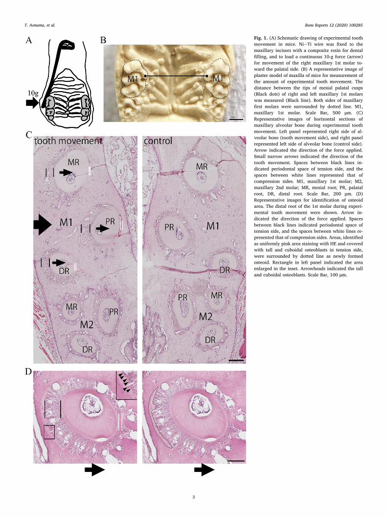
Fig. 1. (A) Schematic drawing of experimental toothmovement in mice. NieTi wire was fixed to themaxillary incisors with a composite resin for dentalfilling, and to load a continuous 10-g force (arrow)for movement of the right maxillary 1st molar to-ward the palatal side. (B) A representative image ofplaster model of maxilla of mice for measurement ofthe amount of experimental tooth movement. Thedistance between the tips of mesial palatal cusps(Black dots) of right and left maxillary 1st molarswas measured (Black line). Both sides of maxillaryfirst molars were surrounded by dotted line. M1,maxillary 1st molar. Scale Bar, 500 μm. (C)Representative images of horizontal sections ofmaxillary alveolar bone during experimental toothmovement. Left panel represented right side of al-veolar bone (tooth movement side), and right panelrepresented left side of alveolar bone (control side).Arrow indicated the direction of the force applied.Small narrow arrows indicated the direction of thetooth movement. Spaces between black lines in-dicated periodontal space of tension side, and thespaces between white lines represented that ofcompression sides. M1, maxillary 1st molar; M2,maxillary 2nd molar; MR, mesial root; PR, palatalroot, DR, distal root. Scale Bar, 200 μm. (D)Representative images for identification of osteoidarea. The distal root of the 1st molar during experi-mental tooth movement were shown. Arrow in-dicated the direction of the force applied. Spacesbetween black lines indicated periodontal space oftension side, and the spaces between white lines re-presented that of compression sides. Areas, identifiedas uniformly pink area staining with HE and coveredwith tall and cuboidal osteoblasts in tension side,were surrounded by dotted line as newly formedosteoid. Rectangle in left panel indicated the areaenlarged in the inset. Arrowheads indicated the talland cuboidal osteoblasts. Scale Bar, 100 μm.
T. Aonuma, et al. Bone Reports 12 (2020) 100285
3
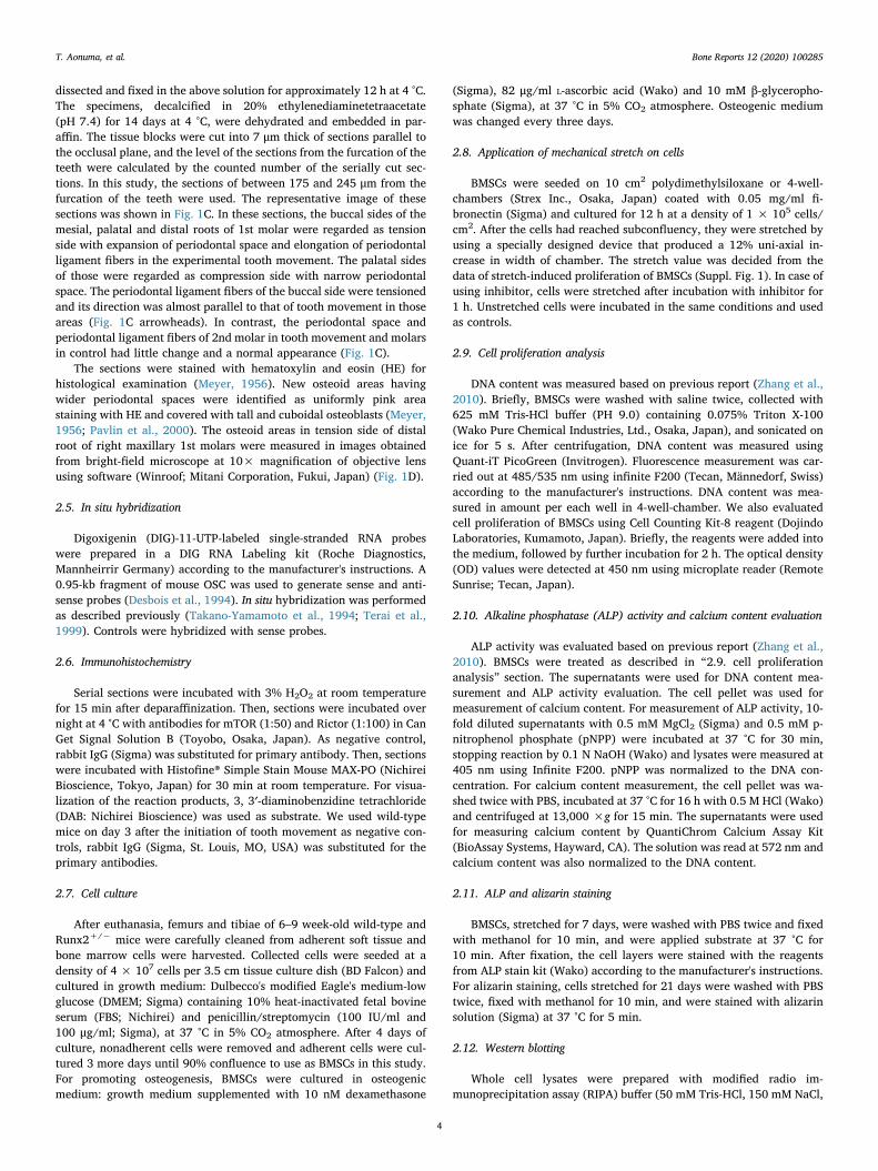
dissected and fixed in the above solution for approximately 12 h at 4 °C.The specimens, decalcified in 20% ethylenediaminetetraacetate(pH 7.4) for 14 days at 4 °C, were dehydrated and embedded in par-affin. The tissue blocks were cut into 7 μm thick of sections parallel tothe occlusal plane, and the level of the sections from the furcation of theteeth were calculated by the counted number of the serially cut sec-tions. In this study, the sections of between 175 and 245 μm from thefurcation of the teeth were used. The representative image of thesesections was shown in Fig. 1C. In these sections, the buccal sides of themesial, palatal and distal roots of 1st molar were regarded as tensionside with expansion of periodontal space and elongation of periodontalligament fibers in the experimental tooth movement. The palatal sidesof those were regarded as compression side with narrow periodontalspace. The periodontal ligament fibers of the buccal side were tensionedand its direction was almost parallel to that of tooth movement in thoseareas (Fig. 1C arrowheads). In contrast, the periodontal space andperiodontal ligament fibers of 2nd molar in tooth movement and molarsin control had little change and a normal appearance (Fig. 1C).
The sections were stained with hematoxylin and eosin (HE) forhistological examination (Meyer, 1956). New osteoid areas havingwider periodontal spaces were identified as uniformly pink areastaining with HE and covered with tall and cuboidal osteoblasts (Meyer,1956; Pavlin et al., 2000). The osteoid areas in tension side of distalroot of right maxillary 1st molars were measured in images obtainedfrom bright-field microscope at 10× magnification of objective lensusing software (Winroof; Mitani Corporation, Fukui, Japan) (Fig. 1D).
2.5. In situ hybridization
Digoxigenin (DIG)-11-UTP-labeled single-stranded RNA probeswere prepared in a DIG RNA Labeling kit (Roche Diagnostics,Mannheirrir Germany) according to the manufacturer's instructions. A0.95-kb fragment of mouse OSC was used to generate sense and anti-sense probes (Desbois et al., 1994). In situ hybridization was performedas described previously (Takano-Yamamoto et al., 1994; Terai et al.,1999). Controls were hybridized with sense probes.
2.6. Immunohistochemistry
Serial sections were incubated with 3% H2O2 at room temperaturefor 15 min after deparaffinization. Then, sections were incubated overnight at 4 °C with antibodies for mTOR (1:50) and Rictor (1:100) in CanGet Signal Solution B (Toyobo, Osaka, Japan). As negative control,rabbit IgG (Sigma) was substituted for primary antibody. Then, sectionswere incubated with Histofine® Simple Stain Mouse MAX-PO (NichireiBioscience, Tokyo, Japan) for 30 min at room temperature. For visua-lization of the reaction products, 3, 3′-diaminobenzidine tetrachloride(DAB: Nichirei Bioscience) was used as substrate. We used wild-typemice on day 3 after the initiation of tooth movement as negative con-trols, rabbit IgG (Sigma, St. Louis, MO, USA) was substituted for theprimary antibodies.
2.7. Cell culture
After euthanasia, femurs and tibiae of 6–9 week-old wild-type andRunx2+/− mice were carefully cleaned from adherent soft tissue andbone marrow cells were harvested. Collected cells were seeded at adensity of 4 × 10⁷ cells per 3.5 cm tissue culture dish (BD Falcon) andcultured in growth medium: Dulbecco's modified Eagle's medium-lowglucose (DMEM; Sigma) containing 10% heat-inactivated fetal bovineserum (FBS; Nichirei) and penicillin/streptomycin (100 IU/ml and100 μg/ml; Sigma), at 37 °C in 5% CO2 atmosphere. After 4 days ofculture, nonadherent cells were removed and adherent cells were cul-tured 3 more days until 90% confluence to use as BMSCs in this study.For promoting osteogenesis, BMSCs were cultured in osteogenicmedium: growth medium supplemented with 10 nM dexamethasone
(Sigma), 82 μg/ml L-ascorbic acid (Wako) and 10 mM β-glyceropho-sphate (Sigma), at 37 °C in 5% CO2 atmosphere. Osteogenic mediumwas changed every three days.
2.8. Application of mechanical stretch on cells
BMSCs were seeded on 10 cm2 polydimethylsiloxane or 4-well-chambers (Strex Inc., Osaka, Japan) coated with 0.05 mg/ml fi-bronectin (Sigma) and cultured for 12 h at a density of 1 × 10⁵ cells/cm2. After the cells had reached subconfluency, they were stretched byusing a specially designed device that produced a 12% uni-axial in-crease in width of chamber. The stretch value was decided from thedata of stretch-induced proliferation of BMSCs (Suppl. Fig. 1). In case ofusing inhibitor, cells were stretched after incubation with inhibitor for1 h. Unstretched cells were incubated in the same conditions and usedas controls.
2.9. Cell proliferation analysis
DNA content was measured based on previous report (Zhang et al.,2010). Briefly, BMSCs were washed with saline twice, collected with625 mM Tris-HCl buffer (PH 9.0) containing 0.075% Triton X-100(Wako Pure Chemical Industries, Ltd., Osaka, Japan), and sonicated onice for 5 s. After centrifugation, DNA content was measured usingQuant-iT PicoGreen (Invitrogen). Fluorescence measurement was car-ried out at 485/535 nm using infinite F200 (Tecan, Männedorf, Swiss)according to the manufacturer's instructions. DNA content was mea-sured in amount per each well in 4-well-chamber. We also evaluatedcell proliferation of BMSCs using Cell Counting Kit-8 reagent (DojindoLaboratories, Kumamoto, Japan). Briefly, the reagents were added intothe medium, followed by further incubation for 2 h. The optical density(OD) values were detected at 450 nm using microplate reader (RemoteSunrise; Tecan, Japan).
2.10. Alkaline phosphatase (ALP) activity and calcium content evaluation
ALP activity was evaluated based on previous report (Zhang et al.,2010). BMSCs were treated as described in “2.9. cell proliferationanalysis” section. The supernatants were used for DNA content mea-surement and ALP activity evaluation. The cell pellet was used formeasurement of calcium content. For measurement of ALP activity, 10-fold diluted supernatants with 0.5 mM MgCl2 (Sigma) and 0.5 mM p-nitrophenol phosphate (pNPP) were incubated at 37 °C for 30 min,stopping reaction by 0.1 N NaOH (Wako) and lysates were measured at405 nm using Infinite F200. pNPP was normalized to the DNA con-centration. For calcium content measurement, the cell pellet was wa-shed twice with PBS, incubated at 37 °C for 16 h with 0.5 M HCl (Wako)and centrifuged at 13,000 ×g for 15 min. The supernatants were usedfor measuring calcium content by QuantiChrom Calcium Assay Kit(BioAssay Systems, Hayward, CA). The solution was read at 572 nm andcalcium content was also normalized to the DNA content.
2.11. ALP and alizarin staining
BMSCs, stretched for 7 days, were washed with PBS twice and fixedwith methanol for 10 min, and were applied substrate at 37 °C for10 min. After fixation, the cell layers were stained with the reagentsfrom ALP stain kit (Wako) according to the manufacturer's instructions.For alizarin staining, cells stretched for 21 days were washed with PBStwice, fixed with methanol for 10 min, and were stained with alizarinsolution (Sigma) at 37 °C for 5 min.
2.12. Western blotting
Whole cell lysates were prepared with modified radio im-munoprecipitation assay (RIPA) buffer (50 mM Tris-HCl, 150 mM NaCl,
T. Aonuma, et al. Bone Reports 12 (2020) 100285
4
1% Triton X-100, 1% Na-deoxycholate, 0.1% SDS, 1 mM EDTA, 1 mMNaF, pH 7.5) containing protease inhibitor cocktail (Sigma aldrich, St,Louis, MO). Protein (30 μg) was loaded to Tris-Glycine SDS-PAGE gel(Bio-Rad Laboratories Inc., CA, USA) and transferred to nitrocelluroseor polyvinylidene difluoride membrane using Trans-Blot® Turbo™Transfer System (Bio-Rad Laboratories Inc.). The membranes were in-cubated with primary antibody (1:1000) at 4 °C overnight, followed byincubation with secondary antibody conjugated with horseradish per-oxidase (1:5000). Proteins were detected with SuperSignal West FemtoChemiluminescent Substrate (Pierce Chemical Co, CA, USA). Theimages were acquired using VersaDoc5000MP (Bio-Rad LaboratoriesInc.).
2.13. RNA interference
For knockdown of the expression of Rictor, we employed an RNAinterference method (Sen et al., 2014). siRictor against Rictor and itscontrol siRNAs were purchased from Invitrogen (Carlsbad, CA). Cellswere transfected with specific siRNA or control siRNA (10 nM) usingPepMute Plus (SignaGen Lab, Rockville, MD) at 18 h after plating cells.Mechanical stretch was applied at 24 h after transfection. The siRNAsequences for Rictor were 5′-UCAUCUUUCUGACUAAGCGAAGGGC andfor the control (nucleotide change within same sequence) were5′-GCCCUCGUUGACUGAAAGAAUCUGA (Sen et al., 2014).
2.14. Real-time RT-PCR
The total RNA was extracted from BMSCs using TRIzol reagent®(Invitrogen) according to the manufacturer's instructions. Total RNA(1 μg) was reverse transcribed to generate complementary DNA(cDNA). Real-time PCR was performed using Thermal Cycler Dice RealTime System (Takara Bio. Inc., Shiga, Japan) with SYBR premix Ex Taq(Takara). Amplification reactions were conducted with thermal dena-turation 95 °C for 10 min followed by 40 cycles at annealing 95 °C for15 s and extension reaction at 60 °C for 60 s using the primer sets(Suppl. Table 1). Each sample was subjected to triplicate PCR reactionsand the threshold cycle value of samples was normalized by expressionof GAPDH as an endogenous housekeeping gene.
2.15. Statistical evaluations
The results were presented as means± SD. Statistical analysis wasperformed using two-way ANOVA in vivo and two-way ANOVA in vitrofollowed by Sheffe's test. P values 0.05 were considered significant. Allexperiments were performed at least two times.
3. Results
3.1. Small amount of experimental tooth movement in Runx2+/− mice
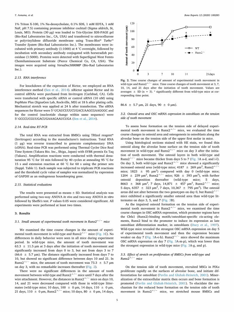
We examined the time course changes in the amount of experi-mental tooth movement in wild-type and Runx2+/− mice (Fig. 1C). Nodifferences in daily behavior were seen in all mice during experimentperiod. In wild-type mice, the amount of tooth movement was63.3 ± 11.5 μm at 3 days after the initiation of tooth movement andsignificantly increased from days 0 to 3, but not from days 3 to 7(66.6 ± 5.7 μm). The distance significantly increased from days 7 to10, but showed no significant difference between days 10 and 21. InRunx2+/− mice, the amount of tooth movement was 73.3 ± 5.7 μmon day 3, with no remarkable increases thereafter (Fig. 2).
There were no significant differences in the amount of toothmovement between wild-type and Runx2+/− mice until 7 days after thewire attachment. However, the distances in Runx2+/− mice on days 10,14, and 21 were decreased compared with those in wild-type litter-mates (wild-type mice; 10 days, 100 ± 0 μm, 14 days, 110 ± 0 μm,21 days, 110 ± 0 μm, Runx2+/− mice; 10 days, 80 ± 0 μm, 14 days,
86.6 ± 5.7 μm, 21 days, 90 ± 0 μm).
3.2. Osteoid area and OSC mRNA expression in osteoblasts on the tensionside of tooth movement
To assess bone formation on the tension side of delayed experi-mental tooth movement in Runx2+/− mice, we evaluated the timecourse changes in osteoid area and osteogenesis in osteoblasts along thealveolar bone on the tension side of the upper first molar in mice.
Using histological sections stained with HE stain, we found thinosteoid along the alveolar bone surface on the tension side of toothmovement in wild-type and Runx2+/− mice on day 3 after the initia-tion of tooth movement. The osteoid layers in both wild-type andRunx2+/− mice became thicker from days 5 to 7 (Fig. 3A a-d, and i-l).On day 3, both wild-type and Runx2+/− mice showed a significantlyincreased osteoid area (wild-type mice; 4187 ± 269 μm2, Runx2+/−
mice; 1823 ± 95 μm2) compared with day 0 (wild-type mice;1204 ± 239 μm2, Runx2+/− mice; 926 ± 393 μm2), with furtherosteoid deposition thereafter (wild-type mice; 5 days,8663 ± 368 μm2, 7 days, 14,871 ± 457 μm2, Runx2+/− mice;5 days, 6357 ± 323 μm2, 7 days, 10,367 ± 795 μm2). The osteoidareas did not alter between the two genotypes on day 0, but Runx2+/−
mice exhibited a significantly smaller osteoid area than wild-type lit-termates on days 3, 5, and 7 (Fig. 3B).
As the impaired osteoid formation on the tension side of experi-mental tooth movement in Runx2+/− mice, we examined the timecourse changes in OSC mRNA expression, which promoter regions havethe Cbfa1 (Runx2)-binding motifs/osteoblast-specific cis-acting ele-ments, Runx2 bind to the promoter to induce its expression as lateosteoblast differentiation marker, in osteoblasts (Ducy et al., 1997).Wild-type mice revealed the strongest OSC mRNA expression on day 5of experimental tooth movement and then the expression becameweaker on day 7 (Fig. 3A-e-h). Runx2+/− mice showed the maximumOSC mRNA expression on day 7 (Fig. 3A-m-p), which was lower thanthe strongest expression in wild-type mice (Fig. 3A-g, and p).
3.3. Effect of stretch on proliferation of BMSCs from wild-type andRunx2+/− mice
On the tension side of tooth movement, recruited MSCs in PDLsproliferate rapidly on the surfaces of alveolar bone, and initiate dif-ferentiation for osteoblast (Pavlin and Gluhak-Heinrich, 2001). Miner-alization of the extracellular matrix then occurs and bone formation ispromoted (Pavlin and Gluhak-Heinrich, 2001). To elucidate the me-chanism for the reduced bone formation on the tension side of toothmovement in Runx2+/− mice, we stretched mouse BMSCs and
Fig. 2. Time course changes of amount of experimental tooth movement inwild-type and Runx2+/− mice. Time course changes of tooth movement at 3, 7,10, 14, and 21 days after the initiation of tooth movement. Values areaverages ± SD (n = 3). * significantly different from wild-type mice at cor-responding time point.
T. Aonuma, et al. Bone Reports 12 (2020) 100285
5
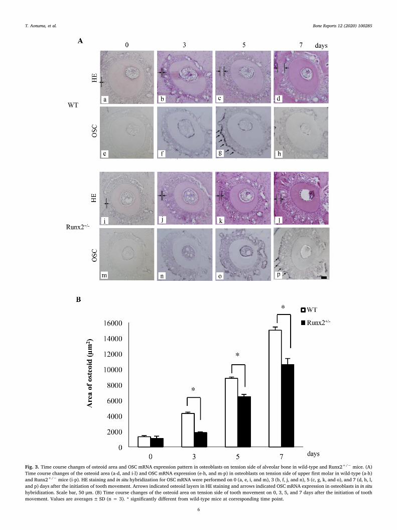
Fig. 3. Time course changes of osteoid area and OSC mRNA expression pattern in osteoblasts on tension side of alveolar bone in wild-type and Runx2+/− mice. (A)Time course changes of the osteoid area (a-d, and i-l) and OSC mRNA expression (e-h, and m-p) in osteoblasts on tension side of upper first molar in wild-type (a-h)and Runx2+/− mice (i-p). HE staining and in situ hybridization for OSC mRNA were performed on 0 (a, e, i, and m), 3 (b, f, j, and n), 5 (c, g, k, and o), and 7 (d, h, l,and p) days after the initiation of tooth movement. Arrows indicated osteoid layers in HE staining and arrows indicated OSC mRNA expression in osteoblasts in in situhybridization. Scale bar, 50 μm. (B) Time course changes of the osteoid area on tension side of tooth movement on 0, 3, 5, and 7 days after the initiation of toothmovement. Values are averages± SD (n = 3). * significantly different from wild-type mice at corresponding time point.
T. Aonuma, et al. Bone Reports 12 (2020) 100285
6
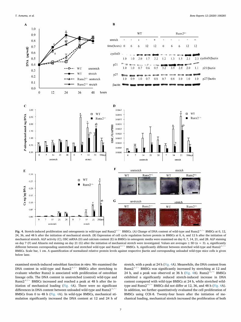
examined stretch-induced osteoblast function in vitro. We examined theDNA content in wild-type and Runx2+/− BMSCs after stretching toevaluate whether Runx2 is associated with proliferation of osteoblastlineage cells. The DNA content in unstretched (control) wild-type andRunx2+/− BMSCs increased and reached a peak at 48 h after the in-itiation of mechanical loading (Fig. 4A). There were no significantdifferences in DNA content between unloaded wild-type and Runx2+/−
BMSCs from 0 to 48 h (Fig. 4A). In wild-type BMSCs, mechanical sti-mulation significantly increased the DNA content at 12 and 24 h of
stretch, with a peak at 24 h (Fig. 4A). Meanwhile, the DNA content fromRunx2+/− BMSCs was significantly increased by stretching at 12 and24 h, and a peak was observed at 36 h (Fig. 4A). Runx2+/− BMSCsexhibited a significantly reduced stretch-induced increase in DNAcontent compared with wild-type BMSCs at 24 h, while stretched wild-type and Runx2+/− BMSCs did not differ at 12, 36, and 48 h (Fig. 4A).In addition, we further quantitatively evaluated the cell proliferation ofBMSCs using CCK-8. Twenty-four hours after the initiation of me-chanical loading, mechanical stretch increased the proliferation of both
Fig. 4. Stretch-induced proliferation and osteogenesis in wild-type and Runx2+/− BMSCs. (A) Change of DNA content of wild-type and Runx2+/− BMSCs at 0, 12,24, 36, and 48 h after the initiation of mechanical stretch. (B) Expression of cell cycle regulation factors protein in BMSCs at 0, 6, and 12 h after the initiation ofmechanical stretch. ALP activity (C), OSC mRNA (D) and calcium content (E) in BMSCs in osteogenic media were examined on day 0, 7, 14, 21, and 28. ALP stainingon day 7 (F) and Alizarin red staining on day 21 (G) after the initiation of mechanical stretch were investigated. Values are averages± SD (n = 3). a, significantlydifferent between corresponding unstretched and stretched wild-type and Runx2+/− BMSCs. b, significantly different between stretched wild-type and Runx2+/−
BMSCs. Scale bar, 1 cm. A quantification of normalized relative protein levels against respective βactin and corresponding unloaded wild-type mice cells is givenbelow lane.
T. Aonuma, et al. Bone Reports 12 (2020) 100285
7
wild-type and Runx2+/− BMSCs, and more importantly stretchedRunx2+/− BMSCs significantly proliferated compared with wild-typeBMSCs (Suppl. Fig. 2).
Cyclins and cyclin-dependent kinases (cdks) are integrators ofgrowth factor-mediated signals that drive the cell cycle (Baldin et al.,1993; Lim and Kaldis, 2013). Growth factors promote the G1 phase ofthe cell cycle and D-type cyclins act primarily as growth factor sensors(Baldin et al., 1993; Lim and Kaldis, 2013). p21 and p27 are tight-binding inhibitors of cyclin D-cdk4 complex and inhibit G1 progression(Harper et al., 1993; Polyak et al., 1994). We evaluated the expressionof cell cycle regulatory proteins in wild-type and Runx2+/− BMSCs at 6and 12 h after the initiation of stretch to examine the association of cellcycle progression with stretch. Cyclin D was enhanced in unstretchedwild-type and Runx2+/− BMSCs at 12 h compared with 0 h (Fig. 4B).
Stretching remarkably induced cyclin D in wild-type BMSCs at 6 h, andin Runx2+/− BMSCs at 12 h (Fig. 4B). p21 protein expression in un-stretched wild-type and Runx2+/− BMSCs did not change from 0 to 6 h,but was reduced from 6 to 12 h, although Runx2+/− BMSCs showedremarkably higher p21 expression from 0 to 12 h than wild-type BMSCsregardless of mechanical loading (Fig. 4B). In wild-type BMSCs,stretching attenuated p21 expression at 6 h, but not at 12 h, while inRunx2+/− BMSCs, stretching attenuated p21 expression at both 6 and12 h (Fig. 4B). There was little difference in p27 expression in BMSCsfrom 0 to 12 h, regardless of loading and Runx2 gene dosage (Fig. 4B).
These findings indicated that stretch increased the proliferation ofBMSCs and regulated cell cycle progression, and that the reduction ofRunx2 gene dosage delayed mechanical stimulation-induced prolifera-tion of BMSCs.
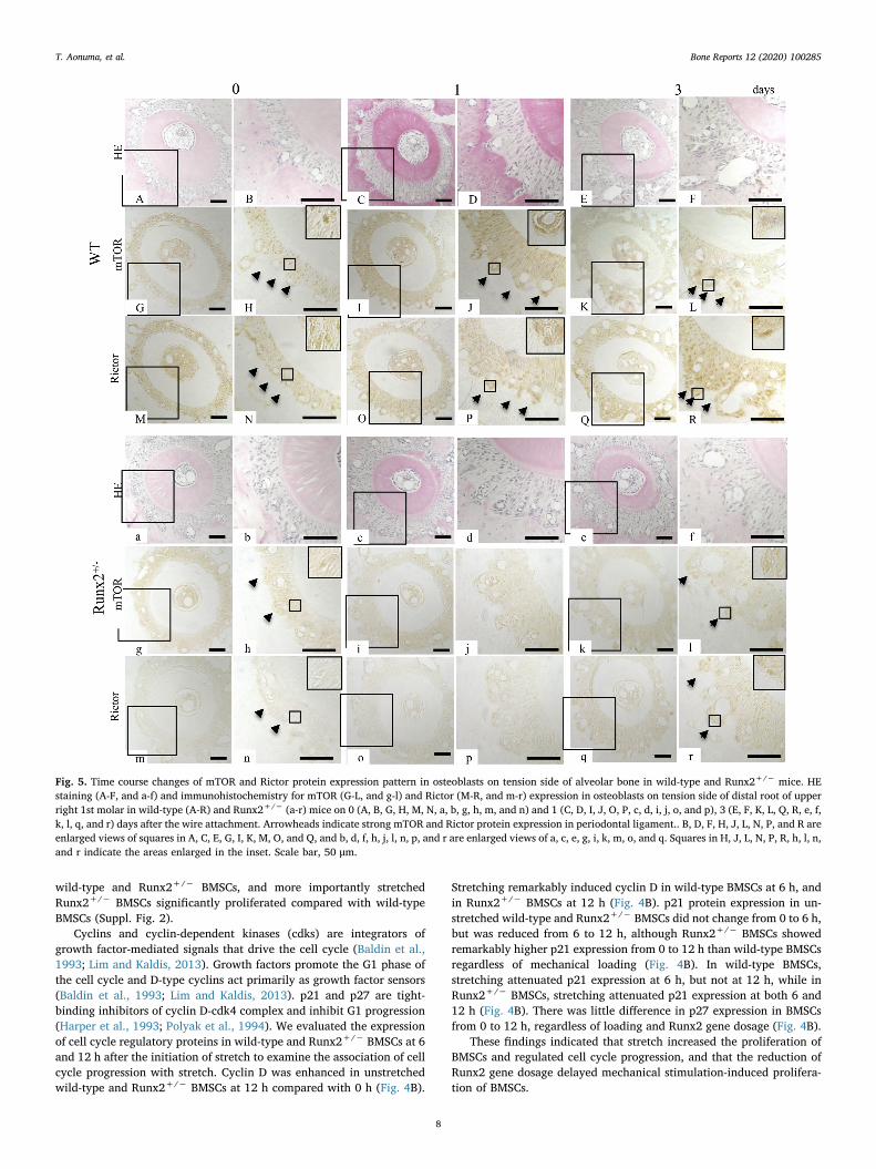
Fig. 5. Time course changes of mTOR and Rictor protein expression pattern in osteoblasts on tension side of alveolar bone in wild-type and Runx2+/− mice. HEstaining (A-F, and a-f) and immunohistochemistry for mTOR (G-L, and g-l) and Rictor (M-R, and m-r) expression in osteoblasts on tension side of distal root of upperright 1st molar in wild-type (A-R) and Runx2+/− (a-r) mice on 0 (A, B, G, H, M, N, a, b, g, h, m, and n) and 1 (C, D, I, J, O, P, c, d, i, j, o, and p), 3 (E, F, K, L, Q, R, e, f,k, l, q, and r) days after the wire attachment. Arrowheads indicate strong mTOR and Rictor protein expression in periodontal ligament.. B, D, F, H, J, L, N, P, and R areenlarged views of squares in A, C, E, G, I, K, M, O, and Q, and b, d, f, h, j, l, n, p, and r are enlarged views of a, c, e, g, i, k, m, o, and q. Squares in H, J, L, N, P, R, h, l, n,and r indicate the areas enlarged in the inset. Scale bar, 50 μm.
T. Aonuma, et al. Bone Reports 12 (2020) 100285
8
(caption on next page)
T. Aonuma, et al. Bone Reports 12 (2020) 100285
9
3.4. Effect of stretch on osteogenesis of BMSCs from wild-type andRunx2+/− mice
Next, we loaded stretch onto wild-type and Runx2+/− BMSCs inosteogenic medium to investigate the effect of Runx2 on stretch-in-duced osteoblast differentiation. In unstretched wild-type and Runx2+/
− BMSCs, ALP activity, an early osteogenesis marker, gradually in-creased from 0 to 21 days after the initiation of stretching and de-creased thereafter, with no significant differences between the twogenotypes until day 28 (Fig. 4C). In wild-type BMSCs, stretching sig-nificantly increased ALP activity on days 7 and 14, and the peak of ALPactivity occurred on day 7 (Fig. 4C). In Runx2+/− BMSCs, mechanicalstimulation significantly enhanced ALP activity on day 14, but not onday 7, and the peak of ALP activity was observed on day 21. Further-more, the heterozygous Runx2 deficiency significantly reduced stretch-induced ALP activity in BMSCs on day 14 (Fig. 4C). Stretch-inducedpeak of ALP activity in Runx2+/− BMSCs was significantly lower thanthat in wild-type BMSCs (Fig. 4C).
Mechanical loading significantly increased OSC mRNA expression inwild-type BMSCs on day 14 after the initiation of stretch, and thestretch-induced OSC mRNA expression peaked on day 21 and decreasedthereafter (Fig. 4D). In contrast, stretch significantly induced OSCmRNA expression in Runx2+/− BMSCs on day 21, but not on day 14(Fig. 4D). Runx2+/− BMSCs showed a significantly lower peak ofstretch-induced OSC mRNA expression than wild-type BMSCs (Fig. 4D).
We further examined stretch-induced matrix-deposited calcium, alate marker of osteogenesis, in BMSCs from wild-type and Runx2+/−
mice (Fig. 4E). In unloaded wild-type and Runx2+/− BMSCs, the cal-cium content increased constantly until day 28 after the initiation ofstretch, but there was no significant difference between wild-type andRunx2+/− (Fig. 4E). Mechanical loading significantly enhanced thecalcium content in wild-type BMSCs on days 21 and 28, and in Runx2+/
− BMSCs on day 28, but not on day 21 (Fig. 4E). On day 28, the stretch-induced calcium content in Runx2+/− BMSCs was significantly lowerthan in wild-type BMSCs (Fig. 4E).
In addition, wild-type and Runx2+/− BMSCs were stained for ALPon day 7 and alizarin red on day 21 after the initiation of mechanicalstimulation (Fig. 4F and G). Stretching caused remarkable staining ofALP and alizarin red in both genotypes (Fig. 4F and G). Unstretched andstretched Runx2+/− BMSCs showed less staining than the corre-sponding wild-type BMSCs (Fig. 4F and G).
Taken together, we found that stretch enhanced osteogenesis inboth wild-type and Runx2+/− BMSCs, but Runx2+/− BMSCs showedreduced and delayed stretch-induced osteoblast differentiation com-pared with wild-type BMSCs.
3.5. mTOR and Rictor protein expression in osteoblasts on the tension sideof alveolar bone
To assess the association of mTORC2 with bone formation on thetension side of delayed tooth movement in Runx2+/− mice, we ex-amined the protein expression of mTOR and Rictor in osteoblasts on thetension side of experimental tooth movement.
Before the initiation of experimental tooth movement (day 0),mTOR and Rictor were expressed in the PDL in both wild-type andRunx2+/− mice, although Runx2+/− mice showed weaker expressionof both proteins compared with wild-type littermates (Fig. 5H, N, h, and
n, arrowheads). Enhanced mTOR and Rictor expression in osteoblastson the tension side of experimental tooth movement in wild-type micewas detected on days 1 and 3 of experimental tooth movement (Fig. 5J,P, L, and R, arrows), while Runx2+/− mice exhibited weaker expres-sions of these proteins on day 3 (Fig. 5j, p, l, and r, arrows). Expressionof mTOR and Rictor was not detected in osteoblasts of Runx2+/− miceon day 1 (Fig. 5i, j, o, and p). We used wild-type mice on day 3 after theinitiation of tooth movement as negative controls, and there was noexpression (data not shown).
3.6. Association of mTORC2 activation with stretch-induced proliferation ofBMSCs
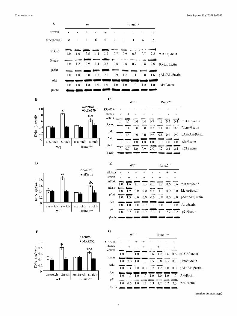
To assess whether delayed stretch-induced proliferation of Runx2+/
− BMSCs was caused by failure of mTORC2 activation, we evaluatedmTORC2 activation in BMSCs after mechanical loading. In unstretchedwild-type and Runx2+/− BMSCs, there was little change in mTORprotein expression from 0 to 6 h after the initiation of stretching(Fig. 6A). Mechanical stimulation remarkably increased mTOR proteinexpression in wild-type BMSCs at both 1 and 6 h, and in Runx2+/−
BMSCs at 6 h, but not at 1 h (Fig. 6A). The Runx2 heterozygous defi-ciency reduced the expression of mTOR in BMSCs regardless of stretch(Fig. 6A). Similar results were confirmed for Rictor, an essential com-ponent factor of mTORC2, and Akt phosphorylation at serine 473, atarget of mTORC2 (Fig. 6A).
To examine the effect of mTORC2 on stretch-induced cell pro-liferation, we inhibited component factors of mTORC2 and Akt phos-phorylation. KU63794, an inhibitor of mTOR activation, did not changethe DNA content in unstretched wild-type and Runx2+/− BMSCs butdramatically inhibited the stretch induced-increase in DNA content(Fig. 6B). By immunoblot analysis, we found that KU63794 down-regulated mTOR expression and Akt phosphorylation in wild-type andRunx2+/− BMSCs regardless of stretching, and inhibited both stretch-induced Rictor expression and stretch-induced reduction of p21 ex-pression (Fig. 6C). To confirm the involvement of mTORC2, but notmTORC1, in the stretch-induced cell proliferation, we silenced Rictorby short interfering RNA (siRNA). siRictor inhibited the stretch-inducedDNA content in wild-type and Runx2+/− BMSCs (Fig. 6D). Immunoblotanalysis revealed that siRictor silenced Rictor expression and Aktphosphorylation in both wild-type and Runx2+/− BMSCs and inhibitedstretch-regulated mTOR and p21 expression (Fig. 6E). Furthermore,MK2206, an Akt phosphorylation inhibitor, down-regulated the in-crease in DNA content and weakened p21 expression in response tostretching (Fig. 6F and G). Interestingly, MK2206 also inhibited stretch-induced mTOR and Rictor expression (Fig. 6G).
3.7. PI3K signaling on stretch-induced proliferation of BMSCs
Growth factors such as insulin-like growth factor (IGF), cytokinesand stretch activate PI3K and phosphorylate Akt (Vanhaesebroecket al., 2010; Watabe et al., 2011). PI3K/Akt signaling regulates cellproliferation and differentiation of bone-related cells such as MSCs,osteoblasts, and chondrocytes (Ghosh-Choudhury et al., 2002; Yunet al., 2008; Hidaka et al., 2001).
To examine the involvement of PI3K and mTORC2 signaling instretch-induced cell proliferation, we stretched wild-type and Runx2+/
− BMSCs after treatment with LY294002, a PI3K inhibitor. We found
Fig. 6. Stretch-induced mTORC2/Akt signal and proliferation of wild-type and Runx2+/− BMSCs. (A) Change of mTOR and Rictor protein expression and Aktphosphorylation in BMSCs after 0, 1, and 6 h after the initiation of stretch. DNA content of BMSCs treated with KU63794 (5 μM) (B), siRictor (D), and MK2206(5 μM), and stretched for 24 h (F). Values are averages± SD (n = 3). a, significantly different between corresponding unstretched and stretched wild-type andRunx2+/− BMSCs. b, significantly different between stretched wild-type and Runx2+/− BMSCs. c significantly different between control and inhibitor or siRNAtreated stretched wild-type and Runx2+/− BMSCs. p21 and mTORC2 related protein expression pattern in BMSCs treated with KU63794 (5 μM) (C), siRictor (E), andMK2206 (5 μM) (G) and stretched for 6 h. A quantification of normalized relative protein levels against respective βactin and corresponding unloaded wild-type micecells is given below lane.
T. Aonuma, et al. Bone Reports 12 (2020) 100285
10
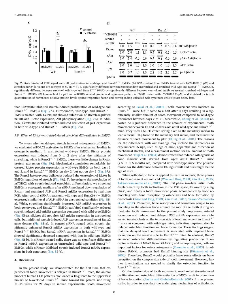
that LY294002 inhibited stretch-induced proliferation of wild-type andRunx2+/− BMSCs (Fig. 7A). Furthermore, wild-type and Runx2+/−
BMSCs treated with LY294002 showed inhibition of stretch-regulatedmTOR and Rictor expression, Akt phosphorylation (Fig. 7B). In addi-tion, LY294002 inhibited stretch-induced reduction of p21 expressionin both wild-type and Runx2+/− BMSCs (Fig. 7B).
3.8. Effect of Rictor on stretch-induced osteoblast differentiation in BMSCs
To assess whether delayed stretch induced osteogenesis of BMSCs,we evaluated mTORC2 activation in BMSCs after mechanical loading inosteogenic medium. In unstretched wild-type BMSCs, Rictor proteinexpression was induced from 0 to 2 days after the initiation ofstretching, while in Runx2+/− BMSCs, there was little change in Rictorprotein expression (Fig. 8A). Mechanical stimulation remarkably in-creased Rictor protein expression in wild-type BMSCs on both days 1and 2, and in Runx2+/− BMSCs on day 2, but not on day 1 (Fig. 8A).The Runx2 heterozygous deficiency reduced the expression of Rictor inBMSCs regardless of stretch (Fig. 8A). To investigate the association ofmTORC2 with stretch-induced osteoblast differentiation, we stretchedBMSCs in osteogenic medium after siRNA-mediated down-regulation ofRictor, and examined ALP and Runx2 mRNA expression by real-timePCR. After control siRNA treatment, wild-type and Runx2+/− BMSCsexpressed similar level of ALP mRNA in unstretched condition (Fig. 8B-a). While, stretching significantly increased ALP mRNA expression inboth genotypes, and Runx2+/− BMSCs exhibited significantly reducedstretch-induced ALP mRNA expression compared with wild-type BMSCs(Fig. 8B-a). siRictor did not alter ALP mRNA expression in unstretchedcells, but inhibited stretch-induced ALP expression regardless of Runx2gene dosage (Fig. 8B-a). In control siRNA-treated cells, stretch sig-nificantly enhanced Runx2 mRNA expression in both wild-type andRunx2+/− BMSCs, but Runx2 mRNA expression in Runx2+/− BMSCsshowed significantly decresed compared with that in wild-type BMSCs(Fig. 8B-b). In siRictor-treated cells, there was no significant differencein Runx2 mRNA expression in unstretched wild-type and Runx2+/−
BMSCs, while siRictor inhibited stretch-induced Runx2 mRNA expres-sions in both genotypes (Fig. 8B-b).
4. Discussion
In the present study, we demonstrated for the first time that ex-perimental tooth movement is delayed in Runx2+/− mice, the animalmodel of human CCD patients. We loaded a 10-g force to the upper firstmolars of 6-week-old Runx2+/− mice toward the palatal side usingNieTi wires for 21 days to induce experimental tooth movement
according to Sakai et al. (2009). Tooth movement was initiated inRunx2+/− mice but it came to a halt after 3 days resulting in a sig-nificantly smaller amount of tooth movement compared to wild-typelittermates between days 7 to 21. Meanwhile, Chung et al. (2004) re-ported no significant difference in the amount of experimental toothmovement between 13 and 22-week-old adult wild-type and Runx2+/−
mice. They used a NieTi coiled spring fixed to the maxillary incisor toload a mesial 10-g force on the maxillary first molar, and measured thedistance of tooth movement by μCT (Chung et al., 2004). The reasonsfor the differences with our findings may include the differences inexperimental design, such as age of mice, apparatus and direction ofmechanical stretch, and measurement method for the amount of toothmovement. Tsuji et al. (2004) demonstrated that reduced osteogensis ofbone marrow cells derived from aged adult Runx2+/− mice(7.5 ± 0.5 months old) compared with wild-type mice. The possiblereason for the difference between Chung's result and ours might be theage of mice.
When orthodontic force is applied to teeth in rodents, three phasesof tooth movement are induced (Wise and King, 2008; Yan et al., 2015;Takano-Yamamoto et al., 2017). The first phase involves initial toothdisplacement by tooth inclination in the PDL space, followed by a lagphase, and finally a tooth movement phase accompanied by bone re-modeling with bone resorption by osteoclasts and bone formation byosteoblasts (Wise and King, 2008; Yan et al., 2015; Takano-Yamamotoet al., 2017). Therefore, bone resorption and formation couple to re-modeling in the alveolar bone around the root of the tooth during or-thodontic tooth movement. In the present study, suppressed osteoidformation and reduced and delayed OSC mRNA expression were ob-served in osteoblasts on the tension side of tooth movement in Runx2+/
− mice as compared with wild-type mice, indicating impaired stretch-induced osteoblast function and bone formation. These findings suggestthat the delayed tooth movement is associated with impaired boneformation on the tension side in Runx2+/− mice. In contrast, Runx2enhanced osteoclast differentiation by regulating production of re-ceptor activator of NF-κB ligand (RANKL) and osteoprotegerin, both areimportant factors for osteoclastogenesis (Enomoto et al., 2003). In ad-dition, RANKL promoter had Runx2 binding site (Kitazawa et al.,2003). Therefore, Runx2 would probably have some effects on boneresorption on the compression side of tooth movement. However, fur-ther investigations are needed to clarify the osteoclast function inRunx2+/− mice.
On the tension side of tooth movement, mechanical stress-inducedproliferation and osteoblast differentiation of MSCs result in promotionof bone formation (Pavlin and Gluhak-Heinrich, 2001). In the presentstudy, in order to elucidate the underlying mechanism of orthodontic
Fig. 7. Stretch-induced PI3K signal and cell proliferation in wild-type and Runx2+/− BMSCs. (A) DNA content from BMSCs treated with LY294002 (5 μM) andstretched for 24 h. Values are averages± SD (n = 3). a, significantly different between corresponding unstretched and stretched wild-type and Runx2+/− BMSCs. b,significantly different between stretched wild-type and Runx2+/− BMSCs. c significantly different between control and inhibitor treated stretched wild-type andRunx2+/− BMSCs. (B) Immunoblot for p21 and mTORC2 related protein and expression pattern in BMSC treated with LY294002 (5 μM) and stretched for 6 h. Aquantification of normalized relative protein levels against respective βactin and corresponding unloaded wild-type mice cells is given below lane.
T. Aonuma, et al. Bone Reports 12 (2020) 100285
11

force-induced impaired bone formation in Runx2+/− mice, we cul-tured mouse BMSCs in silicon chambers and loaded continuous uniaxialstretching to the chambers, which simulates the tension side of toothmovement in vivo, and investigated cell proliferation in growth mediumand osteogenesis in osteogenic medium.
It has been reported that chondrocyte proliferation in the growthplates of tibia and femur is reduced in Runx2 knockout embryos com-pared with wild-type mice, and that Runx2 promotes chondrocyteproliferation through induction of Indian hedgehog and Tcf7 (Yoshidaet al., 2004; Mikasa et al., 2011). In addition, recent studies found thatRunx2 phosphorylation increased the proliferation of human bonemarrow endothelial cells (hECs) (Qiao et al., 2006; Pierce et al., 2012).We found that the stretch-induced growth rate of BMSCs was sig-nificantly delayed in Runx2+/− mice compared with wild-type litter-mates, suggesting for the first time an association of Runx2 with stretch-induced proliferation of BMSCs. Contrary to our results, it was reportedthat Runx2 overexpression suppressed the proliferation of MC3T3mouse preosteoblastic cells and C2C12 mouse mesenchymal cells, andthat Runx2 overexpression in MC3T3 cells caused a delay in G1 phaseprogression (Galindo et al., 2005). On the contrary, calvarial osteo-blastic cells from Runx2-deficient mice also exhibited an increased cellgrowth rate compared with those from wild-type littermates (Pratapet al., 2003). In the present study, stretch elevated cyclin D (G1) ex-pression in wild-type and Runx2+/− BMSCs, it did not promote os-teoblastic phenotypes (Suppl. Fig. 3), indicating that Runx2 response
for mechanical stretch enhanced proliferation of BMSCs but did notpromote osteoblast differentiation. Consistent with our findings, Hataet al. (2013) showed that uniaxial stretch increased proliferation ofstem cells isolated from dental pulp of Sprague-Dawlay rats, while itsosteogenic differentiation was inhibited. Therefore, the influences ofRunx2 in cell proliferation may be dependent on osteoblastic differ-entiation-stages. It is suggested that Runx2 dosage might influencemechanical stress induced-proliferation of BMSCs at early stage of os-teogenesis.
It was reported that Rictor-deficient mice show suppressed axialloading-induced mineral apposition on the periosteal surface of thetibia compared with wild-type littermates (Sen et al., 2014). A bi-axialcyclic stretch elevated mTOR and Rictor protein expression to mTORC2activation for cytoskeletal reorganization of MSCs (Tandon et al.,2014). Tandon et al. (2014) revealed that recruitment of Runx2 to thepromoter of mTOR activated PI3K/Akt signaling via mTORC2 to inducesurvival in a human breast cancer cell line. Therefore, Runx2 might beassociated with mechanical loading-induced osteoblast function viamTORC2 activation. In the present study, for the first time, we foundreduced and delayed expression of mTOR and Rictor in osteoblasts onthe tension side of tooth movement in Runx2+/− mice compared withwild-type littermates. Therefore, we suggest the possible associationbetween Runx2 and mTORC2 in bone formation on the tension side oftooth movement.
In the present study, reduced and delayed expression of mTOR and
Fig. 8. Stretch-induced osteoblast differentiation in wild-type and Runx2+/− BMSCs treated by siRictor. (A) BMSCs were down-regulated with siRictor, and stretchedfor 1 and 2 days after 1 h followed by change of osteogenic medium. (B) mRNA expression of ALP (a), and Runx2 (b) in BMSCs in osteogenic medium on 2 days aftermechanical stretch. Values are averages± SD (n = 3). a, significantly different between corresponding unstretched and stretched wild-type and Runx2+/− BMSCs.b, significantly different between stretched wild-type and Runx2+/− BMSCs. c, significantly different between control and inhibitor treated stretched wild-type andRunx2+/− BMSCs. A quantification of normalized relative protein levels against respective βactin and corresponding unloaded wild-type mice cells is given belowlane.
T. Aonuma, et al. Bone Reports 12 (2020) 100285
12

Rictor, were observed in osteoblasts on the tension side of toothmovement in Runx2+/− mice as compared with wild-type littermates invivo and in vitro. In addition, we demonstrated that silenced mTOR andRictor expression, and inhibition of Akt phosphorylation completelyinhibited stretch-induced proliferation of BMSCs in vitro, indicating thatstretch-induced mTORC2/Akt signaling promoted BMSC proliferation.Taken together, these findings suggest that mTORC2 activation wasinvolved in delayed mechanical stress-induced tooth movement inRUNX2+/− mouse (CCD) model.
Next, we investigated cell cycle regulators to elucidate the me-chanism of the delayed stretch-induced proliferation of Runx2+/−
BMSCs. It was reported that glucose treatment facilitated Runx2 DNA-binding activity and cell cycle progression in hECs, and that targetedknockdown of Runx2 delayed progression in G1 phase (Pierce et al.,2012). Consistent with these findings, we revealed delayed stretch-elevated expression of cyclin D, a G1 cell cycle progression marker, inRunx2+/− BMSCs compared with that in wild-type BMSCs. Akt phos-phorylation promotes cell proliferation through inhibition of p21 ex-pression, an inhibitor of cdk and G1 transition (Gu et al., 2011).Phosphorylated Runx2 is translocated into the nucleus and recruited tothe promoter of p21 to suppress its expression in hECs and mouse os-teoblastic cell lines (Pierce et al., 2012; Westendorf et al., 2002). Fur-thermore, Runx2 is a direct substrate of Akt and is phosphorylated inhuman breast cancer cells (Pande et al., 2013). In the present study,stretch-induced mTORC2/Akt activation was inhibited in Runx2+/−
BMSCs compared with that in wild-type BMSCs. In addition, suppres-sion of p21 expression induced by stretch was abolished by inhibition ofmTORC2 and Akt phosphorylation. These findings suggest that Runx2 isassociated with stretch-regulated expression of cell cycle regulators inBMSCs, and affects stretch-induced cell proliferation. It is likely thatdown-regulation of p21 by stretching via Runx2/mTORC2/Akt axisregulated cell cycle progression. We propose that there may be twounderlying mechanisms, one involving indirect inhibition by Runx2 forp21 expression via mTORC2/Akt activation, and the other involvingdirect inhibition by recruitment of Akt-phosphorylated Runx2 to thep21 promoter. PI3K is a kinase that phosphorylates Akt and inducesmTORC2 activation (Masui et al., 2014). In the present study, inhibitionof PI3K suppressed stretch-induced mTOR and Rictor expression andAkt phosphorylation, and also inhibited stretch-increased cell pro-liferation, suggesting that regulation of Runx2 via PI3K-dependentmTOR/Akt signaling plays a critical role in stretch-induced osteoblasticcell proliferation (Fig. 9A). Interestingly, we found that inhibition ofAkt phosphorylation downstream of mTORC2 down-regulated stretch-enhanced mTOR and Rictor expression. It is suggested that anotherupstream mechanism of Akt and mTORC2 for stretch-induced mTORC2activity may be involved, but this remains to be clarified.
Several studies have revealed that mechanical stress activatesRunx2 to promote osteogenesis in osteoblastic cells (Liu et al., 2011a;Ziros et al., 2002; Kanno et al., 2007; Li et al., 2012;
Salingcarnboriboon et al., 2006; Liu et al., 2011b). Consistent withthese past reports, we showed that Runx2+/− BMSCs in osteogenicmedium exhibited delayed and reduced stretch-induced early and lateosteoblast differentiation compared with wild-type BMSCs. Over-expression of Runx2 up-regulated Akt phosphorylation, a target ofmTORC2, in MC3T3 cells (Fujita et al., 2004). However, there are noreports on the association of mTORC2 with osteoblast differentiationpromoted by Runx2. Akt, a downstream of mTORC2, was phosphory-lated by overexpression of Runx2 in MC3T3 cells (Fujita et al., 2004). Inthe present study, the expression of stretch-enhanced mTOR and Rictorin Runx2+/− BMSCs cultured in osteogenic medium was delayed andreduced compared with that in wild-type BMSCs. Akt phosphorylationpromoted BMP-induced osteoblast differentiation and enhanced Runx2mRNA and protein expression (Choi et al., 2014), and IGF phosphory-lated Akt and elevated ALP activity during osteoblast differentiation(Fujita et al., 2004). We found that silencing of Rictor suppressedstretch-elevated mRNA expression of Runx2 and ALP in wild-type andRunx2+/− BMSCs in osteogenic medium in vitro, indicating stretch-promoted osteoblast differentiation in BMSCs via mTORC2 activation.Taken together, we suggest that the bidirectional interaction of Runx2and mTORC2 affects stretch-induced osteoblast differentiation(Fig. 9B).
Resently, Dai et al. (2017) have reported that preosteoblast-specificknockout of mTOR impaired mouse osteoblast differentiation and causeCCD-like bone phenotype. They showed ablation of Raptor, specificfactor of mTORC1, reduced Runx2 expression in osteoblasts and sup-pressed osteoblast differentiation. In our preliminary data, stretch in-duced Raptor protein expression in BMSCs and Runx2+/− BMSCsshowed delayed and reduced stretch-induced expression, as is the casewith Rictror (data not shown). There is a possibility association ofmTORC1 as well as mTORC2 for mechanical stress-induced osteoblastfunction through Runx2 activation, but it remains to be studied. Inaddition, several studies have reported involvement of other signalingpathway, such as MAPK, BMP/Smad and Wnt signal, for mechanicalstimuli-induced Runx2 activation and osteoblast function (Kanno et al.,2007; Liu et al., 2011b; Yan et al., 2016). Therefore, these signalingpathways would probably have some effects on bone formation on thetension side of experimental tooth movement in Runx2+/− mice.However, further investigations are needed to clarify the involvementof other signaling pathways in mechanical stress-induced bone forma-tion in Runx2+/− mice.
5. Conclusions
We have shown for the first time that tooth movement is delayed inRunx2+/− mice compared with wild-type mice. This prompted us touse Runx2+/− mice as a model for delayed orthodontic tooth move-ment in CCD patients having RUNX2+/−. In addition, we have alsoshown that Runx2+/− BMSCs decreased stretch-induced proliferation,
Fig. 9. Runx2 regulates stretch-induced osteoblast function via mTORC2. (A) Runx2 regulates PI3K/mTORC2/Akt signaling axis in stretch-induced proliferation ofBMSCs. (B) Runx2 interacts with mTORC2 in stretch-induced osteoblast differentiation.
T. Aonuma, et al. Bone Reports 12 (2020) 100285
13

and differentiation to osteoblasts via mTORC2 activation. Our findingssuggest that the association of Runx2 with mTORC2/Akt activation isone of critical role for stretch-induced proliferation and differentiationto osteoblasts in osteogenesis during tooth movement.
Author contribution
T.T.-Y. contributed to the conception and design of the study.N.Tamamura, Y.S., T.F., N.Takeshita, T.Y., I.T. and T.T.-Y. contributedto the in vivo studies. T.A. T.F., S.S. and T.T.-Y. contributed to the invitro studies. All statistical analysis was carried out by T.A.. T.T.-Y. andT.A. prepared the manuscript. All authors read the manuscript andprovided comments and revisions.
Declaration of competing interest
All authors declare that they have no competing interest.
Acknowledgments
We thank Dr. E. Fukumoto (Tohoku University), Dr. H. Kawaki(Asahi University), Dr. E. Ikeda (RIKEN Center for DevelopmentalBiology), and Dr. Kitaura (Tohoku University) for excellent technicalsupport, and Biomedical Research Unit of Tohoku University Hospitalfor technical equipment support.
This study was supported by a grant-in Aid for scientific Researchfrom Ministry of Education, Culture, Sports, Science and Technology,Japan (15659491, 17209064, 20249081, 23249085, 15K11335 and15H05048 to T.T.-Y., 15K11335 and 18K09827 to T.F., 24792270 and26861768 to T.A.).
Appendix A. Supplementary data
Supplementary data to this article can be found online at https://doi.org/10.1016/j.bonr.2020.100285.
References
Aberg, T., Cavender, A., Gaikwad, J.S., et al., 2004. Phenotypic changes in dentition ofRunx2 homozygote-null mutant mice. J. Histochem. Cytochem. 52 (1), 131–139.
Baldin, V., Lukas, J., Marcote, M.J., et al., 1993. Cyclin D1 is a nuclear protein requiredfor cell cycle progression in G1. Genes Dev. 7 (5), 812–821.
Becker, A., Lustmann, J., Shteyer, A., 1997a. Cleidocranial dysplasia: part 1–generalprinciples of the orthodontic and surgical treatment modality. Am. J. Orthod.Dentofac. Orthop. 111 (1), 28–33.
Becker, A., Shteyer, A., Bimstein, E., Lustmann, J., 1997b. Cleidocranial dysplasia: part 2–treatment protocol for the orthodontic and surgical modality. Am. J. Orthod.Dentofac. Orthop. 111 (2), 173–183.
Beertsen, W., McCulloch, C.A., Sodek, J., 1997. The periodontal ligament: unique, mul-tifunctional connective tissue. Periodontol 2000 (13), 20–40.
Bhaskar, P.T., Hay, N., 2007. The two TORCs and Akt. Dev. Cell 12 (4), 487–502.Chen, J., Holguin, N., Shi, Y., et al., 2015. mTORC2 signaling promotes skeletal growth
and bone formation in mice. J. Bone Miner. Res. 30 (2), 369–378.Choi, Y.H., Kim, Y.J., Jeong, H.M., et al., 2014. Akt enhances Runx2 protein stability by
regulating Smurf2 function during osteoblast differentiation. FEBS J. 281 (16),3656–3666.
Chung, C.R., Tsuji, K., Nifuji, A., et al., 2004. Micro-CT evaluation of tooth, calvaria andmechanical stress-induced tooth movement in adult Runx2/Cbfa1 heterozygousknock-out mice. J Med Dent Sci 51 (1), 105–113.
Dai, Q., Xu, Z., Ma, X., et al., 2017. mTOR/raptor signaling is critical for skeletogenesis inmice through the regulation of Runx2 expression. Cell Death Differ. 24 (11),1886–1899.
Davidovitch, Z., 1991. Tooth movement. Crit. Rev. Oral Biol. Med. 2 (24), 411–450.Desbois, C., Hogue, D.A., Karsenty, G., 1994. The mouse osteocalcin gene cluster contains
three genes with two separate spatial and temporal patterns of expression. J. Biol.Chem. 269 (2), 1183–1190.
Ducy, P., Zhang, R., Geoffroy, V., et al., 1997. Osf2/Cbfa1: a transcriptional activator ofosteoblast differentiation. Cell 89 (5), 747–754.
Enomoto, H., Shiojiri, S., Hoshi, K., et al., 2003. Induction of osteoclast differentiation byRunx2 through receptor activator of nuclear factor-kappa B ligand (RANKL) andosteoprotegerin regulation and partial rescue of osteoclastogenesis in Runx2−/−mice by RANKL transgene. J. Biol. Chem. 278 (26), 23971–23977.
Fujita, T., Azuma, Y., Fukuyama, R., et al., 2004. Runx2 induces osteoblast and chon-drocyte differentiation and enhances their migration by coupling with PI3K-Akt
signaling. J. Cell Biol. 166 (1), 85–95.Galindo, M., Pratap, J., Young, D.W., et al., 2005. The bone-specific expression of Runx2
oscillates during the cell cycle to support a G1-related antiproliferative function inosteoblasts. J. Biol. Chem. 280 (21), 20274–20285.
García-Martínez, J.M., Moran, J., Clarke, R.G., et al., 2009. Ku-0063794 is a specific in-hibitor of the mammalian target of rapamycin (mTOR). Biochem. J. 421 (1), 29–42.
Ghosh-Choudhury, N., Abboud, S.L., Nishimura, R., et al., 2002. Requirement of BMP-2-induced phosphatidylinositol 3-kinase and Akt serine/threonine kinase in osteoblastdifferentiation and Smad-dependent BMP-2 gene transcription. J. Biol. Chem. 277(36), 33361–33368.
Gu, Y., Lindner, J., Kumar, A., et al., 2011. Rictor/mTORC2 is essential fining a balancebetween beta-cell proliferation and cell size. Diabetes 60 (3), 827–837.
Harper, J.W., Adami, G.R., Wei, N., et al., 1993. The p21 Cdk-interacting protein Cip1 is apotent inhibitor of G1 cyclin-dependent kinases. Cell 75 (4), 805–816.
Hata, M., Naruse, K., Ozawa, S., et al., 2013. Mechanical stretch increases the pro-liferation while inhibiting the osteogenic differentiation in dental pulp stem cells.Tissue Eng Part A 19 (5–6), 625–633.
Hidaka, K., Kanematsu, T., Takeuchi, H., et al., 2001. Involvement of the phosphoinosi-tide 3-kinase/protein kinase B signaling pathway in insulin/IGF-I-induced chon-drogenesis of the mouse embryonal carcinoma-derived cell line ATDC5. Int. J.Biochem. Cell Biol. 33 (11), 1094–1103.
Kanno, T., Takahashi, T., Tsujisawa, T., et al., 2007. Mechanical stress-mediated Runx2activation is dependent on Ras/ERK1/2 MAPK signaling in osteoblasts. J. Cell.Biochem. 101 (5), 1266–1277.
Kitazawa, S., Kajimoto, K., Kondo, T., et al., 2003. Vitamin D3 supports osteoclastogenesisvia functional vitamin D response element of human RANKL gene promoter. J. Cell.Biochem. 89 (4), 771–777.
Komori, T., Yagi, H., Nomura, S., Yamaguchi, A., et al., 1997. Targeted disruption ofCbfa1 results in a complete lack of bone formation owing to maturational arrest ofosteoblasts. Cell 89 (5), 755–764.
Lekic, P., McCulloch, C.A., 1996. Periodontal ligament cell population: the central role offibroblasts in creating a unique tissue. Anat. Rec. 245 (2), 327–341.
Li, Y., Ge, C., Long, J.P., et al., 2012. Biomechanical stimulation of osteoblast gene ex-pression requires phosphorylation of the Runx2 transcription factor. J. Bone Miner.Res. 27 (6), 1263–1274.
Lim, S., Kaldis, P., 2013. Cdks, cyclins and CKIs: roles beyond cell cycle regulation.Development 140 (15), 3079–3093.
Liu, L., Shao, L., Li, B., Zong, C., et al., 2011a. Extracellular signal regulated kinase1/2activated by fluid shear stress promotes osteogeneic differentiaiton of human bonemarrow-derived mesenchymal stem cells through novel signaling pathways. Int. J.Biochem. Cell Biol. 43 (11), 1591–1601.
Liu, L., Shao, L., Li, B., Zong, C., et al., 2011b. Extracellular signal regulated kinase1/2activated by fluid shear stress promotes osteogeneic differentiaiton of human bonemarrow-derived mesenchymal stem cells through novel signaling pathways. Int. J.Biochem. Cell Biol. 43 (11), 1591–1601.
Malagu, K., Duggan, H., Menear, K., et al., 2009. The discovery and optimisation of pyrido[2,3-d]pyrimidine-2,4-diamines as potent and selective inhibitors of mTOR kinase.Bioorg. Med. Chem. Lett. 19 (20), 5950–5953.
Masui, K., Cavenee, W.K., Mischel, P.S., 2014. mTORC2 in the center of cancer metabolicreprogramming. Trends Endocrinol. Metab. 25 (6), 364–373.
Meyer, P.C., 1956. The histological identification of osteoid tissue. J Pathol Bacteriol 71(2), 325–333.
Mikasa, M., Rokutanda, S., Komori, H., et al., 2011. Regulation of Tcf7 by Runx2 inchondrocyte maturation and proliferation. J. Bone Miner. Metab. 29 (3), 291–299.
Mundlos, S., 1999. Cleidocranial dysplasia: clinical and molecular genetics. J. Med.Genet. 36 (3), 177–182.
Otto, F., Thornell, A.P., Crompton, T., et al., 1997. Cbfa1, a candidate gene forCleidocranial dysplasia syndrome, is essential for osteoblast differentiation and bonedevelopment. Cell 89 (5), 765–771.
Pande, S., Browne, G., Padmanabhan, S., et al., 2013. Oncogenic Cooperation BetweenPI3K/Akt Signaling and Transcription Factor Runx2 Promotes the Invasive Propertiesof Metastatic Breast Cancer Cells. J. Cell. Physiol. 228 (8), 1784–1792.
Pavlin, D., Gluhak-Heinrich, J., 2001. Effect of mechanical loading on periodontal cells.Crit. Rev. Oral Biol. Med. 12 (5), 414–424.
Pavlin, D., Goldman, E.S., Gluhak-Heinrich, J., et al., 2000. Orthodontically stressedperiodontium of transgenic mouse as a model for studying mechanical response inbone: the effect on the number of osteoblasts. Clin. Orthod. Res. 3 (2), 55–66.
Pierce, A.D., Anglin, I.E., Vitolo, M.I., et al., 2012. Glucose-activated RUNX2 phosphor-ylation promotes endothelial cell proliferation and an angiogenic phenotype. J. Cell.Biochem. 113 (1), 282–292.
Polyak, K., Kato, J.Y., Solomon, M.J., et al., 1994. p27Kip1, a cyclin-Cdk inhibitor, linkstransforming growth factor-beta and contact inhibition to cell cycle arrest. GenesDev. 8 (1), 9–22.
Pratap, J., Galindo, M., Zaidi, S.K., et al., 2003. Cell growth regulatory role of Runx2during proliferative expansion of preosteoblasts. Cancer Res. 63 (17), 5357–5362.
Qiao, M., Shapiro, P., Fosbrink, M., et al., 2006. Cell cycle-dependent phosphorylation ofthe RUNX2 transcription factor by cdc2 regulates endothelial cell proliferation. J.Biol. Chem. 2281 (11), 7118–7128.
Sakai, Y., Balam, T.A., Kuroda, S., et al., 2009. CTGF and apoptosis in mouse osteocytesinduced by tooth movement. J. Dent. Res. 88 (4), 345–350.
Salingcarnboriboon, R., Tsuji, K., Komori, T., et al., 2006. Runx2 is a target of mechanicalunloading to alter osteoblastic activity and bone formation in vivo. Endocrinology147 (5), 2296–2305.
Sen, B., Xie, Z., Case, N., et al., 2014. mTORC2 regulates mechanically induced cytos-keletal reorganization and lineage selection in marrow-derived mesenchymal stemcells. J. Bone Miner. Res. 29 (1), 78–89.
T. Aonuma, et al. Bone Reports 12 (2020) 100285
14
Takano-Yamamoto, T., Takemura, T., Kitamura, Y., et al., 1994. Site-specific expression ofmRNAs for osteonectin, osteocalcin, and osteopontin revealed by in situ hybridiza-tion in rat periodontal ligament during physiological tooth movement. J. Histochem.Cytochem. 42 (7), 885–896.
Takano-Yamamoto, T., Sasaki, K., Fatemeh, G., et al., 2017. Synergistic acceleration ofexperimental tooth movement by supplementary high-frequency vibration appliedwith a static force in rats. Sci. Rep. 7 (1), 13969.
Takimoto, A., Kawatsu, M., Yoshimoto, Y., et al., 2015. Scleraxis and osterix antag-onistically regulate tensile force-responsive remodeling of the periodontal ligamentand alveolar bone. Development 142 (4), 787–796.
Tandon, M., Chen, Z., Pratap, J., 2014. Runx2 activates PI3K/Akt signaling via mTORC2regulation in invasive breast cancer cells. Breast Cancer Res. 16 (1), R16.
Terai, K., Takano-Yamamoto, T., Ohba, Y., et al., 1999. Role of osteopontin in bone re-modeling caused by mechanical stress. J. Bone Miner. Res. 14 (6), 839–849.
Tsuji, K., Komori, T., Noda, M., 2004. Aged mice require full transcription factor, Runx2/Cbfa1, gene dosage for cancellous bone regeneration after bone marrow ablation. J.Bone Miner. Res. 19 (9) (1481-1289).
Vanhaesebroeck, B., Guillermet-Guibert, J., Graupera, M., Bilanges, B., 2010. The emer-ging mechanisms of isoform-specific PI3K signalling. Nat. Rev. Mol. Cell Biol. 11 (5),329–341.
Vlahos, C.J., Matter, W.F., Hui, K.Y., et al., 1994. A specific inhibitor of phosphatidyli-nositol 3-kinase, 2-(4-morpholinyl)-8-phenyl-4H-1-benzopyran-4-one (LY294002). J.Biol. Chem. 269 (7), 5241–5248.
Watabe, H., Furuhama, T., Tani-Ishii, N., Mikuni-Takagaki, Y., 2011.Mechanotransduction activates α₅β₁ integrin and PI3K/Akt signaling pathways inmandibular osteoblasts. Exp. Cell Res. 317 (18), 2642–2649.
Westendorf, J.J., Zaidi, S.K., Cascino, J.E., et al., 2002. Runx2 (Cbfa1, AML-3) interactswith histone deacetylase 6 and represses the p21(CIP1/WAF1) promoter. Mol. Cell.Biol. 22 (22), 7982–7992.
Wise, G.E., King, G.J., 2008. Mechanisms of tooth eruption and orthodontic toothmovement. J. Dent. Res. 87 (5), 414–434.
Yan, L., May 2009. MK-2206: A potent oral allosteric AKT inhibitor. In: AACR AnnualMeeting-Apr 18–22, 2009, Denver, CO Published.
Yan, Y., Liu, F., Kou, X., et al., 2015. T cells are required for orthodontic tooth movement.J. Dent. Res. 94 (10), 1463–1470.
Yan, Y., Sun, H., Gong, Y., et al., 2016. Mechanical strain promotes osteoblastic differ-entiation through integrin-β1-mediated β-catenin signaling. Int. J. Mol. Med. 38 (2),594–600.
Yoshida, C.A., Yamamoto, H., Fujita, T., et al., 2004. Runx2 and Runx3 are essential forchondrocyte maturation, and Runx2 regulates limb growth through induction ofIndian hedgehog. Genes Dev. 18 (8), 952–963.
Yun, S.P., Lee, M.Y., Ryu, J.M., et al., 2008. Role of HIF-1alpha and VEGF in humanmesenchymal stem cell proliferation by 17beta-estradiol: involvement of PKC, PI3K/Akt, and MAPKs. Am. J. Phys. Cell Phys. 296 (2), C317–C326.
Zhang, Y., Deng, X., Scheller, E.L., et al., 2010. The effects of Runx2 immobilization onpoly (epsilon-caprolactone) on osteoblast differentiation of bone marrow stromalcells in vitro. Biomaterials 31 (12), 3231–3236.
Ziros, P.G., Gil, A.P., Georgakopoulos, T., et al., 2002. The bone-specific transcriptionalregulator Cbfa1 is a target of mechanical signals in osteoblastic cells. J. Biol. Chem.277 (26), 23934–23941.
Zoncu, R., Efeyan, A., Sabatini, D.M., 2011. mTOR: from growth signal integration tocancer, diabetes and ageing. Nat. Rev. Mol. Cell Biol. (1), 21–35.
T. Aonuma, et al. Bone Reports 12 (2020) 100285
15